
Submitted:
24 September 2026
Posted:
24 September 2026
You are already at the latest version
Abstract
Background: Effective empirical therapy and antimicrobial stewardship rely on up-to-date local data on respiratory pathogens and antimicrobial resistance trends. In Kazakhstan, contemporary microbiological surveillance remains limited, particularly for hospitalized patients with chronic pulmonary disease (COPD) and pneumonia. This study aimed to characterize the microbial profiles and antimicrobial susceptibility patterns in respiratory specimens from these patient groups in Almaty, Kazakhstan. Methods: This retrospective observational study included adult patients hospitalized at a tertiary referral center between January 2024 and December 2025. Respiratory specimens (sputum and bronchoalveolar lavage) underwent bacterial culture, species identification using MALDI-TOF MS, and antimicrobial susceptibility testing via EU-CAST disk diffusion. Susceptibility was interpreted according to EUCAST categories (S, I, R). Group comparisons were performed using Pearson’s chi-square or Fisher’s exact test with Benjamini-Hochberg correction. Results: Among 713 patients (255 COPD exacerbation; 458 pneumonia), Streptococcus pyogenes was the most frequently isolated organism in both groups, followed by viridans group streptococci (VGS) and Streptococcus pneumoniae. Gram-negative isolates included most frequently Klebsiella pneumoniae, Klebsiella aerogenes, and Pseudomonas aeruginosa. After correction for multiple testing, bacterial distribution did not differ significantly between COPD and pneumonia. Streptococcal isolates demonstrated consistently high susceptibility to β-lactams, while susceptibility to macrolides, clindamycin, and doxycycline was lower. Gram-negative organisms demonstrated more variable resistance profiles. MDR phe-notypes were common among K. pneumoniae (16.7% COPD; 31.0% pneumonia) and P. aeruginosa (22.2% COPD; 17.9% pneumonia), with no significant between group dif-ferences. Conclusions: COPD exacerbation and pneumonia patients showed broadly similar bacterial profiles, whereas antimicrobial susceptibility varied substantially across species. High β-lactam activity against streptococci contrasted with complex resistance patterns and notable MDR/XDR phenotypes among Gram-negative organisms. These findings highlight the importance of ongoing institution-specific surveillance to support antimicrobial stewardship and guide empirical treatment decisions.
Keywords:
chronic obstructive pulmonary disease
; pneumonia
; respiratory specimens
; antimicrobial resistance
; antimicrobial susceptibility
; multidrug resistance
; Klebsiella pneumoniae
; Pseudomonas aeruginosa
; Kazakhstan
1. Introduction
Respiratory diseases remain one of the leading causes of morbidity, disability, and mortality worldwide. According to recent Global Burden of Disease estimates, chronic respiratory diseases affect hundreds of millions of people and account for approximately four million deaths annually [1,2,3]. Chronic obstructive pulmonary disease (COPD) is the largest contributor to mortality among chronic respiratory diseases [2,4], while lower respiratory tract infections continue to impose a substantial healthcare burden across all age groups [5]. Despite gradual reductions in age-standardized mortality rates over recent decades, population growth and aging have resulted in an increasing absolute number of affected individuals, emphasizing the continuing importance of respiratory diseases as a global public health challenge [1,3].
Bacterial infections play a central role in the development of lower respiratory tract and lung parenchymal infections, both community-acquired and hospital-acquired, and are among the most common causes of acute exacerbations of COPD [6]. Accurate identification of causative pathogens is essential for selecting appropriate antimicrobial therapy, improving clinical outcomes, and limiting unnecessary antibiotic exposure. However, the microbial spectrum of respiratory infections varies considerably according to geographical region, healthcare setting, patient population, and local prescribing practices. Consequently, empirical treatment recommendations should be supported by contemporary regional microbiological surveillance rather than relying solely on international data.
The rapid emergence and global spread of antimicrobial resistance (AMR) have become major threats to modern healthcare. Recent estimates indicate that bacterial AMR was associated with approximately five million deaths worldwide in 2019 [5], highlighting its enormous clinical and economic impact. Increasing resistance among respiratory pathogens compromises the effectiveness of empirical antibiotic therapy, prolongs hospitalization, increases healthcare costs, and contributes to higher mortality [5]. Of particular concern are multidrug-resistant (MDR) organisms, including Staphylococcus aureus, Klebsiella pneumoniae, Acinetobacter baumannii, Pseudomonas aeruginosa, and Escherichia coli, which account for a substantial proportion of AMR-related deaths worldwide [7] and are recognized by the World Health Organization (WHO) as priority bacterial pathogens [8].
Continuous local surveillance of respiratory pathogens and their antimicrobial susceptibility patterns represents a cornerstone of antimicrobial stewardship. Because resistance profiles may differ substantially even between hospitals within the same country, institution-specific epidemiological data are essential for optimizing empirical antimicrobial therapy and supporting infection prevention strategies. Such surveillance also facilitates early detection of emerging resistance trends and provides evidence for updating local treatment guidelines.
In Kazakhstan, information regarding the microbiological epidemiology of respiratory infections remains limited. Although recent nationwide investigations have provided valuable data on healthcare-associated infections and antimicrobial use, respiratory infections, particularly pneumonia, remain among the most frequently reported hospital-acquired infections, while third-generation cephalosporins continue to be the most prescribed antimicrobial agents [9, 10]. Furthermore, available studies indicate considerable AMR among respiratory pathogens, but published evidence remains geographically fragmented and methodologically heterogeneous. These findings underscore the need for additional regional surveillance studies capable of providing detailed information on pathogen distribution and local antimicrobial susceptibility patterns.
The present study was designed to characterize the local epidemiology of respiratory bacterial pathogens and describe antimicrobial susceptibility patterns among isolates obtained from hospitalized patients in a tertiary hospital in Almaty, Kazakhstan. In addition, we compared the distribution of respiratory pathogens and resistance profiles between patients with COPD exacerbation and those hospitalized with pneumonia (complicated /severe pneumonia) to provide clinically relevant information for empirical antimicrobial therapy and local antimicrobial stewardship programs.
2. Results
A total of 713 hospitalized patients were included in the analysis, comprising 255 patients with COPD exacerbation and 458 patients with complicated or severe pneumonia. Patients with COPD were predominantly male (71.4% vs. 59.0%, p<0.001). Mean age was 63.2±8.7 years in the COPD group and 61.6±15.7 years in the pneumonia group, with no significant between-group difference (p=0.133). Smoking exposure was substantially greater in the COPD group, with a higher mean number of pack-years (35.9±14.3 vs. 17.2±11.8, p<0.001) and a higher proportion of current or former smokers (69.0% vs. 26.9%, p<0.001). Among patients with COPD, the mean post-bronchodilator FEV1 was 42.5±13.3% of predicted, and the mean post-bronchodilator FEV1/FVC ratio was 0.61±0.24 (Table 1).
The distribution of bacterial isolates in patients with COPD and pneumonia is presented in Table 2 and Figure 1. Streptococcus pyogenes was the most frequently identified microorganism in both groups, accounting for 44.7% of patients with COPD and 40.2% of those with pneumonia. Other commonly identified Gram-positive organisms included Streptococcus pneumoniae (11.8% and 19.2%, respectively) and viridans group streptococci (VGS) (14.5% and 13.1%, respectively).
Among Gram-negative organisms, Klebsiella pneumoniae was identified in 7.1% of patients with COPD and 6.3% of patients with pneumonia, while Klebsiella aerogenes accounted for 4.7% and 6.1%, respectively. Pseudomonas aeruginosa was detected in 3.5% of the COPD group and 6.1% of the pneumonia group. Less frequently isolated organisms included Staphylococcus aureus, Enterococcus spp., Klebsiella oxytoca, Enterobacter cloacae, Escherichia coli, Citrobacter spp., Proteus mirabilis, and Acinetobacter spp.
Nominal differences between the two groups were observed for S. pneumoniae (p=0.011) and Acinetobacter spp. (p=0.040); however, neither remained statistically significant after correction for multiple comparisons (adjusted p=0.154 and p=0.280, respectively). Overall, no statistically significant differences in the distribution of individual bacterial species between COPD and pneumonia were identified after Benjamini–Hochberg correction.
The antimicrobial susceptibility profiles of the major Gram-positive isolates are presented in Tables S3-S5 and S8-S9. S. pyogenes demonstrated high susceptibility to β-lactam antibiotics. Susceptibility to ceftriaxone was 97.3% among COPD isolates and 93.4% among pneumonia isolates, while susceptibility to amoxicillin/clavulanate reached 94.6% and 95.0%, respectively. Amoxicillin susceptibility was also relatively high (81.3% and 85.2%). In contrast, substantially lower susceptibility was observed for clindamycin (35.8% and 42.9%), clarithromycin (35.8% and 55.0%), and azithromycin (49.6% and 55.4%). No statistically significant between-group differences were observed after adjustment for multiple comparisons.
S. pneumoniae showed high susceptibility to β-lactam agents, particularly amoxicillin/clavulanate and ceftriaxone. Among pneumonia isolates tested against amoxicillin and amoxicillin/clavulanate, all were classified as susceptible, whereas susceptibility among COPD isolates was 82.3% and 96.2%, respectively. Ceftriaxone susceptibility was 93.1% in COPD and 98.9% in pneumonia. Lower susceptibility rates were observed for clindamycin, azithromycin, clarithromycin, and doxycycline. Levofloxacin retained relatively high activity in both groups (82.8% and 80.5%, respectively). None of the between-group differences remained statistically significant after correction for multiple comparisons.
Among S. aureus isolates, susceptibility patterns were more heterogeneous. All COPD isolates tested against vancomycin were susceptible, compared with 80.0% of tested pneumonia isolates. Levofloxacin susceptibility was 85.7% and 87.5%, respectively. Susceptibility to macrolides and clindamycin varied between the groups, although the relatively small number of isolates limited the precision of these comparisons. No statistically significant differences were detected.
VGS demonstrated high susceptibility to amoxicillin (94.4% in COPD and 90.0% in pneumonia), amoxicillin/clavulanate (97.2% and 98.3%), and ceftriaxone (88.9% and 85.0%). In contrast, susceptibility to clindamycin was low in both groups (28.6% and 28.1%), and reduced susceptibility was also observed for azithromycin and doxycycline. No significant differences between the COPD and pneumonia groups were identified.
K. pneumoniae exhibited a heterogeneous antimicrobial susceptibility profile (Table S6). All tested COPD isolates and 88.0% of tested pneumonia isolates were resistant to ampicillin. Relatively high susceptibility was observed for meropenem (83.3% and 93.1%), levofloxacin (83.3% and 82.8%), and gentamicin (70.6% and 86.2%). Susceptibility to cephalosporins was lower and varied according to the antimicrobial agent tested. Ceftriaxone susceptibility was 50.0% in both groups, whereas cefepime susceptibility was 58.8% in COPD and 65.6% in pneumonia. No statistically significant between-group differences were identified after multiple-comparison adjustment.
P. aeruginosa demonstrated a limited susceptibility profile (Table S7). The highest susceptibility rates were observed for gentamicin (77.8% in COPD and 78.6% in pneumonia), meropenem (66.7% and 71.4%), and tobramycin (66.7% and 64.3%). Levofloxacin susceptibility was moderate (55.6% in COPD; 57.1% in pneumonia). Lower susceptibility was recorded for several cephalosporins. No statistically significant differences in susceptibility profiles were observed between COPD and pneumonia isolates.
K. aerogenes showed high susceptibility to gentamicin, meropenem, levofloxacin, and cefepime, although susceptibility to several other cephalosporins was considerably lower. COPD isolates demonstrated greater resistance to ceftriaxone and ceftazidime than pneumonia isolates, with adjusted p=0.035 for both comparisons. These were the principal statistically significant differences in antimicrobial susceptibility identified between the two clinical groups.
Figure 2 summarizes antimicrobial susceptibility among the major bacterial isolates, providing a visual overview of susceptibility patterns across the principal pathogen–antimicrobial combinations. Overall, Gram-positive streptococcal isolates demonstrated the highest susceptibility to β-lactam agents, particularly amoxicillin/clavulanate and ceftriaxone. In contrast, susceptibility to macrolides, clindamycin, and doxycycline was lower and varied across species.
Among the major Gram-negative isolates, meropenem, levofloxacin, and aminoglycosides generally retained greater in-vitro activity than several cephalosporins, although considerable species-specific variation was observed. P. aeruginosa exhibited one of the lowest overall susceptibility profiles among the major isolates evaluated.
The prevalence of MDR and XDR isolates among bacterial species eligible for classification is presented in Table 10. MDR phenotypes were identified in both clinical groups, with considerable species-specific variation. Among K. pneumoniae, MDR isolates accounted for 16.7% (3/18) in the COPD group and 31.0% (9/29) in the pneumonia group, while XDR isolates represented 5.6% (1/18) and 6.9% (2/29), respectively. Among P. aeruginosa, MDR phenotypes were identified in 22.2% (2/9) and 17.9% (5/28) of isolates, and XDR phenotypes in 11.1% (1/9) and 14.3% (4/28), respectively. MDR phenotypes were also observed in several other Enterobacterales species, although the number of evaluable isolates was limited. Overall, XDR phenotypes were uncommon, and no statistically significant differences in MDR or XDR prevalence were observed between the COPD and pneumonia groups for any of the evaluated bacterial species (all p>0.05).
Overall, the findings demonstrate broadly comparable distributions of major respiratory bacterial isolates between hospitalized patients with COPD and pneumonia, accompanied by substantial pathogen-specific variation in antimicrobial susceptibility and the presence of MDR and XDR phenotypes among clinically important Gram-negative organisms.
3. Discussion
The present study provides a detailed characterization of respiratory bacterial profiles and antimicrobial susceptibility patterns among hospitalized patients with COPD and pneumonia in a tertiary hospital in Almaty, Kazakhstan. Several findings merit particular attention. First, the overall distribution of bacterial isolates was broadly comparable between the two clinical groups, with no significant differences in the frequency of individual microorganisms after adjustment for multiple comparisons. Second, Gram-positive organisms, particularly streptococci, accounted for a substantial proportion of recovered isolates. Third, antimicrobial susceptibility was strongly species dependent: streptococcal isolates generally retained high susceptibility to β-lactam agents, whereas susceptibility to macrolides, lincosamides, and tetracyclines was substantially lower. In contrast, clinically important Gram-negative organisms, particularly Klebsiella pneumoniae and Pseudomonas aeruginosa, demonstrated more heterogeneous susceptibility profiles and included MDR and XDR phenotypes. Together, these observations emphasize the importance of local microbiological surveillance rather than relying exclusively on resistance estimates derived from other geographical settings.
The bacterial spectrum observed in the present cohort included both Gram-positive and Gram-negative organisms traditionally associated with respiratory tract infections, although their relative frequencies differed considerably. Streptococcus pyogenes was the most frequently identified organism in both COPD and pneumonia, followed by VGS and S. pneumoniae among the major Gram-positive isolates. Among Gram-negative bacteria, K. pneumoniae, Klebsiella aerogenes, and P. aeruginosa were among the most frequently recovered species.
Although nominal differences were observed for S. pneumoniae and Acinetobacter spp., these associations did not remain statistically significant after Benjamini–Hochberg correction. Thus, the principal finding is not a disease-specific separation of the microbial spectra but rather their overall similarity among hospitalized patients with COPD and pneumonia. This result may reflect the specific characteristics of the patient population receiving specialized (tertiary-level) surgical care; factors such as prior healthcare encounters, antimicrobial therapy, recurrent disease, and the severity of the current illness can influence the composition of the upper-tract microbiota. Most patients were admitted to the tertiary care hospital due to conditions resulting from treatment failure in outpatient settings or at primary and secondary care hospitals. The duration of pre-admission antimicrobial therapy ranged from 3 to 7 days. Additionally, the majority of patients had a history of at least one hospitalization involving antibiotic use within the preceding year.
However, these factors were not comprehensively assessed in the current analysis and warrant a separate, in-depth study.
Recent studies demonstrate substantial geographic and methodological heterogeneity in the respiratory microbial profiles reported in patients with COPD. In a recent study by Liu et al. [11], Gram-negative bacteria predominated among potentially pathogenic microorganisms and were more frequent in patients with COPD than in those without COPD (55.91% vs. 45.53%). At the species level, S. pneumoniae (21.62%), P. aeruginosa (21.62%), K. pneumoniae (16.22%), and Haemophilus influenzae (15.44%) were among the most frequently detected organisms in patients with COPD. These findings differ from our results, in which Gram-positive organisms, particularly S. pyogenes, VGS, and S. pneumoniae, constituted a substantial proportion of isolates. Such differences may reflect geographic variation in respiratory microbial ecology, differences in disease severity and previous antimicrobial exposure, as well as differences in specimen type and microbiological detection methods.
A prospective study from Vietnam by Dao et al. [12] provides a particularly relevant comparison with our findings. Among patients with acute exacerbations of COPD with and without pneumonia, 26 bacterial species were identified, and no substantial difference in bacterial distribution between the two groups was observed. The predominant organisms were K. pneumoniae, H. influenzae, Moraxella catarrhalis, and S. pneumoniae, followed by A. baumannii and S. mitis. Differences in AMR between the groups were also generally limited. Although the predominant species differed from those identified in our cohort, the absence of a pronounced disease-specific separation is broadly consistent with our observation that differences in individual bacterial isolates between COPD and pneumonia did not remain significant after correction for multiple comparisons. Together, these findings suggest that, among hospitalized patients with respiratory disease, microbial distributions may overlap substantially across clinical diagnostic categories.
The prominence of S. pyogenes in our cohort deserves particular consideration. It accounted for 44.7% of isolates in patients with COPD and 40.2% in those with pneumonia, which represents an unusual distribution for respiratory bacterial cultures. Importantly, recovery of a microorganism from a respiratory specimen does not necessarily establish its etiological role. This distinction is particularly relevant for expectorated sputum, where upper airway colonization or contamination during specimen collection may influence culture results. Consequently, the present findings should be interpreted as a description of the microorganisms recovered from lower respiratory tract specimens in routine clinical practice rather than definitive evidence that each isolate represented the causative pathogen of the corresponding respiratory episode.
Importantly, considerable heterogeneity in the microbial composition of COPD has also been demonstrated using culture-independent approaches. A recent analysis of bronchoalveolar lavage fluid from patients with acute exacerbations of COPD found Streptococcus, Prevotella, Veillonella, and Rothia among the most abundant bacterial genera, while patients with more severe airflow limitation showed enrichment of Moraxella, Haemophilus, Pseudomonas, and Klebsiella [13]. Similarly, contemporary reviews of the COPD respiratory microbiome identify Streptococcus among the dominant genera in bronchoalveolar lavage samples, together with Veillonella, Prevotella, Pseudomonas, and other taxa [14]. Thus, although the predominance of S. pyogenes in our culture-based data set is unusual and should be interpreted cautiously, the substantial representation of streptococci at the genus level is consistent with the broader literature on the respiratory microbial ecology of COPD [13, 14].
The absence of H. influenzae and M. catarrhalis among the recovered isolates may be partly attributed to the characteristics of the study population and the specific features of the culture-based diagnostic methods employed. Our facility is a specialized tertiary care center, and the majority of patients had received antimicrobial therapy, either in an outpatient setting or during prior hospitalizations, before being referred to us. One possible explanation for the absence of H. influenzae and M. catarrhalis in the isolates is precisely this prior treatment and its failure, which led to clinical deterioration and necessitated urgent admission to a tertiary care hospital. Prior antibiotic therapy could have reduced the likelihood of isolating antibiotic-susceptible respiratory bacteria while increasing the probability of recovering less susceptible strains.
In addition, bacterial identification by MALDI-TOF MS was performed on colonies recovered after primary culture; therefore, organisms that failed to grow under the routine culture conditions or were not selected for subsequent identification would not have been detected. These factors may be particularly relevant for fastidious organisms such as H. influenzae. Consequently, the absence of H. influenzae and M. catarrhalis in this dataset should not be interpreted as evidence that these microorganisms are absent from the respiratory microbiota or do not play a role in the chain of events leading to respiratory infections in the patients enrolled to this study or in the general population.
Importantly, discrepancies between our findings and those of previous studies should not necessarily be interpreted as evidence of inconsistency. Respiratory microbial composition may vary substantially depending on the population studied, disease severity, respiratory specimen type, sampling site, and microbiological detection methods [15, 16]. Therefore, direct comparisons of microbial frequencies across studies should be interpreted cautiously, particularly when patient selection and microbiological methodologies differ.
An important finding of this study was the generally preserved in-vitro activity of β-lactam antibiotics against the major streptococcal isolates. S. pyogenes showed high susceptibility to ceftriaxone and amoxicillin/clavulanate in both clinical groups, while susceptibility to amoxicillin was also relatively high. Similarly, S. pneumoniae demonstrated high susceptibility to amoxicillin/clavulanate and ceftriaxone, and VGS showed consistently high susceptibility profiles to these agents.
In contrast, substantially lower susceptibility was observed for several non-β-lactam agents. Reduced susceptibility to azithromycin, clarithromycin, clindamycin, and doxycycline was observed across several streptococcal groups. This pattern is clinically relevant because macrolide resistance in respiratory streptococci remains an important component of the global AMR burden. The updated 2024 WHO Bacterial Priority Pathogens List specifically includes macrolide-resistant group A streptococci and macrolide-resistant S. pneumoniae among its medium-priority resistant pathogens. Our findings therefore support the value of continued local surveillance of macrolide and lincosamide susceptibility, particularly when these agents are considered as alternatives to β-lactam therapy.
The susceptibility profile of S. aureus was more heterogeneous, although interpretation is limited by the relatively small number of isolates. No significant differences between COPD and pneumonia were demonstrated. The clinical importance of monitoring resistant S. aureus remains substantial, as methicillin-resistant S. aureus is classified by WHO among high-priority antimicrobial-resistant pathogens. The small number of S. aureus isolates in the present study, however, precludes robust conclusions regarding local resistance epidemiology for this species.
The Gram-negative organisms demonstrated more complex resistance profiles. Among K. pneumoniae isolates, meropenem, levofloxacin, and gentamicin retained relatively high in-vitro activity, whereas susceptibility to several cephalosporins was lower. Ceftriaxone susceptibility was only 50% in both clinical groups, while cefepime susceptibility was 58.8% in COPD and 65.6% in pneumonia. These findings are clinically relevant in the context of the growing international concern regarding antimicrobial-resistant Enterobacterales. The WHO 2024 priority pathogen framework classifies third-generation cephalosporin-resistant and carbapenem-resistant Enterobacterales among the critical-priority resistant bacterial groups.
The susceptibility profile of P. aeruginosa was markedly limited compared with that of the major Gram-positive isolates. Gentamicin, meropenem, and tobramycin demonstrated the highest observed susceptibility rates, whereas activity of several other tested agents was lower. This finding is relevant because P. aeruginosa represents an important healthcare-associated pathogen with a considerable capacity to acquire AMR. Carbapenem-resistant P. aeruginosa remains a high-priority pathogen in the WHO 2024 classification. However, the number of P. aeruginosa isolates in the COPD group was small, and individual percentages should therefore be interpreted cautiously.
The prominence of K. pneumoniae among Gram-negative respiratory isolates is consistent with observations from other hospital-based populations. In a recent study of lower respiratory tract infections, K. pneumoniae was the most frequently recovered bacterial species (30.4%), followed by S. pneumoniae (20.6%) and P. aeruginosa (15.7%); nearly half (47.1%) of the isolates exhibited multidrug resistance [15]. Studies of pneumonia have also reported substantial resistance among Gram-negative organisms, particularly Klebsiella spp., while several major respiratory pathogens retained relatively high susceptibility to amoxicillin/clavulanate and ceftriaxone [16]. These observations are consistent with the heterogeneous susceptibility profiles observed among Gram-negative isolates in our cohort and reinforce the importance of local AMR surveillance, particularly for K. pneumoniae and P. aeruginosa. The clinical relevance of such surveillance is further supported by recent global evidence demonstrating a substantial AMR-related burden associated with K. pneumoniae lower respiratory tract infections, with carbapenem-resistant strains representing a particularly important public health concern [17].
K. aerogenes represented another noteworthy Gram-negative isolate. In contrast to most pathogen–antimicrobial comparisons, greater resistance to ceftriaxone and ceftazidime was observed among COPD isolates than among pneumonia isolates, with these differences remaining statistically significant after multiple-comparison adjustment. Nevertheless, the relatively limited number of isolates makes this finding exploratory. Furthermore, K. aerogenes possesses clinically important intrinsic and inducible β-lactam resistance mechanisms, which complicate interpretation of cephalosporin susceptibility. Confirmation in larger cohorts and with detailed characterization of resistance mechanisms would therefore be valuable.
The presence of MDR and XDR phenotypes among clinically important Gram-negative organisms represents an important finding of the present study. Among K. pneumoniae isolates, MDR phenotypes were identified in 16.7% of isolates from patients with COPD and 31.0% of those from patients with pneumonia, while XDR phenotypes accounted for 5.6% and 6.9%, respectively. Among P. aeruginosa, MDR phenotypes were detected in 22.2% of COPD isolates and 17.9% of pneumonia isolates, whereas XDR phenotypes were identified in 11.1% and 14.3%, respectively. MDR phenotypes were also observed among several other Enterobacterales species, although the small numbers of isolates in these subgroups limit the precision of the corresponding estimates. Despite species-specific numerical variation, no statistically significant differences in MDR or XDR prevalence were observed between the COPD and pneumonia groups.
Accordingly, the principal observation is not that either COPD or pneumonia was associated with a significantly higher prevalence of multidrug resistance, but rather that MDR and, less frequently, XDR phenotypes were present among clinically important Gram-negative respiratory isolates in both populations. This is particularly relevant for K. pneumoniae and P. aeruginosa, given their recognized importance in the global AMR agenda. WHO emphasizes that resistant Gram-negative bacteria are of particular concern because of limited treatment options, their capacity to acquire resistance determinants, and their ability to disseminate resistance mechanisms.
These results also reinforce the importance of distinguishing between overall susceptibility to individual agents and multidrug-resistance phenotypes. An isolate may retain susceptibility to one or more clinically useful antimicrobials while simultaneously demonstrating resistance across several antimicrobial categories. For antimicrobial stewardship programs, therefore, both organism-specific antibiograms and surveillance of MDR/XDR phenotypes provide complementary information.
The present findings have several potential implications for local antimicrobial stewardship. First, the marked variation in susceptibility profiles across bacterial species indicates that antimicrobial activity cannot be inferred reliably from clinical diagnosis alone. Second, the broadly similar microbial distributions observed in COPD and pneumonia suggest that diagnostic category by itself may provide limited discrimination of the expected cultured organism in this hospitalized population. Third, the coexistence of relatively preserved β-lactam susceptibility among major streptococcal isolates with more complex resistance patterns among Gram-negative organisms underscores the importance of obtaining appropriate microbiological specimens in hospitalized patients at increased risk of resistant infection.
The findings should not, however, be interpreted as establishing a specific empirical antibiotic regimen. Selection of empirical therapy requires consideration of infection severity, community- versus healthcare-associated acquisition, previous antimicrobial exposure, individual risk factors for resistant organisms, renal and hepatic function, and local treatment guidelines in addition to aggregate susceptibility data. Rather, the present results provide institution-specific evidence that may contribute to the development and periodic revision of local antibiograms and antimicrobial stewardship strategies.
This is consistent with the broader surveillance approach advocated internationally. WHO emphasizes the need to contextualize global AMR priorities according to regional differences in pathogen distribution and resistance ecology, while the GLASS framework supports systematic collection of species identification and antimicrobial susceptibility data from routine clinical microbiology. Local hospital-level surveillance may therefore complement national and international AMR monitoring by identifying resistance patterns that are directly relevant to empirical prescribing within individual healthcare settings.
This study has several strengths. It included a relatively large cohort of 713 hospitalized patients over a two-year period and evaluated real-world microbiological data from a tertiary respiratory care center. The analysis incorporated a broad range of bacterial species and antimicrobial agents, comparing two major respiratory diagnostic groups, and applied false discovery rate adjustment to reduce the risk of type I error arising from multiple comparisons. The inclusion of MDR and XDR phenotypes provides additional information beyond conventional reporting of susceptibility to individual antimicrobial agents.
Several limitations should also be acknowledged. First, the retrospective, single-center design limits generalizability, and the observed microbial spectrum may not represent other hospitals, outpatient populations, or other regions of Kazakhstan. Second, the number of isolates tested varied across antimicrobial agents, reflecting routine clinical laboratory practice rather than a uniform research AST panel. Consequently, susceptibility estimates for agents tested in small numbers should be interpreted cautiously. Third, the recovery of bacteria from respiratory specimens does not establish causality, particularly for sputum specimens, and colonization or contamination by upper respiratory tract flora cannot be completely excluded. This consideration is particularly important when interpreting the high frequency of S. pyogenes and VGS.
Fourth, H. influenzae and M. catarrhalis were not recovered in the study population. Because these organisms are recognized respiratory pathogens, their absence may reflect characteristics of the study population or methodological factors related to routine specimen collection, transport, culture, or previous antimicrobial exposure and should therefore be interpreted cautiously. Fifth, the relatively small numbers of several Gram-negative species, including P. aeruginosa in the COPD group, reduced statistical power for between-group comparisons and produced relatively imprecise resistance estimates.
Finally, the present analysis was primarily microbiological and did not comprehensively evaluate associations between resistance phenotypes and individual clinical exposures or outcomes. In particular: prior antimicrobial use, repeated hospitalization, disease severity, intensive care exposure, and other potential determinants of resistant-organism acquisition were not incorporated into the current analysis. Future prospective multicenter studies combining standardized microbiological sampling with detailed clinical data would be valuable for identifying independent predictors of MDR/XDR respiratory isolates and determining their impact on treatment response, length of hospitalization, and mortality.
4. Materials and Methods
4.1. Study Design and Setting
This retrospective observational study was conducted at City Clinical Hospital No. 1, a tertiary referral center in Almaty, Kazakhstan, providing specialized respiratory care for patients from Almaty and the surrounding region. The hospital includes three pulmonology departments, a thoracic surgery department, and a department for elderly and geriatric patients. The hospital serves as a clinical base for Al-Farabi Kazakh National University, faculty of Medicine and Healthcare.
Respiratory microbiological isolates obtained from hospitalized adult patients with COPD and pneumonia between January 2024 and December 2025 were retrospectively analyzed. The study aimed to characterize the local distribution of respiratory bacterial pathogens and their antimicrobial susceptibility profiles to compare microbiological findings between patients with COPD and with pneumonia.
4.2. Study Population
All consecutive adult patients admitted to the pulmonology departments of City Clinical Hospital No. 1 (Almaty, Kazakhstan) between January 2024 and December 2025 who underwent microbiological examination of lower respiratory tract specimens were screened for eligibility.
Patients were included if they were ≥18 years of age, had a diagnosis of COPD exacerbation or severe and/or complicated pneumonia established according to national and international clinical guidelines, and had at least one lower respiratory tract specimen (sputum or bronchoalveolar lavage) submitted for bacterial culture and antimicrobial susceptibility testing.
Virtually all patients were referred to emergency hospitalization due to worsening of their condition while undergoing treatment in an outpatient day-hospital setting or at outpatient medical centers. In the 3–7 days prior to hospitalization, most patients had received antibiotic therapy (amoxicillin and other beta-lactam antibiotics, and/or macrolides).
Exclusion criteria comprised incomplete microbiological records and contaminated or non-interpretable respiratory specimens that precluded reliable microbiological evaluation.
4.3. Data Collection
Clinical and microbiological data were retrospectively retrieved from electronic medical records and the microbiology laboratory database. Demographic variables included age and sex. Clinical information comprised the primary respiratory diagnosis (COPD or pneumonia), smoking status, and the type of respiratory specimen submitted for microbiological examination (sputum or bronchoalveolar lavage).
Microbiological data included the bacterial species isolated from each respiratory specimen, the total number of isolates, and antimicrobial susceptibility results for all tested antimicrobial agents. Susceptibility was recorded as susceptible (S), susceptible by increased exposure (I), or resistant (R). Whenever applicable, isolates were further classified as multidrug-resistant (MDR) or extensively drug-resistant (XDR) according to internationally accepted definitions.
To avoid duplication and overestimation of pathogen prevalence, only the first non-duplicate bacterial isolate obtained from each patient during a single hospitalization was included in the analysis. All microbiological analyses were performed at the centralized reference laboratory of the City Infectious Diseases Hospital.
4.4. Microbiological Procedures
Whenever clinically feasible, respiratory specimens were collected before initiation of in-hospital antimicrobial therapy.
4.5. Microbial Identification
Respiratory specimens were inoculated directly into culture media and incubated at 37 °C for 24 h under standard aerobic conditions. After incubation, colonies were assessed by routine macroscopic and microscopic examination. Species identification was performed using MALDI TOF MS (“Microflex LT”, Bruker Daltonics) following the manufacturer’s protocols for sample preparation and spectral acquisition. Instrument performance was verified through routine calibration with Bruker reference standards.
4.6. Antimicrobial Susceptibility Testing
Antimicrobial susceptibility testing was performed using EUCAST disk diffusion on Mueller-Hinton agar. Zone diameters were measured after 16-20 h incubation and interpreted according to the corresponding EUCAST version. Results were categorized as S, I, or R.
MDR and XDR phenotypes were classified according to the standardized definitions proposed by Magiorakos et al. [18]. MDR was defined as acquired non-susceptibility to at least one antimicrobial agent in three or more antimicrobial categories, whereas XDR was defined as non-susceptibility to at least one agent in all but two or fewer relevant antimicrobial categories. Species-specific intrinsic resistance was excluded when applying these definitions. MDR/XDR classification was restricted to bacterial species covered by the Magiorakos et al. framework and for which sufficiently complete antimicrobial susceptibility data were available [18]. Because the contemporary EUCAST category I denotes “susceptible, increased exposure,” isolates categorized as I were not considered resistant for MDR/XDR classification.
For Enterobacterales isolates corresponding to the Enterobacteriaceae group covered by Magiorakos et al. (Escherichia coli, Klebsiella pneumoniae, K. oxytoca, K. aerogenes, Enterobacter cloacae, and Proteus mirabilis), MDR/XDR phenotypes were determined using the antimicrobial categories specified in Table S3 of Magiorakos et al., after exclusion of species-specific intrinsic resistance [18].
4.7. Statistical Analysis
Statistical analyses were performed using IBM SPSS Statistics version 31.0.2.0 (IBM Corp., Armonk, NY, USA). Continuous variables are presented as mean ± standard deviation (SD) and were compared between groups using the unpaired two-tailed Student's t-test. Categorical variables are presented as frequencies and percentages and were compared using Pearson's chi-square test or Fisher's exact test, as appropriate. Differences in MDR and XDR prevalence between COPD and pneumonia isolates were assessed using two-sided Fisher’s exact test.
To control false-positive findings associated with multiple comparison testing, p-values were adjusted using the Benjamini-Hochberg false discovery rate procedure.
All statistical tests were two-sided, and adjusted p-values <0.05 were considered statistically significant.
4.8. Ethics Statement
The study was conducted in accordance with the ethical principles of the Declaration of Helsinki. Owing to its retrospective design and the use of anonymized clinical and microbiological data, informed consent was waived in accordance with institutional policy. The study protocol was approved by the Local Ethics Committee of Al-Farabi Kazakh National University, Almaty, Kazakhstan (IRB00010790 IRB#1, code: IRB-A403, dated from 21 March 2022).
5. Conclusions
In this hospital-based cohort from Almaty, the overall distribution of respiratory bacterial isolates was broadly similar among patients hospitalized with COPD and pneumonia, while antimicrobial susceptibility varied substantially according to bacterial species. Major streptococcal isolates generally retained high in-vitro susceptibility to β-lactam agents but demonstrated lower susceptibility to several macrolides, lincosamides, and tetracyclines. Gram-negative organisms, particularly K. pneumoniae and P. aeruginosa, exhibited more complex resistance profiles and included clinically relevant MDR and XDR phenotypes. These findings provide locally derived evidence on respiratory AMR in Kazakhstan and support continued institution-specific surveillance to inform antimicrobial stewardship and empirical treatment policies.
Supplementary Materials
The following supporting information can be downloaded at the website of this paper posted on Preprints.org, Table S3: Antimicrobial Susceptibility Profile of Streptococcus pyogenes Isolates; Table S4: Antimicrobial Susceptibility Profile of Streptococcus pneumoniae Isolates; Table S5: Antimicrobial Susceptibility Profile of Staphylococcus aureus Isolates; Table S6: Antimicrobial Susceptibility Profile of Klebsiella pneumoniae Isolates; Table S7: Antimicrobial Susceptibility Profile of Pseudomonas aeruginosa Isolates; Table S8: Antimicrobial Susceptibility Profile of VGS Isolates; Table S9: Antimicrobial Susceptibility Profile of Klebsiella aerogenes Isolates.
Author Contributions
Conceptualization, G.K. and A.A.; methodology, Zh.Zh. and M.M.; investigation, M.M. and U.S.; resources, N.Z., L.A., N.B., M.Zh. and A.T.; data curation, L.A., U.S. and M.Zh.; writing - original draft preparation, M.M. and A.A.; writing - review and editing, G.K., A.A. and Zh.Zh.; visualization, A.A. and N.Z.; supervision, G.K.; project administration, G.K. and A.A.; funding acquisition, G.K. and A.A. All authors have read and agreed to the published version of the manuscript.
Funding
This research was funded by the Science Committee of the Ministry of Science and Higher Education of the Republic of Kazakhstan (Grant No. AP14870934).
Institutional Review Board Statement
The study was conducted in accordance with the ethical principles of the Declaration of Helsinki. Owing to its retrospective design and the use of anonymized clinical and microbiological data, informed consent was waived in accordance with institutional policy. The study protocol was approved by the Local Ethics Committee of Al-Farabi Kazakh National University, Almaty, Kazakhstan (IRB00010790 IRB#1, code: IRB-A403, dated from 21 March 2022).
Informed Consent Statement
Individual informed consent was waived because the study used anonymized routinely collected clinical and microbiological data for non-profit research purposes.
Data Availability Statement
The raw data supporting the conclusions of this article will be made available by the authors, without undue reservation.
Acknowledgments
The authors are extremely grateful to the staff of the Pulmonology Departments of the City Clinical Hospital No 1 in Almaty, Kazakhstan.
Conflicts of Interest
The author(s) declared that this work was conducted in the absence of any commercial or financial relationships that could be construed as a potential conflict of interest.
Abbreviations
The following abbreviations are used in this manuscript:
| COPD | chronic obstructive pulmonary disease |
| MALDI-TOF MS | matrix-assisted laser desorption/ionization-time of flight mass spectrometry |
| EUCAST | European Committee on Antimicrobial Susceptibility Testing |
| S | susceptible |
| I | susceptible, increased exposure |
| R | resistant |
| VGS | viridans group streptococci |
| MDR | multidrug-resistant |
| XDR | extensively drug-resistant |
| AMR | Antimicrobial resistance |
| WHO | World Health Organization |
| GLASS | Global Antimicrobial Resistance and Use Surveillance System |
References
- Soriano, J.; Kendrick, P.; Paulson, K.; et al. Prevalence and attributable health burden of chronic respiratory diseases, 1990–2017: a systematic analysis for the Global Burden of Disease Study 2017. Lancet Respir. Med. 8 (6) 2020, 585–596. [Google Scholar] [CrossRef] [PubMed]
- Forum of International Respiratory Societies. The global impact of respiratory disease. Third Edition. European Respiratory Society 2021. Available online: firsnet.org/images/publications/FIRS_Master_09202021.pdf. (accessed on 22 September 2021).
- Cao, Z.; He, L.; Luo, Y.; Tong, X.; Zhao, J.; Huang, K.; et al. Burden of chronic respiratory diseases and their attributable risk factors in 204 countries and territories, 1990–2021: Results from the global burden of disease study 2021. Chin. Med. J. Pulm. Crit. Care Med. 2025, 3(2), 100–110. [Google Scholar] [CrossRef] [PubMed]
- Li, X.; Cao, X.; Guo, M.; Xie, M.; Liu, X. Trends and risk factors of mortality and disability adjusted life years for chronic respiratory diseases from 1990 to 2017: systematic analysis for the Global Burden of Disease Study 2017. BMJ Published correction appears in BMJ 2020, 370:m3150.. 2020, 368, m234. [Google Scholar] [CrossRef] [PubMed]
- Lozano, R.; Naghavi, M.; Foreman, K.; et al. Global and regional mortality from 235 causes of death for 20 age groups in 1990 and 2010: a systematic analysis for the Global Burden of Disease Study 2010. Lancet 380 2012, 2095–2128. [Google Scholar] [CrossRef] [PubMed]
- Mandell, L.A.; Read, R.C. Infections of the lower respiratory tract. Antibiot. Chemother. 2010, 9, 574–88. [Google Scholar] [CrossRef]
- Murray, C.; Ikuta, K.; Sharara, F.; et al. Global burden of bacterial antimicrobial resistance in 2019: a systematic analysis. The Lancet 2022, 399, 629–655. [Google Scholar] [CrossRef] [PubMed]
- Ho, C.S.; Wong, C.T.H.; Aung, T.T.; et al. Antimicrobial resistance: a concise update. Lancet Microbe 2025, 6(1), 100947. [Google Scholar] [CrossRef] [PubMed]
- Smagul, M.; Yessmagambetova, A.; Semenova, Y.; et al. Prevalence of healthcare-associated infections and antimicrobial use in Kazakhstan: results of the first nationwide survey in 2023. Antimicrob. Resist Infect. Control 2025, 14(1), 150. [Google Scholar] [CrossRef] [PubMed]
- Sarsenov, R.; Solomadin, M.; Lavrinenko, A.; et al. Microbial Etiology and Antimicrobial Resistance in Pneumonia Among Hospitalized Patients in Kazakhstan: A Systematic Review and Single-Arm Meta-Analysis of Prevalence Data. Health Sci. Rep. 2026, 9, e72236. [Google Scholar] [CrossRef] [PubMed]
- Liu, C.; Liu, C.; Liu, H.; Lin, S. Characteristics of pathogenic microorganisms in COPD-related infections: prognostic correlations and implications. Front. Cell. Infect. Microbiol. 15 2026, 1739849. [Google Scholar] [CrossRef] [PubMed]
- Dao, D.T.; Le, H.Y.; Nguyen, M.H.; et al. Spectrum and antimicrobial resistance in acute exacerbation of chronic obstructive pulmonary disease with pneumonia: a cross-sectional prospective study from Vietnam. BMC Infect. Dis. 2024, 24, 622. [Google Scholar] [CrossRef] [PubMed]
- Bahetjan, K.; Yu-Xia; Lin, S.; et al. Analysis of the bronchoalveolar lavage fluid microbial flora in COPD patients at different lung function during acute exacerbation. Sci. Rep. 2025, 15, 13179. [Google Scholar] [CrossRef] [PubMed]
- Ciesielska-Markowska, I.; Mycroft-Rzeszotarska, K.; Korczyński, P.; Pulik, K.; Górska, K. Characteristics of Respiratory Microbiome in COPD—A Literature Review. Adv. Respir. Med. 2026, 94, 37. [Google Scholar] [CrossRef] [PubMed]
- Yimer, O.; Abebaw, A.; Adugna, A.; et al. Bacterial profile, antimicrobial susceptibility patterns, and associated factors among lower respiratory tract infection patients attending at Debre Markos comprehensive specialized hospital, Northwest, Ethiopia. BMC Infect. Dis. 2025, 25, 266. [Google Scholar] [CrossRef] [PubMed]
- Jose, W.E.; Baby, T.L.; Kumar, R.C.K. Bacteriological profile and antibiotic sensitivity pattern in patients with community-acquired pneumonia. Eur. J. Cardiovasc. Med. 2023, 13(3), 2389–2393. [Google Scholar]
- Chen, H.; Wang, S.; Yuan, M.; Liu, Z.; Li, Z. Global Trends in Antimicrobial Resistance-Related Mortality from Klebsiella pneumoniae-Associated Lower Respiratory Infections: A GBD 2021 Analysis, 1990–2021. Trop. Med. Infect. Dis. 11 2026, 222. [Google Scholar] [CrossRef] [PubMed]
- Magiorakos, A.; Srinivasan, A.; Carey, R. Multidrug-resistant, extensively drug-resistant and pandrug-resistant bacteria: an international expert proposal for interim standard definitions for acquired resistance. Clin. Microbiol. Infect. 2012, 18, 268–281. [Google Scholar] [CrossRef] [PubMed]
Figure 1.
Distribution of the most frequently isolated respiratory microorganisms in patients with COPD and pneumonia.
Figure 1.
Distribution of the most frequently isolated respiratory microorganisms in patients with COPD and pneumonia.
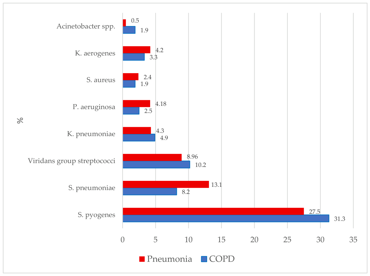
Figure 2.
Heatmap of antimicrobial susceptibility among major respiratory bacterial isolates. Cell values represent the percentage of susceptible isolates (%S) among all isolates tested for each bacterial species-antimicrobial combination. A fixed color scale ranging from 0% to 100% was applied across the entire heatmap, allowing direct comparison of susceptibility levels between bacterial species and antimicrobial agents. Gray cells indicate combinations that were not tested or for which susceptibility data were unavailable. Abbreviations: AMX, amoxicillin; AMC, amoxicillin/clavulanate; CLI, clindamycin; AZM, azithromycin; CLR, clarithromycin; FOS, fosfomycin; CFZ, cefazolin; CAZ, ceftazidime; CXM, cefuroxime; CRO, ceftriaxone; FEP, cefepime; CSL, cefoperazone/sulbactam; LVX, levofloxacin; OFX, ofloxacin; CIP, ciprofloxacin; GEN, gentamicin; AMK, amikacin; DOX, doxycycline; TCY, tetracycline; VAN, vancomycin; MEM, meropenem; IPM, imipenem; TOB, tobramycin; PMB, polymyxin B.
Figure 2.
Heatmap of antimicrobial susceptibility among major respiratory bacterial isolates. Cell values represent the percentage of susceptible isolates (%S) among all isolates tested for each bacterial species-antimicrobial combination. A fixed color scale ranging from 0% to 100% was applied across the entire heatmap, allowing direct comparison of susceptibility levels between bacterial species and antimicrobial agents. Gray cells indicate combinations that were not tested or for which susceptibility data were unavailable. Abbreviations: AMX, amoxicillin; AMC, amoxicillin/clavulanate; CLI, clindamycin; AZM, azithromycin; CLR, clarithromycin; FOS, fosfomycin; CFZ, cefazolin; CAZ, ceftazidime; CXM, cefuroxime; CRO, ceftriaxone; FEP, cefepime; CSL, cefoperazone/sulbactam; LVX, levofloxacin; OFX, ofloxacin; CIP, ciprofloxacin; GEN, gentamicin; AMK, amikacin; DOX, doxycycline; TCY, tetracycline; VAN, vancomycin; MEM, meropenem; IPM, imipenem; TOB, tobramycin; PMB, polymyxin B.
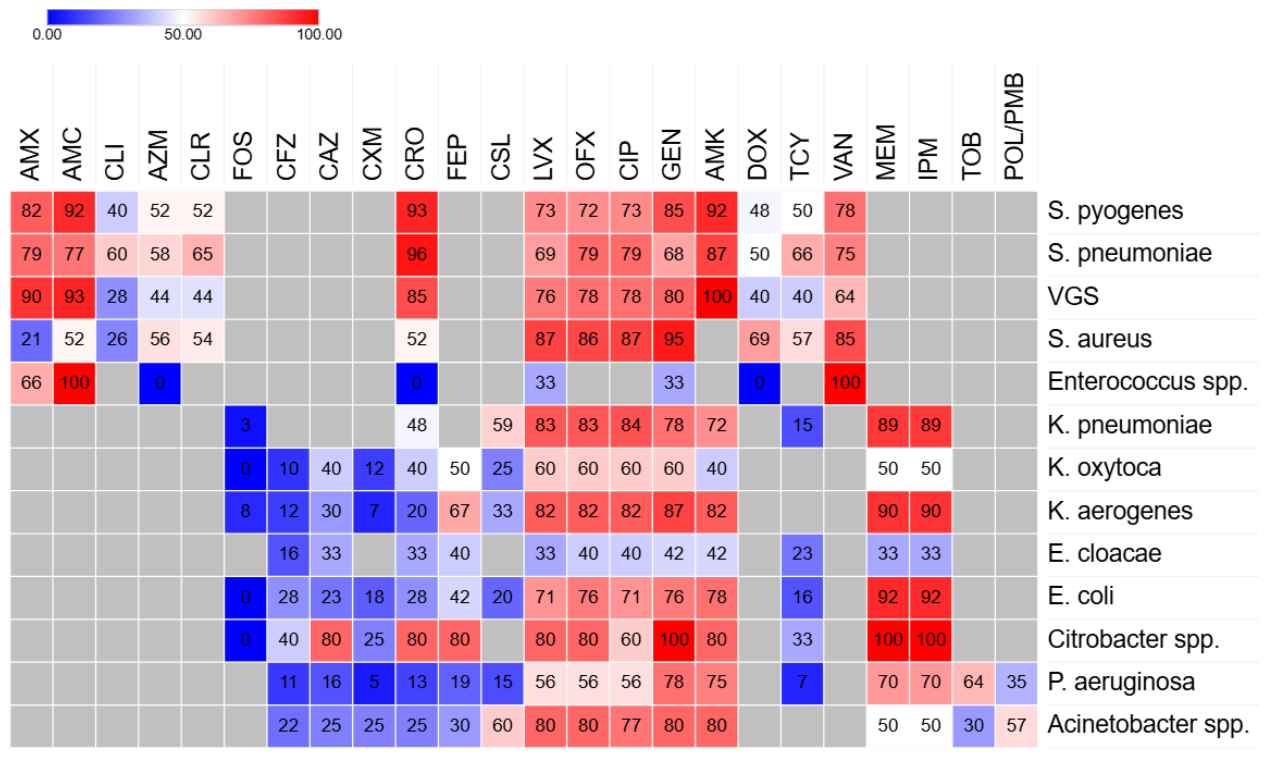

Table 1.
Clinical characteristics of the study population.
| Variables |
COPD (n=255) |
Pneumonia (n=458) | p-value |
| Male, n (%) | 182 (71.4%) | 270 (59.0%) | <0.001 |
| Female, n (%) | 73 (28.6%) | 188 (41.0%) | |
| Age, years (mean ± SD1) | 63.2±8.7 | 61.6±15.7 | 0.133 |
| Pack-years smoked (mean ± SD) | 35.9±14.3 | 17.2±11.8 | < 0.001 |
| Smoking status (%) | |||
| Current and former smokers, n (%) | 176 (69.0%) | 123 (26.9%) | <0.001 |
| Non-smokers, n (%) | 79 (31.0%) | 335 (73.1%) | |
| Post-FEV12 % (mean ±SD) | 42.5±13.3 | ||
| Post-FEV1/FVC3 ratio (mean ±SD) | 0.61±0.24 |
1 SD, standard deviation; 2FEV1, forced expiratory volume in 1 second; 3FVC, forced vital capacity.
Table 2.
Spectrum of respiratory microorganisms isolated from patients with COPD and pneumonia.
| Pathogen |
COPD N=255 |
Pneumonia N=458 |
p-value | Adjusted p-value |
| Streptococcus pyogenes | 114 (44.7%) | 184 (40.2%) | 0.240 | 0.480 |
| Streptococcus pneumoniae | 30 (11.8%) | 88 (19.2%) | 0.011 | 0.154 |
| VGS | 37 (14.5%) | 60 (13.1%) | 0.599 | 0.699 |
| Staphylococcus aureus | 7 (2.7%) | 16 (3.5%) | 0.588 | 0.699 |
| Enterococcus spp. | 2 (0.8%) | 1 (0.2%) | 0.292 | 0.511 |
| Klebsiella pneumoniae | 18 (7.1%) | 29 (6.3%) | 0.708 | 0.763 |
| Klebsiella oxytoca | 6 (2.4%) | 4 (0.9%) | 0.180 | 0.420 |
| Klebsiella aerogenes | 12 (4.7%) | 28 (6.1%) | 0.434 | 0.608 |
| Enterobacter cloacae | 1 (0.4%) | 6 (1.3%) | 0.431 | 0.608 |
| Escherichia coli | 5 (2.0%) | 9 (2.0%) | 0.997 | 0.997 |
| Citrobacter spp. | 5 (2.0%) | 2 (0.4%) | 0.104 | 0.380 |
| Proteus mirabilis | 2 (0.8%) | 0 | 0.128 | 0.380 |
| Pseudomonas aeruginosa | 9 (3.5%) | 28 (6.1%) | 0.136 | 0.380 |
| Acinetobacter spp. | 7 (2.7%) | 3 (0.7%) | 0.040 | 0.280 |
Table 10.
Prevalence of multidrug- and extensively drug-resistant isolates by bacterial species.
| Pathogen | Evaluable isolates | MDR, n (%) | XDR, n (%) | |||||
| COPD | Pneumonia | COPD | Pneumonia | p | COPD | Pneumonia | p | |
| Staphylococcus aureus | 7 | 16 | 0 | 1 (6.3%) | 1.000 | 0 | 1 (6.3%) | 1.000 |
| Enterococcus spp. | 2 | 1 | 2 (100%) | 0 | 0.333 | 0 | 0 | - |
| Escherichia coli | 5 | 9 | 3 (60%) | 2 (22.2%) | 0.266 | 0 | 0 | - |
| Klebsiella pneumoniae | 18 | 29 | 3 (16.7%) | 9 (31.0%) | 0.324 | 1 (5.6%) | 2 (6.9%) | 1.000 |
| Klebsiella oxytoca | 6 | 4 | 3 (50%) | 1 (25%) | 0.571 | 1 (16.7%) | 1 (25%) | 1.000 |
| Klebsiella aerogenes | 12 | 28 | 3 (25%) | 2 (7.1%) | 0.149 | 0 | 0 | - |
| Enterobacter cloacae | 1 | 6 | 0 | 4 (66.7%) | 0.429 | 0 | 2 (33.3%) | 1.000 |
| Proteus mirabilis | 2 | 0 | 0 | 0 | 0 | 0 | - | |
| Pseudomonas aeruginosa | 9 | 28 | 2 (22.2%) | 5 (17.9%) | 1.000 | 1 (11.1%) | 4 (14.3%) | 1.000 |
| Acinetobacter spp. | 7 | 3 | 2 (28.6%) | 1 (33.3%) | 1.000 | 1 (14.3%) | 0 | 1.000 |
Disclaimer/Publisher’s Note: The statements, opinions and data contained in all publications are solely those of the individual author(s) and contributor(s) and not of MDPI and/or the editor(s). MDPI and/or the editor(s) disclaim responsibility for any injury to people or property resulting from any ideas, methods, instructions or products referred to in the content. |
© 2026 by the authors. Licensee MDPI, Basel, Switzerland. This article is an open access article distributed under the terms and conditions of the Creative Commons Attribution (CC BY) license (http://creativecommons.org/licenses/by/4.0/).
Copyright: This open access article is published under a Creative Commons CC BY 4.0 license, which permit the free download, distribution, and reuse, provided that the author and preprint are cited in any reuse.