

Submitted:
16 September 2026
Posted:
23 September 2026
You are already at the latest version
Abstract
No drug has yet been shown to stop an aortic aneurysm (AA, abdominal AAA or thoracic TAA) from enlarging, and many patients are recognised only once the wall is already failing. Alongside the classical risk factors, three conditions that patients and clinicians can actually change—chronic cold exposure, lack of exercise and severe infection—have each been tied to faster progression, although their molecular overlap is rarely discussed together. This narrative review draws on 80 sources from PubMed, Web of Science and CNKI (2003–2026). Upstream the three insults look quite different: cold acts through extracellular cold-inducible RNA-binding protein (eCIRP)–TLR4 signalling and the sympathetic nervous system, inactivity through low and oscillatory wall shear stress, and infection through pathogen-associated molecular patterns. Downstream they meet on NF-κB, MMP-2/9, oxidative stress and loss of vascular smooth muscle cells, with the NLRP3 inflammasome as the shared bottleneck—its caspase-1 both releases IL-1β/IL-18 and cleaves pro-MMP-9 directly. We use this convergence to re-read the still piecemeal single-omics evidence and weigh NLRP3-, IL-1β- and CIRP-directed drugs against exercise, warmth and vaccination. The clearest gaps are multifactorial models, paired multi-omics cohorts and trials that follow expansion rate.
Keywords:
aortic aneurysm
; cold exposure
; cold-inducible RNA-binding protein
; physical inactivity
; wall shear stress
; NLRP3 inflammasome
; matrix metalloproteinase 9
1. Introduction
Aortic aneurysm is a permanent, focal dilatation of a weakened wall, named abdominal (AAA) or thoracic (TAA) by its location. Roughly 4%–7% of men over 65 carry an AAA, and aneurysm disease kills about 200,000 people each year 1. It is usually silent, so a sizeable minority first present with rupture, which kills more than 80% of those affected 2. Open or endovascular repair is still the only treatment that clearly works; no drug has been shown to slow expansion 1,2. The familiar risks are age, male sex, smoking, hypertension, atherosclerosis and inherited connective-tissue disease, and the occasional coexistence of AAA with abdominal-wall hernia hints at an inborn weakness of the wall 3.
Three modifiable exposures have had less attention than they deserve. Cold is one. Time-stratified case-crossover work from northeast China, Japan and the United States all tie low temperature to dissection and rupture 4,5,6. Sedentary living is another: cohort and meta-analytic evidence links habitual activity and fitness to a lower chance of AAA 7. Infection is the third, whether as direct bacterial invasion of the wall (an infected or “mycotic” aneurysm) or as sepsis that speeds an existing aneurysm along 8–10. In real life these overlap. Winter brings cold and respiratory infection at the same time, and the endothelial and metabolic fallout of inactivity leaves the wall less able to tolerate either. Our aim is to read the three together—to trace how cold, inactivity and infection accelerate aneurysm through shared inflammatory circuits, where those circuits converge, and what they mean for treatment and for the omics work still missing. We write as a narrative rather than a systematic review and make no attempt at meta-analysis.
3. Chronic Cold Exposure: The eCIRP–TLR4 Axis and Sympathetic Activation
3.1. Epidemiological Evidence
The pattern holds across quite different settings. A 2026 retrospective series from northeast China found that sustained cold and abrupt day-to-day falls both raised dissection risk, the effect lingering for more than a week 4. A Japanese case-crossover study then pulled two events apart: dissection tracked sudden drops whereas rupture tracked prolonged cold, a hint that the two do not share identical physiology 5. Nationwide US CDC WONDER data point the same way for mortality 6.
3.2. The CIRP–TLR4–NF-κB Axis
Cold-inducible RNA-binding protein (CIRP) is a stress protein with fingers in several cardiac and vascular processes 24,25. Cold or hypoxia moves it out of the cell as extracellular CIRP (eCIRP), where it behaves as a damage-associated molecular pattern 24. Macrophages and endothelial cells read eCIRP through TLR4-MD2 24 and through TREM-1 26; TLR4 then uses MyD88 to switch on NF-κB and the TNF-α/IL-1β/IL-6/MCP-1 programme 27,28. In BAPN-treated mice, acute cold (4 ± 1 °C) drove vascular inflammation and MMP-2 through eCIRP–TLR4 and raised dissection risk, and adding CIRP to endothelial cells reproduced the effect in vitro 27.
Li and colleagues supply the most complete AAA chain. Human AAA tissue held 5.6-fold more CIRP mRNA and 93% more CIRP protein, and serum CIRP tracked severity; in elastase-treated rats, anti-CIRP antibody (1 mg/kg) held dilatation down and cut MMP-2/9, TNF-α, MCP-1 and CD68+ infiltration; recombinant CIRP raised macrophage MMP-9 mRNA as much as 5.5-fold and migration by about 2.7-fold 29. A caveat matters: these are acute models, whereas a human aneurysm develops over years, and CIRP’s role under genuinely chronic cold is still untested.
3.3. Sympathetic Activation, Oxidative Stress and a Prothrombotic State
Cold also fires up the sympathetic nervous system. Catecholamines rise, the heart speeds, peripheral vessels clamp down, and blood pressure and platelet activity go up with them 30,31. By Laplace’s law wall tension scales with pressure times radius, so even a modest pressure rise loads an existing aneurysm more heavily. Sympathetic activation recruits renin–angiotensin too; angiotensin II generates NADPH-derived ROS and pushes VSMC switching and MMP expression 2. Oxidant production outruns antioxidant capacity and feeds NF-κB and MAPK 32. Fibrinogen can rise by as much as 23% in winter, viscosity rises, and the intraluminal thrombus that follows becomes a slow, luminal source of macrophage and neutrophil proteases 30.
4. Physical Inactivity: Disturbed Haemodynamics and Mechanotransduction
4.1. Epidemiological Evidence
Across cohorts and meta-analyses, active and fit people develop less AAA and carry a smaller burden 7,33–36. That pattern raises a mechanical question: how does a sedentary life actually reach the aortic wall?
4.2. Low and Oscillatory Wall Shear Stress
Wall shear stress (WSS), the friction of flowing blood on the endothelium, is a major homeostatic cue; healthy aortic WSS runs around 1–7 Pa in one direction. A weak leg-muscle pump slows distal flow and leaves the infrarenal aorta bathed in low (<0.4 Pa), oscillating shear 37,38. Dalman’s group tested the consequence in elastase-treated rats, pairing an arteriovenous fistula (WSS up ~300%) against iliac ligation (WSS down ~60%). Low-flow aneurysms ended up larger; high-flow walls kept more endothelial and smooth muscle cells, made more VEGF-D and PDGF-B, and favoured proliferation over apoptosis 38. Low flow also encouraged adventitial neovascularisation, GM-CSF and macrophages, whereas high flow retained CD34+ progenitors 39—the experimental case for moving the legs to move blood past the aneurysm.
4.3. Endothelial Dysfunction, Mechanotransduction and Interstitial Flow
Laminar flow keeps eNOS running through PI3K/Akt, and NO holds the vessel relaxed and anti-inflammatory; low or oscillating shear instead activates NF-κB and ICAM-1, VCAM-1 and E-selectin, inviting monocytes in 37. A damaged glycocalyx compounds the signalling failure 40, and loss of the junctional VE-cadherin/AmotL2 pathway is itself enough to provoke inflammation and AAA 41; how such intramural forces bear on the thoracic aorta has been reviewed elsewhere 42. Luminal shear is not the only flow at stake: interstitial flow within the wall lowers SM-MHC, smoothelin and calponin and nudges VSMCs toward the synthetic state 43. Inactivity also travels with obesity and disordered sphingolipid metabolism 44; exercise, conversely, improves lipids and lowers high-sensitivity CRP 7, while sedentary metabolism accumulates the oxLDL and cholesterol crystals that switch NLRP3 on 21.
5. Severe Infection: The PAMP–NLRP3–MMP-9 Axis and the Septic Surge
5.1. Epidemiology, Pathogens and Routes of Invasion
An infected (“mycotic”) aneurysm forms when bacteria or fungi breach the wall. It is uncommon—0.7%–2.6% of all aneurysms—but aggressive, with mortality of 30%–50% and thoracic cases that are easy to miss 8,45. Its patients skew younger than degenerative AAA, often under 40 9. Staphylococcus aureus and Salmonella (the nontyphoidal link to aortitis is especially well documented) lead, followed by streptococci and E. coli; fungi appear mainly in the immunosuppressed 8,10,46. Organisms reach the wall by colonising an atheromatous plaque, riding septic emboli into the vasa vasorum, spreading from adjacent infection, entering through trauma or an iatrogenic device, or arriving in endocarditis emboli 10.
5.2. The Inflammatory–Protease Cascade and Sepsis
Once inside, pathogen-associated molecular patterns (PAMPs)—LPS, peptidoglycan, bacterial DNA—are caught by TLR and NOD receptors 8. Neutrophils dump MMP-8, MMP-9 and neutrophil gelatinase-associated lipocalin (NGAL) onto the matrix 8, while macrophages read LPS through TLR4, activate NF-κB and amplify TNF-α, IL-1β and IL-6 11. PAMPs, bacterial DNA, ATP and ROS then engage NLRP3; gasdermin D (GSDMD) pores, circRNA-governed macrophage pyroptosis and VSMC putrescine synthesis have all been implicated in AAA, and here again caspase-1 activates MMP-9 13,47,48. Proteases, ROS and toxins necrose the wall, collapse its elastin and collagen and end in pseudoaneurysm or rupture 9. Even without invasion, sepsis floods the circulation with cytokines that drive MMPs and apoptosis in wall cells 11, and hypotension and vasopressors worsen perfusion; that this raises rupture risk is mechanistically plausible but still awaits a proper cohort.
5.3. IL-1β as the Connecting Cytokine
IL-1β is where infection and aneurysm most clearly meet. Knocking IL-1β out, or blocking its receptor with anakinra, curbed experimental TAA and AAA 49; and CANTOS showed that canakinumab, an anti-IL-1β antibody, lowers systemic inflammation and atherothrombotic events—indirect support, but real, for the same move in aneurysm 21.
6. A Convergent, Synergistic Network with NLRP3 at Its Core
The upstream stories differ; the downstream wiring does not, and Table 1 and Figure 1 lay this out. Cold arrives by eCIRP–TLR4 and sympathetic stress, inactivity by low, oscillating shear and a dysfunctional endothelium, infection by PAMP–TLR/NOD. Each feeds NF-κB, NLRP3, MMP-2/9, oxidant stress and VSMC dysfunction, and NLRP3-derived caspase-1 is the choke point at which mature cytokines and direct MMP-9 activation meet.
Winter makes the synergy concrete. Cold leaves CIRP and sympathetic pathways switched on, and a respiratory infection on top drives PAMP–NLRP3 signalling hard, spiking IL-1β and MMP-9 enough to tip a vulnerable wall into dissection or rupture—consistent with the winter peak in events 4. Inactivity works the other side of the same coin: with less NO and more oxLDL, the wall simply has less reserve against cold or infection. Combined, the effect is networked rather than additive.
7. From Single-Omics to Integrated Multi-Omics
7.1. What Single-Omics Has Revealed—And Where It Falls Short
Each omics layer has been turned on aneurysm, one at a time. scRNA-seq found Malat1+ VSMCs and other disease subsets 50; spatial transcriptomics placed GPNMB+ macrophages where the elastic lamina was most damaged, driving VSMC switching through CD44 51, and mapped distinct changes around CRP deposits 52. Proteomics named CXCR4 a shared marker of inflammatory cells and vessels for imaging 53, and ECM proteomics logged changes in collagen XII, thrombospondin-2 and periostin while catching MMP-12 as a substrate for several matrix glycoproteins 54. Taurine reprogramming and lactylation joined the metabolic side 55,56, and the microRNA layer has been reviewed on its own 57. Useful as these are, one layer shows only one dimension, and a transcript is a poor proxy for a protein’s amount or timing; single-omics can neither reconstruct the cell–microenvironment web nor tell a driver from a bystander 58.
7.2. Integrated Multi-Omics in Other Vascular Diseases
scRNA-seq has already catalogued VSMC and endothelial subsets across AAA and dissection 18,59,60. Atherosclerosis shows what adding layers can buy. Joint CITE-seq (transcriptome plus surface protein) and scRNA-seq resolved dozens of multimodal states, TREM2+ and SMC-derived foam cells among them, and set symptomatic against asymptomatic plaque 61; fate mapping combined with scRNA-seq and human genetics traced the SMC-to-“SEM” intermediate and flagged retinoic-acid signalling as a lever 62. The comparative point is simple: cross-layer analysis exposes what any single layer misses.
7.3. Current State and Gaps in Aneurysm
Aneurysm still lags. Most studies hold to one transcriptome or one metabolome and never cross genome, transcript, protein and metabolite. Pairing scRNA-seq with micro-mechanical devices implicated Piezo1 in VSMC mechanosensing 63; pairing it with scATAC-seq showed DNA damage pushing VSMCs from contractile to inflammatory through STING–IRF3 64; and complement and precision-medicine ideas are only now arriving 65. scRNA-seq also throws away position and often needs pooled donors, gaps spatial methods partly fill 66. Some open questions are still basic: do TAA and AAA, or aneurysms driven by infection, cold, trauma or sedentary flow, even carry distinguishable signatures? Most human-relevant comparisons rest on a single acute animal model.
7.4. Application Prospects
Risk stratification is the first payoff within reach: layered signatures might tell an indolent aneurysm from a fast-growing one when the two look identical on imaging. Running the same omics in parallel across stressors could map wall-degeneration modules and settle whether the risk factors share a module or use separate routes. Combined plasma proteomics, metabolomics and lipidomics could supply non-invasive markers, alongside MMP-9, desmosine and 18F-FDG PET, which already predict growth of small AAA 67–70; tying GWAS hits to particular cell-type transcriptomes gives a target genetic backing 71. Around 23 AAA loci are known 72,73; lipid genetics and Mendelian randomisation argue for causal LDL-C and triglyceride effects and implicate HMGCR, CETP, PCSK9 and the IL-6R, PLAU and PSMA4 loci 74,75, while proteomic genetics added neogenin and KIT ligand 76. Enthusiasm should be tempered: CITE-seq, scATAC-seq and spatial methods are expensive and underpowered, and stable signatures await large, multicentre, paired tissue–blood cohorts.
8. Therapeutic Targets and Translational Challenges
A handful of candidates have reached animals or the clinic (Table 2). Doxycycline, roxithromycin and the statins have not produced a drug that stops expansion 77,78. NLRP3, sitting at the convergence point, draws the most interest: the selective inhibitor MCC950 limited dilatation, dissection and rupture in hyperlipidaemic, Ang II-infused mice, partly by blocking caspase-1-driven MMP-9 activation 13; colchicine blocks NLRP3 assembly through microtubules and has cardiovascular outcome-trial support (COLCOT, LoDoCo2), though no direct AAA test exists; and the approved sulphonylurea glyburide inhibits NLRP3 and lessened Ang II-induced AAA/dissection 20. Downstream, canakinumab and anakinra target IL-1β 21,49, anti-CIRP antibody limited rat AAA 29, and the eCIRP-derived M3 peptide, which interrupts eCIRP–TREM-1, lifted survival from 40% to 80% in intestinal ischaemia–reperfusion 26.
8.1. NLRP3 Inhibitors
None of this is straightforward. MCC950 stalled after hepatic safety signals. Colchicine has a narrow window, with gastrointestinal and muscle toxicity in older patients and several cardiovascular interactions. Glyburide would need doses above its hypoglycaemic range to hit NLRP3, bringing hypoglycaemia straight back. Because NLRP3 also defends against infection, chronic blockade carries an infectious cost, and a dedicated AAA randomised trial does not yet exist.
8.2. CIRP-Directed Therapy
Anti-CIRP antibody protects rats 29 and M3 disrupts eCIRP–TREM-1 26, but neither has reached patients, and CIRP’s everyday role in coping with stress argues against blocking it for long. Serum CIRP may prove more useful sooner as a marker of cold-related risk than as a drug target.
8.3. Exercise, Stratified by Aneurysm Status
Regular leg exercise raises flow and WSS, helps the endothelium, lowers systemic inflammation and improved inflammatory markers in small-AAA training studies 79. Above 5.5 cm, or during rapid expansion, hard exercise and Valsalva manoeuvres are best avoided; today’s guidance leans on expert consensus 80, and diameter-specific safety data simply do not exist.
8.4. IL-1β Blockade
The best IL-1β evidence still comes from coronary disease rather than AAA: canakinumab cut recurrent events after myocardial infarction in CANTOS 21, and anakinra checked experimental TAA/AAA 49. CANTOS also saw more fatal infections on treatment, a genuine worry in older patients; canakinumab is costly and anakinra must be injected daily. Whether either helps in AAA needs its own trial.
9. Limitations and Future Directions
Much of the synergy here is inferred from mechanism and pairwise evidence; no model yet puts single and combined exposures head to head. Cold evidence is epidemiological or based on acute exposure, inactivity evidence leans on questionnaires, and AAA drug evidence lacks large trials—and, as a narrative review, we cannot exclude selection bias. Technically, integrated single-cell and spatial omics, aortic organoids and organ-on-chip devices could compare stressors and screen drugs with fewer animals; models that combine cold, infection and disturbed flow are needed to measure synergy, thresholds and any window of susceptibility. The most realistic near-term moves are serum CIRP as a predictor and phase-II trials of colchicine or glyburide in 3.0–4.5 cm AAA with expansion rate as the primary endpoint, alongside graded exercise prescriptions and risk models that fold temperature, activity and infection history in with the standard factors.
10. Conclusions
Cold, inactivity and infection start in different places and finish in much the same one: NF-κB, NLRP3, MMP-2/9, oxidant stress and a wall stripped of contractile VSMCs. NLRP3-derived caspase-1 is the unusual step, releasing IL-1β/IL-18 and activating MMP-9 at the same moment. Winter’s “double hit” and the sedentary wall’s loss of reserve are simply that network in everyday life.
The drugs remain preclinical or borrowed from other diseases, and their problems—hepatic toxicity, narrow windows, infection risk—are unresolved; exercise, keeping warm in winter and respiratory vaccination are available now. The next stage, in our view, is less about adding single-exposure observations and more about multifactorial models, multicentre multi-omics cohorts and early trials judged by how fast the aneurysm grows.
Author Contributions
Cheng Hu: conceptualization, literature retrieval and screening, data curation, formal analysis, writing–original draft, writing–review & editing, and supervision.
Funding
This research did not receive any specific grant from funding agencies in the public, commercial or not-for-profit sectors. [Replace if a grant number is to be declared.].
Acknowledgments
The author thanks the Wu Lien-teh Honors Program of Harbin Medical University for academic training.
Declaration of generative AI and AI-assisted technologies in the writing process: During the preparation of this work the authors used [name of AI tool/service] in order to improve English language and readability. After using this tool, the authors reviewed and edited the content as needed and take full responsibility for the content of the publication.
References
- Liu, B.; Granville, D.J.; Golledge, J.; Kassiri, Z. Pathogenic mechanisms and the potential of drug therapies for aortic aneurysm. Am. J. Physiol. Heart Circ. Physiol. 2020, 318(3), H652–H670. [Google Scholar] [CrossRef]
- Gao, J.; Cao, H.; Hu, G.; et al. The mechanism and therapy of aortic aneurysms. Signal Transduct. Target Ther. 2023, 8(1), 132. [Google Scholar] [CrossRef]
- Kessler, V.; Klopf, J.; Eilenberg, W.; Neumayer, C.; Brostjan, C. AAA Revisited: A Comprehensive Review of Risk Factors, Management, and Hallmarks of Pathogenesis. Biomedicines 2022, 10(1), 94. [Google Scholar] [CrossRef]
- Liao, Z.; Liu, Z.; Ding, Y.; et al. Low ambient temperature and sudden temperature drop increases the incidence of acute aortic dissection: a retrospective analysis from northeast China. Front Public Health 2026, 14, 1848286. [Google Scholar] [CrossRef]
- Jimba, T.; Kohsaka, S.; Takahashi, T.; et al. Distinct temporal effects of ambient temperature on acute aortic dissection and aneurysm rupture: a time-stratified case-crossover study. Eur. J. Prev. Cardiol. 2026, zwag049. [Google Scholar] [CrossRef]
- Wang, H.; Wang, S.; Fan, K.; et al. Spatiotemporal mortality patterns and temperature-associated risk of aortic dissection in the United States: a national CDC WONDER database analysis. Vasc. Health Risk Manag. 2026, 22, 569639. [Google Scholar] [CrossRef]
- Li, R.; Lei, F.; Liu, B.; et al. Association between questionnaire- and accelerometer-based physical activity and abdominal aortic aneurysm incidence. J. Am. Heart Assoc. 2026, 15(7), e043217. [Google Scholar] [CrossRef]
- Raavi, L.; Garg, P.; Hussain, M.W.A.; et al. Mycotic thoracic aortic aneurysm: epidemiology, pathophysiology, diagnosis, and management. Cureus 2022, 14(11), e31010. [Google Scholar] [CrossRef]
- Mycotic Aneurysm. StatPearls. NCBI Bookshelf 2025. [CrossRef]
- Hakim, S.; Davila, F.; Amin, M.; et al. Infectious aortitis: a life-threatening endovascular complication of nontyphoidal Salmonella bacteremia. Case Rep. Med. 2018, 2018, 6845617. [Google Scholar] [CrossRef]
- Fan, W.; Tang, T.; Zeng, Q.; et al. The role of IL-1β in aortic aneurysm. Clin. Chim. Acta 2020, 504, 7–14. [Google Scholar] [CrossRef]
- Atkinson, G.; Bianco, R.; Di Gregoli, K.; Johnson, J.L. The contribution of matrix metalloproteinases and their inhibitors to the development, progression, and rupture of abdominal aortic aneurysms. Front Cardiovasc Med. 2023, 10, 1248561. [Google Scholar] [CrossRef]
- Ren, P.; Wu, D.; Appel, R.; et al. Targeting the NLRP3 Inflammasome With Inhibitor MCC950 Prevents Aortic Aneurysms and Dissections in Mice. J. Am. Heart Assoc. 2020, 9(11), e014044. [Google Scholar] [CrossRef]
- Milewicz, D.M.; Trybus, K.M.; Guo, D.; et al. Altered SMC Force Generation as Driver of Thoracic Aortic Aneurysms and Dissections. Arterioscler. Thromb. Vasc. Biol. 2017, 37(1), 26–33. [Google Scholar] [CrossRef]
- Leng, S.; Li, H.; Zhang, P.; et al. SGK1-Mediated VSMC Phenotypic Transformation Promotes Thoracic Aortic Dissection. Arterioscler. Thromb. Vasc. Biol. 2024, 44(7), e168–e183. [Google Scholar] [CrossRef]
- Hu, Y.; Cai, Z.; He, B. Smooth Muscle Heterogeneity and Plasticity in Health and Aortic Aneurysmal Disease. Int. J. Mol. Sci. 2024, 25(3), 1842. [Google Scholar] [CrossRef]
- Lu, H.; Du, W.; Ren, L.; et al. Vascular Smooth Muscle Cells in Aortic Aneurysm: From Genetics to Mechanisms. J. Am. Heart Assoc. 2022, 11(15), e025783. [Google Scholar] [CrossRef]
- Cao, G.; Xuan, X.; Li, Y.; et al. VSMC phenotypic landscape in aortic aneurysm by scRNA-seq. Cell Commun. Signal. 2023, 21(1), 160. [Google Scholar]
- Chen, Y.; Zhang, T.; Yao, F.; et al. Dysregulation of interaction between LOXhigh fibroblast and smooth muscle cells contributes to the pathogenesis of aortic dissection. Theranostics 2022, 12(5), 2174–2190. [Google Scholar] [CrossRef]
- Wu, D.; Ren, P.; Zheng, Y.; et al. NLRP3-Caspase-1 Inflammasome Degrades Contractile Proteins: Implications for Aortic Biomechanical Dysfunction and Aneurysm and Dissection Formation. Arterioscler. Thromb. Vasc. Biol. 2017, 37(8), 1548–1559. [Google Scholar]
- Takahashi, M. NLRP3 inflammasome as a key driver of vascular disease. Cardiovasc Res. 2021, 117(6), 1462–1475. [Google Scholar]
- Duewell, P.; Kono, H.; Rayner, K.J.; et al. NLRP3 inflammasomes are required for atherogenesis and activated by cholesterol crystals. Nature 2010, 464(7293), 1357–1361. [Google Scholar] [CrossRef]
- Gaul, S.; Schaeffer, K.M.; Opitz, L.; et al. Extracellular NLRP3 inflammasome particles are internalized by human coronary artery smooth muscle cells. Sci. Rep. 2021, 11(1), 16100. [Google Scholar] [CrossRef]
- Lujan, D.A.; Ochoa, J.L.; Hartley, R.S. Cold-Inducible RNA Binding Protein in Cancer and Inflammation. Wiley Interdiscip. Rev. RNA 2018, 9(3), e1476. [Google Scholar] [CrossRef]
- Zhong, P.; Peng, J.; Bian, Z.; Huang, H. The Role of Cold Inducible RNA-Binding Protein in Cardiac Physiology and Diseases. Front Pharmacol. 2021, 12, 610792. [Google Scholar] [CrossRef]
- Denning, N.; Aziz, M.; Ochani, M.; et al. Inhibition of a triggering receptor expressed on myeloid cells-1 (TREM-1) with an extracellular cold-inducible RNA-binding protein (eCIRP)-derived peptide protects mice from intestinal ischemia-reperfusion injury. Surgery 2020, 168(4), 692–700. [Google Scholar] [CrossRef]
- Tsai, H.Y.; Chien, W.C.; Chung, C.H.; et al. Cold exposure increases aortic dissection risk through extracellular cold inducible RNA binding protein and toll-like receptor 4 signaling. Sci. Rep. 2026, 16(1), Article 38164. [Google Scholar] [CrossRef]
- Zhang, R.; Fang, K.; Mu, C.; Zhang, L. Cold-inducible RNA-binding protein induces inflammatory responses via NF-κB signaling pathway in normal human bronchial epithelial cells infected with streptococcus pneumoniae. Int. Immunopharmacol. 2023, 123, 110748. [Google Scholar] [CrossRef]
- Li, G.; Yang, L.; Yuan, H.; et al. Cold-inducible RNA-binding protein plays a central role in the pathogenesis of abdominal aortic aneurysm in a murine experimental model. Surgery 2016, 159(5), 1336–1346. [Google Scholar] [CrossRef]
- Li, Y.; Wu, J.; Xu, Y.; et al. Cold exposure and the cardiovascular system: from physiological adaptation to pathological risk. Front Physiol. 2026, 16, 1740919. [Google Scholar] [CrossRef]
- Liu, C.; Yavar, Z.; Sun, Q. Cardiovascular response to thermoregulatory challenges. Am. J. Physiol. Heart Circ. Physiol. 2015, 309(11), H1793–H1812. [Google Scholar] [CrossRef]
- Ma, G.; Cai, H.; Li, Z.; et al. Advancements in understanding inflammatory responses and the development of cardiovascular diseases under cold stimulation. Frigid Zone Med. 2023, 3(4), 209–215. [Google Scholar] [CrossRef]
- Forsdahl, S.H.; Singh, K.; Solberg, S.; Jacobsen, B.K. Risk Factors for Abdominal Aortic Aneurysms: The Tromsø Study. Circulation 2009, 119(17), 2307–2315. [Google Scholar] [CrossRef]
- Aune, D.; Sen, A.; Kobeissi, E.; Hamer, M.; Norat, T.; Riboli, E. Physical activity and the risk of abdominal aortic aneurysm: a meta-analysis. Sci. Rep. 2020, 10(1), 20755. [Google Scholar] [CrossRef]
- Perissiou, M.; Bailey, T.G.; Saynor, Z.L.; et al. Cardiorespiratory fitness in people with abdominal aortic aneurysm. Exp. Physiol. 2022, 107(4), 413–425. [Google Scholar] [CrossRef]
- Singh, K.; Bønaa, K.H.; Jacobsen, B.K.; Bjørk, L.; Solberg, S. Prevalence of and Risk Factors for Abdominal Aortic Aneurysms: The Tromsø Study. Am. J. Epidemiol. 2001, 154(3), 236–244. [Google Scholar] [CrossRef]
- Dua, M.M.; Dalman, R.L. Hemodynamic Influences on Abdominal Aortic Aneurysm Disease: Application of Biomechanics to Aneurysm Pathophysiology. Vasc. Pharmacol. 2009, 51(2-3), 100–107. [Google Scholar] [CrossRef]
- Hoshina, K.; Sho, E.; Sho, M.; et al. Wall shear stress and strain modulate experimental aneurysm cellularity. J. Vasc. Surg. 2003, 37(6), 1302–1308. [Google Scholar] [CrossRef]
- Sho, E.; Sho, M.; Nanjo, H.; et al. Hemodynamic Regulation of CD34⁺ Cell Localization and Differentiation in Experimental Aneurysms. Arterioscler. Thromb. Vasc. Biol. 2004, 24(10), 1916–1921. [Google Scholar] [CrossRef]
- Liu, J.; Kang, H.; Ma, X.; et al. Vascular Cell Glycocalyx-Mediated Vascular Remodeling Induced by Hemodynamic Environmental Alteration. Hypertension 2018, 71(3), 481–490. [Google Scholar] [CrossRef]
- Zhang, Y.; Zhang, Y.; Hutterer, E.; et al. The VE-cadherin/AmotL2 mechanosensory pathway suppresses aortic inflammation and the formation of abdominal aortic aneurysms. Nat. Cardiovasc Res. 2023, 2(6), 542–557. [Google Scholar] [CrossRef]
- Humphrey, J.D.; Schwartz, M.A.; Tellides, G.; Milewicz, D.M. Mechanotransduction in Vascular Biology: Thoracic Aortic Aneurysms and Dissections. Circ. Res. 2015, 116(8), 1448–1461. [Google Scholar]
- Fukui, W.; Ujihara, Y.; Nakamura, M.; et al. Direct visualization of interstitial flow distribution in aortic walls. Sci. Rep. 2022, 12, 5763. [Google Scholar] [CrossRef]
- Okrzeja, J.; Karwowska, A.; Bachnio-Zabielska, A. The Role of Obesity, Inflammation and Sphingolipids in the Development of an Abdominal Aortic Aneurysm. Nutrients 2022, 14(12), 2438. [Google Scholar] [CrossRef]
- Malouf, J.F.; Chandrasekaran, K.; Orszulak, T.A. Mycotic Aneurysms of the Thoracic Aorta: A Diagnostic Challenge. Am. J. Med. 2001, 111(6), 489–494. [Google Scholar]
- Hohmann, E.L. Nontyphoidal Salmonellosis. Clin. Infect. Dis. 2001, 32(2), 263–269. [Google Scholar] [CrossRef]
- Cai, D.; Li, C.; Zhang, Y.; et al. CircHipk3 serves a dual role in macrophage pyroptosis by promoting NLRP3 transcription and inhibition of autophagy to induce abdominal aortic aneurysm formation. Clin. Transl. Med. 2024, 14(12), e1732. [Google Scholar] [CrossRef]
- Gao, J.; Chen, Y.; Wang, H.; et al. Gasdermin D Deficiency in VSMCs Ameliorates AAA Through Reducing Putrescine Synthesis. Adv. Sci. 2023, 10(7), e2205771. [Google Scholar] [CrossRef]
- Johnston, W.F.; Salmon, M.; Pope, N.H.; et al. Inhibition of interleukin-1β decreases aneurysm formation and progression in a novel model of thoracic aortic aneurysms. Circulation 2014, 130((11) Suppl 1, S51–S59. [Google Scholar] [CrossRef]
- Yu, L.; Zhang, J.; Gao, A.; et al. An intersegmental single-cell profile reveals aortic heterogeneity and identifies a novel Malat1+ vascular smooth muscle subtype involved in abdominal aortic aneurysm formation. Signal Transduct. Target Ther. 2022, 7(1), 183. [Google Scholar] [CrossRef]
- Zhao, G.; Choo, C.S.; Liu, H.; et al. Single-cell spatial transcriptomics unravels the cellular landscape of abdominal aortic aneurysm. JCI Insight 2025, 10(5), e190534. [Google Scholar] [CrossRef]
- Kim, E.N.; Seok, H.Y.; Lim, J.S.; et al. CRP deposition in human abdominal aortic aneurysm is associated with transcriptome alterations toward aneurysmal pathogenesis: insights from in situ spatial whole transcriptomic analysis. Front Immunol. 2024, 15, 1362548. [Google Scholar] [CrossRef]
- Cao, G.; Zhang, R.; Jia, X.; et al. CXCR4-targeted sensitive magnetic particle imaging for abdominal aortic aneurysm early detection and prognosis evaluation by recognizing total inflammatory cells. Cardiovasc Res. 2025, 121(2), 324–337. [Google Scholar] [CrossRef]
- Didangelos, A.; Yin, X.; Mandal, K.; et al. Extracellular matrix composition and remodeling in human abdominal aortic aneurysms: a proteomics approach. Mol. Cell Proteom. 2011, 10(8), M111.008128. [Google Scholar] [CrossRef]
- Sun, X.; Du, C.; Chen, Y.; et al. MS4A1-PTGS2 axis induces taurine metabolic reprogramming to exacerbate abdominal aortic aneurysm progression. Int. J. Med. Sci. 2024, 21(4), 1452–1467. [Google Scholar] [CrossRef]
- Li, J.; Huang, Z.; He, J.; et al. Integrative analysis of lactylation-associated features in abdominal aortic aneurysm and its immune microenvironment utilizing scRNA-seq and bulk RNA sequencing. Circ. J. 2024, 88(5), 412–425. [Google Scholar] [CrossRef]
- Maegdefessel, L.; Spin, J.M.; Adam, M.; et al. Micromanaging Abdominal Aortic Aneurysms. Arterioscler. Thromb. Vasc. Biol. 2014, 34(4), 729–734. [Google Scholar]
- Chiva-Blanch, G.; Mayr, M. Lessons from spatiotemporal RNA vs proteins during the cell cycle. Cardiovasc Res. 2021, 117(10), 2237–2240. [Google Scholar] [CrossRef]
- Li, Y.; LeMaire, S.A.; Shen, Y.H. Molecular and Cellular Dynamics of Aortic Aneurysms by scRNA-seq. Arterioscler. Thromb. Vasc. Biol. 2021, 41(11), 2579–2591. [Google Scholar] [CrossRef]
- Zhao, G.; Lu, H.; Chang, Z.; et al. Cellular heterogeneity of aneurysmal infrarenal abdominal aorta by scRNA-seq. Cardiovasc Res. 2020, 116(14), 2314–2327. [Google Scholar]
- Bashore, A.C.; Yan, H.; Xue, C.; et al. High-dimensional single-cell multimodal landscape of human carotid atherosclerosis. Circulation 2023, 148(12), 919–934. [Google Scholar]
- Pan, H.; Xue, C.; Auerbach, B.J.; et al. Single-cell genomics reveals a novel cell state during smooth muscle cell phenotypic switching and potential therapeutic targets for atherosclerosis in mouse and human. Circulation 2020, 142(22), 2167–2183. [Google Scholar] [CrossRef]
- Qian, W.; Hadj, T.; Silvestro, M.; et al. Microskeletal stiffness promotes aortic aneurysm by sustaining pathological vascular smooth muscle cell mechanosensation via Piezo1. Nat. Commun. 2021, 12(1), 7220. [Google Scholar] [CrossRef]
- Chakraborty, A.; Li, Y.; Zhang, C.; et al. Epigenetic induction of smooth muscle cell phenotypic alterations in aortic aneurysms and dissections. Circulation 2020, 142(17), 1658–1673. [Google Scholar]
- Dreher, L.; Kuehl, M.B.; Wenzel, U.O.; Kylies, D. Aortic aneurysm and dissection: complement and precision medicine in aortic disease. Am. J. Physiol. Heart Circ. Physiol. 2025, 328(4), H814–H829. [Google Scholar] [CrossRef]
- Sawada, H.; Lu, H.S.; Daugherty, A. Single-cell transcriptomics as a building block for determining mechanistic insight of abdominal aortic aneurysm formation. Cardiovasc Res. 2021, 117(5), 1243–1245. [Google Scholar] [CrossRef]
- Forsythe, R.O.; Newby, D.E.; Robson, J.M. Monitoring the biological activity of abdominal aortic aneurysms: Beyond Ultrasound. Heart 2016, 102(12), 927–934. [Google Scholar] [CrossRef]
- Mordi, I.R.; Forsythe, R.O.; Gellatly, C.; et al. Plasma Desmosine and Abdominal Aortic Aneurysm Disease. J. Am. Heart Assoc. 2020, 9(5), e013743. [Google Scholar] [CrossRef]
- Lindholt, J.S.; Vammen, S.; Fasting, H.; Henneberg, E.W. Plasma MMP-9 may predict natural history of small AAA. Eur. J. Vasc. Endovasc. Surg. 2001, 21(5), 427–431. [Google Scholar] [CrossRef]
- Bruls, S.; Musumeci, L.; Courtois, A.; et al. Biomarkers and PET Imaging Predict AAA Growth Rate. J. Clin. Med. 2024, 13(9), 2485. [Google Scholar] [CrossRef]
- Raghavan, A.; Pirruccello, J.P.; Ellinor, P.T.; et al. Using genomics to identify novel therapeutic targets for aortic disease. Arterioscler. Thromb. Vasc. Biol. 2024, 44(6), e123–e134. [Google Scholar] [CrossRef]
- Pinard, A.; Jones, G.T.; Milewicz, D.M. Aortic aneurysms compendium: genetics of thoracic and abdominal aortic diseases, aneurysms, dissections, and ruptures. Circ. Res. 2019, 124(4), 588–606. [Google Scholar]
- Klarin, D.; Cho, K.; Sun, Y.V.; et al. Genetic architecture of abdominal aortic aneurysm in the Million Veteran Program. Circulation 2020, 142(18), 1734–1746. [Google Scholar] [CrossRef]
- Harrison, S.C.; Holmes, M.V.; Burgess, S.; et al. Genetic association of lipids and lipid drug targets with abdominal aortic aneurysm: a meta-analysis. JAMA Cardiol. 2018, 3(3), 211–219. [Google Scholar] [CrossRef]
- Chen, Y.; Xu, X.; Wang, L.; et al. Genetic insights into therapeutic targets for aortic aneurysms: a Mendelian randomization study. eBioMedicine 2022, 81, 104124. [Google Scholar]
- Steffen, B.T.; Pankow, J.S.; Norby, F.L.; et al. Proteomics analysis of genetic liability of abdominal aortic aneurysm identifies plasma neogenin and kit ligand: the ARIC study. Arterioscler. Thromb. Vasc. Biol. 2023, 43(3), e89–e100. [Google Scholar] [CrossRef]
- Assar, A.N. Medical treatment of small abdominal aortic aneurysm. J. Cardiovasc Surg. 2012, 53(4), 463–472. [Google Scholar]
- Chen, J.; Hu, L.; Liu, Z. Medical treatments for abdominal aortic aneurysm: an overview of clinical trials. Expert Opin. Investig. Drugs 2024, 33(4), 327–338. [Google Scholar] [CrossRef]
- Niebauer, S.; Niebauer, J.; Dalman, R.; Myers, J. Exercise Training on Vascular Markers in Small Abdominal Aortic Aneurysms. Am. J. Med. 2020, 133(12), 1478–1486. [Google Scholar] [CrossRef]
- Chaikof, E.L.; Dalman, R.L.; Eskandari, M.K.; et al. The Society for Vascular Surgery Practice Guidelines for AAA. J. Vasc. Surg. 2018, 67(2), 377–384. [Google Scholar] [CrossRef]
Figure 1.
How three modifiable factors converge to accelerate aortic aneurysm. Cold engages eCIRP–TLR4/TREM-1 and sympathetic stress; inactivity generates low, oscillatory wall shear stress and endothelial dysfunction; infection presents PAMPs to TLR/NOD receptors. Different as these are upstream, all reach the NLRP3 inflammasome/caspase-1, which releases IL-1β/IL-18, drives pyroptosis and cleaves pro-MMP-9. Inflammation, MMP-2/9 activity, VSMC loss and oxidant stress then reinforce one another and progressively destroy the wall, ending in expansion, dissection and rupture. Drawn by the authors from references 7,8,13,20,21,27.
Figure 1.
How three modifiable factors converge to accelerate aortic aneurysm. Cold engages eCIRP–TLR4/TREM-1 and sympathetic stress; inactivity generates low, oscillatory wall shear stress and endothelial dysfunction; infection presents PAMPs to TLR/NOD receptors. Different as these are upstream, all reach the NLRP3 inflammasome/caspase-1, which releases IL-1β/IL-18, drives pyroptosis and cleaves pro-MMP-9. Inflammation, MMP-2/9 activity, VSMC loss and oxidant stress then reinforce one another and progressively destroy the wall, ending in expansion, dissection and rupture. Drawn by the authors from references 7,8,13,20,21,27.
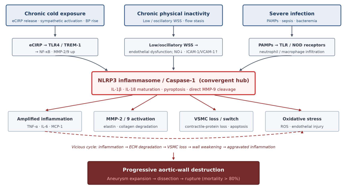

Table 1.
How the three modifiable factors activate shared pathogenic pathways in the aortic wall.
| Shared pathway | Chronic cold | Physical inactivity | Severe infection |
| NF-κB inflammation | eCIRP→TLR4→MyD88→NF-κB 24,27 | Low/oscillatory WSS→endothelial NF-κB 37 | PAMPs→TLR/NOD→NF-κB 8,11 |
| NLRP3 inflammasome | ROS + DAMP (eCIRP), indirect 32 | oxLDL/cholesterol crystals + mechanical stress 21 | PAMPs + bacterial DNA + ATP, direct 13 |
| MMP-2/9 up-regulation | CIRP induces endothelial MMP-2, macrophage MMP-9 27,29 | Low WSS→VSMC switch→MMP secretion 16,38 | Leukocyte degranulation + caspase-1 activation 8,13 |
| Oxidative stress | NADPH oxidase + antioxidant deficit 32 | eNOS uncoupling + ROS 37 | Neutrophil burst + mitochondrial ROS 8 |
| VSMC dysfunction | Sympathetic/Ang II→switch, apoptosis 2,30 | Altered interstitial flow→loss of contractile proteins 43 | IL-1β/TNF-α→caspase-1 degrades contractile proteins 20 |
Table 2.
Candidate interventions for aortic aneurysm and their stage of development.
| Strategy | Representative agent | Mechanism | Stage |
| NLRP3 inhibitor | MCC950 (CRID3) | Blocks inflammasome assembly, caspase-1 and MMP-9 cleavage 13 | Preclinical (mouse AAA) |
| NLRP3 inhibitor | Colchicine | Microtubule inhibition of NLRP3; lowers IL-1β/MMP-9 | CV outcome trials (COLCOT, LoDoCo2); AAA RCT lacking |
| NLRP3 inhibitor | Glyburide | Approved sulphonylurea that inhibits NLRP3; reduces Ang II-induced AAA/dissection 20 | Preclinical; repurposing candidate |
| IL-1β target | Canakinumab | Anti-IL-1β antibody; lowers vascular inflammation/events in CANTOS 21 | Phase III (CV); AAA untested |
| IL-1β target | Anakinra | IL-1 receptor antagonist; limits experimental TAA/AAA 49 | Preclinical; approved (RA) |
| CIRP target | Anti-CIRP antibody | Blocks eCIRP–TLR4/TREM-1; limits AAA and MMP-2/9 29 | Preclinical (rat AAA) |
| CIRP target | M3 peptide | Disrupts eCIRP–TREM-1; reduces systemic inflammation 26 | Preclinical (ischaemia–reperfusion) |
| Non-pharmacological | Aerobic exercise | Raises WSS, improves endothelial function, lowers hs-CRP and lipids 7,38,79 | Epidemiological evidence; RCT needed |
| Non-pharmacological | Thermoprotection + vaccination | Reduces cold exposure; influenza/pneumococcal vaccination limits inflammatory surges 4 | Recommended; low cost |
Disclaimer/Publisher’s Note: The statements, opinions and data contained in all publications are solely those of the individual author(s) and contributor(s) and not of MDPI and/or the editor(s). MDPI and/or the editor(s) disclaim responsibility for any injury to people or property resulting from any ideas, methods, instructions or products referred to in the content. |
© 2026 by the author. Licensee MDPI, Basel, Switzerland. This article is an open access article distributed under the terms and conditions of the Creative Commons Attribution (CC BY) license.
Copyright: This open access article is published under a Creative Commons CC BY 4.0 license, which permit the free download, distribution, and reuse, provided that the author and preprint are cited in any reuse.