
Submitted:
22 September 2026
Posted:
22 September 2026
You are already at the latest version
Abstract
Background/Objectives: Cryptorchidism occurs in numerous genetic developmental disorders, yet these associations are generally considered syndrome by syndrome, and the biological basis for their recurrence across molecularly heterogeneous conditions remains poorly understood. We aimed to characterize the clinical and molecular findings and develop a hypothesis-generating framework of developmental convergence. Methods: We retrospectively evaluated children with genetically confirmed rare developmental disorders who underwent surgical treatment for cryptorchidism at a tertiary pediatric urology center between 2015 and 2025. Clinical, operative, and molecular data were analyzed at patient and testis levels, and disorder-specific associations with cryptorchidism were evaluated through targeted literature review. Results: Among 94 children screened, 26 met the inclusion criteria, contributing 41 undescended testes. Cryptorchidism was bilateral in 57.7% of patients, 51.2% of testes were nonpalpable, and 51.2% were located at a high inguinal or intra-abdominal level. Despite substantial molecular heterogeneity, the underlying abnormalities could be organized into five interconnected developmental domains involving gene-expression regulation, signal sensing and integration, neural information transmission, neuromuscular execution and fetal mechanobiology, and mesenchymal and structural implementation. Conclusions: We propose that genetically distinct developmental disorders may perturb different levels of the biological system required for testicular descent while converging on shared downstream morphogenetic processes. This developmental-convergence model is hypothesis-generating and provides a testable framework linking human genetics, developmental biology, and pediatric urology.
Keywords:
cryptorchidism
; testicular descent
; developmental convergence
; genotype–phenotype
; gubernaculum
; developmental biology
1. Introduction
Cryptorchidism is among the most common congenital anomalies of the male genitourinary system and is associated with impaired fertility and an increased risk of testicular malignancy later in life [1,2]. Although its anatomical presentation is readily recognized, its developmental etiology remains incompletely understood. Testicular descent is a complex, temporally coordinated morphogenetic process traditionally divided into transabdominal and inguinoscrotal phases [3,4]. Experimental and human studies have established important roles for insulin-like peptide 3 (INSL3) signaling through its receptor RXFP2, androgen-dependent mechanisms, the gubernaculum, and neural and local tissue interactions [3,4,5]. Nevertheless, pathogenic abnormalities involving these classical regulatory pathways explain only a minority of human cryptorchidism, suggesting that successful descent depends on a broader developmental architecture [4,6].
This broader perspective is biologically plausible because testicular descent requires more than the presence of appropriate endocrine signals. The developing gubernaculum and surrounding inguinal tissues must receive, integrate, and translate hormonal and neural information into coordinated cellular proliferation, extracellular-matrix remodeling, tissue differentiation, and migration [4,5,7]. These processes, in turn, depend on upstream regulation of developmental gene expression, morphogenetic signal integration, neural competence, neuromuscular function, and mesenchymal organization. Disruption at different levels of this developmental hierarchy could therefore potentially lead to a common anatomical endpoint even when the primary molecular abnormalities are unrelated.
Rare developmental disorders provide a unique opportunity to examine this possibility. Cryptorchidism is a recognized component of several genetic syndromes, including disorders affecting chromatin and transcriptional regulation, while genital abnormalities have also been described in ciliopathies, neuromuscular syndromes, neurodevelopmental disorders, and chromosomal abnormalities [8,9,10]. However, these associations have largely been described syndrome by syndrome, with cryptorchidism treated as one feature within a broader phenotype. Consequently, little attention has been directed toward whether genetically heterogeneous disorders associated with testicular maldescent might converge on shared developmental processes. Moreover, in many recently characterized developmental disorders, male genital phenotypes remain incompletely documented, making it difficult to distinguish established associations from under-recognized manifestations or coincidental findings.
We therefore evaluated the clinical, operative, and molecular characteristics of children with genetically confirmed rare developmental disorders undergoing surgical treatment for cryptorchidism. In addition to defining the testicular phenotype and examining existing evidence for disorder-specific associations, we classified the underlying molecular abnormalities according to the predominant developmental processes they affect. We hypothesized that genetically heterogeneous disorders associated with cryptorchidism could be organized into a limited number of interconnected developmental layers that ultimately converge on the morphogenetic processes required for normal testicular descent. By integrating detailed pediatric urological phenotyping with human genetics and established developmental biology, we aimed to develop a hypothesis-generating framework of developmental convergence that could provide a basis for future clinical and mechanistic investigation.
2. Materials and Methods
2.1. Study Design
This retrospective observational cohort study evaluated children with genetically confirmed rare developmental disorders who underwent surgical treatment for cryptorchidism at a tertiary referral pediatric urology center between January 2015 and December 2025. The primary objective was to characterize the clinical phenotype of cryptorchidism across genetically heterogeneous developmental disorders and to explore potential biological mechanisms through a developmental systems framework. The study was conducted in accordance with the principles of the Declaration of Helsinki and was approved by the University of Health Sciences Erzurum Medical Faculty Ethical Committee (approval no. E-37732058-514.99; date of approval: 05-09-2022). The requirement for informed consent was waived by the Ethics Committee because of the retrospective nature of the study.
2.2. Patient Selection
Children referred to or assessed at our pediatric urology center for possible cryptorchidism during the study period were retrospectively screened for eligibility. Patients were included if they fulfilled all of the following criteria: (1) genetically confirmed diagnosis of a rare developmental disorder established by chromosomal microarray analysis, whole-exome sequencing, targeted molecular testing, or conventional cytogenetic analysis, according to the underlying disorder; (2) surgically confirmed undescended testis; (3) complete clinical, operative, and genetic records available for review. Patients with retractile testes, acquired cryptorchidism, or incomplete clinical or genetic documentation were excluded. The analysis was restricted to selected developmental disorders; Prader–Willi, Noonan, and Klinefelter syndromes were excluded from its diagnostic scope. The final study cohort comprised 26 children with 41 undescended testes. Patient selection is summarized in Figure 1.
2.3. Clinical Evaluation
Clinical data were extracted from electronic medical records and operative reports. Recorded variables included age at surgery, laterality, preoperative clinical palpability, anatomical position of the undescended testis, operative findings, and definitive surgical procedure. For bilateral cryptorchidism, each testis was evaluated independently for testis-level analyses, whereas patient-level analyses considered each child as a single observational unit.
Testicular location was classified intraoperatively as mid-inguinal, high inguinal, or intra-abdominal according to operative findings. Surgical management was categorized as conventional inguinal orchiopexy, laparoscopic Fowler–Stephens orchiopexy (single-stage), or laparoscopic Fowler–Stephens orchiopexy (two-stage), depending on intraoperative anatomy and vascular characteristics.
2.4. Genetic Evaluation
Genetic diagnoses were established using clinically indicated molecular investigations performed as part of routine patient care. Depending on the underlying disorder, diagnosis was confirmed by chromosomal microarray analysis, whole-exome sequencing, targeted gene sequencing, or conventional karyotype analysis. Copy-number variants were interpreted according to genomic coordinates based on the GRCh38 reference genome where available. Molecular diagnoses were reviewed together with the corresponding clinical phenotype to ensure diagnostic consistency prior to inclusion in the study.
2.5. Developmental Framework Classification
Following completion of clinical and genetic data collection, each disorder was assigned to a predominant developmental layer according to the principal biological function of the causative gene or genomic abnormality. Classification was informed by a targeted review of the developmental biology and human genetics literature and organized into five hierarchical developmental layers: (1) regulation of developmental gene expression, (2) developmental signal sensing and integration, (3) neural specification and information transmission, (4) neuromuscular execution and fetal mechanobiology, and (5) mesenchymal patterning and structural implementation. Chromosomal copy-number abnormalities were additionally considered cross-cutting genomic perturbations capable of affecting multiple developmental layers simultaneously. This classification was developed to facilitate biological interpretation of genotype–phenotype relationships rather than to establish causal molecular mechanisms.
2.6. Literature Review
A targeted literature review was performed to identify previously reported associations between each disorder and cryptorchidism and to characterize established developmental pathways involved in normal testicular descent. The literature review was used to construct the genotype–phenotype evidence maps and to support biological interpretation of the findings within the proposed developmental framework.
2.7. Statistical Analysis
Descriptive statistics were used to summarize demographic, clinical, operative, and genetic characteristics. Continuous variables are presented as median (range), whereas categorical variables are presented as frequencies and percentages. Patient-level and testis-level analyses were performed separately according to the predefined analytical framework.
3. Results
3.1. Study Cohort
Between January 2015 and December 2025, 94 children referred for or assessed for possible cryptorchidism were screened for eligibility. Of these, 68 were excluded: 22 had retractile testes, 9 had acquired cryptorchidism, 18 had incomplete clinical or genetic documentation, and 19 had diagnoses outside the selected study scope (Prader–Willi syndrome, n = 8; Noonan syndrome, n = 6; Klinefelter syndrome, n = 5). The final analysis included 26 children with genetically confirmed rare developmental disorders, contributing 41 undescended testes (Figure 1).
The median age at surgery was 2 years (range, 1–5 years). Bilateral cryptorchidism was present in 15 patients (57.7%), whereas 11 (42.3%) had unilateral disease, including 7 right-sided (26.9%) and 4 left-sided (15.4%) cases. Diagnostic laparoscopy was the initial operative approach in 13 patients (50.0%), while the remaining 13 (50.0%) underwent primary inguinal exploration. Definitive surgical management consisted of conventional inguinal orchiopexy in 14 patients (53.8%), laparoscopic Fowler–Stephens orchiopexy (single-stage) in 9 (34.6%), and laparoscopic Fowler–Stephens orchiopexy (two-stage) in 3 (11.5%).
Two-stage Fowler–Stephens orchiopexy was performed in three patients with bilateral intra-abdominal testes. Single-stage Fowler–Stephens orchiopexy was performed in two patients with unilateral intra-abdominal testes, one with a unilateral high-inguinal testis, and six with bilateral high-inguinal testes. These procedures involved 12 patients and 21 testes: eight intra-abdominal and 13 high-inguinal testes.

Table 1.
Baseline demographic and clinical characteristics of the study cohort. Percentages are calculated per patient (n = 26). Age refers to the age at definitive surgical intervention. Initial surgical approach describes the first operative access (inguinal exploration or laparoscopy), whereas definitive operative procedure refers to the final orchiopexy technique performed. Surgical technique counts represent patients, not individual testes.
Table 1.
Baseline demographic and clinical characteristics of the study cohort. Percentages are calculated per patient (n = 26). Age refers to the age at definitive surgical intervention. Initial surgical approach describes the first operative access (inguinal exploration or laparoscopy), whereas definitive operative procedure refers to the final orchiopexy technique performed. Surgical technique counts represent patients, not individual testes.
| Characteristic | Value |
| Number of patients, n | 26 |
| Number of undescended testes, n | 41 |
| Age at surgery (years) | Median 2 (range 1–5) |
| Laterality, n (%) | |
| • Bilateral cryptorchidism | 15 (57.7%) |
| • Right unilateral cryptorchidism | 7 (26.9%) |
| • Left unilateral cryptorchidism | 4 (15.4%) |
| Initial surgical approach, n (%) | |
| • Primary inguinal exploration | 13 (50.0%) |
| • Initial laparoscopy | 13 (50.0%) |
| Definitive orchiopexy technique, n (%) | |
| • Inguinal orchiopexy | 14 (53.8%) |
| • Laparoscopic Fowler–Stephens orchiopexy (single-stage) | 9 (34.6%) |
| • Laparoscopic Fowler–Stephens orchiopexy (two-stage) | 3 (11.5%) |
3.2. Clinical Phenotype of the Undescended Testes
Analysis at the testis level included 41 undescended testes, comprising 22 right (53.7%) and 19 left (46.3%) testes. Clinical examination demonstrated that 20 testes (48.8%) were palpable, whereas 21 (51.2%) were nonpalpable.
Operative findings demonstrated a predominantly proximal anatomical distribution. Twenty testes (48.8%) were located at the mid-inguinal level, 13 (31.7%) at the high inguinal level, and 8 (19.5%) were intra-abdominal. Consequently, 21 of 41 testes (51.2%) were situated at either a high inguinal or intra-abdominal position.
In all 15 patients with bilateral cryptorchidism, both testes were located within the same anatomical category: intra-abdominal in 3 patients, high inguinal in 6, and mid-inguinal in 6.
Table 2.
Testis-level clinical and anatomical characteristics of the undescended testes. Percentages are calculated per testis (n = 41). In bilateral cases, each testis was analyzed independently. Anatomical position refers to the intraoperative location documented during surgical exploration.
Table 2.
Testis-level clinical and anatomical characteristics of the undescended testes. Percentages are calculated per testis (n = 41). In bilateral cases, each testis was analyzed independently. Anatomical position refers to the intraoperative location documented during surgical exploration.
| Characteristic | Value |
| Side, n (%) | |
| • Right | 22 (53.7%) |
| • Left | 19 (46.3%) |
| Clinical palpability, n (%) | |
| • Palpable | 20 (48.8%) |
| • Nonpalpable | 21 (51.2%) |
| Intraoperative anatomical position, n (%) | |
| • Mid-inguinal | 20 (48.8%) |
| • High inguinal | 13 (31.7%) |
| • Intra-abdominal | 8 (19.5%) |
| Characteristic | Value |
| Side, n (%) |
3.3. Genotype–Phenotype Characteristics
The cohort comprised children with a broad spectrum of genetically confirmed developmental disorders, including single-gene disorders, chromatin-remodeling disorders, ciliopathies, neuromuscular disorders, neurodevelopmental syndromes, and chromosomal copy-number abnormalities. Disorders with previously recognized associations with cryptorchidism, including Rubinstein–Taybi syndrome, KAT6B-related disorders, Escobar syndrome, and FG syndrome, were represented alongside several disorders in which cryptorchidism has been only occasionally reported or has not been systematically characterized.
Chromosomal abnormalities included Cri du Chat syndrome associated with a terminal 5p15.33–p15.1 deletion, 16p13.11p12.3 duplication syndrome, and a large 5q duplication, further expanding the molecular diversity of the cohort.
The distribution of disorders according to previously reported evidence for cryptorchidism and their genotype–phenotype characteristics is summarized in Table 3.
3.4. Developmental Framework Classification
Following developmental classification, the included disorders were distributed across five principal hierarchical developmental layers according to the predominant biological function of the causative gene or genomic abnormality. The largest category consisted of disorders affecting regulation of developmental gene expression, including chromatin modification, chromatin remodeling, transcriptional regulation, and post-transcriptional control. Additional categories included developmental signal sensing and integration, neural specification and information transmission, neuromuscular execution and fetal mechanobiology, and mesenchymal patterning and structural implementation. Chromosomal copy-number abnormalities were classified separately as cross-cutting genomic dosage perturbations capable of influencing multiple developmental layers simultaneously.
Despite substantial molecular heterogeneity, the cohort demonstrated a recurring clinical pattern characterized by frequent bilateral involvement, proximal anatomical localization, and a slight predominance of nonpalpable testes. The developmental classification of the disorders included in the present cohort is presented in Table 4.
4. Discussion
4.1. Principal Findings and Clinical Phenotype
The present study demonstrates that genetically heterogeneous developmental disorders may be accompanied by a recurring cryptorchid phenotype despite substantial differences in their underlying molecular etiologies. In this cohort of 26 children with 41 undescended testes, bilateral involvement predominated, affecting 57.7% of patients, while 51.2% of testes were nonpalpable. Anatomically, 31.7% of testes were located at the high inguinal level and 19.5% were intra-abdominal; thus, more than half of all undescended testes were situated at a high inguinal or intra-abdominal position. An additional notable observation was anatomical concordance in all 15 bilateral cases, with both testes occupying the same recorded anatomical category. Taken together, these findings define a clinical and operative phenotype characterized by frequent bilateral involvement and relatively proximal testicular localization across an otherwise highly heterogeneous group of developmental disorders.
This pattern is relevant because normal testicular descent is itself a multistep developmental process rather than a simple anatomical displacement of the gonad. The transabdominal and inguinoscrotal phases require coordinated hormonal, neural, and tissue-specific responses. Experimental models have established an essential role for INSL3 signaling through RXFP2 in gubernacular development during the transabdominal phase, whereas androgen-dependent mechanisms are particularly important during inguinoscrotal descent. The latter involves interactions among the genitofemoral nerve, calcitonin gene-related peptide (CGRP), and the developing gubernaculum, although the precise contribution of neural mechanisms in humans remains less completely defined than in experimental models [3,4,5]. Importantly, pathogenic variants directly involving INSL3, RXFP2, the androgen receptor, or other established components of these pathways account for only a minority of human cryptorchidism, supporting a considerably broader and probably multifactorial developmental architecture [4,6].
The gubernaculum provides an important anatomical interface between these regulatory systems and the final position of the testis. Rather than functioning as a passive ligament, it undergoes temporally regulated growth, extracellular-matrix remodeling, changes in cellular composition, and migration during fetal development. Neural and endocrine signals must therefore be translated into coordinated cellular and structural responses within the gubernaculum and surrounding inguinal tissues. Experimental evidence supports involvement of the genitofemoral nerve and CGRP in this process, particularly in rodents, but more recent analyses emphasize important interspecies differences; the human gubernaculum contains abundant extracellular matrix and relatively less musculature, making direct extrapolation of a simple CGRP-mediated traction mechanism inappropriate [4,30]. This distinction is particularly relevant to the present study because it suggests that abnormal descent may result not only from failure of a single hormonal signal but also from disruption of the tissues and developmental programs required to receive and execute that signal.
Against this biological background, the molecular composition of our cohort is striking. The included disorders affect fundamentally different processes, including chromatin modification and remodeling, transcriptional and post-transcriptional regulation, ciliary signal integration, neuronal specification and trafficking, synaptic transmission, fetal neuromuscular function, mesenchymal patterning, extracellular-matrix biology, and chromosomal gene dosage. Nevertheless, these molecularly diverse conditions converged on a comparatively restricted anatomical outcome. This observation differs conceptually from the conventional gene-to-phenotype model in which cryptorchidism is considered independently within each syndrome. Instead, our findings raise the possibility that different upstream genetic disturbances may enter the developmental system at different levels while ultimately interfering with a smaller number of downstream processes required for successful testicular descent.
The predominance of bilateral disease provides additional, although indirect, support for considering a systemic developmental contribution. Bilateral cryptorchidism is particularly relevant in disorders capable of influencing endocrine or developmental processes affecting both sides simultaneously. In the present series, the additional observation that both testes occupied the same anatomical category in all bilateral cases is intriguing because it is compatible with a shared developmental disturbance acting on both sides during a similar developmental interval. However, anatomical concordance alone cannot identify the responsible mechanism, and this observation should not be interpreted as evidence of a specific molecular pathway. Rather, it provides a clinical phenotype against which the biological plausibility of the underlying disorders can be examined.
Accordingly, the principal contribution of this study is not the identification of a single novel cryptorchidism gene or pathway. It is the recognition that a broad spectrum of genetic abnormalities affecting distinct biological functions may be organized into a limited number of interconnected developmental processes relevant to normal testicular descent. We therefore interpret the observed phenotype as a potential example of developmental convergence: molecularly different upstream perturbations may ultimately reach the same anatomical endpoint through disruption of shared downstream morphogenetic requirements. This concept remains hypothesis-generating and does not establish causality for any individual disorder. The following sections examine the existing syndrome-specific evidence and then consider whether contemporary developmental biology provides a coherent mechanistic framework for this convergence.
4.2. Current Evidence and Phenotypic Expansion Across Rare Developmental Disorders
The relationship between genetic developmental disorders and cryptorchidism is heterogeneous. In some conditions represented in our cohort, cryptorchidism is a recognized component of the clinical spectrum, whereas in others it has been reported only sporadically or is not currently considered a characteristic manifestation. This distinction is important because the present findings should not be interpreted as identifying cryptorchidism de novo across all of these disorders. Rather, the cohort brings together established and less well-characterized associations within a common clinical and developmental context (Table 3).
The strongest prior clinical associations in our cohort involve disorders in which genital abnormalities form part of a broader congenital phenotype. Rubinstein–Taybi syndrome, caused most commonly by pathogenic variants in CREBBP, is a multisystem developmental disorder in which cryptorchidism has long been recognized among the abnormalities affecting males [11,12]. KAT6B-related disorders provide an even more informative example. Genitopatellar syndrome and Say–Barber–Biesecker–Young–Simpson syndrome result from pathogenic KAT6B variants and combine neurodevelopmental, skeletal, patellar, craniofacial, and genital abnormalities; cryptorchidism and scrotal abnormalities are well-recognized components of the male phenotype [13,14]. Similarly, genital anomalies including cryptorchidism occur within MED12-related FG/Opitz–Kaveggia syndrome [18]. These disorders therefore provide established clinical precedents demonstrating that perturbation of broadly acting developmental regulators can include abnormal testicular descent within a multisystem congenital phenotype.
Coffin–Siris syndrome provides a related but molecularly distinct example. The SMARCB1-related disorder represented in the present cohort belongs to the spectrum of BAF/SWI–SNF chromatin-remodeling disorders. Coffin–Siris syndromes are characterized by developmental impairment together with variable congenital abnormalities involving multiple organ systems, including the genitourinary tract. Importantly, SMARCB1, CREBBP, KAT6B, and ANKRD11 should not be regarded simply as unrelated “syndrome genes.” Contemporary human genetics increasingly recognizes substantial phenotypic overlap among disorders of chromatin and transcriptional regulation, reflecting disruption of interacting molecular processes governing developmental gene expression [8,9]. Rubinstein–Taybi, Coffin–Siris, KBG, and related chromatinopathies have consequently been discussed together as developmental disorders of transcriptional or chromatin regulation [8,9]. More recent molecular work continues to demonstrate substantial clinical overlap among these chromatinopathies, consistent with convergence at the level of developmental regulation rather than individual organ-specific pathways.
The situation is less clearly defined for KBG syndrome. ANKRD11 regulates chromatin-associated transcriptional processes and histone acetylation, and KBG syndrome is characterized predominantly by neurodevelopmental, craniofacial, dental, and skeletal abnormalities. Genitourinary findings, including cryptorchidism, have been reported in KBG syndrome, although they are not among its defining manifestations [16]. The presence of cryptorchidism in two patients with KBG syndrome in the present cohort therefore adds to an existing but variably expressed association. These observations do not establish increased penetrance but support systematic documentation of testicular position in larger ANKRD11 cohorts.
An even greater degree of caution is required for disorders such as CNOT3-related neurodevelopmental disorder, EBF3-related disorder, AP-4 deficiency syndrome, STXBP1-related developmental and epileptic encephalopathy, PPM1D-related Jansen–de Vries syndrome, and PLP1-related Pelizaeus–Merzbacher disease. EBF3-related disorder occupies an intermediate position: cryptorchidism has been documented in several genetically confirmed patients but remains incompletely characterized within the broader HADDS phenotype [21,22]. By contrast, for CNOT3-related disorder, AP-4 deficiency syndrome, STXBP1-related developmental and epileptic encephalopathy, and PLP1-related Pelizaeus–Merzbacher disease, cryptorchidism is not currently established or has not been systematically evaluated in available clinical series. Cryptorchidism has also been reported in PPM1D-related Jansen–de Vries syndrome, although its frequency and developmental mechanism remain incompletely characterized [23]. Their presence in the current cohort is therefore hypothesis-generating. In particular, the observation of cryptorchidism should not be taken to imply that the corresponding gene directly participates in testicular descent. It instead identifies these disorders as candidates for future genotype–phenotype studies in which male genital findings are recorded prospectively and systematically.
The neuromuscular disorders illustrate another level of evidence. CHRNG-related Escobar syndrome provides the strongest association within the neuromuscular group, with cryptorchidism recognized within the multiple-pterygium phenotype [10,26]. Cryptorchidism has also been reported in severe RYR1-associated fetal-akinesia and lethal multiple-pterygium phenotypes [24,25], although it is not established across the broader RYR1-related congenital-myopathy spectrum. By contrast, cryptorchidism is not currently established as part of IGHMBP2-related SMARD1. These disorders therefore differ substantially in evidential strength despite sharing a broad neuromuscular classification.
TMEM67-related Joubert syndrome similarly requires distinction between the molecular disorder and the broader phenotypic spectrum. TMEM67 encodes a ciliary transition-zone protein and can cause phenotypes ranging from Joubert syndrome to more severe Meckel–Gruber-spectrum ciliopathies. Genital abnormalities have been described within severe ciliopathy phenotypes, but cryptorchidism is not a uniform feature of TMEM67-related Joubert syndrome [19,20]. The present observation therefore extends the clinical context in which abnormal testicular descent may occur without establishing a TMEM67-specific mechanism. This distinction is particularly relevant because ciliary dysfunction can perturb multiple developmental signaling pathways and organ systems simultaneously.
Chromosomal disorders add further complexity because their phenotypes cannot generally be attributed to a single affected gene. The patient with Cri du Chat syndrome carried a 16.39-Mb terminal 5p15.33–p15.1 deletion, and male genital abnormalities, including cryptorchidism, have previously been described within the 5p deletion spectrum [28]. Recurrent 16p13.11p12.3 copy-number changes demonstrate marked variable expressivity and incomplete penetrance, and cryptorchidism has recently been reported among the urogenital manifestations of this CNV spectrum [29]. In such disorders, altered dosage of multiple genes may influence several developmental systems simultaneously, and assigning cryptorchidism to one candidate gene would be premature. Copy-number abnormalities are therefore particularly compatible with a multimodular developmental interpretation but provide limited evidence for any single molecular pathway.
Taken together, the literature comparison reveals three distinct evidential groups within the present cohort: disorders with an established association with cryptorchidism, disorders in which cryptorchidism has been reported but remains incompletely characterized, and disorders for which the present observations may represent potential phenotype expansion. This spectrum is itself informative. It suggests that cryptorchidism occurs not only in disorders already recognized to involve genital development but also across molecular conditions whose primary phenotypes involve chromatin regulation, neurodevelopment, ciliary biology, neuromuscular function, or broader tissue morphogenesis. The next question is therefore not whether all of these genes independently “cause” cryptorchidism, but whether their diverse molecular effects intersect with developmental processes already known to be required for normal testicular descent.
4.3. Developmental Biology of Normal Testicular Descent
Interpretation of cryptorchidism across genetically heterogeneous disorders requires consideration of testicular descent as an integrated morphogenetic process rather than a simple movement of the gonad from the abdomen to the scrotum. Classical descriptions divide descent into transabdominal and inguinoscrotal phases, but contemporary experimental evidence indicates substantial biological interaction between the endocrine, neural, mesenchymal, and mechanical processes operating across these stages (Figure 2). The gubernaculum lies at the center of this system: it is a transient fetal organ whose growth, extracellular-matrix composition, cellular differentiation, and spatial remodeling determine the relationship between the testis, abdominal wall, inguinal canal, and developing scrotum [3,4].
The transabdominal phase is most strongly associated with signaling through insulin-like peptide 3 (INSL3) and its receptor RXFP2. INSL3 is produced by differentiated fetal Leydig cells and acts on RXFP2-expressing cells within the gubernaculum. Experimental inactivation of either Insl3 or Rxfp2 in mice produces bilateral intra-abdominal cryptorchidism associated with failure of normal gubernacular development, providing some of the strongest causal evidence for a molecular pathway regulating testicular descent [5]. Conversely, INSL3 overexpression can promote gubernacular development and abnormal gonadal displacement in experimental models. Human fetal studies demonstrate circulating INSL3 during the developmental period in which gubernacular enlargement and testicular descent occur, supporting conservation of this endocrine system in human development, although pathogenic INSL3/RXFP2 variants account for only a small proportion of human cryptorchidism [3,4,6].
The biological effect of INSL3 is broader than a simple change in gubernacular size. RXFP2 activation influences cellular proliferation and extracellular-matrix organization and interacts with developmental signaling networks involving WNT, BMP, β-catenin, Notch, cytoskeletal, and neural pathways. Thus, even one of the best-established hormonal mechanisms of testicular descent ultimately depends on the capacity of mesenchymal cells to receive an extracellular signal and translate it into coordinated changes in proliferation, differentiation, matrix composition, and tissue architecture. This provides an important developmental principle: an intact endocrine signal is insufficient if the target tissue is unable to interpret or execute that signal appropriately [4,5].
The inguinoscrotal phase is particularly androgen dependent. Clinical observations in disorders of androgen action and experimental androgen-receptor models strongly support this requirement, although the precise cellular targets through which androgens regulate descent remain incompletely resolved. Proposed mechanisms include direct androgen-receptor signaling within gubernacular or cremasteric tissues and indirect actions involving androgen-sensitive structures surrounding the gubernaculum. Studies in boys with nonsyndromic cryptorchidism have identified altered androgen-receptor and myosin-heavy-chain expression in cremaster muscle, while experimental models demonstrate regional differences in androgen-receptor expression, extracellular-matrix composition, and biomechanical properties within the developing gubernaculum [1,30]. These findings reinforce the view that androgen action during descent is mediated through a developing tissue system rather than through a single isolated molecular target.
Neural signaling adds another layer of regulation. Experimental studies, predominantly in rodents, implicate the genitofemoral nerve and its sensory neuropeptide calcitonin gene-related peptide (CGRP) in gubernacular proliferation, motility, and inguinoscrotal migration. Androgen exposure appears to interact with this pathway, providing a potential neuroendocrine interface between hormonal signaling and local tissue behavior [4]. Nevertheless, extrapolation to human development requires caution. The human fetal gubernaculum differs structurally from its rodent counterpart and contains abundant extracellular matrix relative to musculature. Furthermore, direct human evidence for a dominant CGRP-driven mechanism remains limited, and pathogenic variants within the CGRP pathway have not emerged as a common explanation for human cryptorchidism. The genitofemoral nerve–CGRP axis is therefore best regarded as an experimentally supported component of a broader neuroendocrine system rather than a complete explanation for human inguinoscrotal descent [31].
The extracellular matrix is particularly important in bridging molecular signaling and anatomy. During fetal development, the gubernaculum undergoes substantial changes in glycosaminoglycans, collagen, extracellular-matrix volume, cellular composition, and mechanical properties. These changes determine not only its dimensions but also its stiffness, deformability, and interaction with surrounding tissues. Experimental studies demonstrate that endocrine signaling alters matrix composition and biomechanical properties within the gubernaculum, while structural studies of cryptorchid tissues show differences from normally developing fetal gubernacula. The extracellular matrix should therefore not be considered a passive scaffold; it forms part of the mechanism through which molecular signals are converted into morphogenetic behavior [30,31].
A similar principle applies to fetal neuromuscular function. Movement and mechanical force are fundamental components of embryonic and fetal morphogenesis throughout the musculoskeletal system. In testicular descent, however, the contribution of muscle should not be reduced to the historical concept of a structure physically “pulling” the testis into the scrotum. A more contemporary interpretation is that neuromuscular development contributes to the mechanical environment within which the abdominal wall, inguinal canal, cremasteric apparatus, gubernaculum, and surrounding mesenchyme mature. Changes in muscle differentiation, contractile competence, tissue loading, or fetal movement could therefore modify morphogenesis without directly altering INSL3 or androgen signaling. Evidence from cryptorchid animal models demonstrating regional variation in gubernacular biomechanical properties and from human studies showing altered cremasteric muscle gene expression supports the relevance of this broader mechanical environment, although a direct causal role for fetal movement in human testicular descent has not been established [1,30].
Normal testicular descent therefore appears to require successful completion of several interdependent biological tasks: appropriate developmental gene expression; reception and integration of extracellular signals; endocrine regulation; neural communication; neuromuscular competence; mesenchymal proliferation and differentiation; extracellular-matrix remodeling; and coordinated structural morphogenesis. Failure at one level may potentially be compensated by other components of the system, whereas broader or sufficiently severe disturbances may ultimately impair the common downstream process of gubernacular development and migration.
This integrated view provides the physiological foundation for interpreting the present cohort. Importantly, none of the established pathways described above predict that every child with cryptorchidism should carry a defect in INSL3, RXFP2, androgen signaling, or the genitofemoral nerve. Instead, they demonstrate that testicular descent depends on a network of interacting tissues and developmental processes. The molecular disorders represented in our cohort can therefore be considered not only according to whether their causative genes belong to a classical “cryptorchidism pathway,” but according to which level of this developmental system they are capable of perturbing.
4.4. Hierarchical Developmental Convergence: From Molecular Heterogeneity to A Shared Anatomical Phenotype
The molecular heterogeneity observed in the present cohort appears, at first sight, difficult to reconcile with a single clinical phenotype. The causative abnormalities range from defects of chromatin modification and transcriptional regulation to ciliopathy, neuronal dysfunction, impaired neuromuscular transmission, skeletal muscle disease, mesenchymal patterning abnormalities, and large chromosomal copy-number changes. These processes are too biologically diverse to support a conventional single-pathway explanation for cryptorchidism. However, when considered according to their position within the sequence of developmental events required to generate an anatomical structure, a different pattern emerges. We propose that these disorders may enter the developmental process at different hierarchical levels yet converge downstream on impaired gubernacular morphogenesis and testicular descent (Table 4; Figure 3).
The most upstream level of this model is regulation of developmental gene expression. Several disorders in the cohort affect proteins that control chromatin accessibility, histone modification, transcriptional regulation, or transcript stability, including CREBBP, KAT6B, SMARCB1, ANKRD11, MED12, CNOT3, and PPM1D. These proteins do not constitute a testicular-descent pathway in the conventional sense. Rather, they regulate when, where, and to what extent numerous downstream developmental genes are expressed. This distinction is fundamental. Developmental regulators acting at the level of chromatin or transcription can influence multiple embryonic tissues simultaneously, and phenotypic overlap among Rubinstein–Taybi, Coffin–Siris, KBG, and related disorders has increasingly been interpreted as reflecting disruption of interacting transcriptional and chromatin-regulatory processes. Thus, cryptorchidism in these conditions may represent one component of a broader disturbance in developmental programming rather than failure of an isolated gonadal pathway.
This concept is particularly compelling for CREBBP, KAT6B, and SMARCB1, which affect different components of gene-expression control. CREBBP and KAT6B encode lysine acetyltransferases involved in chromatin regulation, whereas SMARCB1 is a core component of the ATP-dependent BAF/SWI–SNF chromatin-remodeling complex. Despite these distinct molecular functions, disorders involving these genes share developmental abnormalities affecting multiple organ systems. Contemporary classifications of developmental disorders increasingly recognize such chromatinopathies as biologically overlapping rather than entirely separate entities. In this context, the occurrence of cryptorchidism across several chromatin-regulatory disorders in our cohort suggests that correct testicular descent may be sensitive to disturbances in upstream developmental gene regulation even when the canonical INSL3/RXFP2 and androgen pathways themselves are intact.
A second level involves developmental signal sensing and integration. TMEM67-related Joubert syndrome provides the clearest example in the present cohort. TMEM67 encodes meckelin, a component of the ciliary transition zone. Primary cilia act as signaling compartments through which developing cells interpret extracellular information, including Hedgehog-related and other morphogenetic signals, while also contributing to cellular polarity and tissue organization [32,33,34]. A ciliary defect therefore differs conceptually from loss of a single morphogen: the signal may be present, but the cell's ability to receive, spatially interpret, or appropriately respond to developmental information may be impaired. TMEM67-related cryptorchidism, if mechanistically related, could therefore arise through disturbed integration of developmental signals across several tissues rather than through direct alteration of a recognized endocrine pathway controlling descent. This remains a hypothesis, but ciliopathies provide established examples elsewhere in embryology of how defective signal integration can produce multisystem structural malformations.
The third level is neural specification and information transmission. Several genes represented in the cohort affect different components of neuronal development or function: EBF3 participates in neuronal specification and transcriptional regulation; AP4S1 is involved in neuronal cargo trafficking; PLP1 is essential for myelin–axon integrity; and STXBP1/Munc18-1 is a core component of presynaptic vesicle-fusion machinery [21,22,35,36,37]. These disorders do not constitute a known cryptorchidism pathway. Their relevance emerges instead from the broader requirement for neural competence within the developmental system governing inguinoscrotal descent. The genitofemoral nerve–CGRP model provides experimental proof of principle that neural information can influence gubernacular behavior, although direct involvement of the genes represented in our cohort in this pathway has not been demonstrated. We therefore interpret these disorders as neural-competence candidates, rather than as evidence that their causative genes directly regulate the genitofemoral nerve or CGRP.
The fourth level is neuromuscular execution and fetal mechanobiology, represented most clearly by CHRNG, RYR1, and IGHMBP2 [38,39,40,41,42,43]. These genes affect three different points along the motor pathway: fetal neuromuscular transmission, skeletal-muscle excitation–contraction coupling, and motor-neuron maintenance, respectively. CHRNG is especially informative because the fetal acetylcholine-receptor γ-subunit has a temporally specific role during prenatal neuromuscular development, and pathogenic variants cause Escobar/multiple-pterygium syndrome through impaired fetal neuromuscular transmission and reduced movement [10,38,39]. RYR1 dysfunction can likewise impair skeletal-muscle excitation–contraction coupling and, in severe recessive phenotypes, produce fetal akinesia and multiple-pterygium syndromes [24,25,40]. IGHMBP2-related motor-neuron dysfunction represents a more exploratory example; a specific prenatal mechanism linking SMARD1 to testicular descent has not been established [41,42]. These disorders illustrate how different molecular defects can converge on reduced neuromuscular output. We do not propose that muscle contraction mechanically pulls the testis into the scrotum. Rather, impaired neuromuscular competence may alter the biomechanical environment in which the abdominal wall, inguinal canal, cremasteric apparatus, gubernaculum, and surrounding mesenchyme develop. CHRNG provides the strongest clinical support for this level because cryptorchidism is already recognized within the multiple-pterygium phenotype; RYR1 and IGHMBP2 remain progressively more exploratory examples.
The fifth level is mesenchymal patterning and structural implementation. Normal developmental signals can generate an anatomical outcome only if responding tissues possess the proliferative, migratory, extracellular-matrix, and mechanical competence required to execute them. FOXC1, RECQL4, and the 7q11.23 deletion underlying Williams syndrome affect distinct aspects of tissue patterning and structural competence. FOXC1 is required for normal development and differentiation of neural-crest-derived mesenchymal tissues [44], while RECQL4 is required for DNA replication, cellular proliferation, and normal osteoblast expansion [45], whereas ELN haploinsufficiency alters elastic-fiber and connective-tissue biology [46]. More broadly, mesenchymal cells and extracellular-matrix networks provide both structural support and developmental signals that influence tissue morphogenesis [47,48]. None of these abnormalities has been shown to directly disrupt the human gubernaculum. Their relevance is instead conceptual: they illustrate molecular routes through which tissues responsible for implementing developmental instructions may become structurally or morphogenetically incompetent.
Chromosomal copy-number abnormalities require a different interpretation and are therefore positioned across rather than within the hierarchy. The 5p deletion causing Cri du Chat syndrome, the 16p13.11p12.3 duplication, and the large 5q duplication alter the dosage of multiple genes simultaneously. Such variants may affect transcriptional regulation, neural development, cellular proliferation, cytoskeletal function, extracellular-matrix biology, or other developmental processes in parallel. Their phenotypes are also characterized by variable expressivity and, for some recurrent copy-number variants, incomplete penetrance. Assigning cryptorchidism to a single gene within these intervals would therefore be speculative. In the proposed framework, chromosomal dosage abnormalities are better considered multimodular perturbations capable of lowering developmental robustness at several levels simultaneously.
The hierarchical organization proposed here should not be interpreted as a rigid linear pathway. Development is characterized by extensive feedback, redundancy, crosstalk, and reciprocal signaling between tissues. Chromatin regulation influences ciliary, neural, muscular, and mesenchymal gene expression; ciliary signaling influences cell polarity and tissue morphogenesis; neural activity interacts with muscle and target tissues; and extracellular-matrix properties can themselves modify cellular signaling. Individual genes may therefore act across more than one level. The value of the hierarchy is not to assign each disorder permanently to one box, but to identify the predominant level at which its molecular defect enters the developmental system.
This distinction also introduces the concept of developmental robustness. Embryonic systems are capable of buffering many genetic and environmental perturbations through redundant pathways and compensatory interactions. A molecular abnormality therefore does not inevitably produce cryptorchidism, even when it affects a biologically relevant developmental process. This may help explain the variable penetrance of cryptorchidism within many of the syndromes represented in our cohort. A defect may become clinically manifest only when its magnitude, developmental timing, tissue distribution, or interaction with additional genetic or environmental modifiers exceeds the buffering capacity of the system. Such a threshold model is particularly attractive for copy-number variants and pleiotropic developmental disorders, although it cannot be directly tested in the present study.
A developmental-convergence perspective is increasingly applied to genetically heterogeneous congenital disorders, in which distinct upstream molecular abnormalities may ultimately perturb shared morphogenetic processes [49,50]. Rather than assuming a simple one-gene/one-malformation relationship, such approaches emphasize the developmental programs on which molecular abnormalities converge. A similar framework may be useful for cryptorchidism. The relevant endpoint is not necessarily disruption of one “cryptorchidism gene,” but failure to complete a morphogenetic program that depends on coordinated endocrine signaling, cellular competence, neural communication, mechanical execution, and tissue remodeling.
Accordingly, we propose a hierarchical developmental-convergence model of cryptorchidism (Figure 3). In this model, genetically heterogeneous disorders can perturb developmental gene regulation, signal integration, neural communication, neuromuscular execution, or structural implementation independently or in combination. These disturbances ultimately converge on the gubernaculum and its surrounding developmental environment, where successful integration of endocrine, neural, mesenchymal, and mechanical information is required for normal testicular descent. The model does not imply that each gene represented in the cohort is causally involved in cryptorchidism, nor does it establish equivalence between disorders with strong and weak syndrome-level associations. Rather, it provides a testable framework for organizing genetic heterogeneity and generating mechanistic hypotheses that can be evaluated in larger clinical cohorts and experimental models.
4.5. Clinical Implications of A Developmental-Convergence Perspective
The proposed developmental-convergence framework is primarily biological, but it also has potential clinical implications. Cryptorchidism is usually approached as an isolated congenital anomaly, particularly when unilateral and otherwise uncomplicated. This remains appropriate for the majority of affected boys. The present findings do not support routine genetic investigation of all children with undescended testes, nor do they modify established recommendations for the timing or surgical management of cryptorchidism. Rather, they suggest that the phenotypic context in which cryptorchidism occurs deserves greater attention, particularly when bilateral disease, nonpalpable testes, other congenital anomalies, neurodevelopmental abnormalities, dysmorphic features, skeletal abnormalities, or a known genetic disorder coexist.
Current clinical guidance already recognizes that some cryptorchid phenotypes require evaluation beyond the testis itself. Bilateral nonpalpable testes in a phenotypic male newborn require prompt assessment for disorders/differences of sex development, and the combination of cryptorchidism with severe proximal hypospadias similarly warrants endocrine and genetic consideration. Thus, the principle that testicular position may provide information about a broader developmental or endocrine disorder is already embedded within pediatric endocrine and urological practice [51].
Our cohort suggests that this principle may extend beyond classical disorders of sex development and endocrine abnormalities. More than half of the children had bilateral cryptorchidism, more than half of the testes were nonpalpable, and more than half were situated at high inguinal or intra-abdominal levels. In a child who also demonstrates developmental delay, dysmorphism, congenital skeletal abnormalities, neurological disease, multiple malformations, or another unexplained multisystem phenotype, such a presentation should encourage the clinician to consider whether cryptorchidism represents one component of a broader developmental disorder rather than an unrelated isolated finding. This does not imply that the cryptorchidism itself establishes an indication for genomic testing; instead, it may contribute to the cumulative phenotypic evidence supporting referral for clinical genetic assessment.
The converse is equally relevant. Children with established genetic or developmental syndromes may receive intensive neurological, cardiological, respiratory, nutritional, or orthopedic care, while genital examination receives comparatively less attention. For disorders in which cryptorchidism is not traditionally emphasized, an undescended testis may consequently be documented inconsistently or recognized late. Our findings support systematic documentation of testicular position as part of the phenotypic assessment of boys with rare developmental disorders. This is particularly important because timely recognition of cryptorchidism remains clinically relevant irrespective of its genetic background, and management should continue to follow established pediatric urological principles.
The developmental framework may also influence how genetic findings are interpreted after a molecular diagnosis has been established. In contemporary genomic practice, identification of a pathogenic variant is often followed by comparison of the patient's phenotype with the previously reported syndrome spectrum. When cryptorchidism is absent from standard descriptions, it may be dismissed as coincidental. Table 3 demonstrates why a more nuanced approach may be useful. A genital finding observed in a single patient should not automatically be attributed to the causative gene, but repeated observations across independent patients may eventually identify a previously under-recognized component of the phenotype. This is especially relevant for newly described neurodevelopmental disorders, for which published cohorts may be small and phenotyping may have focused primarily on neurological or dysmorphic features.
Accordingly, detailed phenotyping is as important as increasingly sophisticated sequencing. The value of a molecular diagnosis depends partly on the quality of the clinical information attached to it. For cryptorchidism, useful phenotypic descriptors extend beyond the binary presence or absence of an undescended testis and include laterality, palpability, anatomical position, associated genital abnormalities, age at spontaneous or surgical descent, and operative findings. Our observation that both testes occupied the same anatomical category in all bilateral cases illustrates the type of information that would be lost if cryptorchidism were recorded only as a syndromic checklist item. Standardized phenotyping across genetic cohorts could permit future analyses to determine whether particular molecular classes are associated not merely with cryptorchidism, but with specific patterns of testicular maldescent.
This has implications for collaboration between pediatric urology and clinical genetics. Pediatric urologists encounter the anatomical endpoint and have access to information that may not be available to the geneticist, including precise testicular location and operative anatomy. Geneticists, in turn, can identify molecular abnormalities that place an apparently isolated surgical finding within a broader developmental context. Prospective integration of these datasets may be particularly valuable for rare disorders, in which no single institution is likely to accumulate sufficient numbers to define the full phenotypic spectrum.
The framework may also provide a more disciplined way to interpret unusual genotype–phenotype observations. Rather than asking only whether a particular gene has previously been reported in association with cryptorchidism, clinicians and investigators can ask two sequential questions: first, is the association recurrent and clinically credible; and second, does the known biological function of the affected gene intersect with a developmental process required for normal testicular descent? Concordance between these two lines of evidence would strengthen biological plausibility, whereas biological plausibility without recurrent clinical observations should remain hypothesis-generating. This distinction is particularly important in rare-disease genomics, where coincidental findings can otherwise be overinterpreted.
Finally, a developmental-convergence perspective should not alter the fundamental surgical objective. Once cryptorchidism is diagnosed, the indication and timing of orchiopexy remain governed by established clinical principles rather than by the developmental category of the underlying disorder [51,52]. The potential value of the framework lies elsewhere: in recognizing when cryptorchidism may be a marker of broader developmental disturbance, improving phenotypic characterization of rare genetic disorders, facilitating communication between specialties, and identifying patient groups in whom future mechanistic investigation may be most informative.
Thus, the clinical translation of the present study is not a new genetic-testing algorithm for cryptorchidism. It is a proposal for more developmentally informed phenotyping: to view testicular maldescent within the context of the whole child, while preserving the distinction between established clinical recommendations, recurrent genotype–phenotype associations, and biologically plausible but as yet unproven mechanisms.
4.6. Strengths and Limitations
The principal strength of this study is the integration of detailed pediatric urological phenotyping with molecularly confirmed developmental disorders. Cryptorchidism is frequently recorded in genetic literature simply as present or absent, whereas the present cohort was characterized using clinically and surgically relevant variables including laterality, palpability, precise anatomical location, operative approach, and surgical procedure. Analysis at both the patient and individual-testis levels allowed bilateral cases to be examined without losing side-specific anatomical information. This level of phenotypic resolution is particularly valuable in rare disorders, in which genital findings may be incompletely characterized in syndrome-focused reports.
A second strength is the molecular diversity of the cohort. Rather than examining a single syndrome or gene, the study includes monogenic disorders affecting different biological processes together with chromosomal copy-number abnormalities. At first sight, such heterogeneity could be considered a limitation. However, for the hypothesis addressed here it also provides an opportunity: the recurrence of a relatively similar anatomical phenotype across molecularly distinct disorders permits exploration of shared developmental processes that would not be apparent in a single-syndrome series. Importantly, the proposed classification was based on the established predominant biological function of the affected gene or genomic abnormality rather than on the presence of cryptorchidism itself, reducing—but not eliminating—the risk of circular interpretation.
Several limitations nevertheless require emphasis. First, this is a retrospective, single-center study with a small sample size. Although 26 patients represent a clinically informative series in the context of individually rare developmental disorders, the cohort is insufficient to estimate syndrome-specific prevalence, penetrance, or relative risk of cryptorchidism. The number of patients with most individual molecular diagnoses was one or very small, and observations concerning potential phenotype expansion must therefore be considered preliminary. No inference should be made that cryptorchidism occurs more frequently in a particular disorder solely because that disorder is represented in this cohort.
Second, the cohort is subject to substantial ascertainment and referral bias. Patients were identified among children referred to or assessed in a pediatric urology service for possible cryptorchidism, with inclusion restricted to surgically confirmed cases, rather than through unselected cohorts of children with the corresponding genetic disorders. The diagnosis-specific exclusion of Prader–Willi, Noonan, and Klinefelter syndromes further limits the representativeness of the cohort and may influence its clinical and molecular distribution. The study therefore asks which developmental disorders were encountered among children with surgically treated cryptorchidism; it cannot determine how commonly cryptorchidism occurs among all children carrying those molecular diagnoses. This distinction is critical. Confirmation of a genotype–phenotype association requires the reverse study design: systematic examination of sufficiently large, independently ascertained cohorts with each genetic disorder to determine the frequency and pattern of testicular maldescent.
Third, there is no nonsyndromic cryptorchidism control group. Consequently, the relatively high frequencies of bilateral disease, nonpalpability, and proximal testicular location observed in the cohort cannot be formally attributed to the presence of a developmental disorder. Comparisons with published cryptorchidism populations may provide context but cannot substitute for a contemporaneous matched control group because referral patterns, age at surgery, definitions of anatomical location, and clinical practice vary among studies. The anatomical pattern identified here should therefore be considered descriptive rather than comparative.
Fourth, genetic evaluation was not uniform across the cohort. Molecular diagnoses were established according to clinical indication using different technologies, including chromosomal microarray analysis, whole-exome sequencing, targeted molecular testing, and cytogenetic methods. This reflects real-world clinical practice over the 2015–2025 study period but introduces differences in diagnostic resolution. Furthermore, the presence of a molecular diagnosis does not exclude additional genetic modifiers that were not detected by the test performed. This issue may be particularly relevant to variable phenotypes such as cryptorchidism, in which penetrance could be influenced by variants outside the primary disease-causing locus.
Fifth, the study was not designed to evaluate classical endocrine determinants of testicular descent. Systematic fetal or postnatal measurements of INSL3, testosterone, gonadotropins, anti-Müllerian hormone, or androgen responsiveness were not available, and endocrine assessment was not standardized retrospectively across disorders. We therefore cannot determine whether cryptorchidism in individual patients resulted from altered endocrine signaling, impaired tissue responsiveness, or mechanisms independent of measurable endocrine abnormalities. Similarly, direct histological, transcriptomic, or molecular analysis of gubernacular, cremasteric, or inguinal tissues was not performed. The proposed developmental links consequently remain biological interpretations rather than demonstrated patient-specific mechanisms.
Sixth, the hierarchical developmental classification necessarily simplifies complex gene functions. Pleiotropic developmental genes rarely operate within a single biological compartment. A chromatin regulator may influence neuronal, muscular, and mesenchymal differentiation simultaneously; ciliary proteins participate in several signaling pathways; and neural, muscular, and extracellular-matrix development interact reciprocally. Assignment to a predominant developmental layer was therefore intended as an analytical framework rather than a definitive ontology. Alternative classifications are possible, and future experimental evidence may require individual genes to be repositioned or assigned to multiple interacting modules.
The same caution applies to the concept of developmental convergence itself. The present study demonstrates phenotypic convergence—genetically heterogeneous disorders occurring with a shared anatomical abnormality—but it does not experimentally demonstrate mechanistic convergence on the gubernaculum or any specific downstream pathway. Figure 3 should therefore be interpreted as a hypothesis-generating model derived from the combination of our clinical observations and established developmental biology. Direct demonstration of mechanistic convergence would require functional studies showing that different genetic perturbations produce measurable abnormalities within shared cell populations, signaling networks, or morphogenetic processes involved in testicular descent.
Finally, the absence or rarity of published cryptorchidism in some disorders requires careful interpretation. Rare-disease phenotypes are influenced not only by biological penetrance but also by what clinicians choose to examine and report. Genital findings may be under-recorded in cohorts assembled primarily for neurological, developmental, or dysmorphology phenotyping. Conversely, because the present cohort was assembled in a pediatric urology setting, cryptorchidism is necessarily over-represented. Neither source of evidence alone can establish the true association. The most reliable future evidence will require prospective, standardized phenotyping across independently recruited genetic cohorts.
These limitations define the appropriate scope of the present study. The data support detailed description of the cryptorchid phenotype in this selected cohort and identify several potentially under-recognized genotype–phenotype associations. They also provide a clinical observation from which a biologically plausible developmental framework can be generated. They do not establish syndrome-specific prevalence, prove causality for individual genes, or demonstrate the molecular mechanism of cryptorchidism. We consider this distinction a strength rather than a weakness of the proposed model: by separating what is observed, what is already established, and what is hypothesized, the framework generates specific questions that can now be tested experimentally and clinically.
4.7. Future Directions: From Developmental Framework to Testable Mechanisms
The proposed developmental-convergence model generates several testable hypotheses and therefore provides a framework for future investigation rather than an endpoint. The first priority should be independent clinical validation. For disorders in which cryptorchidism is already recognized, larger genotype-defined cohorts could determine whether particular molecular subtypes are associated with bilateral disease, nonpalpability, or proximal testicular location. For disorders in which the association is uncertain or potentially novel, the appropriate strategy is reverse phenotyping: starting with independently recruited patients carrying the relevant molecular diagnosis and systematically documenting testicular position and operative anatomy. Multicenter collaboration will be essential because individual disorders are too rare for meaningful syndrome-specific estimates to be generated by most single institutions.
Such studies would benefit from standardized phenotyping. Recording cryptorchidism simply as present or absent discards potentially informative developmental information. Future genetic cohorts should ideally document laterality, palpability, anatomical level, associated genital abnormalities, spontaneous descent when applicable, age at surgery, operative findings, and the surgical procedure required. Where bilateral disease is present, each testis should be recorded independently. Incorporation of standardized Human Phenotype Ontology terminology could facilitate comparison between genomic datasets and permit subsequent aggregation across international rare-disease cohorts.
A second research direction concerns developmental timing and tissue specificity. A gene can be biologically relevant to testicular descent only if its perturbation can plausibly influence the appropriate tissue or interacting developmental system during the relevant fetal interval. Publicly available developmental transcriptomic datasets therefore provide an immediate opportunity to refine the model. Expression of candidate genes could be mapped across fetal testis, gubernacular mesenchyme, inguinal tissues, peripheral and sensory neurons, skeletal muscle, and relevant supporting cell populations. Developmental-stage-specific expression would be particularly informative because transient fetal expression may be more relevant than expression measured in postnatal or adult tissues.
Single-cell and spatial transcriptomic approaches could take this considerably further [53]. Normal testicular descent requires communication among cell populations rather than activity of one isolated cell type. Defining the cellular composition of the human fetal gubernaculum and adjacent inguinal region, and mapping receptors, transcriptional regulators, extracellular-matrix genes, neural markers, and mechanosensitive pathways within their spatial context, could identify the cellular interfaces through which apparently unrelated developmental disorders might converge. Such datasets could also determine whether genes currently classified within different modules are expressed within shared cell populations or influence common transcriptional states.
The third step is functional convergence analysis. If the model is correct, molecularly distinct perturbations need not produce identical upstream changes; they should instead generate overlapping downstream abnormalities in developmental processes required for descent. Patient-derived induced pluripotent stem cells, gene-edited cellular models, organoid systems, and appropriate animal models could be used to examine functional convergence [50]. Relevant outcomes could include impaired mesenchymal proliferation, altered extracellular-matrix composition, abnormal cellular migration, disrupted response to INSL3 or androgenic signaling, altered neural–mesenchymal communication, or changes in cellular mechanical properties. The critical experiment would therefore not simply ask whether a candidate gene “causes cryptorchidism,” but whether disruption of different genes produces a reproducible shared defect within a biologically relevant developmental process.
The gubernaculum represents an obvious candidate tissue for such investigation, but the proposed model predicts that it should not be studied in isolation. Its development depends on signals originating from the fetal testis, interactions with neural structures, differentiation of surrounding mesenchyme and cremasteric tissues, and the evolving mechanical environment of the abdominal wall and inguinal region. Future experimental systems capable of reproducing interactions among these compartments may therefore be more informative than single-cell-type models. Ex vivo tissue systems or multicellular developmental models could eventually permit direct testing of how endocrine, neural, extracellular-matrix, and mechanical signals are integrated during gubernacular morphogenesis.
The developmental robustness hypothesis is also experimentally testable. If cryptorchidism emerges when developmental buffering is exceeded, individuals with the same primary pathogenic variant but discordant testicular phenotypes may be particularly informative. Comparative genomic, epigenomic, or transcriptomic analysis of affected and unaffected males within the same molecular disorder could identify modifier variants, differences in pathway activity, or compensatory transcriptional responses associated with preservation of normal descent. Such studies may ultimately explain why cryptorchidism shows incomplete penetrance in many pleiotropic developmental disorders.
Copy-number variants offer another opportunity to test the framework. Rather than attempting to assign the phenotype immediately to one gene within a deleted or duplicated interval, integrated dosage-sensitive network analysis could identify groups of genes affecting several developmental modules simultaneously. Comparison of patients with overlapping but differently sized copy-number variants may then help narrow candidate regions and determine whether cryptorchidism correlates with disruption of particular developmental networks. This approach is especially relevant to chromosomal disorders because their biology may be intrinsically multimodular.
An additional question concerns whether the anatomical level of arrest contains information about developmental timing. In principle, disruption predominantly affecting early gubernacular development might be expected to produce a different anatomical pattern from disturbances acting later during inguinoscrotal migration. The classical association of severe INSL3/RXFP2 disruption with high intra-abdominal testes in experimental models provides biological precedent for this possibility. Our present cohort is too small and molecularly heterogeneous to test such relationships, but sufficiently large future datasets could examine whether developmental module, gestational timing of gene action, and final testicular position are associated. If confirmed, operative anatomy could become not merely a surgical description but a developmental phenotype.
Ultimately, the strongest test of the proposed framework would be prospective and integrative. A multicenter cohort of boys with genetically defined developmental disorders could combine standardized pediatric urological phenotyping with genomic data, endocrine measurements where clinically appropriate, and developmental-functional annotation of the affected genes. Rather than grouping patients exclusively by syndrome, analyses could also group them according to the biological level predicted to be disrupted. Demonstration that molecularly unrelated disorders assigned to the same or interacting developmental modules share reproducible cryptorchid phenotypes or downstream molecular signatures would provide evidence for true mechanistic convergence.
Conversely, the model must remain falsifiable. If larger independently recruited cohorts fail to confirm the proposed genotype–phenotype associations, if candidate genes are not expressed in relevant developmental tissues or interacting systems, or if experimental perturbations fail to produce convergent downstream effects, the hierarchy should be revised accordingly. This is an important feature of the framework: its purpose is not to accommodate every genetic disorder associated with cryptorchidism, but to organize current observations into hypotheses that can be rejected, refined, or strengthened by subsequent evidence.
The longer-term objective is therefore not the construction of an ever-expanding list of “cryptorchidism genes.” A more informative goal may be to define the limited number of developmental states whose disruption can prevent successful testicular descent. Such an approach could eventually connect clinical anatomy with human genetics, developmental transcriptomics, cell biology, and mechanobiology, transforming cryptorchidism from an anatomical endpoint into a model through which the integration of multiple developmental systems can be studied.
5. Conclusions
Cryptorchidism in genetically heterogeneous developmental disorders may represent more than the coincidental recurrence of a common congenital anomaly. In this cohort, markedly different molecular abnormalities were associated with a comparatively consistent phenotype characterized by frequent bilateral involvement and proximal testicular maldescent. Viewed alongside contemporary developmental biology, these observations support a model in which disturbances arising at different levels of development—from regulation of gene expression and signal integration to neural communication, neuromuscular competence, and mesenchymal implementation—may ultimately converge on the morphogenetic processes required for normal testicular descent. This developmental-convergence model remains hypothesis-generating and requires independent clinical and experimental validation. Its significance lies not in defining an expanding catalogue of cryptorchidism-associated genes, but in shifting the question from which genes cause cryptorchidism to which developmental processes must remain intact for testicular descent to succeed. Such a perspective may provide a bridge between pediatric urology, human genetics, and developmental biology, and a framework through which the molecular heterogeneity of cryptorchidism can be investigated systematically.
Data Availability Statement: The de-identified data that support the findings of this study are available from the corresponding author upon reasonable request, subject to institutional ethical requirements and restrictions related to the protection of participant confidentiality.
Author Contributions
H.D. conceived and designed the study, performed the clinical and surgical evaluation, curated and interpreted the data, developed the developmental-convergence framework, and drafted the manuscript. Ö.Y.B. contributed to genetic evaluation, interpretation of molecular diagnoses and genotype–phenotype relationships, and critical revision of the manuscript from a clinical genetics perspective. B.G.F. contributed to clinical data evaluation, interpretation of the findings, and critical revision of the manuscript. M.K. contributed to clinical interpretation, study supervision, and critical revision of the manuscript. All authors reviewed and approved the final version of the manuscript.
Funding
AI/LLM disclosure: During preparation of this manuscript, the authors used ChatGPT (OpenAI) for language refinement, editorial organization, and improvement of clarity and readability. The authors critically reviewed and revised all AI-assisted content and take full responsibility for the accuracy, integrity, interpretation, references, and conclusions of the final manuscript. The AI tool was not used to generate or analyze the primary study data.
Institutional Review Board Statement
The study was conducted in accordance with the Declaration of Helsinki and approved by the University of Health Sciences Erzurum Medical Faculty Ethical Committee (approval no. E-37732058-514.99; date of approval: 05-09-2022).
Informed Consent Statement
The requirement for informed consent was waived by the University of Health Sciences Erzurum Medical Faculty Ethical Committee because of the retrospective nature of the study.
Conflicts of Interest
The authors declare no conflicts of interest.
References
- Barthold, J.S.; Wang, Y.; Reilly, A.; Robbins, A.; Figueroa, T.E.; Banihani, A.; et al. Reduced expression of androgen receptor and myosin heavy chain mRNA in cremaster muscle of boys with nonsyndromic cryptorchidism. J. Urol. 2012, 188, 1411–1416. [Google Scholar] [CrossRef]
- Kuiri-Hänninen, T.; Koskenniemi, J.J.; Dunkel, L.; Toppari, J.; Sankilampi, U. Postnatal testicular activity in healthy boys and boys with cryptorchidism. Front. Endocrinol. 2019, 10, 489. [Google Scholar] [CrossRef]
- Esteban-López, M.; Agoulnik, A.I. Diverse functions of insulin-like 3 peptide. J. Endocrinol. 2020, 247, R1–R12. [Google Scholar] [CrossRef]
- Barthold, J.S.; Ivell, R. Perspective: a neuro-hormonal systems approach to understanding the complexity of cryptorchidism susceptibility. Front. Endocrinol. 2018, 9, 401. [Google Scholar] [CrossRef]
- Huang, Z.; Rivas, B.; Agoulnik, A.I. Insulin-like 3 signaling is important for testicular descent but dispensable for spermatogenesis and germ cell survival in adult mice. Biol. Reprod. 2012, 87, 143. [Google Scholar] [CrossRef]
- Ayers, K.; Kumar, R.; Robevska, G.; Bruell, S.; Bell, K.; Malik, M.A.; et al. Familial bilateral cryptorchidism is caused by recessive variants in RXFP2. J. Med. Genet. 2019, 56, 727–733. [Google Scholar] [CrossRef]
- Barthold, J.S.; McCahan, S.M.; Singh, A.V.; Knudsen, T.B.; Si, X.; Campion, L.; et al. Altered expression of muscle- and cytoskeleton-related genes in a rat strain with inherited cryptorchidism. J. Androl. 2008, 29, 352–366. [Google Scholar] [CrossRef]
- Larizza, L.; Finelli, P. Developmental disorders with intellectual disability driven by chromatin dysregulation: clinical overlaps and molecular mechanisms. Clin. Genet. 2019, 95, 231–240. [Google Scholar] [CrossRef]
- Koparir, A.; Kerkhof, J.; Rzasa, J.; Metzger, E.; Carbajal, P.B.; Kolokotronis, K.; et al. Chromatinopathies: clinically overlapping disorders, revealing novel variants and their DNA methylation signatures. Clin. Epigenetics. 2026, 18, 69. [Google Scholar] [CrossRef]
- Morgan, N.V.; Brueton, L.A.; Cox, P.; Greally, M.T.; Tolmie, J.; Pasha, S.; et al. Mutations in the embryonal subunit of the acetylcholine receptor (CHRNG) cause lethal and Escobar variants of multiple pterygium syndrome. Am. J. Hum. Genet. 2006, 79, 390–395. [Google Scholar] [CrossRef]
- Panigrahi, I.; Suthar, R.; Marwaha, R.K.; Gupta, K. Rubinstein-Taybi syndrome: clinical profile of 11 patients and review of literature. Indian J. Hum. Genet. 2012, 18, 161–166. [Google Scholar] [CrossRef]
- Hennekam, R.C.M.; Van Den Boogaard, M.J.; Sibbles, B.J.; Van Spijker, H.G. Rubinstein–Taybi syndrome in the Netherlands. Am. J. Med. Genet. 1990, 6, 17–29. [Google Scholar] [CrossRef]
- Lonardo, F.; Lonardo, M.S.; Acquaviva, F.; Della Monica, M.; Scarano, F. Say-Barber-Biesecker-Young-Simpson syndrome and genitopatellar syndrome: lumping or splitting? Clin. Genet. 2019, 95, 253–261. [Google Scholar] [CrossRef]
- Maglione, V.; Pizzuti, A.; Mastromoro, G.; Cresta, E.; Favata, P.; Digilio, M.C.; et al. Phenotypic characterization of seven pediatric patients diagnosed with KAT6B-related disorders: case series and review of the literature. Am. J. Med. Genet. A 2025, 197, e64100. [Google Scholar] [CrossRef]
- Kosho, T.; Okamoto, N.; Ohashi, H.; Tsurusaki, Y.; Imai, Y.; Hibi-Ko, Y.; et al. Clinical correlations of mutations affecting six components of the SWI/SNF complex: detailed description of 21 patients and a review of the literature. Am. J. Med. Genet. A 2013, 161A, 1221–1237. [Google Scholar] [CrossRef]
- Miyatake, S.; Okamoto, N.; Stark, Z.; Nabetani, M.; Tsurusaki, Y.; Nakashima, M.; et al. ANKRD11 variants cause variable clinical features associated with KBG syndrome and Coffin–Siris-like syndrome. J. Hum. Genet. 2017, 62, 741–746. [Google Scholar] [CrossRef]
- Gao, F.; Zhao, X.; Cao, B.; Fan, X.; Li, X.; Li, L.; et al. Genetic and phenotypic spectrum of KBG syndrome: a report of 13 new Chinese cases and a review of the literature. J. Pers. Med. 2022, 12, 407. [Google Scholar] [CrossRef]
- Rump, P.; Niessen, R.C.; Verbruggen, K.T.; Brouwer, O.F.; de Raad, M.; Hordijk, R. A novel mutation in MED12 causes FG syndrome (Opitz-Kaveggia syndrome). Clin. Genet. 2011, 79, 183–188. [Google Scholar] [CrossRef]
- Stembalska, A.; Rydzanicz, M.; Pollak, A.; Kostrzewa, G.; Stawinski, P.; Biela, M.; et al. Prenatal versus postnatal diagnosis of Meckel–Gruber and Joubert syndrome in patients with TMEM67 mutations. Genes 2021, 12, 1078. [Google Scholar] [CrossRef]
- Hartill, V.; Szymanska, K.; Sharif, S.M.; Wheway, G.; Johnson, C.A. Meckel–Gruber syndrome: an update on diagnosis, clinical management, and research advances. Front. Pediatr. 2017, 5, 244. [Google Scholar] [CrossRef]
- Zhu, J.; Li, W.; Yu, S.; Lu, W.; Xu, Q.; Wang, S.; et al. Further delineation of EBF3-related syndromic neurodevelopmental disorder in twelve Chinese patients. Front. Pediatr. 2023, 11, 1091532. [Google Scholar] [CrossRef]
- Huang, Y.; Mei, L.; Wang, Y.; Ye, H.; Ma, X.; Zhang, J.; et al. A novel de novo mutation in EBF3 associated with hypotonia, ataxia, and delayed development syndrome in a Chinese boy. Front. Genet. 2021, 12, 676832. [Google Scholar] [CrossRef]
- Li, Z.; Du, C.; Zhang, C.; Zhang, M.; Ying, Y.; Liang, Y.; et al. Novel truncating variant of PPM1D penultimate exon in a Chinese patient with Jansen-de Vries syndrome. Mol. Genet. Genom. Med. 2020, 8, e1120. [Google Scholar] [CrossRef]
- McKie, A.B.; Alsaedi, A.; Vogt, J.; Stuurman, K.E.; Weiss, M.M.; Shakeel, H.; et al. Germline mutations in RYR1 are associated with foetal akinesia deformation sequence/lethal multiple pterygium syndrome. Acta Neuropathol. Commun. 2014, 2, 148. [Google Scholar] [CrossRef]
- Kariminejad, A.; Ghaderi-Sohi, S.; Nedai, H.H.N.; Varasteh, V.; Moslemi, A.R.; Tajsharghi, H. Lethal multiple pterygium syndrome, the extreme end of the RYR1 spectrum. BMC Musculoskelet. Disord. 2016, 17, 109. [Google Scholar] [CrossRef]
- Robinson, K.G.; Viereck, M.J.; Margiotta, M.V.; Gripp, K.W.; Abdul-Rahman, O.A.; Akins, R.E. Neuromotor synapses in Escobar syndrome. Am. J. Med. Genet. A 2013, 161A, 3042–3048. [Google Scholar] [CrossRef]
- Sammour, Z.M.; Gomes, C.M.; deBessa, Jr J; Pinheiro, M.S.; Kim, C.A.E.; Hisano, M.; et al. Congenital genitourinary abnormalities in children with Williams–Beuren syndrome. J. Pediatr. Urol. 2014, 10, 804–809. [Google Scholar] [CrossRef]
- Mainardi, P.C. Cri du Chat syndrome. Orphanet J. Rare Dis. 2006, 1, 33. [Google Scholar] [CrossRef]
- Tang, X.; Su, J.; Li, W.; Zhou, C.; Wei, Y.; Lu, W.; et al. 16p13.11 deletion/duplication: a large cohort study on prenatal diagnosis, postnatal outcomes, and phenotypic manifestations. BMC Pregnancy Childbirth 2025, 25, 1317. [Google Scholar] [CrossRef]
- Morgan, J.T.; Robbins, A.K.; Mateson, A.B.; Sawamoto, K.; Tomatsu, S.; Gray, D.R.; et al. Regional variation in androgen receptor expression and biomechanical properties may contribute to cryptorchidism susceptibility in the LE/orl rat. Front. Endocrinol. 2018, 9, 738. [Google Scholar] [CrossRef]
- Favorito, L.A.; Favorito, L.M.M.; Morais, A.R.M.; Sampaio, F.J.B. Role of gubernaculum testis innervation during the process of testicular migration in human fetuses. Int. Braz. J. Urol. 2024, 50, 519–529. [Google Scholar] [CrossRef]
- Owens, J.W.; Hopkin, R.J.; Martin, L.J.; Kodani, A.; Simpson, B.N. Phenotypic variability in Joubert syndrome is partially explained by ciliary pathophysiology. Ann. Hum. Genet. 2024, 88, 86–100. [Google Scholar] [CrossRef]
- Jensen, V.L.; Lambacher, N.J.; Li, C.; Mohan, S.; Williams, C.L.; Inglis, P.N.; et al. Role for intraflagellar transport in building a functional transition zone. EMBO Rep. 2018, 19, e45862. [Google Scholar] [CrossRef]
- Kim, S.E.; Nechipurenko, I.; Christensen, S.T. Editorial: Signaling by primary cilia in development and disease. Front. Cell Dev. Biol. 2023, 11, 1186367. [Google Scholar] [CrossRef]
- Vadisiute, A.; Meijer, E.; Szabo, F.; Hoerder-Suabedissen, A.; Kawashita, E.; Hayashi, S.; et al. The role of SNARE proteins in cortical development. Dev. Neurobiol. 2022, 82, 457–475. [Google Scholar] [CrossRef]
- Hobson, G.M.; Garbern, J.Y. Pelizaeus–Merzbacher disease, Pelizaeus–Merzbacher-like disease 1, and related hypomyelinating disorders. Semin. Neurol. 2012, 32, 62–67. [Google Scholar] [CrossRef]
- Behne, R.; Teinert, J.; Wimmer, M.; D’Amore, A.; Davies, A.K.; Scarrott, J.M.; et al. Adaptor protein complex 4 deficiency: a paradigm of childhood-onset hereditary spastic paraplegia caused by defective protein trafficking. Hum. Mol. Genet. 2020, 29, 320–334. [Google Scholar] [CrossRef]
- Çetin, H.; Beeson, D.; Vincent, A.; Webster, R. The structure, function, and physiology of the fetal and adult acetylcholine receptor in muscle. Front. Mol. Neurosci. 2020, 13, 581097. [Google Scholar] [CrossRef]
- Michalk, A.; Stricker, S.; Becker, J.; Rupps, R.; Pantzar, T.; Miertus, J.; et al. Acetylcholine receptor pathway mutations explain various fetal akinesia deformation sequence disorders. Am. J. Hum. Genet. 2008, 82, 464–476. [Google Scholar] [CrossRef]
- Alkhunaizi, E.; Shuster, S.; Shannon, P.; Siu, V.M.; Darilek, S.; Mohila, C.A.; et al. Homozygous/compound heterozygote RYR1 gene variants: expanding the clinical spectrum. Am. J. Med. Genet. A 2019, 179, 386–396. [Google Scholar] [CrossRef]
- Krieger, F.; Elflein, N.; Ruiz, R.; Guerra, J.; Serrano, A.L.; Asan, E.; et al. Fast motor axon loss in SMARD1 does not correspond to morphological and functional alterations of the neuromuscular junction. Neurobiol. Dis. 2013, 54, 169–182. [Google Scholar] [CrossRef]
- Krieger, F.; Metzger, F.; Jablonka, S. Differentiation defects in primary motoneurons from a SMARD1 mouse model that are insensitive to treatment with low dose PEGylated IGF1. Rare Dis. 2014, 2, e29415. [Google Scholar] [CrossRef]
- Ravenscroft, G.; Clayton, J.S.; Faiz, F.; Sivadorai, P.; Milnes, D.; Cincotta, R.; et al. Neurogenetic fetal akinesia and arthrogryposis: genetics, expanding genotype-phenotypes and functional genomics. J. Med. Genet. 2021, 58, 609–618. [Google Scholar] [CrossRef]
- Seo, S.; Chen, L.; Liu, W.; Zhao, D.; Schultz, K.M.; Sasman, A.; et al. Foxc1 and Foxc2 in the neural crest are required for ocular anterior segment development. Invest. Ophthalmol. Vis. Sci. 2017, 58, 1368–1377. [Google Scholar] [CrossRef]
- Ng, A.J.M.; Walia, M.K.; Smeets, M.F.; Mutsaers, A.J.; Sims, N.A.; Purton, L.E.; et al. The DNA helicase Recql4 is required for normal osteoblast expansion and osteosarcoma formation. PLoS Genet. 2015, 11, e1005160. [Google Scholar] [CrossRef]
- Baldwin, A.K.; Simpson, A.; Steer, R.; Cain, S.A.; Kielty, C.M. Elastic fibres in health and disease. Expert Rev. Mol. Med. 2013, 15, e8. [Google Scholar] [CrossRef]
- Schüler, S.C.; Liu, Y.; Dumontier, S.; Grandbois, M.; LeMoal, E.; Cornelison, D.; et al. Extracellular matrix: brick and mortar in the skeletal muscle stem cell niche. Front. Cell Dev. Biol. 2022, 10, 1056523. [Google Scholar] [CrossRef]
- Tokita, M.; Abe, T.; Suzuki, K. The developmental basis of bat wing muscle. Nat. Commun. 2012, 3, 1302. [Google Scholar] [CrossRef]
- Rana, M.S.; Christoffels, V.M.; Moorman, A.F.M. A molecular and genetic outline of cardiac morphogenesis. Acta Physiol. 2013, 207, 588–615. [Google Scholar] [CrossRef]
- Soldner, F.; Jaenisch, R. Stem cells, genome editing, and the path to translational medicine. Cell 2018, 175, 615–632. [Google Scholar] [CrossRef]
- Kolon, T.F.; Herndon, C.D.A.; Baker, L.A.; Baskin, L.S.; Baxter, C.G.; Cheng, E.Y.; et al. Evaluation and treatment of cryptorchidism: AUA guideline. J. Urol. 2014, 192, 337–345. [Google Scholar] [CrossRef]
- Shin, J.H.; Jeon, G.W. Comparison of diagnostic and treatment guidelines for undescended testis. Clin. Exp. Pediatr. 2020, 63, 415–421. [Google Scholar] [CrossRef]
- Pflugfelder, M.; Eberts, A.; Koch, V.; Martynov, I.; Maj, C.; Seitz, G.; et al. Multi-organ single-cell analysis of preferential expression of CAKUT genes. BMC Nephrol. 2026, 27, 271. [Google Scholar] [CrossRef]
Figure 1.
Study flowchart illustrating patient selection and final cohort assembly. Children referred to or assessed at our pediatric urology center for possible cryptorchidism during the study period were retrospectively screened for eligibility. Patients with genetically confirmed rare developmental disorders and complete clinical, operative, and molecular data were included. The 19 diagnosis-specific exclusions comprised Prader–Willi syndrome (n = 8), Noonan syndrome (n = 6), and Klinefelter syndrome (n = 5). The final cohort comprised 26 children with 41 undescended testes, which were analyzed at the patient level (Table 1), testis level (Table 2), and according to genotype–phenotype relationships and developmental mechanisms (Table 3 and Table 4).
Figure 1.
Study flowchart illustrating patient selection and final cohort assembly. Children referred to or assessed at our pediatric urology center for possible cryptorchidism during the study period were retrospectively screened for eligibility. Patients with genetically confirmed rare developmental disorders and complete clinical, operative, and molecular data were included. The 19 diagnosis-specific exclusions comprised Prader–Willi syndrome (n = 8), Noonan syndrome (n = 6), and Klinefelter syndrome (n = 5). The final cohort comprised 26 children with 41 undescended testes, which were analyzed at the patient level (Table 1), testis level (Table 2), and according to genotype–phenotype relationships and developmental mechanisms (Table 3 and Table 4).
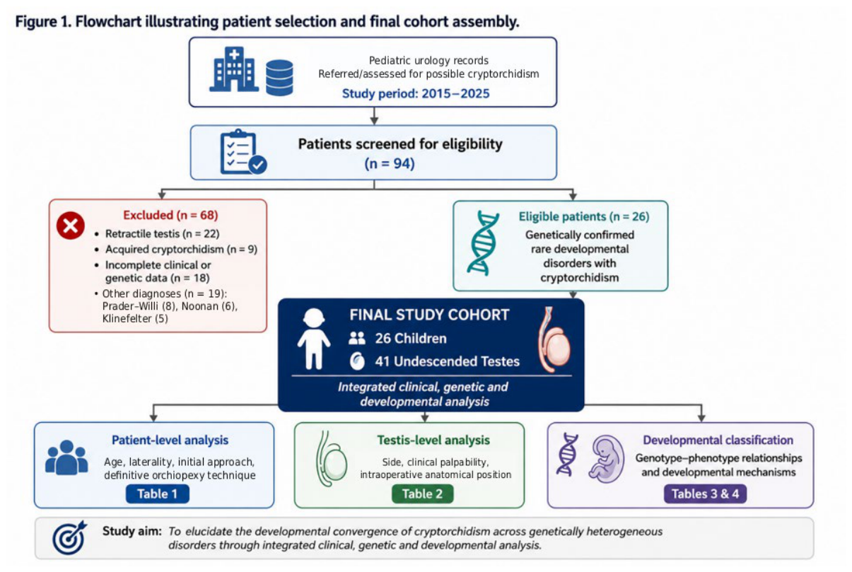
Figure 2.
Testicular descent is a multistage morphogenetic process requiring coordinated endocrine, neural, mesenchymal, and mechanical interactions. During the transabdominal phase, insulin-like peptide 3 (INSL3), produced by fetal Leydig cells, signals through its receptor RXFP2 within the gubernaculum and promotes gubernacular growth, cellular proliferation, and extracellular-matrix remodeling. The subsequent inguinoscrotal phase is predominantly androgen dependent and involves further gubernacular remodeling and migration through the inguinal canal toward the scrotum. Experimental evidence, particularly from rodent models, also supports contributions from the genitofemoral nerve and calcitonin gene-related peptide (CGRP) to gubernacular proliferation and motility. Successful descent therefore requires not only intact endocrine signaling but also appropriate signal reception, neural communication, mesenchymal responsiveness, extracellular-matrix remodeling, and coordinated morphogenesis of the gubernaculum and surrounding inguinal tissues. The schematic summarizes established and experimentally supported components of normal testicular descent and provides the developmental reference framework for the proposed model shown in Figure 3. GFN, genitofemoral nerve; CGRP, calcitonin gene-related peptide; INSL3, insulin-like peptide 3; RXFP2, relaxin family peptide receptor 2.
Figure 2.
Testicular descent is a multistage morphogenetic process requiring coordinated endocrine, neural, mesenchymal, and mechanical interactions. During the transabdominal phase, insulin-like peptide 3 (INSL3), produced by fetal Leydig cells, signals through its receptor RXFP2 within the gubernaculum and promotes gubernacular growth, cellular proliferation, and extracellular-matrix remodeling. The subsequent inguinoscrotal phase is predominantly androgen dependent and involves further gubernacular remodeling and migration through the inguinal canal toward the scrotum. Experimental evidence, particularly from rodent models, also supports contributions from the genitofemoral nerve and calcitonin gene-related peptide (CGRP) to gubernacular proliferation and motility. Successful descent therefore requires not only intact endocrine signaling but also appropriate signal reception, neural communication, mesenchymal responsiveness, extracellular-matrix remodeling, and coordinated morphogenesis of the gubernaculum and surrounding inguinal tissues. The schematic summarizes established and experimentally supported components of normal testicular descent and provides the developmental reference framework for the proposed model shown in Figure 3. GFN, genitofemoral nerve; CGRP, calcitonin gene-related peptide; INSL3, insulin-like peptide 3; RXFP2, relaxin family peptide receptor 2.
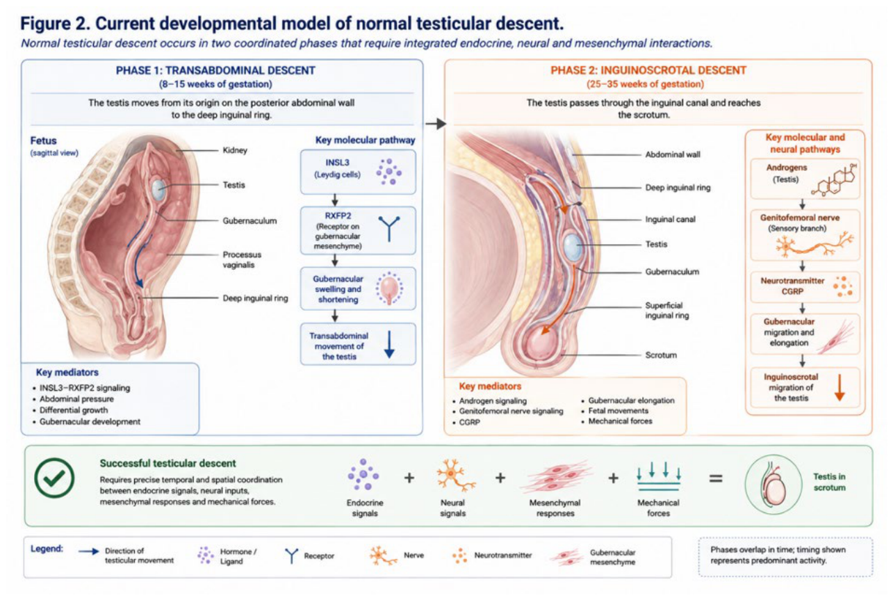
Figure 3.
The model proposes that genetically heterogeneous developmental disorders may perturb different hierarchical levels of the biological system required for normal testicular descent while converging on a shared anatomical phenotype. The principal levels comprise: (1) regulation of developmental gene expression, including chromatin modification, chromatin remodeling, transcriptional regulation, and post-transcriptional control; (2) developmental signal sensing and integration; (3) neural specification and information transmission; (4) neuromuscular execution and fetal mechanobiology; and (5) mesenchymal patterning and structural implementation. Chromosomal copy-number abnormalities are represented as cross-cutting genomic perturbations because altered dosage of multiple genes may influence several developmental levels simultaneously. These processes are biologically interconnected rather than strictly linear, and individual genes may influence more than one level. Disturbances arising at different positions within this hierarchy are hypothesized to converge downstream on impaired gubernacular development, tissue remodeling, and/or inguinoscrotal migration, ultimately resulting in cryptorchidism. The framework integrates the genotype–phenotype observations of the present cohort with established principles of testicular developmental biology; it is intended as a hypothesis-generating and testable model rather than evidence of a causal molecular pathway for any individual disorder.
Figure 3.
The model proposes that genetically heterogeneous developmental disorders may perturb different hierarchical levels of the biological system required for normal testicular descent while converging on a shared anatomical phenotype. The principal levels comprise: (1) regulation of developmental gene expression, including chromatin modification, chromatin remodeling, transcriptional regulation, and post-transcriptional control; (2) developmental signal sensing and integration; (3) neural specification and information transmission; (4) neuromuscular execution and fetal mechanobiology; and (5) mesenchymal patterning and structural implementation. Chromosomal copy-number abnormalities are represented as cross-cutting genomic perturbations because altered dosage of multiple genes may influence several developmental levels simultaneously. These processes are biologically interconnected rather than strictly linear, and individual genes may influence more than one level. Disturbances arising at different positions within this hierarchy are hypothesized to converge downstream on impaired gubernacular development, tissue remodeling, and/or inguinoscrotal migration, ultimately resulting in cryptorchidism. The framework integrates the genotype–phenotype observations of the present cohort with established principles of testicular developmental biology; it is intended as a hypothesis-generating and testable model rather than evidence of a causal molecular pathway for any individual disorder.
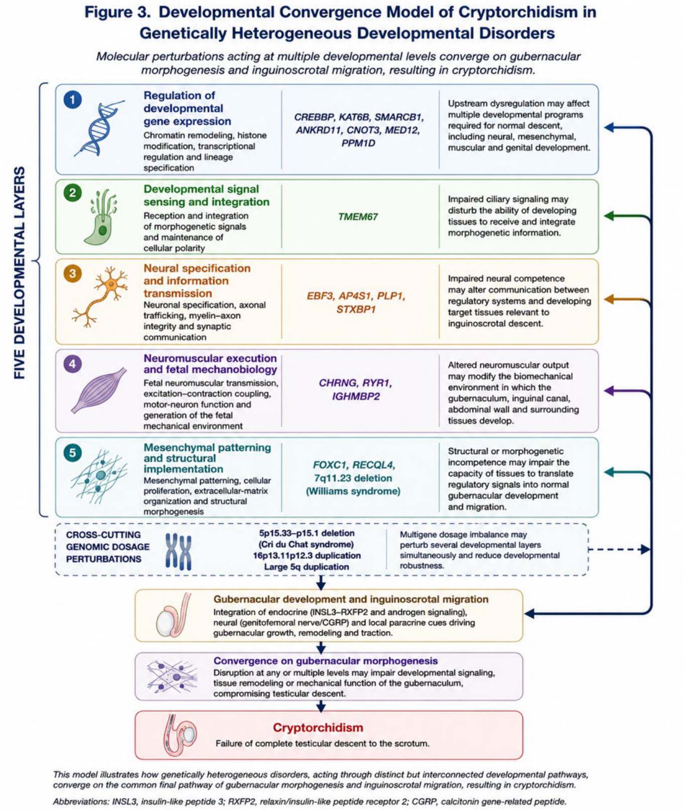
Table 3.
Previous evidence for cryptorchidism was categorized as well established, reported/recognized, not established, or unknown according to the available literature. “Potential phenotype expansion” denotes an observation for which cryptorchidism is not currently an established component of the reported disorder spectrum and should not be interpreted as evidence of causality. Copy-number abnormalities are reported according to the molecular information available in the clinical genetic record.
Table 3.
Previous evidence for cryptorchidism was categorized as well established, reported/recognized, not established, or unknown according to the available literature. “Potential phenotype expansion” denotes an observation for which cryptorchidism is not currently an established component of the reported disorder spectrum and should not be interpreted as evidence of causality. Copy-number abnormalities are reported according to the molecular information available in the clinical genetic record.
| Disorder | Molecular diagnosis | Previous evidence for cryptorchidism | Interpretation in the present study |
| Rubinstein–Taybi syndrome | CREBBP | Well established [11,12] | Consistent with the recognized phenotypic spectrum |
| KAT6B-related disorder | KAT6B | Well established [13,14] | Consistent with the recognized phenotypic spectrum |
| Coffin–Siris syndrome | SMARCB1 | Reported within the Coffin–Siris spectrum [8,9,15] | Supports an existing but incompletely characterized association |
| KBG syndrome | ANKRD11 | Reported / recognized within the phenotypic spectrum [16,17] | Consistent with an existing but variably expressed association |
| CNOT3-related disorder | CNOT3 | Not established | Potential phenotype expansion; hypothesis-generating |
| Lujan–Fryns syndrome | MED12 | Not established / very limited | Potential phenotype expansion; hypothesis-generating |
| Opitz–Kaveggia (FG) syndrome | MED12 | Well established [18] | Consistent with the recognized phenotypic spectrum |
| Joubert syndrome | TMEM67 | Reported within the TMEM67-associated ciliopathy spectrum, particularly Meckel-Gruber phenotypes [19,20] | Supports an existing but incompletely characterized association |
| Pelizaeus–Merzbacher disease | PLP1 | Not established | Potential phenotype expansion; hypothesis-generating |
| AP-4 deficiency syndrome | AP4S1 | Not established | Potential phenotype expansion; hypothesis-generating |
| EBF3-related disorder | EBF3 | Reported [21,22] | Supports an existing but incompletely characterized association |
| Jansen–de Vries syndrome | PPM1D | Reported in prior clinical literature [23] | Consistent with a reported but incompletely characterized association |
| STXBP1-related developmental and epileptic encephalopathy | STXBP1 | Not established | Potential phenotype expansion; hypothesis-generating |
| RYR1-related congenital myopathy | RYR1 | Reported in severe fetal-akinesia/multiple-pterygium phenotypes [24,25] | Supports a reported association within the severe RYR1 spectrum; relevance to broader RYR1-related congenital myopathy remains incompletely characterized |
| Escobar syndrome | CHRNG | Well established [10,26] | Consistent with the recognized phenotypic spectrum |
| SMARD1 | IGHMBP2 | Not established | Potential phenotype expansion; hypothesis-generating |
| Axenfeld–Rieger syndrome | FOXC1 | Not established | Potential phenotype expansion; hypothesis-generating |
| Baller–Gerold syndrome | RECQL4 | Not established | Potential phenotype expansion; hypothesis-generating |
| Williams syndrome | 7q11.23 deletion | Recognized / reported in dedicated clinical cohorts [27] | Consistent with a recognized genitourinary manifestation of the syndrome |
| Cri du Chat syndrome | arr(GRCh38) 5p15.33p15.1(113461–16502731)x1 | Reported [28] | Consistent with a previously reported association within the 5p deletion spectrum |
| 16p13.11p12.3 duplication syndrome | arr(GRCh38) 16p13.11p12.3 duplication (~2.9 Mb) | Recently reported [29] | Supports an emerging but incompletely characterized association |
| Chromosome 5q duplication | Large pathogenic duplication | Unknown | Association currently unclassified; hypothesis-generating |
Table 4.
The proposed hierarchy organizes molecular diagnoses according to the predominant level at which their biological effect is considered to enter the developmental system. The layers are interconnected rather than strictly linear, and several genes have functions spanning more than one developmental domain. Assignment to a layer therefore represents an analytical framework rather than a definitive molecular ontology. Cross-cutting copy-number abnormalities are considered separately because altered dosage of multiple genes may influence several layers simultaneously. †Physiological regulatory inputs represent established components of normal testicular descent and are included as a reference framework; they are not necessarily directly altered in the disorders represented in the cohort. Proposed disorder-specific links to cryptorchidism remain hypothesis-generating unless supported by direct experimental evidence.
Table 4.
The proposed hierarchy organizes molecular diagnoses according to the predominant level at which their biological effect is considered to enter the developmental system. The layers are interconnected rather than strictly linear, and several genes have functions spanning more than one developmental domain. Assignment to a layer therefore represents an analytical framework rather than a definitive molecular ontology. Cross-cutting copy-number abnormalities are considered separately because altered dosage of multiple genes may influence several layers simultaneously. †Physiological regulatory inputs represent established components of normal testicular descent and are included as a reference framework; they are not necessarily directly altered in the disorders represented in the cohort. Proposed disorder-specific links to cryptorchidism remain hypothesis-generating unless supported by direct experimental evidence.
| Developmental layer | Principal biological function | Representative molecular diagnoses in the cohort | Potential relevance to testicular descent | Evidence status |
| 1. Regulation of developmental gene expression | Chromatin accessibility, histone modification, transcriptional regulation, lineage specification, and transcript stability during embryogenesis | CREBBP, KAT6B, SMARCB1, ANKRD11, CNOT3, MED12, PPM1D | Upstream dysregulation may affect multiple developmental programs required for normal descent, including neural, mesenchymal, muscular, and genital development | Established molecular functions; proposed relationship to descent is predominantly hypothesis-generating |
| 2. Developmental signal sensing and integration | Reception and integration of morphogenetic signals and maintenance of cellular polarity | TMEM67 | Impaired ciliary signaling may disturb the ability of developing tissues to receive and integrate morphogenetic information | Strong evidence for ciliary dysfunction; direct mechanism in testicular descent not established |
| 3. Neural specification and information transmission | Neuronal specification, axonal trafficking, myelin–axon integrity, and synaptic communication | EBF3, AP4S1, PLP1, STXBP1 | Impaired neural competence may alter communication between regulatory systems and developing target tissues relevant to inguinoscrotal descent | Established neurological functions; relationship to testicular descent remains indirect |
| 4. Neuromuscular execution and fetal mechanobiology | Fetal neuromuscular transmission, excitation–contraction coupling, motor-neuron function, and generation of the fetal mechanical environment | CHRNG, RYR1, IGHMBP2 | Altered neuromuscular output may modify the biomechanical environment within which the gubernaculum, inguinal canal, abdominal wall, and surrounding tissues develop | Strongest clinical support for CHRNG; RYR1 and IGHMBP2 remain exploratory |
| 5. Mesenchymal patterning and structural implementation | Mesenchymal patterning, cellular proliferation, extracellular-matrix organization, connective-tissue competence, and structural morphogenesis | FOXC1, RECQL4, 7q11.23 deletion/ Williams syndrome | Structural or morphogenetic incompetence may impair the capacity of developing tissues to translate regulatory signals into normal gubernacular development and migration | Strong developmental plausibility; limited disorder-specific mechanistic evidence |
| Cross-cutting genomic dosage perturbations | Simultaneous alteration of multiple dosage-sensitive genes and developmental networks | 5p15.33–p15.1 deletion/Cri du Chat syndrome; 16p13.11p12.3 duplication; large 5q duplication | Multigene dosage imbalance may perturb several developmental layers simultaneously and reduce developmental robustness | Established genomic principle; individual contributions to cryptorchidism remain unresolved |
| Physiological regulatory inputs† | Coordination of gubernacular development and inguinoscrotal migration | INSL3–RXFP2 signaling, androgen signaling, genitofemoral nerve/CGRP | Provides the established physiological context through which upstream developmental disturbances may ultimately influence descent | Established experimental and clinical evidence, with species-dependent limitations for some neural mechanisms. |
Disclaimer/Publisher’s Note: The statements, opinions and data contained in all publications are solely those of the individual author(s) and contributor(s) and not of MDPI and/or the editor(s). MDPI and/or the editor(s) disclaim responsibility for any injury to people or property resulting from any ideas, methods, instructions or products referred to in the content. |
© 2026 by the authors. Licensee MDPI, Basel, Switzerland. This article is an open access article distributed under the terms and conditions of the Creative Commons Attribution (CC BY) license.
Copyright: This open access article is published under a Creative Commons CC BY 4.0 license, which permit the free download, distribution, and reuse, provided that the author and preprint are cited in any reuse.