

Submitted:
21 September 2026
Posted:
22 September 2026
You are already at the latest version
Abstract
Cellular stress is not inherently detrimental; its biological consequences depend on intensity, duration, context, and the capacity for recovery. This narrative review proposes the Burn-ON/Burn-OUT paradigm as a hypothesis-generating framework describing the transition from adaptive cellular activation to progressive loss of cellular resilience. A structured literature search was conducted for English-language articles published between 2021 and 2026 in PubMed, Web of Science, and Scopus, focusing on cellular stress adaptation, mitochondrial dysfunction, integrated stress response, autophagy and mitophagy, redox signaling, inflammasome activation, metabolic flexibility, senescence, and nutritional modulation. In the proposed model, Burn-ON represents a state of increased cellular demand in which mitochondrial adaptation, stress-response signaling, autophagic quality control, redox homeostasis, and metabolic flexibility remain sufficiently preserved to sustain function and allow recovery. With persistent or repetitive stress, these adaptive mechanisms may progressively lose reversibility, defining a potential tipping point toward Burn-OUT. Burn-OUT is characterized conceptually by impaired mitochondrial quality control, reduced energetic reserve, defective autophagic flux, persistent inflammatory signaling, redox imbalance, metabolic inflexibility, and loss of functional recovery, with senescence or regulated cell death representing possible downstream outcomes. Nutrition and lifestyle may modulate this continuum by influencing nutrient sensing, substrate load, mitochondrial metabolism, autophagy, and inflammatory pathways. The Burn-ON/Burn-OUT model frames cellular stress as a double-edged biological process, in which adaptation and dysfunction represent different positions along a dynamic continuum. Longitudinal studies are needed to determine whether recovery kinetics and multidimensional biomarker profiles can distinguish these proposed states.
Keywords:
cellular stress
; Burn-ON
; Burn-OUT
; cellular resilience
; mitochondrial dysfunction
; integrated stress response
; autophagy
; mitophagy
; NLRP3 inflammasome
; metabolic flexibility
1. Introduction
Cells are continuously exposed to fluctuations in nutrient availability, energy demand, redox balance, oxygen tension, inflammatory cues, and environmental stressors. These challenges do not inevitably lead to injury. In many contexts, transient or moderate stress activates adaptive programs that preserve proteostasis, maintain redox balance, reprogram metabolism, and improve cellular resilience. The concept of mitohormesis illustrates this principle well: mild mitochondrial perturbation can trigger adaptive responses aimed at restoring cellular metabolism, cytosolic proteostasis, and redox homeostasis, sometimes leaving the cell less vulnerable to subsequent stress [1].
A central component of this adaptive machinery is the integrated stress response (ISR). The ISR is activated by several forms of stress, including amino acid deprivation, endoplasmic reticulum stress, and mitochondrial dysfunction, and acts through eukaryotic translation initiation factor 2 alpha (eIF2α)-dependent translational control and downstream transcriptional programs, activating transcription factor 4 (ATF4). Its physiological role is fundamentally adaptive: it reduces global protein synthesis while selectively inducing stress-responsive genes and metabolic pathways that support recovery [2]. ISR signaling is intrinsically context-dependent; chronic or excessive activation can also become maladaptive and promote cell-death programs [2].
Recent mechanistic evidence further supports the idea that cellular adaptation depends not only on pathway activation, but also on response plasticity and reversibility. Avelar et al. showed that normal cells can undergo ISR-mediated transcriptional reprogramming and homeostatic recovery under chronic stress, whereas irreversible ISR activation is associated with failure of adaptation and cell death. The fate of the cell depends on the nature, duration, magnitude, and reversibility of the stress response [3].
Autophagy provides another striking example of this dynamic behavior. In a recent study, acute stress activated neuronal autophagy, whereas chronic stress suppressed it, indicating that the same quality-control pathway may shift from adaptive activation to maladaptation according to the duration of stress exposure [4]. The authors further demonstrated opposing AMP-activated protein kinase (AMPK) and mechanistic target of rapamycin (mTOR) signaling patterns, with acute stress activating AMPK-dependent autophagy and chronic stress activating mTOR and suppressing autophagy [4].
This adaptive-to-maladaptive transition is also highly relevant in metabolic disease. Human studies in obesity, type 2 diabetes, and metabolic dysfunction-associated steatotic liver disease indicate that autophagy may initially increase as a compensatory mechanism to remove damaged proteins and organelles, whereas prolonged hyperglycemia and disrupted insulin signaling can impair autophagic flux and promote the accumulation of dysfunctional cellular components [5]. This dynamic is consistent with the broader observation that metabolic diseases share mitochondrial dysfunction, oxidative and endoplasmic reticulum stress, chronic inflammation, and altered autophagy.
Persistent metabolic stress can also engage inflammatory pathways. The NLR family pyrin domain containing 3 (NLRP3) inflammasome inflammasome–IL-1β axis is activated by metabolic signals including glucose, non-esterified fatty acids (NEFAs), cholesterol, uric acid, adenosine triphosphate (ATP), reactive oxygen species (ROS), and hypoxia, thereby linking nutrient and metabolic overload with innate immune activation [6]. This pathway also illustrates the double-edged nature of stress signaling. Acute and transient interleukin-1 beta (IL-1β) signaling contributes to physiological insulin secretion and adaptation, whereas chronic low-grade activation is associated with insulin resistance, β-cell dysfunction, and metabolic deterioration [6].
At the whole-body level, loss of adaptability is also reflected by metabolic inflexibility. A recent systematic review and meta-analysis of 65 studies reported lower insulin-stimulated changes in respiratory exchange ratio in individuals with overweight/obesity and type 2 diabetes compared with lean individuals, although substantial heterogeneity suggests that metabolic inflexibility should not be considered a single universal state [7]. Cellular senescence may represent another downstream manifestation of chronic metabolic stress. A systematic review and meta-analysis found a reciprocal association between obesity and senescence, with increased pro-inflammatory and pro-senescence signaling reported in obesity-related states [8].
Mitochondrial quality control appears to be sensitive to stress duration. In an experimental model of acute and chronic stress, Zhang et al. demonstrated progressive mitochondrial structural damage, excessive fission, impaired fusion, altered mitophagy, reduced mitochondrial content, increased ROS, and reduced ATP production under chronic stress [9] These findings support the concept that stress responses may initially remain compensatory but become progressively maladaptive when exposure persists.
Based on this converging evidence, we propose the Burn-ON/Burn-OUT paradigm as a hypothesis-generating framework describing the transition from adaptive cellular activation to progressive loss of cellular resilience. In this model, Burn-ON represents a state in which stress-response pathways, mitochondrial adaptation, autophagy, redox control, and metabolic flexibility remain sufficiently preserved to maintain function despite increased cellular demand. By contrast, Burn-OUT represents a proposed state in which persistent stress exceeds repair and recovery capacity, leading to impaired mitochondrial quality control, autophagic dysfunction, sustained inflammatory signaling, metabolic inflexibility, and eventually cellular senescence or loss of function.
Burn-OUT is not proposed as a newly established molecular entity, nor is it synonymous with occupational or psychological burnout. Rather, the Burn-ON/Burn-OUT continuum is intended as an integrative framework connecting established mechanisms that are currently investigated largely in isolation. We conceptualize cellular stress as a double-edged sword: the same adaptive machinery that promotes resilience during transient challenge may, when persistently engaged without adequate recovery, contribute to progressive cellular dysfunction.
2. Materials and Methods
2.1. Review Design and Reporting Framework
This study was designed as a structured, hypothesis-generating narrative review. The methodological approach was guided by the Scale for the Assessment of Narrative Review Articles (SANRA), with emphasis on the justification of the review, transparency of the literature search, appropriate referencing, scientific reasoning, and balanced presentation of the available evidence [10].
The objective was not to perform a systematic or scoping review, but to integrate current clinical, translational, and mechanistic evidence relevant to the transition from adaptive cellular stress responses to progressive loss of cellular resilience. Attention was given to the temporal behavior, reversibility, and recovery of stress-response pathways.
The terms Burn-ON and Burn-OUT were not used as search terms because they represent the conceptual framework proposed in this review. Instead, established biological processes underlying cellular adaptation, persistent stress, impaired recovery, and loss of resilience were investigated.
2.2. Literature Search Strategy
A structured literature search was conducted in PubMed/MEDLINE, Scopus, and Web of Science for English-language publications published between January 2021 and September 2026.
Of the 70 references included in the final review, 68 were directly retrieved through the PubMed electronic search strategies (Table S1), comprising 30 from the primary stress-domain search and 38 from nine additional targeted searches addressing complementary mechanistic and nutritional domains not captured by the primary strategy. The remaining 2 references were identified through targeted retrieval of foundational methodological and conceptual publications (the SANRA quality-assessment instrument and an earlier psychological stress–mitochondria review), consistent with SANRA guidance for narrative reviewsThe search was organized around two complementary domains. The first addressed chronic systemic and psychological stress and included terms related to burnout, professional burnout, occupational burnout, emotional exhaustion, chronic stress, psychological stress, occupational stress, and allostatic load. The second addressed cellular adaptation and dysfunction and included mitochondrial function and quality control, oxidative and redox signaling, hormesis and mitohormesis, cellular resilience, autophagy, mitophagy, Nrf2, AMPK, mTOR, unfolded protein response, integrated stress response, endoplasmic reticulum stress, inflammasome activation, and metabolic inflexibility.
Because the base search required co-occurrence of a stress-domain term (e.g., burnout, chronic stress, psychological stress, allostatic load) with a cellular/metabolic term, mechanistic or nutritional literature lacking explicit stress-related terminology was not captured by the primary strategy. Additional targeted searches were therefore performed to address these domains, including nutritional modulation, metabolic flexibility, mitochondrial allostatic load, cellular senescence, regulated cell death, circadian disruption, recovery kinetics, inflammasome signaling in metabolic disease, autophagy and mitophagy in obesity and metabolic dysfunction-associated steatotic liver disease (MASLD), exercise-induced mitochondrial adaptation (AMPK/mTOR signaling), gut microbiota–drug interactions, micronutrient modulation (e.g., vitamin D), and sleep–insulin sensitivity relationships. Reference lists of relevant reviews and key primary studies were also screened to identify additional publications of direct relevance.
The complete database-specific search strategies, Boolean operators, and filters are provided in Supplementary Table S1 and are not duplicated in the main text.
2.3. Study Selection and Eligibility Criteria
Publications were considered eligible when they provided evidence relevant to at least one component of the proposed cellular stress continuum: adaptive stress signaling, mitochondrial remodeling and bioenergetics, autophagy or mitophagy, redox regulation, inflammatory signaling, metabolic flexibility, cellular senescence, nutritional or lifestyle modulation, psychological or circadian stress, or recovery following stress exposure.
Priority was given to systematic reviews and meta-analyses, randomized controlled trials, human observational studies, and recent mechanistic studies. Experimental cellular and animal studies were included when they provided mechanistic or temporal information that could not be adequately obtained from human studies, particularly for mitochondrial dynamics, autophagic flux, mitophagy, inflammasome activation, cellular senescence, and regulated cell-death pathways.
Earlier seminal studies were retained when they were necessary to define established concepts or mechanisms not adequately represented by the 2021–2026 literature.
Studies were excluded when they were not relevant to the biological domains examined, lacked sufficient mechanistic or clinical information, or duplicated evidence more comprehensively represented by another source. Studies addressing psychological or occupational burnout without biological, metabolic, neuroendocrine, inflammatory, redox, or mitochondrial outcomes were not used as direct evidence for the proposed cellular Burn-OUT construct.
2.4. Study Selection and Author Involvement
The literature search and initial organization of the retrieved material were coordinated by S.I., S.M., V.M., M.D.-G., and A.C.F.F.S., with overall scientific supervision from S.I., M.R., D.T., and L.-A.T. Titles and abstracts were screened for relevance by S.I., S.M., V.M., M.D.-G., A.C.F.F.S., and V.I. according to the predefined thematic domains of the review.
Retrieved records were manually screened by title and abstract for thematic relevance. Potentially relevant articles were subsequently assessed in full text. Studies were retained when they provided evidence relevant to cellular stress adaptation, mitochondrial function and quality control, integrated stress-response signaling, autophagy and mitophagy, redox regulation, inflammatory signaling, metabolic flexibility, cellular senescence, nutritional and lifestyle modulation, gut–brain interactions, psychological or circadian stress, or recovery following stress exposure. Duplicate records and clearly irrelevant publications were excluded. Because this was a narrative review, no formal PRISMA-based screening process or risk-of-bias assessment was performed.
S.I., S.M., V.M., M.D.-G., and A.C.F.F.S. contributed to literature retrieval, screening, evidence organization, and thematic classification. D.T. contributed to the interpretation of cellular stress, resilience, recovery, and longevity-related mechanisms. A.G. contributed to the metabolic and cardiometabolic interpretation of the evidence. M.R. and D.T. contributed to the metabolic, inflammatory, and translational interpretation of the literature. V.I. contributed to the cellular and anatomical interpretation of the evidence and to the visualization of the proposed framework. L.-A.T. contributed to the clinical, metabolic, and translational interpretation of the evidence.
Uncertainties regarding article relevance, thematic classification, or interpretation were resolved through discussion among the authors, with final scientific oversight by S.I., M.R., D.T., and L.-A.T.
2.5. Evidence Classification and Synthesis
Evidence was synthesized narratively because of substantial heterogeneity in study populations, tissues, experimental models, stress paradigms, interventions, and outcome measures. No quantitative pooling was performed.
To avoid conflating different levels of evidence, findings were interpreted according to their source. Human clinical and intervention studies were used where available to support translational relevance. Experimental cellular and animal studies were used primarily to define mechanistic and temporal relationships. Evidence derived from occupational or psychological stress studies was considered supportive of potential biological overlap but was not interpreted as direct validation of cellular Burn-OUT.
The evidence was subsequently organized into major biological domains: stress-response plasticity and reversibility; mitochondrial quality control and bioenergetics; autophagy and mitophagy; redox regulation; inflammasome and inflammatory signaling; metabolic flexibility; cellular senescence and regulated cell death; nutritional and lifestyle modulation; and psychological and circadian stress.
2.6. Development of the Burn-ON/Burn-OUT Framework
The conceptual framework was developed after synthesis of the retrieved evidence rather than being used as an a priori classification system for study selection. Findings describing coordinated, reversible responses with preserved functional recovery were considered compatible with the proposed Burn-ON state. Findings describing delayed recovery, prolonged signaling, increasing energetic cost, or incomplete restoration of cellular homeostasis informed the proposed tipping point. Persistent dysfunction across mitochondrial, proteostatic, redox, inflammatory, or metabolic systems was considered relevant to the proposed Burn-OUT state.
The resulting Burn-ON/Burn-OUT continuum represents an integrative, hypothesis-generating interpretation of established biological mechanisms rather than a validated classification system. No individual study was interpreted as demonstrating Burn-ON or Burn-OUT directly, because these terms have not yet been experimentally validated. Emphasis was placed on recovery kinetics and reversibility as potential features capable of distinguishing adaptive from maladaptive cellular stress in future longitudinal studies.
2.7. Methodological Considerations
Because this work is a structured narrative review and conceptual synthesis, no formal meta-analysis or risk-of-bias assessment was performed. The review was not intended to provide an exhaustive systematic synthesis of all studies related to cellular stress.
The heterogeneity of experimental models and biological outcomes limits direct comparison across studies. Mechanistic evidence derived from cellular and animal models was therefore distinguished from human evidence throughout the interpretation. Associations were not interpreted as causal when direct experimental support was unavailable.
3. When Adaptation Becomes Maladaptation
3.1. Loss of Stress-Response Reversibility
The transition from Burn-ON toward Burn-OUT is unlikely to be defined by a single molecular event. It may emerge when stress-response pathways lose their capacity to return to baseline after the initial challenge has resolved. In this context, reversibility may be one of the most important characteristics distinguishing adaptive from maladaptive cellular stress [3].
Persistent activation of the integrated stress response provides a useful model for this transition. While transient ISR activation can reduce translational burden and promote cellular adaptation, prolonged signaling may sustain ATF4- and C/EBP homologous protein (CHOP)-dependent transcriptional programs and alter cell fate [11]. Experimental work has shown that the temporal architecture of ISR signaling is tightly regulated, suggesting that duration itself is a determinant of whether the response remains adaptive or becomes maladaptive [12].
This distinction may be relevant under conditions of repeated stress. Cells that retain regulatory flexibility can recalibrate protein synthesis, redox balance, and energy utilization after each challenge [3]. Temporal regulation of the stress response is critical for maintaining adaptive cellular function [12]. By contrast, persistent or unresolved ISR signaling may interfere with the restoration of cellular homeostasis [11]. In the Burn-ON/Burn-OUT framework, the tipping point may represent a progressive loss of stress-response plasticity.
Metabolic adaptation studies support this interpretation. Chronic variable stress can induce substantial remodeling of myocardial metabolic pathways, indicating that tissues attempt to maintain function under prolonged demand [13]. This compensation may carry an energetic cost and may become unsustainable when recovery is incomplete or when multiple stressors converge.
Defining feature of the Burn-ON-to-Burn-OUT transition is the progressive loss of reversibility: signaling pathways remain activated beyond the period required for adaptation, cellular recovery becomes incomplete, and homeostatic restoration progressively fails.
3.2. Mitochondrial Quality-Control Failure and Energetic Decline
Mitochondria may represent one of the most sensitive biological systems marking the transition from adaptive activation to loss of resilience. During adaptive stress, mitochondrial remodeling and coordinated fission–fusion dynamics contribute to the maintenance of mitochondrial function and cellular homeostasis [9]. Mitophagy provides an additional quality-control mechanism by selectively removing damaged mitochondria and supporting adaptation to sustained cellular stress [14]. Persistent stress may progressively disrupt mitochondrial surveillance and compromise these quality-control mechanisms [15].
Recent experimental evidence shows that chronic stress can disrupt mitochondrial status and mitophagy in the frontal cortex, supporting the concept that prolonged exposure compromises mitochondrial surveillance [15]. Similarly, dynamic changes in autophagy and mitophagy have been associated with behavioral adaptation to chronic stress, suggesting that mitochondrial quality control remains closely linked to the ability to cope with sustained challenge [14].
The fission–fusion balance is relevant to mitochondrial adaptation. Mitochondrial fission can facilitate the segregation of damaged mitochondrial components, thereby supporting their subsequent removal through mitophagy [14]. Persistent mitochondrial fragmentation, when accompanied by insufficient biogenesis or clearance, may progressively compromise mitochondrial reserve [16]. Under chronic stress, this loss of mitochondrial homeostasis may be accompanied by reduced membrane potential, increased ROS production, decreased ATP generation, and progressive structural mitochondrial damage [9].
Chronic stress may also affect mitochondrial biogenesis through AMPK–PGC-1α signaling. Experimental studies have reported stress-induced impairment of mitochondrial biogenesis through alterations in the AMPK–PGC-1α axis, suggesting that failure to replenish the mitochondrial pool may further accelerate loss of energetic resilience [17].
The mitochondrial component of the tipping point may be characterized by a shift from adaptive mitochondrial remodeling to progressive quality-control failure. Declining mitochondrial quality control can compromise energetic capacity and the effective removal and replacement of damaged mitochondria [15]. Within the proposed Burn-ON/Burn-OUT framework, this state may represent a transitional phase in which the cell remains viable but becomes progressively less capable of recovering from subsequent challenges.
3.3. Redox Imbalance and Persistent Inflammatory Signaling
The transition from adaptive stress signaling to cellular dysfunction may also involve a progressive loss of redox control. At low or transient levels, reactive oxygen species can function as signaling molecules that contribute to cellular adaptation and stress responses [1]. The biological effects of ROS are context-dependent, with controlled redox signaling supporting cellular regulation while persistent oxidative imbalance can promote metabolic inflammation. Persistent mitochondrial dysfunction may instead promote excessive ROS generation, accompanied by declining ATP production and progressive loss of mitochondrial homeostasis [9]. Impaired autophagic and lysosomal clearance may further favor the accumulation of dysfunctional cellular components and oxidative stress [18]. Within the proposed Burn-ON/Burn-OUT framework, this may represent a shift from regulatory redox signaling toward a self-amplifying state of cellular damage.
Mitochondrial dysfunction and inflammatory signaling are closely interconnected. Damaged mitochondria can increase ROS production and generate mitochondrial-derived danger signals that influence innate immune responses [9]. Metabolic and mitochondrial danger signals, including ROS and extracellular ATP, can promote activation of the NLRP3 inflammasome [6]. Persistent NLRP3 activation may subsequently sustain IL-1β/IL-18 signaling and contribute to chronic sterile inflammation [19]. Within the Burn-ON/Burn-OUT framework, this mitochondria–inflammasome interaction may provide a mechanistic bridge between declining energetic resilience and persistent inflammatory signaling.
The NLRP3 inflammasome is relevant to the proposed tipping point because it responds to several metabolic signals, including excess glucose, non-esterified fatty acids, cholesterol, ATP, ROS, uric acid, and hypoxia [6]. This pathway is not intrinsically pathological. IL-1β participates in physiological metabolic regulation, including meal-associated insulin secretion, whereas chronic low-grade activation can progressively impair β-cell responsiveness and promote metabolic dysfunction [6].
The dual behavior of inflammatory signaling makes the NLRP3 inflammasome relevant to the Burn-ON/Burn-OUT framework. Transient IL-1β signaling can participate in physiological metabolic adaptation, whereas persistent activation of the NLRP3–IL-1β axis is associated with metabolic dysfunction and chronic inflammation [6]. Failure to terminate NLRP3 signaling may further sustain inflammasome activity under prolonged stress conditions [20]. Within the proposed framework, Burn-ON may involve transient and resolvable inflammatory signaling, whereas the tipping point may be characterized by progressively incomplete inflammatory resolution.
The biological transition should not be reduced to increased ROS or inflammasome activation alone. We propose that the tipping point emerges when redox regulation, mitochondrial quality control, and inflammatory resolution begin to fail simultaneously. This convergence may progressively reduce the capacity of the cell to recover from subsequent metabolic challenges.
3.4. Metabolic Inflexibility and the Progressive Loss of Cellular Resilience
A further manifestation of declining adaptive capacity may be the progressive loss of metabolic flexibility. Metabolic flexibility describes the capacity to adjust substrate utilization and oxidation in response to changes in nutrient availability, hormonal signals, and energetic demand [7]. Within the proposed Burn-ON/Burn-OUT framework, preserved metabolic flexibility may be considered a functional expression of cellular resilience, allowing adaptation to changing metabolic conditions. Progressive inflexibility may indicate a narrowing adaptive reserve.
Recent human evidence supports an association between excess adiposity and reduced metabolic flexibility. In a systematic review and meta-analysis including 65 studies, insulin-stimulated changes in respiratory exchange ratio were greater in lean individuals than in participants with overweight/obesity or type 2 diabetes [7]. Intervention studies further indicate that metabolic flexibility is sensitive to the nature of the nutritional challenge. In a randomized trial, time-restricted eating did not improve metabolic flexibility compared with unrestricted eating and was associated with lower metabolic flexibility than caloric restriction [21]. The considerable between-study heterogeneity and the absence of a universal metabolic-inflexibility threshold indicate that this phenotype should be interpreted as a continuum.
This observation is relevant to the Burn-ON/Burn-OUT paradigm. During adaptive stress, cells may retain the capacity to adjust substrate utilization and energy production according to metabolic demand [7]. With persistent metabolic stress, impaired insulin signaling and mitochondrial dysfunction may progressively restrict this adaptive metabolic repertoire [5]. Within the proposed framework, these changes may contribute to the transition from preserved metabolic adaptability toward declining cellular resilience.
Human evidence indicates that overweight/obesity and type 2 diabetes are associated with reduced insulin-stimulated metabolic flexibility, although substantial heterogeneity exists across studies [7]. We propose metabolic inflexibility not as a defining biomarker of Burn-OUT, but as a potential functional phenotype of declining adaptive reserve.
Cellular senescence may represent a later stage of this trajectory. Senescent cells remain metabolically active but undergo stable cell-cycle arrest and can develop a senescence-associated secretory phenotype (SASP), producing inflammatory mediators capable of influencing neighboring cells and tissue function. In obesity, chronic inflammation and oxidative stress have been associated with senescence-related changes, and systematic evidence suggests a reciprocal relationship between obesity and cellular aging [8].
Senescence should not be equated with Burn-OUT. It may represent one possible downstream phenotype occurring when prolonged metabolic and inflammatory stress is no longer adequately resolved. Similarly, apoptosis, pyroptosis, ferroptosis, or necroptosis should be considered potential late outcomes.
3.5. A Proposed Biological Definition of the Tipping Point
A major challenge is determining whether a single molecular threshold separates Burn-ON from Burn-OUT. Available evidence suggests that cellular adaptation involves several biological systems whose responses vary with stress intensity, duration, and reversibility [3]. We propose the tipping point as a transitional zone in which cellular demand begins to exceed repair, quality-control, and recovery capacity.
This transition may involve progressive loss of stress-response reversibility, deterioration of organelle quality control, declining energetic and redox reserve, and reduced metabolic flexibility. These processes are discussed in the preceding sections and summarized across the proposed continuum in Table 1. Their relevance may depend on their persistence and on the ability of the cell to restore baseline function after the stressor is reduced or removed.
Experimental evidence provides an example of this transition. Under prolonged stress, mitochondrial remodeling may progress toward persistent fission, impaired fusion, structural mitochondrial damage, reduced mitochondrial content, increased ROS production, and decreased ATP generation [9]. These changes illustrate how an adaptive response may gradually become insufficient to maintain mitochondrial and cellular homeostasis.
The proposed transition from cellular homeostasis through Burn-ON and the tipping point toward Burn-OUT is summarized in Figure 1.
Conceptual model illustrates the proposed transition from cellular homeostasis to an adaptive Burn-ON state, followed by a tipping point and, when stress persists or recovery becomes insufficient, progression toward Burn-OUT. Burn-ON represents a predominantly adaptive and potentially reversible state characterized by transient stress-response activation, mitochondrial remodeling, controlled reactive oxygen species (ROS) signaling, preserved metabolic flexibility, and effective recovery mechanisms. The tipping point reflects progressive loss of reversibility associated with persistent or repeated stress, prolonged ISR/ATF4 signaling, mitochondrial fragmentation, impaired autophagic clearance, increasing ROS burden, and declining metabolic flexibility. Burn-OUT is proposed as a state of persistent cellular dysfunction in which the cell remains viable but shows reduced energetic reserve, mitochondrial dysfunction, impaired autophagic flux, persistent inflammasome signaling, redox imbalance, metabolic inflexibility, and defective recovery. The transition across the continuum is hypothesized to depend on the interaction between stress intensity, stress duration, and recovery capacity. Cellular senescence and regulated cell-death pathways, including ferroptosis, pyroptosis, necroptosis, and apoptosis, are presented as potential downstream outcomes and should not be considered synonymous with the Burn-OUT state itself. The model is hypothesis-generating and does not imply that all cells follow a linear or irreversible trajectory.
4. Burn-OUT: The Progressive Loss of Cellular Resilience
We define Burn-OUT as a proposed state in which adaptive stress-response systems can no longer adequately restore cellular homeostasis under persistent demand. Burn-OUT is not synonymous with cell death. It represents a stage of declining resilience characterized by impaired energetic capacity, defective organelle quality control, persistent inflammatory signaling, and reduced reversibility. Depending on the cellular context and severity of stress, senescence or regulated cell death may subsequently emerge as downstream outcomes.
4.1. Mitochondrial Dysfunction and Energetic Failure
Mitochondria are central to cellular adaptation because they integrate energetic demand, redox signaling, calcium homeostasis, and stress sensing [16]. Under transient stress, mitochondrial remodeling can support adaptation and preserve cellular function [14]. With sustained stress, mitochondrial dynamics, bioenergetics, and quality-control mechanisms may progressively deteriorate [9].Recent experimental evidence shows that chronic stress can disrupt mitochondrial networks at several levels. Ulecia-Morón et al. reported mitochondrial depolarization, dysregulated mitochondrial dynamics, increased mitophagy markers, and induction of mitochondrial biogenesis pathways in the frontal cortex after chronic mild stress, suggesting an extensive remodeling response to sustained stress [15].
More severe loss of mitochondrial control has been demonstrated in chronic stress models involving excessive mitochondrial fission. Drp1-dependent fission in the hippocampus was associated with impaired bioenergetics, oxidative stress, disrupted mitophagy, and altered synaptic function; inhibition of Drp1 partially reversed these abnormalities [22].
These findings support a distinction between mitochondrial remodeling and mitochondrial failure. Changes in fission, fusion, biogenesis, and mitophagy can initially contribute to mitochondrial adaptation and quality control [16]. Under sustained stress, persistent disruption of mitochondrial dynamics may be accompanied by reduced mitochondrial content, increased ROS production, and declining ATP generation [9]. Within the proposed framework, the former pattern is compatible with Burn-ON, whereas progressive loss of mitochondrial reserve may characterize the transition toward Burn-OUT.
This interpretation is consistent with the concept of mitochondrial allostatic load, recently proposed to describe the transition from adaptive mitochondrial responses to dysfunction when regulatory limits are chronically exceeded [23]. Mitochondrial allostatic load and Burn-OUT should not be treated as synonymous. Mitochondrial allostatic load describes the cumulative burden imposed on mitochondria by repeated or sustained stress [23]. Burn-OUT, as proposed here, is a broader integrative construct encompassing progressive dysfunction across mitochondrial, proteostatic, inflammatory, and metabolic systems.
4.2. Autophagic and Lysosomal Failure
Autophagy and mitophagy are essential components of cellular quality control, removing damaged proteins and organelles and supporting metabolic homeostasis [5]. Autophagic dysfunction, however, cannot be defined simply by reduced activity. Defects may occur at different stages of the pathway, including autophagosome formation, cargo processing, lysosomal fusion, and degradation [18].
Human evidence from metabolic disease illustrates this complexity [5].
Autophagosome formation may initially be increased in obesity and type 2 diabetes, whereas prolonged hyperglycemia and disturbed insulin signaling may impair autophagic flux and lysosomal degradation, leading to accumulation of damaged proteins and organelles [5] This distinction between autophagy activation and effective autophagic clearance is critical. Chronic stress may also impair autophagic flux, further supporting the importance of evaluating functional clearance [24]
Stress-related cardiac injury also appears to involve convergence between ER stress and mitophagy. Ma et al. identified a stress-responsive gene module involving sirtuin 1 (SIRT1), forkhead box O3 (FOXO3), peroxisome proliferator-activated receptor gamma (PPARγ), and activator of transcription 3 (STAT3), linking prolonged psychological stress with ER stress, mitophagy, and immune remodeling [25]. Their findings are relevant because SIRT1–FOXO3 signaling can initially represent a compensatory response, while persistent activation may reflect unresolved organelle stress.
Burn-OUT should not be defined by “low autophagy” alone. A more accurate concept is ineffective cellular clearance despite persistent activation of quality-control pathways. In this state, the cell continues to signal damage but becomes progressively less capable of restoring organelle integrity.
4.3. NLRP3 Inflammasome and Chronic Sterile Inflammation
Persistent inflammatory signaling is another candidate mechanism of Burn-OUT. The NLRP3 inflammasome integrates metabolic, mitochondrial, and damage-associated signals, including glucose, NEFAs, cholesterol, ATP, ROS, and hypoxia [6]. Chronic stress may further sustain NLRP3-dependent inflammatory signaling through interactions with neuroendocrine and cellular stress pathways [19].
Stress-related NLRP3 activation has been increasingly documented beyond classical metabolic disease. A recent review described chronic stress as a driver of neuroimmune dysregulation involving HPA-axis disturbance, glial activation, and NLRP3-dependent IL-1β and IL-18 signaling [19].
Recent mechanistic evidence also shows that inflammasome persistence may depend on post-translational regulation. Gai et al. demonstrated that NLRP3 neddylation stabilizes the protein by limiting its ubiquitin-mediated degradation, thereby augmenting inflammasome activity. Inhibition of this process reduced stress-induced NLRP3 activation and anxiety-like behavior in experimental models [20].
These findings may be relevant to cellular Burn-OUT because they illustrate that chronic inflammation is not simply the consequence of repeated pathway activation. It may also result from failure to terminate inflammatory signaling.
4.4. Cellular Senescence and SASP
Cellular senescence may represent one possible downstream state when chronic stress becomes unresolved. Senescent cells undergo stable cell-cycle arrest while remaining metabolically active and may develop a senescence-associated secretory phenotype (SASP) characterized by inflammatory and tissue-remodeling mediators [26].
Senescence should not be equated with Burn-OUT. Burn-OUT is proposed as a broader state of declining adaptive capacity, whereas senescence represents one possible cellular fate within that trajectory. Metabolic stress may favor the development of senescent phenotypes [8]. In human adipose-derived stem cells, exposure to an obesity-associated inflammatory environment induced cell-cycle arrest, increased p16^INK4A^ and SASP cytokines, and impaired mitochondrial respiration [26]. Mitochondrial dysfunction is also evident in adipose-derived mesenchymal stromal cells obtained directly from individuals with obesity. These cells show altered mitochondrial structure and function together with epigenetic changes in mitochondria-related genes [27].
Recent literature increasingly emphasizes the interaction between oxidative stress, autophagy, and senescence. Trachana highlighted ageing as the consequence of interconnected processes governing damage management and resilience to acute and chronic stress, with autophagy, oxidative stress, and senescence functioning as closely linked systems [28].
This relationship may be a plausible framework in which failure of autophagic and mitochondrial quality control promotes persistent oxidative stress, DNA damage, inflammatory signaling, and eventually senescence-associated phenotypes.
4.5. From Dysfunction to Regulated Cell Death
A key distinction in the Burn-ON/Burn-OUT paradigm is that Burn-OUT should precede, rather than equal, cell death.
A cell may remain viable despite substantial metabolic and functional impairment. With persistent oxidative and mitochondrial injury, regulated cell-death pathways may subsequently be engaged, depending on the cellular context. Chronic stress has been linked to ferroptotic signaling through increased oxidative stress and lipid peroxidation [29]. Stress-induced inflammasome activation may also promote pyroptotic cell death [30].
These findings support the possibility that regulated cell death represents a downstream consequence of failed cellular adaptation rather than a defining feature of Burn-OUT.In chronic stress models, inhibition of NOX4 reduced ROS generation and lipid peroxidation while activating the Nrf2/HO-1/GPX4 axis and suppressing ferroptosis, suggesting that ferroptotic signaling can emerge downstream of severe oxidative stress [31].
Inflammasome activation may culminate in pyroptotic cell death under sustained stress conditions [30]. These processes should be presented as late consequences of failed cellular resilience and not defining components of Burn-OUT itself.
Burn-OUT should be understood as a state of progressive failure of cellular resilience, not as a synonym for cellular death; senescence and regulated cell-death pathways represent potential downstream outcomes when adaptive recovery can no longer be restored.
5. Nutrition and Lifestyle as Modulators of the Burn-ON/Burn-OUT Continuum
5.1. Nutrient Excess, Western Diet, and Metabolic Overload
Nutrition is likely to influence whether metabolic stress remains adaptive or progresses toward dysfunction. Nutrient availability directly regulates mitochondrial substrate utilization, AMPK–mTOR signaling, autophagy, redox balance, and insulin signaling [5]. Chronic nutrient excess can promote mitochondrial stress, lipid accumulation, insulin resistance, and inflammatory activation [32]. Within the proposed Burn-ON/Burn-OUT framework, repeated nutrient overload may therefore reduce the capacity for recovery between metabolic challenges. Experimental evidence supports an interaction between diet and chronic stress. Nicholas et al. showed that a Western diet induced a prediabetic phenotype characterized by increased body weight, hyperinsulinemia, insulin resistance, and hyperleptinemia, while chronic stress independently impaired cardiac function and adaptive resilience [32]. When both stressors were present, cardiovascular and behavioral allostatic load increased further. Proteomic analyses identified mitochondrial function and innate immune pathways among the processes most strongly modified by the combined exposure [32].
These findings suggest that nutritional and psychological stressors can converge on the same cellular systems [32]. A cell exposed to high energetic demand may initially compensate through increased substrate oxidation, mitochondrial activity, and stress-response signaling. However, persistent nutrient surplus can promote lipid accumulation, insulin resistance, mitochondrial dysfunction, and inflammatory signaling [5], progressively limiting the capacity for further metabolic adaptation.
In the proposed framework, nutrient excess may act as a Burn-OUT accelerator. The critical issue may be cumulative load: repeated metabolic challenge without sufficient recovery progressively narrows the adaptive range.
Intermittent nutrient restriction may modulate cellular quality-control pathways in humans. In a randomized trial involving 121 adults with obesity, intermittent fasting combined with time-restricted eating was associated with higher autophagic flux compared with standard care at 6 months, although the findings were exploratory [33].
Caloric restriction has also been associated with metabolic improvement, reduced mitochondrial ROS, and modulation of mitochondrial dynamics and autophagy in individuals with obesity [34].
5.2. Exercise as a Controlled Hormetic Stressor
Exercise provides a clear example of stress producing adaptation rather than damage. Physical activity imposes a transient energetic challenge, while repeated exposure induces mitochondrial adaptations that improve the capacity to respond to subsequent energetic demands [35]. In the proposed framework, exercise can therefore be viewed as a physiological model of controlled Burn-ON.
In experimental chronic-stress models, exercise has been shown to preserve neural function and enhance stress resilience. Yan et al. demonstrated that treadmill exercise maintained medial prefrontal cortical activity and axonal myelination in chronically stressed adolescent mice through modulation of anfragile X messenger ribonucleoprotein (FMRP)–mTOR pathway [36].
mTOR is neither inherently beneficial nor detrimental. Its biological effects depend on cellular and nutritional context and on its interaction with AMPK and autophagy [37]. Exercise-induced energetic stress can engage AMPK–mTOR signaling and promote autophagic and mitochondrial adaptation [38]. In contrast, sustained nutrient-driven mTORC1 activation may suppress autophagy and contribute to metabolic dysfunction [37].
5.3. Nutritional Modulation of Inflammation, Redox Balance, and the Gut–Brain Axis
Dietary patterns and specific nutrients can influence inflammatory and redox signaling [39]. Diet also affects intestinal barrier integrity and gut microbial activity, with downstream effects on microbial metabolites and host inflammatory responses [40]. Human intervention studies further suggest that dietary exposures can modify intestinal permeability, although the evidence remains heterogeneous. Within the proposed Burn-ON/Burn-OUT framework, these interconnected pathways may influence the cellular response to metabolic stress.
A recent systematic review integrating 35 studies reported that chronic stress-related disorders involve interconnected hypothalamic–pituitary–adrenal (HPA) axis activation, cytokine signaling, oxidative stress, mitochondrial dysfunction, and gut microbiota alterations [39]. Nutritional strategies including Mediterranean-type dietary patterns, omega-3 fatty acids, dietary fiber, polyphenols[41], probiotics, and selected micronutrients were associated with modulation of several of these pathways, although the authors emphasized the need for stronger longitudinal and interventional evidence [39].
Dietary exposures can simultaneously influence inflammatory signaling and oxidative balance [39]. Diet-induced changes in the gut microbiota can also modify microbial metabolites, immune signaling, and neuroendocrine pathways [42]. Gut microbiota may also influence metabolic homeostasis through interactions with glucose-lowering therapies, further illustrating the bidirectional relationship between microbial and host metabolic pathways [43].
Human metabolic data further support interactions between glucose-lowering therapy, metabolic outcomes, and the gut microbiota [44].
Within our model, nutritional quality may influence both the intensity of Burn-ON and the probability of crossing the tipping point.
5.4. Micronutrients, Nicotinamide Adenine Dinucleotide (NAD+) Metabolism, and Redox Resilience
Micronutrient availability may also affect cellular resilience by influencing mitochondrial enzymes, antioxidant systems, membrane stability, and energy metabolism. This area requires caution because mechanistic plausibility is often stronger than clinical evidence.
A recent review of chronic variable stress described disruption across the gut–liver–brain axis, including intestinal barrier dysfunction, altered nutrient transport, hepatic lipid accumulation, oxidative stress, and metabolic dysregulation [45]. The authors discussed magnesium-L-theanine as a potential multi-target intervention and highlighted signaling involving NAD+/SIRT1 and PPARγ, which may influence redox balance and metabolic flexibility [45].
Micronutrient effects extend beyond individual supplements. Adequate cofactor availability supports mitochondrial metabolism, antioxidant defense, and cellular repair. NAD+ is particularly relevant because it links cellular redox state, energy metabolism, and NAD+-dependent signaling pathways, including sirtuins [46]. Changes in NAD+ homeostasis can also affect mitochondrial energy metabolism and cellular responses to oxidative and metabolic stress [47]. Human intervention studies further show that NAD+ metabolism can be modified through precursor supplementation. In a randomized, multicenter, double-blind, placebo-controlled trial, NMN supplementation increased blood NAD concentrations in healthy middle-aged adults [48]. Altered NAD+ availability could influence recovery capacity, although this hypothesis requires direct validation.
Human observational evidence also suggests that vitamin D supplementation may be associated with changes in selected metabolic parameters in individuals with type 2 diabetes and obesity [49]. Such pathways should be presented as candidate modulators, not validated therapeutic targets of cellular Burn-OUT.
5.5. Lifestyle Intervention, Recovery, and Biological Reserve
Phytonutrient-rich dietary patterns may also influence recovery-related pathways. In randomized trials, Mediterranean dietary interventions rich in polyphenols have been associated with reductions in oxidative stress and inflammatory biomarkers [50]. These effects appear to involve several plant-derived bioactive compounds and not a single nutrient [51].
Human intervention studies also suggest that dietary quality can influence metabolic adaptation over time. In the DIRECT-PLUS randomized controlled trial, an 18-month polyphenol-rich green Mediterranean diet produced changes in DNA methylation and gene expression [52].
Recovery is not determined by nutrition alone. Sleep, physical activity, circadian alignment, and periods of reduced metabolic demand may interact with dietary exposures to influence the ability of physiological systems to return toward baseline after repeated challenges [53]. Timing may be as important as the nature of intervention. Circadian rhythms regulate metabolic, endocrine, and cardiovascular function, and disruption of these rhythms can alter cardiometabolic homeostasis [54]. Lifestyle interventions should therefore be considered as integrated exposures rather than isolated nutritional treatments.
Within the Burn-ON/Burn-OUT framework, these observations support a testable hypothesis: repeated stress accompanied by adequate recovery may preserve adaptive capacity, whereas repeated challenges without sufficient recovery may progressively narrow the biological reserve. Whether lifestyle interventions can restore cellular recovery after this transition has begun remains unknown.
6. From Psychological Burnout to Cellular Burn-OUT: A Biological Bridge
6.1. Occupational Burnout and Biological Signatures
Occupational burnout is a clinically and psychologically defined syndrome and should not be conflated with the proposed cellular Burn-OUT state. Nevertheless, human studies have reported biological alterations across neuroendocrine, inflammatory, autonomic, metabolic, and oxidative-stress-related domains [55].
Ungur et al. investigated metabolic biomarkers in healthcare professionals exposed to occupational stress and night-shift work. Their analysis identified differences in lipid and steroid metabolism, catecholamines, amino acids, acyl-carnitines, and pathways related to mitochondrial energy metabolism across burnout-related groups [56]. These findings suggest that sustained psychological and circadian stress may leave a detectable metabolic signature.
From the perspective of the Burn-ON/Burn-OUT framework, these observations are relevant because they indicate that chronic systemic stress may influence the same biological domains involved in cellular adaptation: energy metabolism, mitochondrial function, hormonal regulation, and redox homeostasis. The presence of such metabolic changes does not establish a cellular Burn-OUT state. Instead, they provide a human translational relatioship between chronic stress exposure and altered systemic metabolism.
6.2. Burnout Biomarkers: What the Human Evidence Currently Shows
The broader biomarker literature on burnout remains heterogeneous. Butoi et al. conducted a systematic scoping review of biological biomarkers in emergency and acute-care healthcare workers and found that burnout has been investigated using endocrine, inflammatory, autonomic, metabolic, and oxidative-stress-related markers [55].
No single biomarker or biomarker profile currently defines burnout with sufficient consistency across studies. Differences in study design, burnout instruments, timing of sampling, sleep deprivation, shift work, sex, age, and comorbid stressors substantially influence results.
This heterogeneity is important for the present framework. It argues against using circulating cortisol, inflammatory markers, or metabolomic changes as direct proxies for cellular Burn-OUT. Instead, these markers may reflect different components of systemic stress burden.
6.3. Metabolomics, Cortisol, and Mitochondrial Stress
The biological overlap between chronic psychological stress and cellular dysfunction may be mediated partly through neuroendocrine pathways. Glucocorticoid signaling can directly influence mitochondrial gene expression, oxidative phosphorylation, ROS generation, and apoptosis [57].
Recent experimental evidence illustrates this connection. In a model of stress-induced hypothalamic injury, sustained glucocorticoid signaling disrupted mitochondrial quality control through the nuclear receptor subfamily 3 group C member 1 (NR3C1)/ protein kinase cAMP-activated catalytic subunit gamma (PRKACG) axis, promoting excessive mitochondrial fission, altered mitophagy, reduced mitochondrial content, increased ROS, and impaired ATP production under chronic stress conditions [9]. This illustrates how mechanistic example of how systemic stress signaling can be translated into cellular bioenergetic dysfunction.
The same principle may apply in occupational burnout, where altered cortisol dynamics, sleep disruption, and circadian misalignment interact with metabolic load [58]. Sleep deprivation and circadian disruption may further increase cumulative physiological stress through allostatic mechanisms [53]. Current human evidence remains associative, and direct demonstration of a Burn-ON-to-Burn-OUT cellular transition in individuals with occupational burnout is lacking.
This distinction should be emphasized to avoid overextending the model.
6.4. Why Psychological Burnout and Cellular Burn-OUT Are Not Equivalent
The conceptual similarity between the two terms is useful, but also potentially misleading.
Psychological burnout is defined by exhaustion, increased mental distance or cynicism related to work, and reduced professional efficacy [59]. Cellular Burn-OUT, as proposed here, refers to a biological state characterized by progressive loss of adaptive capacity under persistent cellular and metabolic stress.
The proposed Burn-OUT state should not be interpreted as the molecular equivalent of occupational burnout. Human biomarker studies indicate heterogeneous alterations across neuroendocrine, inflammatory, metabolic, and oxidative-stress-related domains in occupational burnout [55]. Chronic psychological stress may also affect mitochondrial function through neuroendocrine and metabolic pathways [60]. These observations provide a possible biological bridge between the two constructs, but do not establish their equivalence.
7. The Burn-ON/Burn-OUT Framework as a Testable Biological Model
The Burn-ON/Burn-OUT paradigm will have scientific value only if it generates measurable and falsifiable hypotheses. The continuum should not be defined by a single biomarker, but by coordinated changes across several biological domains, including stress-response plasticity, mitochondrial function, energetic cost, autophagic competence, redox balance, inflammatory resolution, and metabolic flexibility.
A systems-based approach is appropriate because chronic stress involves coordinated responses across cellular, metabolic, immune, and neuroendocrine systems [61]. This view is consistent with the concept of cellular allostatic load, in which sustained adaptation is associated with increased energetic expenditure and biological aging [62]. At the organismal level, allostatic load is also inherently multisystemic, and its assessment requires integration of biomarkers across several physiological domains [63].
Recent conceptual work has emphasized the transition from intracellular stress sensing to organism-level resilience, integrating mitochondrial, immune, neuroendocrine, and metabolic responses [61]. The concept of cellular allostatic load further supports the idea that cumulative stress can be associated with increased energetic expenditure and accelerated biological aging, suggesting that adaptation itself carries a measurable metabolic cost [62].
7.1. Candidate Biomarkers of Burn-ON
Burn-ON should reflect preserved adaptive capacity under increased demand, not an absence of biological stress. Individual elevations in stress-related markers would not necessarily indicate pathology. The critical issue is whether the cellular response remains coordinated, efficient, and reversible [64].
Potential Burn-ON characteristics may include preserved mitochondrial membrane potential and ATP-generating capacity [16]. Inducible mitochondrial biogenesis may help maintain energetic capacity during increased demand [17]. Balanced mitochondrial fission and fusion contribute to mitochondrial quality control [16]. Effective mitophagy supports the selective removal of damaged mitochondria [14]. Preserved autophagic flux maintains cellular clearance and organelle quality control [18]. Transient ISR activation can support metabolic and proteostatic adaptation [2]. Controlled redox signaling can contribute to adaptive stress responses [1]. Preserved metabolic flexibility allows substrate utilization to adjust to changing energetic demands [7]. Recovery of stress-responsive pathways toward baseline after removal of the stressor reflects preserved response plasticity and reversibility [3].
Experimental evidence supports the distinction between resilience and vulnerability at the mitochondrial and metabolic levels. Brivio et al. demonstrated that susceptibility versus resilience to chronic stress was associated with distinct metabolomic profiles and mitochondrial dynamics, suggesting that stress outcome depends on the capacity to mount an appropriate metabolic response [14]. Related work also showed differences in brain gene expression and mitochondrial dynamics between stress-resilient and stress-vulnerable animals, further supporting the possibility that resilience can be biologically characterized [65].
Thus, a potential Burn-ON signature should not consist of a single high or low marker. It may be defined by the capacity to adapt and subsequently recover.
7.2. Candidate Markers of the Tipping Point
Potential indicators could include delayed return of ATF4/ISR signaling toward baseline [3]. Sustained CHOP expression may indicate prolonged or maladaptive ISR signaling [11]. Impaired recovery of mitochondrial membrane potential and persistent mitochondrial fragmentation may reflect declining mitochondrial quality control [9]. Reduced mitochondrial biogenesis may accompany disruption of AMPK–PGC-1α signaling [17]. Increasing ROS may indicate progressive loss of redox control [9]. Accumulation of p62/SQSTM1 may reflect defective autophagic clearance when interpreted together with measures of autophagic flux [18]. Declining metabolic flexibility may indicate reduced capacity to adapt substrate utilization to changing energetic demands [7]. Increasing energetic expenditure required to maintain cellular function may reflect rising cellular allostatic load [62].
The concept of allostatic load provides a useful methodological precedent. A multi-cohort, multi-system, multi-biomarker individual participant data meta-analysis concluded that allostatic load is inherently multisystemic and requires integration of biomarkers across several physiological domains [63].
A similar principle may be applied to cellular Burn-ON/Burn-OUT. A composite cellular adaptive reserve profile may be more informative, integrating mitochondrial, metabolic, redox, proteostatic, and inflammatory measures.
7.3. Candidate Biomarkers of Burn-OUT
Burn-OUT would be expected to involve persistent dysfunction across several adaptive systems.
At the mitochondrial level, candidate features may include reduced ATP production and loss of mitochondrial membrane potential [9]. Persistent mitochondrial fragmentation may involve excessive dynamin-related protein 1 (DRP1)-dependent fission [22]. Reduced fusion capacity and mitochondrial content may further indicate failure of mitochondrial quality control [9]. Impaired mitochondrial biogenesis may be associated with disruption of PGC-1α signaling [17].
Autophagic dysfunction may be reflected by impaired autophagic flux [24]. Accumulation of p62/SQSTM1 can indicate defective autophagic clearance when interpreted in the context of flux measurements [18]. Lysosomal dysfunction may further compromise degradation of damaged cellular components [18]. Altered mitophagy signaling may accompany persistent mitochondrial stress [15].
Persistent redox imbalance may be reflected by sustained ROS elevation and increased lipid peroxidation [31]. Reduced antioxidant capacity may further indicate failure of redox homeostasis [66]. Alterations in the NAD+/NADH balance may reflect disturbed cellular redox and energy metabolism.
Persistent inflammatory signaling may involve sustained NLRP3 activation [20]. Continued NLRP3 activity can maintain IL-1β and IL-18 signaling [18]. nuclear factor kappa B (NF-κB) activation may further amplify inflammatory responses [67].
Metabolic impairment may be reflected by reduced metabolic flexibility and inefficient substrate switching [7]. Insulin resistance may further restrict the capacity to adapt substrate utilization to changing metabolic demands [7]. At more advanced stages, cellular senescence and regulated cell-death pathways may emerge. Candidate markers include p16^INK4a^, p21, SASP components, and activation of ferroptotic, pyroptotic, necroptotic, or apoptotic pathways.
Recent work provides examples of measurable loss of mitochondrial homeostasis under chronic psychological stress. Wang et al. demonstrated disruption of hepatic oxidative phosphorylation through dysregulation of thephosphoinositide 3-kinase (PI3K)/ phosphoinositide 3-kinase (PI3K)/protein kinase B (AKT)/forkhead box O3a (FoxO3a) axis, providing another example of chronic stress becoming biologically embedded in cellular energy metabolism [29].
7.4. Experimental Designs Needed to Validate the Paradigm
The Burn-ON/Burn-OUT model cannot be validated adequately by cross-sectional measurement alone because the key variable is dynamic recovery. Experimental evidence indicates that cellular outcome depends not only on stress-response activation but also on its duration, plasticity, and reversibility [3].
Future experimental designs should assess baseline, stress exposure, recovery, and repeated stress. A longitudinal approach would allow evaluation of whether the same pathway changes direction over time and whether restoration toward baseline remains possible [3].
A useful experimental sequence would include baseline homeostasis, an initial adaptive stress response, repeated or prolonged exposure with incomplete recovery, and persistent dysfunction after the stressor has been reduced or removed.
What matters is not only whether a pathway is activated, but what happens after the stressor disappears. Cells that retain stress-response plasticity can return toward homeostasis, whereas persistent signaling is associated with failure of adaptation [3]. The duration and temporal pattern of ISR activation also influence cellular outcome [12]. Following these changes over time may help distinguish Burn-ON from Burn-OUT more clearly than a single biomarker measurement.
7.5. Proposed Biological Characteristics Across the Continuum
Based on the evidence discussed above, we propose a set of biological characteristics that may distinguish Burn-ON, the tipping point, and Burn-OUT, as summarized in Table 1. These states should not be interpreted as discrete biological categories. They represent positions along a dynamic continuum in which stress-response reversibility, mitochondrial function, cellular quality control, redox balance, inflammatory signaling, metabolic flexibility, and recovery change as adaptive capacity declines. The supporting references provide the biological basis for each domain, while their organization into the Burn-ON/Burn-OUT continuum represents the hypothesis proposed in this review.
8. The Double-Edged Sword of Cellular Stress
Cellular stress is not intrinsically detrimental; its biological consequences depend on intensity, duration, timing, cellular context, and the capacity for recovery [46]. This biphasic behavior is consistent with hormesis, in which low or moderate levels of stress stimulate adaptive responses [68], whereas excessive or prolonged exposure can exceed compensatory capacity and promote dysfunction [69].
The Burn-ON/Burn-OUT continuum can be interpreted as the cellular expression of this double-edged biology. Burn-ON represents the adaptive edge, in which stress-responsive pathways enhance resilience and maintain homeostasis [64]. Burn-OUT represents the maladaptive edge, in which persistent activation, incomplete recovery, and cumulative energetic cost progressively reduce adaptive capacity.
8.1. The Adaptive Edge: Burn-ON
Under transient or moderate stress, cells activate coordinated protective responses involving metabolic reprogramming, antioxidant defense, autophagy, mitochondrial quality control, translational regulation, and inflammatory signaling. These responses can increase resistance to subsequent challenges [68].
Hormesis provides a biological framework for this state. Mild stress can improve cellular resistance by activating pathways involved in maintenance, repair, and metabolic adaptation [45].Biological resilience similarly depends on the capacity of cells and tissues to recover after perturbation and restore function [69].
The integrated stress response provides a clear example. Transient ISR activation reduces global translation while redirecting resources toward stress-responsive proteins and proteostatic recovery. ISR activity must remain dynamically regulated. Persistent activation may shift the pathway from adaptation toward dysfunction [64].
Autophagy follows a similar pattern. Appropriate activation facilitates the removal of damaged proteins and organelles, substrate recycling, and maintenance of cellular quality control. Its biological effect depends on magnitude, duration, and tissue context, and prolonged or dysregulated activation may become maladaptive [70].
Burn-ON can be considered a state in which stress remains within the adaptive range of the cell. The defining feature is not pathway inactivity, but effective activation followed by resolution.
8.2. The Maladaptive Edge: Burn-OUT
When stress persists, the same biological systems may progressively lose their protective function. Sustained ISR signaling can impair normal translation and promote maladaptive transcriptional programs. Persistent mitochondrial stress may reduce energetic reserve and increase redox imbalance. Prolonged autophagic demand may exceed lysosomal clearance capacity, while chronic inflammatory activation can amplify metabolic dysfunction.
This transition is evident in pathways that have both physiological and pathological functions. NLRP3 inflammasome signaling participates in host defense and tissue homeostasis, yet chronic activation in response to metabolic stress contributes to insulin resistance, β-cell dysfunction, and systemic inflammation [6].
The same principle applies to metabolic reprogramming. Alterations in energy metabolism may initially support survival under adverse conditions, but persistent reprogramming can reinforce inflammatory signaling and maladaptive cell states. Reciprocal interactions between glycolytic reprogramming and NLRP3 activation further illustrate how an initially adaptive metabolic response may become integrated into chronic inflammatory disease [67].
Burn-OUT does not necessarily arise because adaptive mechanisms disappear. In many cases, these mechanisms may remain activated for too long, become uncoupled from recovery, or operate beyond their optimal range. A pathway that is protective in Burn-ON may contribute to pathology in Burn-OUT.
8.3. Intensity, Duration, and Recovery as Determinants of Cellular Fate
We propose that the outcome of cellular stress may be conceptualized through three interacting dimensions: Intensity × Duration × Recovery
Stress intensity influences the magnitude of the cellular challenge, whereas the duration of stress-response signaling can influence cellular fate [12]. Recovery reflects the capacity of cells to restore homeostasis and functional integrity following a perturbation [64].
Mild or transient cellular stress may promote adaptive responses and increase resistance to subsequent challenges through hormetic mechanisms [1]. In contrast, persistent stress may progressively compromise the capacity for homeostatic recovery [3].
This temporal dimension may help explain why activation of the same stress-response pathway can be associated with different biological outcomes. Transient and reversible stress signaling may support adaptation and recovery [3], whereas persistent or irreversible signaling may contribute to maladaptation and loss of cellular viability [3].
Within the proposed Burn-ON/Burn-OUT framework, the decisive biological feature may be whether the cell retains the ability to terminate the stress response, repair accumulated damage and restore functional homeostasis.
9. Discussion
9.1. From Double-Edged Pathways to a Unified Cellular Continuum
The double-edged behavior of individual stress-response pathways suggests that cellular adaptation and dysfunction may represent different positions along a dynamic continuum [69].
Mitohormesis can initially promote adaptive metabolic, proteostatic, and redox responses that support cellular resilience [1]. Transient ISR activation can facilitate adaptation, whereas persistent signaling may progressively alter cellular fate [11]. Autophagy similarly contributes to cellular quality control during adaptive stress but may become impaired during prolonged metabolic stress [5]. Controlled redox signaling can participate in adaptive responses, whereas persistent mitochondrial dysfunction may increase ROS burden and compromise energetic homeostasis [9]. Inflammatory signaling also displays context-dependent effects, with transient IL-1β activity contributing to physiological metabolic regulation and chronic NLRP3–IL-1β activation contributing to metabolic dysfunction [6].
Repeated or unresolved stress may progressively increase the biological cost of maintaining homeostasis [62]. The energetic cost of sustained allostatic responses may become an important constraint on adaptive capacity [62]. As recovery becomes incomplete, mitochondrial quality control may deteriorate [9], autophagic and lysosomal clearance may become impaired [18], and inflammatory signaling may become increasingly persistent [20].
Within the proposed framework, this convergence may define a tipping zone in which cellular resilience progressively declines. Burn-OUT develops when compensatory systems can no longer adequately restore stable homeostasis.
The double-edged nature of cellular stress can be conceptualized as a biological principle in which outcome depends not simply on pathway activation, but on whether adaptive mechanisms remain proportional, reversible, and capable of resolution [3].
9.2. Integration of the Burn-ON/Burn-OUT Paradigm
The Burn-ON/Burn-OUT paradigm does not propose a new stress-response pathway. Its purpose is to organize established mechanisms according to their temporal behavior, reversibility, and capacity for recovery. Cellular adaptation depends on the ability of stress-response programs to remain plastic and to return toward homeostasis after prolonged challenge [3]. Persistent ISR signaling provides an example of how loss of this temporal control can alter cellular fate [11].
The same principle applies to cellular quality control. Autophagy may initially serve a compensatory role under metabolic stress, while impaired autophagic flux can emerge with prolonged metabolic dysfunction [5]. Mitochondrial remodeling can support adaptation, whereas sustained disruption of mitochondrial dynamics is associated with structural damage, increased ROS production, and reduced ATP generation [9].
The proposed model places recovery at the center of this transition. Cellular allostatic load shows that sustained adaptation carries an energetic cost [62]. Metabolic flexibility provides a functional example of adaptive capacity, as the ability to switch substrate utilization is reduced in obesity and type 2 diabetes [7].
Burn-ON and Burn-OUT are therefore not defined by the activation or suppression of a single pathway. We propose that their distinction lies in the coordination, reversibility, and recovery of several adaptive systems over time. Burn-ON describes preserved adaptation under increased demand, whereas Burn-OUT describes the progressive failure to restore stable homeostasis.
9.3. Positioning Burn-ON/Burn-OUT Among Existing Concepts
The Burn-ON/Burn-OUT framework also differs from classical hormesis. Hormesis primarily describes adaptive responses to low or moderate stress [68]. Burn-ON incorporates this adaptive component but extends the temporal model by considering what may occur when stress becomes repetitive or unresolved and recovery progressively deteriorates. We propose a tipping zone in which adaptive responses remain active but become increasingly unable to restore baseline homeostasis. Burn-OUT subsequently represents the proposed state in which cellular adaptive capacity is insufficient to achieve stable recovery.
Nutrition is relevant to this continuum because nutrient availability directly influences several pathways involved in cellular adaptation. Chronic nutrient excess can promote insulin resistance, mitochondrial dysfunction, and inflammatory signaling, when combined with sustained stress [32]. Conversely, controlled metabolic challenges such as physical activity can activate adaptive pathways and enhance stress resilience [36]. Within the proposed framework, the biological effect of nutritional and metabolic exposures may depend not only on their composition or magnitude, but also on duration, repetition, and the opportunity for recovery.
A further distinction is required between cellular Burn-OUT and psychological or occupational burnout. Human metabolomic evidence indicates that occupational stress and burnout can be accompanied by alterations in lipid metabolism, cortisol-related pathways, amino acids, acyl-carnitines, and markers related to mitochondrial energetic metabolism [56]. Broader biomarker evidence demonstrates substantial heterogeneity across endocrine, inflammatory, autonomic, metabolic, and oxidative-stress-related measures in burnout populations [55]. These findings demonstrate biological correlates of chronic systemic stress but do not establish equivalence between occupational burnout and cellular Burn-OUT. The two constructs may intersect through neuroendocrine, metabolic, inflammatory, and mitochondrial pathways while remaining conceptually distinct.
9.4. Limitations and Need for Validation
A major limitation of the proposed Burn-ON/Burn-OUT paradigm is the absence of a validated biomarker panel capable of distinguishing its proposed states. Existing studies generally examine individual pathways within specific tissues, disease models, or stress paradigms. The same molecular response may have different biological consequences depending on cell type, timing, stress intensity, and metabolic context. Static measurements may be insufficient when the central biological variable is recovery.
Longitudinal assessment of recovery kinetics may ultimately be more informative than isolated biomarker concentrations. Experimental validation should evaluate cellular status at baseline, during stress exposure, after removal of the stressor, and following repeated challenge. Such designs could determine whether mitochondrial function, ISR activity, autophagic flux, inflammatory signaling, and metabolic flexibility return toward baseline or progressively lose reversibility.
The Burn-ON/Burn-OUT paradigm reframes cellular stress as a dynamic continuum. Its central proposition is that the biological significance of a stress response depends not simply on whether a pathway is activated, but on whether activation remains proportional, reversible, and capable of resolution. We propose that the transition toward Burn-OUT occurs when the cumulative energetic and molecular demands of adaptation progressively exceed the cell's capacity for repair, recovery, and restoration of homeostasis.
10. Future Directions
The Burn-ON/Burn-OUT paradigm requires validation across multiple biological levels before it can be considered more than a conceptual framework. Future research should focus primarily on dynamic, longitudinal models capable of distinguishing adaptive activation from progressive loss of resilience. Cross-sectional measurements are unlikely to be sufficient because many of the same pathways may be activated in both adaptive and maladaptive states. The critical distinction may instead lie in the kinetics of activation, reversibility, and recovery.
Experimental studies should assess cellular responses at baseline, during acute stress, after recovery, and following repeated or prolonged challenge. Such designs would allow direct evaluation of whether mitochondrial function, ISR activity, autophagic flux, redox balance, inflammatory signaling, and metabolic flexibility return toward baseline or progressively fail to normalize. Recovery kinetics may prove more informative than static biomarker concentrations in defining the transition from Burn-ON to Burn-OUT.
A second priority is the development of multidimensional biomarker panels. Because the proposed continuum integrates several biological systems, no single biomarker is likely to adequately capture cellular adaptive capacity. Future studies should combine markers of mitochondrial function, autophagy and mitophagy, ISR activation, redox state, inflammasome activity, metabolic flexibility, senescence, and energetic reserve. Composite indices may provide a more realistic estimate of cellular resilience than isolated measurements.
Tissue specificity should also be addressed. The same stressor may produce different adaptive responses in hepatocytes, adipocytes, skeletal muscle cells, pancreatic β-cells, endothelial cells, cardiomyocytes, or neurons. Future work should determine whether Burn-ON and Burn-OUT can be defined through a common biological core or whether tissue-specific versions of the paradigm are required. Comparative studies across metabolically active tissues may be especially informative.
Nutrition represents another major area for investigation. Controlled studies should determine how nutrient excess, fasting–feeding cycles, macronutrient composition, micronutrient availability, and dietary quality influence movement along the Burn-ON/Burn-OUT continuum. It will also be important to establish whether interventions such as caloric restriction, time-restricted eating, exercise, or targeted nutritional modulation can restore cellular recovery capacity after maladaptation has begun.
The role of psychological and circadian stress also warrants further study. Existing evidence suggests that chronic psychological stress and night-shift exposure are associated with measurable metabolic and mitochondrial changes, but it remains unclear whether these alterations reflect transient systemic stress or a sustained loss of cellular resilience. Longitudinal human studies integrating psychological assessment with metabolomics, inflammatory markers, mitochondrial biomarkers, sleep measures, and circadian profiling could clarify this relationship.
Another key question is reversibility. Burn-OUT should not be assumed to represent an irreversible state. Future studies should determine whether early Burn-OUT can return to Burn-ON or directly to homeostasis after removal of the stressor or after targeted intervention. Identifying the point beyond which recovery becomes limited would provide important mechanistic and potentially clinical information.
Finally, the Burn-ON/Burn-OUT model should be tested against existing concepts such as hormesis, allostatic load, mitochondrial allostatic load, metabolic inflexibility, and cellular senescence. Its scientific utility will depend on whether it provides additional explanatory or predictive value beyond these established frameworks. The most informative future studies will be those that directly compare these constructs and determine whether Burn-ON/Burn-OUT captures a distinct and measurable dimension of cellular adaptation and failure.
11. Conclusions
Cellular stress should not be viewed as a simple transition from normality to damage. The available evidence instead supports a dynamic process in which cells continuously adjust to changes in nutrient availability, energy demand, redox state, inflammatory signals, and environmental challenges. During transient or moderate stress, these responses can remain adaptive. Mitochondrial remodeling, integrated stress response activation, autophagy, mitophagy, redox signaling, and nutrient-sensing pathways may all contribute to the preservation of cellular function. The critical feature of this adaptive phase is not the absence of stress, but the capacity of the cell to respond effectively and subsequently restore homeostasis.
Within this context, the Burn-ON/Burn-OUT paradigm is proposed as an integrative framework describing the progressive transition from adaptive activation to loss of cellular resilience. Burn-ON represents a state in which cellular demand is increased but compensatory mechanisms remain coordinated, functional, and reversible. Mitochondrial quality control is preserved, autophagic and proteostatic mechanisms remain effective, metabolic flexibility is maintained, and stress-responsive pathways can return toward baseline after the challenge is reduced or removed.
The transition toward Burn-OUT is likely to occur gradually and it is not a single molecular switch. Persistent stress may prolong ISR signaling, disrupt mitochondrial dynamics, impair autophagic flux, increase oxidative and inflammatory burden, and progressively reduce metabolic flexibility. In this phase, the problem is not simply that stress pathways are activated, but that their resolution becomes incomplete. The cell continues to engage compensatory mechanisms, yet recovery becomes increasingly inefficient.
Burn-OUT describes a proposed state in which cellular adaptive capacity is no longer sufficient to restore stable homeostasis under persistent stress. This state may be characterized by mitochondrial dysfunction, impaired quality control, sustained inflammatory signaling, redox imbalance, metabolic inflexibility, and cellular senescence. Burn-OUT should not be considered synonymous with cell death. Cells may remain viable for prolonged periods despite substantial functional impairment, while ferroptosis, pyroptosis, necroptosis, or apoptosis may develop only at later stages when injury becomes more severe or irreversible.
Nutrition is closely integrated into this continuum because nutrient availability, substrate excess, fasting–feeding cycles, micronutrient status, and dietary quality directly influence mitochondrial metabolism, AMPK–mTOR signaling, autophagy, oxidative balance, and inflammasome activation. Lifestyle-related factors such as physical activity, sleep, circadian alignment, and recovery may also influence the capacity of cells to remain within an adaptive range.
Overall, the biological consequences of stress appear to depend not only on its intensity, but also on its duration and, critically, on the capacity for recovery. The central question is not simply whether a cell is exposed to stress, but whether it retains the ability to terminate the stress response, repair accumulated damage, and restore functional equilibrium.
The Burn-ON/Burn-OUT paradigm is not proposed as an established molecular entity, but as a hypothesis-generating model that integrates existing mechanisms of adaptation, metabolic overload, and loss of resilience. Its future validation will require longitudinal and mechanistic studies capable of examining cellular responses across time, including stress induction, adaptation, recovery, repeated challenge, and eventual failure of homeostatic restoration.
Supplementary Materials
The following supporting information can be downloaded at the website of this paper posted on Preprints.org. Table S1: PubMed search strategies by thematic block; Table S2: Web of Science search strategies by thematic block; Table S3: Scopus search strategies by thematic block; Table S4: Inclusion and exclusion criteria.
Author Contributions
Author Contributions: Conceptualization, S.I., D.T., S.M., and L.-A.T.; methodology, S.I., S.M., V.M., M.D.-G., A.C.F.F.S., M.R., and L.-A.T.; validation, S.I., D.T., S.M., A.G., M.R., V.M., and L.-A.T.; investigation, S.I., S.M., V.M., M.D.-G., A.C.F.F.S., and V.I.; data curation, S.I., S.M., V.M., M.D.-G., A.C.F.F.S., and V.I.; writing—original draft preparation, S.I.; writing—review and editing, all authors; visualization, S.I. and V.I.; supervision, S.I., M.R., D.T., and L.-A.T.; project administration, S.I. and S.M. All authors have read and agreed to the published version of the manuscript.
Funding
This research received no external funding.
Informed Consent Statement
Not applicable.
Data Availability Statement
No new data were generated or analyzed in this review. Data sharing is not applicable to this article.
Acknowledgments
During the preparation of this manuscript, the authors used GPT-5.5 (OpenAI) for language editing and grammar correction. The authors have reviewed and edited the output and take full responsibility for the content of this publication.
Conflicts of Interest
The authors declare no conflicts of interest.
Abbreviations
The following abbreviations are used in this manuscript:
| AMPK | AMP-activated protein kinase |
| ATF4 | Activating transcription factor 4 |
| ATP | Adenosine triphosphate |
| BNIP3 | BCL2 interacting protein 3 |
| BNIP3L | BCL2 interacting protein 3 like |
| CHOP | C/EBP homologous protein |
| DRP1 | Dynamin-related protein 1 |
| eIF2α | Eukaryotic translation initiation factor 2 alpha |
| ER | Endoplasmic reticulum |
| FMRP | Fragile X messenger ribonucleoprotein |
| FOXO3 | Forkhead box O3 |
| HPA | Hypothalamic–pituitary–adrenal |
| IL-1β | Interleukin-1 beta |
| IL-18 | Interleukin-18 |
| ISR | Integrated stress response |
| mTOR | Mechanistic target of rapamycin |
| mTORC1 | Mechanistic target of rapamycin complex 1 |
| NAD+ | Nicotinamide adenine dinucleotide, oxidized form |
| NADH | Nicotinamide adenine dinucleotide, reduced form |
| NEFAs | Non-esterified fatty acids |
| NF-κB | Nuclear factor kappa B |
| NLRP3 | NLR family pyrin domain containing 3 |
| NR3C1 | Nuclear receptor subfamily 3 group C member 1 |
| PGC-1α | Peroxisome proliferator-activated receptor gamma coactivator 1-alpha |
| PI3K | Phosphoinositide 3-kinase |
| PPARγ | Peroxisome proliferator-activated receptor gamma |
| PRKACG | Protein kinase cAMP-activated catalytic subunit gamma |
| ROS | Reactive oxygen species |
| SASP | Senescence-associated secretory phenotype |
| SIRT1 | Sirtuin 1 |
| SQSTM1/p62 | Sequestosome 1 / p62 |
| T2D | Type 2 diabetes |
References
- Cheng, Y.-W.; Liu, J.; Finkel, T. Mitohormesis. Cell Metab. 2023, 35, 1872–1886. [Google Scholar] [CrossRef]
- Ryoo, H.D. The Integrated Stress Response in Metabolic Adaptation. J. Biol. Chem. 2024, 300, 107151. [Google Scholar] [CrossRef]
- Avelar, R.; Gupta, R.; Carvette, G.; Da Veiga Leprevost, F.; Jasti, M.; Colina, J.; Teitel, J.; Nesvizhskii, A.I.; O’Connor, C.M.; Hatzoglou, M.; et al. Integrated Stress Response Plasticity Governs Normal Cell Adaptation to Chronic Stress via the PP2A-TFE3-ATF4 Pathway. Cell Death Differ. 2024, 31, 1761–1775. [Google Scholar] [CrossRef]
- Yang, L.; Guo, C.; Zheng, Z.; Dong, Y.; Xie, Q.; Lv, Z.; Li, M.; Lu, Y.; Guo, X.; Deng, R.; et al. Stress Dynamically Modulates Neuronal Autophagy to Gate Depression Onset. Nature 2025, 641, 427–437. [Google Scholar] [CrossRef]
- Jakubek, P.; Pakula, B.; Rossmeisl, M.; Pinton, P.; Rimessi, A.; Wieckowski, M.R. Autophagy Alterations in Obesity, Type 2 Diabetes, and Metabolic Dysfunction-Associated Steatotic Liver Disease: The Evidence from Human Studies. Intern Emerg. Med. 2024, 19, 1473–1491. [Google Scholar] [CrossRef]
- Meier, D.T.; De Paula Souza, J.; Donath, M.Y. Targeting the NLRP3 Inflammasome–IL-1β Pathway in Type 2 Diabetes and Obesity. Diabetologia 2025, 68, 3–16. [Google Scholar] [CrossRef]
- Hansen, M.; Lange, K.K.; Stausholm, M.B.; Dela, F. Are Individuals With Type 2 Diabetes Metabolically Inflexible? A Systematic Review and Meta--Analysis. Endocrino Diabet. Amp Metab. 2025, 8, e70044. [Google Scholar] [CrossRef]
- Widjaja, S.S.; Rusdiana, R.; Helvi, T.M.; Simanullang, R.H.; Jayalie, V.F.; Amelia, R.; Arisa, J. Finding a Link between Obesity and Senescence: A Systematic Review and Meta-Analysis. ijph 2024. [Google Scholar] [CrossRef]
- Zhang, G.; Cong, J.; Feng, X.; Xin, H.; Zhu, W.; Shi, R.; Li, C.; Wang, Y.; Ma, R.; Li, Y.; et al. NR3C1/PRKACG-Mediated Impairment of Mitochondrial Quality Control Underlies Stress-Induced Hypothalamic Neuronal Injury. Commun. Biol. 2026, 9, 1182. [Google Scholar] [CrossRef]
- Baethge, C.; Goldbeck-Wood, S.; Mertens, S. SANRA—a Scale for the Quality Assessment of Narrative Review Articles. Res. Integr. Peer Rev. 2019, 4, 5. [Google Scholar] [CrossRef]
- Mukhopadhyay, S.; Amodeo, M.E.; Lee, A.S.Y. eIF3d Controls the Persistent Integrated Stress Response. Mol. Cell 2023, 83, 3303–3313.e6. [Google Scholar] [CrossRef]
- Klein, P.; Kallenberger, S.M.; Roth, H.; Roth, K.; Ly-Hartig, T.B.N.; Magg, V.; Aleš, J.; Talemi, S.R.; Qiang, Y.; Wolf, S.; et al. Temporal Control of the Integrated Stress Response by a Stochastic Molecular Switch. Sci. Adv. 2022, 8, eabk2022. [Google Scholar] [CrossRef]
- Ostrowska-Leśko, M.; Herbet, M.; Pawłowski, K.; Korga-Plewko, A.; Poleszak, E.; Dudka, J. Pathological Changes and Metabolic Adaptation in the Myocardium of Rats in Response to Chronic Variable Mild Stress. IJMS 2024, 25, 5899. [Google Scholar] [CrossRef]
- Brivio, P.; Gallo, M.T.; Volpari, E.; Gruca, P.; Lason, M.; Litwa, E.; Fumagalli, F.; Papp, M.; Calabrese, F. Ventral Hippocampal Autophagy and Mitophagy Dynamics Shape Behavioral Responses to Chronic Mild Stress in Adult Male Rats. Neurobiol. Stress 2025, 39, 100769. [Google Scholar] [CrossRef]
- Ulecia-Morón, C.; Bris, Á.G.; MacDowell, K.S.; Madrigal, J.L.M.; García-Bueno, B.; Leza, J.C.; Caso, J.R. Chronic Mild Stress Disrupts Mitophagy and Mitochondrial Status in Rat Frontal Cortex. J. Transl. Med. 2025, 23, 580. [Google Scholar] [CrossRef]
- Creus, A.; Mohapatra, S.; Ortega, L.; Sebastián, D. Mitochondrial Quality Control in Health and Disease: Mechanisms and Therapeutic Targets. Sig Transduct. Target Ther. 2026, 11, 295. [Google Scholar] [CrossRef]
- Yang, D.; Sun, Y.; Wen, P.; Chen, Y.; Cao, J.; Sun, X.; Dong, Y. Chronic Stress-Induced Serotonin Impairs Intestinal Epithelial Cell Mitochondrial Biogenesis via the AMPK-PGC-1α Axis. Int. J. Biol. Sci. 2024, 20, 4476–4495. [Google Scholar] [CrossRef]
- Zeng, J.; Acin-Perez, R.; Assali, E.A.; Martin, A.; Brownstein, A.J.; Petcherski, A.; Fernández-del-Rio, L.; Xiao, R.; Lo, C.H.; Shum, M.; et al. Restoration of Lysosomal Acidification Rescues Autophagy and Metabolic Dysfunction in Non-Alcoholic Fatty Liver Disease. Nat. Commun. 2023, 14, 2573. [Google Scholar] [CrossRef]
- Woźny-Rasała, I.; Ogłodek, E.A. NLRP3 Inflammasome in Stress-Related Neuropsychiatric Disorders: Mechanisms of Neuron–Microglia–Astrocyte Crosstalk, HPA Axis Dysregulation, and Therapeutic Perspective. Biomolecules 2025, 15, 1344. [Google Scholar] [CrossRef]
- Gai, W.; Wu, M.; Wu, A.; Jing, Z.; Ye, Z.; Jin, J.; Zhang, Y.; Zhao, M.; Liu, G.; Wang, X.; et al. Neddylation Targets and Stabilizes NLRP3 to Augment Inflammasome--Mediated Colitis and Mood Disorder. Adv. Sci. 2026, 13, e05906. [Google Scholar] [CrossRef]
- Oldenburg, N.; Mashek, D.G.; Harnack, L.; Wang, Q.; Manoogian, E.N.C.; Evanoff, N.; Dengel, D.R.; Taddese, A.; Yentzer, B.P.; Lysne, L.; et al. Time-Restricted Eating, Caloric Reduction, and Unrestricted Eating Effects on Weight and Metabolism: A Randomized Trial. Obesity 2025, 33, 671–684. [Google Scholar] [CrossRef]
- Peng, Q.; Ai, L.; Chen, C.; Zhang, J.; Sun, Q.; Gao, T.; Zhao, M.; Zhang, J. Drp1-Dependent Mitochondrial Fission in the Hippocampus Drives Chronic Stress-Induced Depressive-like Behaviors in Mice. IJMS 2026, 27, 5085. [Google Scholar] [CrossRef]
- Venkatesan, S.; Comi, C.; De Marchi, F.; Esposito, T.; Gramaglia, C.; Smirne, C.; Ola Pour, M.M.; Pirisi, M.; Vaschetto, R.; Zeppegno, P.; et al. Mitochondrial Dysfunction: The Cellular Bridge from Emotional Stress to Disease Onset: A Narrative Review. Biomolecules 2026, 16, 117. [Google Scholar] [CrossRef]
- Zhang, S.-Q.; Deng, Q.; Zhu, Q.; Hu, Z.-L.; Long, L.-H.; Wu, P.-F.; He, J.-G.; Chen, H.-S.; Yue, Z.; Lu, J.-H.; et al. Cell Type-Specific NRBF2 Orchestrates Autophagic Flux and Adult Hippocampal Neurogenesis in Chronic Stress-Induced Depression. Cell Discov. 2023, 9, 90. [Google Scholar] [CrossRef]
- Ma, C.; Gai, W.; Zhang, K.; Qin, C. Mitigating Psychological Stress-Induced Cardiac Injury: The Impact of CB2R Agonists on Endoplasmic Reticulum Stress and Mitophagy. Biochem. Biophys. Rep. 2026, 45, 102425. [Google Scholar] [CrossRef]
- Grun, L.K.; Maurmann, R.M.; Scholl, J.N.; Fogaça, M.E.; Schmitz, C.R.R.; Dias, C.K.; Gasparotto, J.; Padoin, A.V.; Mottin, C.C.; Klamt, F.; et al. Obesity Drives Adipose-Derived Stem Cells into a Senescent and Dysfunctional Phenotype Associated with P38MAPK/NF-KB Axis. Immun. Ageing 2023, 20, 51. [Google Scholar] [CrossRef]
- Eirin, A.; Thaler, R.; Glasstetter, L.M.; Xing, L.; Zhu, X.-Y.; Osborne, A.C.; Mondesir, R.; Bhagwate, A.V.; Lerman, A.; Van Wijnen, A.J.; et al. Obesity-Driven Mitochondrial Dysfunction in Human Adipose Tissue-Derived Mesenchymal Stem/Stromal Cells Involves Epigenetic Changes. Cell Death Dis. 2024, 15, 387. [Google Scholar] [CrossRef]
- Trachana, V. Autophagy, Cellular Senescence and Oxidative Stress in Ageing and Age-Related Diseases. IJMS 2026, 27, 957. [Google Scholar] [CrossRef]
- Wang, Y.; Zhao, R.; Yuan, Z.; Zhai, L.; Ning, X.; Lv, M.; Jin, Z.; Iqbal, H.; Ur-Rehman, U.; Yi, Z.; et al. Chronic Psychological Stress Disrupts Liver Homeostasis by Dysregulating Oxidative Phosphorylation via the PI3K/AKT/FoxO3a Axis. iScience 2026, 29, 115389. [Google Scholar] [CrossRef]
- Li, C.-C.; Munalisa, R.; Lee, H.-Y.; Lien, T.-S.; Chan, H.; Hung, S.-C.; Sun, D.-S.; Cheng, C.-F.; Chang, H.-H. Restraint Stress-Induced Immunosuppression Is Associated with Concurrent Macrophage Pyroptosis Cell Death in Mice. IJMS 2023, 24, 12877. [Google Scholar] [CrossRef]
- Wang, T.; An, J.; Fu, X.; Sun, J.; Li, H.; Han, X.; Yang, W. GLX351322-Loaded Nanoparticles Alleviate Chronic Stress-Induced Depressive Behaviors Through Inhibition of Ferroptosis and Oxidative Stress. IJN 2025, Volume 20, 14033–14055. [Google Scholar] [CrossRef]
- Nicholas, M.; Helman, T.; Lyon, B.; Naghipour, S.; Ybanez, T.; Ingles, J.T.; Kim, C.; Stapelberg, N.J.C.; Peart, J.N.; Headrick, J.P.; et al. Individual and Comorbid Influences of Chronic Stress and a Western Diet on Allostatic Loads and Cardiac Resilience, Adaptation and Proteome Profiles in Male Mice. Compr. Physiol. 2025, 15, e70045. [Google Scholar] [CrossRef]
- Bensalem, J.; Teong, X.T.; Hattersley, K.J.; Hein, L.K.; Fourrier, C.; Dang, L.V.P.; Singh, S.; Liu, K.; Wittert, G.A.; Hutchison, A.T.; et al. Intermittent Time--restricted Eating May Increase Autophagic Flux in Humans: An Exploratory Analysis. J. Physiol. 2025, 603, 3019–3032. [Google Scholar] [CrossRef]
- Abad-Jiménez, Z.; López-Domènech, S.; Pelechá, M.; Perea-Galera, L.; Rovira-Llopis, S.; Bañuls, C.; Blas-García, A.; Apostolova, N.; Morillas, C.; Víctor, V.M.; et al. Calorie Restriction Modulates Mitochondrial Dynamics and Autophagy in Leukocytes of Patients with Obesity. Free Radic. Biol. Med. 2024, 225, 677–686. [Google Scholar] [CrossRef]
- Bishop, D.J.; Lee, M.J.-C.; Picard, M. Exercise as Mitochondrial Medicine: How Does the Exercise Prescription Affect Mitochondrial Adaptations to Training? Annu. Rev. Physiol. 2025, 87, 107–129. [Google Scholar] [CrossRef]
- Yan, L.; Wang, M.; Yang, F.; Wang, Y.; Wang, S.; So, K.-F.; Zhang, L. Physical Exercise Mediates a Cortical FMRP–mTOR Pathway to Improve Resilience against Chronic Stress in Adolescent Mice. Transl. Psychiatry 2023, 13, 16. [Google Scholar] [CrossRef]
- Smiles, W.J.; Ovens, A.J.; Kemp, B.E.; Galic, S.; Petersen, J.; Oakhill, J.S. New Developments in AMPK and mTORC1 Cross-Talk. Essays Biochem. 2024, 68, 321–336. [Google Scholar] [CrossRef]
- Zhang, D.; Lu, C.; Sang, K. Exercise as a Metabolic Regulator: Targeting AMPK/mTOR-Autophagy Crosstalk to Counteract Sarcopenic Obesity. Aging Dis. 2025, 17, 1904–1915. [Google Scholar] [CrossRef]
- Ts, S.; Yücel, Ş.; Gupta, S.K.; Yücel, R.; Datta, S. Neuroinflammation, HPA Axis Dysregulation, Cytokine Activity and Nutritional Modulation in Depression: A PRISMA-Compliant Systematic Review. Ann. Neurosci. 2026, 09727531261444529. [Google Scholar] [CrossRef]
- Matar, A.; Damianos, J.A.; Jencks, K.J.; Camilleri, M. Intestinal Barrier Impairment, Preservation, and Repair: An Update. Nutrients 2024, 16, 3494. [Google Scholar] [CrossRef]
- Nascimento, D.D.S.M.D.; Mota, A.C.C.C.; Carvalho, M.C.D.C.; Andrade, E.D.D.O.; Oliveira, É.P.S.F.D.; Galvão, L.L.P.; Maciel, B.L.L. Can Diet Alter the Intestinal Barrier Permeability in Healthy People? A Systematic Review. Nutrients 2024, 16, 1871. [Google Scholar] [CrossRef]
- Uțu, D.; Nodiți-Cuc, A.-R.; Kiș, A.-M.; Popovici, R.A.; Pitic, D.E.; Trusculescu, L.-M.; Marian, D.; Nan, A.G.; Matin, A.S.; Cîmpian, D.M.; et al. Diet–Microbiome–Brain Axis and Mental Health: Biological Mechanisms and Nutritional Implications. Nutrients 2026, 18, 1412. [Google Scholar] [CrossRef]
- Mindrescu, N.M.; Guja, C.; Jinga, V.; Ispas, S.; Curici, A.; Nelson Twakor, A.; Pantea Stoian, A.M. Interactions between Gut Microbiota and Oral Antihyperglycemic Drugs: A Systematic Review. IJMS 2024, 25, 3540. [Google Scholar] [CrossRef]
- Mindrescu, N.M.; Guja, C.; Jinga, V.; Ispas, S.; Curici, A.; Danciulescu Miulescu, R.E.; Nelson Twakor, A.; Pantea Stoian, A.M. SGLT-2 Inhibitors and Metabolic Outcomes: A Primary Data Study Exploring the Microbiota–Diabetes Connection. Metabolites 2025, 15, 411. [Google Scholar] [CrossRef]
- Sahin, K.; Erek, G. Multisystem Pathology in Chronic Variable Stress: Gut–Liver–Brain Axis Disruption and the Therapeutic Promise of Magnesium-L-Theanine. Ther. Adv. Endocrinol. Metab. 17 20420188261451345. [CrossRef]
- Migaud, M.E.; Ziegler, M.; Baur, J.A. Regulation of and Challenges in Targeting NAD+ Metabolism. Nat. Rev. Mol. Cell Biol. 2024, 25, 822–840. [Google Scholar] [CrossRef]
- Walker, M.A.; Tian, R. NAD Metabolism and Heart Failure: Mechanisms and Therapeutic Potentials. J. Mol. Cell. Cardiol. 2024, 195, 45–54. [Google Scholar] [CrossRef]
- Yi, L.; Maier, A.B.; Tao, R.; Lin, Z.; Vaidya, A.; Pendse, S.; Thasma, S.; Andhalkar, N.; Avhad, G.; Kumbhar, V. The Efficacy and Safety of β-Nicotinamide Mononucleotide (NMN) Supplementation in Healthy Middle-Aged Adults: A Randomized, Multicenter, Double-Blind, Placebo-Controlled, Parallel-Group, Dose-Dependent Clinical Trial. GeroScience 2023, 45, 29–43. [Google Scholar] [CrossRef]
- Hoffmann, K.; Bryl, W.; Bhongade, B.; Avagimyan, A.; El-Tanani, M.; Rabbani, S.A.; Talath, S.; Rangraze, I.R.; Wali, A.F.; Ibraheem, W.; et al. Vitamin D Supplementation and Selected Metabolic Parameters in Patients with Type 2 Diabetes and Obesity: A Prospective Observational Study. Front. Endocrinol. 2026, 16, 1750040. [Google Scholar] [CrossRef]
- Du, H.; Feng, Y.; Zhou, R.; Lei, Y.; Deng, B.; Ruan, T.; Lu, W.; Mu, D. Effects of the Mediterranean Diet on Oxidative Stress: A Systematic Review and Meta-Analysis of Randomized Clinical Trials. J. Transl. Med. 2026, 24, 832. [Google Scholar] [CrossRef]
- Farias-Pereira, R.; Zuk, J.B.; Khavaran, H. Plant Bioactive Compounds from Mediterranean Diet Improve Risk Factors for Metabolic Syndrome. Int. J. Food Sci. Nutr. 2023, 74, 403–423. [Google Scholar] [CrossRef]
- Hoffmann, A.; Meir, A.Y.; Hagemann, T.; Czechowski, P.; Müller, L.; Engelmann, B.; Haange, S.-B.; Rolle-Kampczyk, U.; Tsaban, G.; Zelicha, H.; et al. A Polyphenol-Rich Green Mediterranean Diet Enhances Epigenetic Regulatory Potential: The DIRECT PLUS Randomized Controlled Trial. Metabolism 2023, 145, 155594. [Google Scholar] [CrossRef]
- McEwen, B.S.; Karatsoreos, I.N. Sleep Deprivation and Circadian Disruption Stress, Allostasis, and Allostatic Load. Sleep Med. Clin. 2022, 17, 253–262. [Google Scholar] [CrossRef]
- Satyam, S.M.; Prabhakar, S.; El-Tanani, M.; Bhongade, B.; Wali, A.F.; Rangraze, I.R.; Matalka, I.I.A.; El-Tanani, Y.; Rizzo, M.; Ispas, S.; et al. Chronopharmacology-Driven Precision Therapies for Time-Optimized Cardiometabolic Disease Management. Biology 2026, 15, 241. [Google Scholar] [CrossRef]
- Butoi, M.A.; Belghiru, V.I.; Puticiu, M.I.; Tat, R.; Golea, A.; Rotaru, L.T. Burnout and Biological Biomarkers in Emergency and Acute-Care Healthcare Workers: A Systematic Scoping Review with Evidence Mapping. Medicina 2026, 62, 526. [Google Scholar] [CrossRef]
- Ungur, A.P.; Socaciu, A.-I.; Barsan, M.; Rajnoveanu, A.G.; Ionut, R.; Socaciu, C.; Procopciuc, L.M. Blood Metabolic Biomarkers of Occupational Stress in Healthcare Professionals: Discriminating Burnout Levels and the Impact of Night Shift Work. Clocks Sleep 2025, 7, 36. [Google Scholar] [CrossRef]
- Kokkinopoulou, I.; Moutsatsou, P. Mitochondrial Glucocorticoid Receptors and Their Actions. Int. J. Mol. Sci. 2021, 22, 6054. [Google Scholar] [CrossRef]
- Zuraikat, F.M.; Laferrère, B.; Cheng, B.; Scaccia, S.E.; Cui, Z.; Aggarwal, B.; Jelic, S.; St-Onge, M.-P. Chronic Insufficient Sleep in Women Impairs Insulin Sensitivity Independent of Adiposity Changes: Results of a Randomized Trial. Diabetes Care 2024, 47, 117–125. [Google Scholar] [CrossRef]
- Bianchi, R.; Schonfeld, I.S. Examining the Evidence Base for Burnout. Bull. World Health Org. 2023, 101, 743–745. [Google Scholar] [CrossRef]
- Picard, M.; McEwen, B.S. Psychological Stress and Mitochondria: A Systematic Review. 2018. [Google Scholar] [CrossRef]
- Hartmann, J. From Intracellular Sensors to Systemic Resilience: Reframing the Biology of Stress. Neurobiol. Stress 2025, 39, 100755. [Google Scholar] [CrossRef]
- Bobba-Alves, N.; Sturm, G.; Lin, J.; Ware, S.A.; Karan, K.R.; Monzel, A.S.; Bris, C.; Procaccio, V.; Lenaers, G.; Higgins-Chen, A.; et al. Cellular Allostatic Load Is Linked to Increased Energy Expenditure and Accelerated Biological Aging. Psychoneuroendocrinology 2023, 155, 106322. [Google Scholar] [CrossRef]
- McCrory, C.; McLoughlin, S.; Layte, R.; NiCheallaigh, C.; O’Halloran, A.M.; Barros, H.; Berkman, L.F.; Bochud, M.; M. Crimmins, E.; T. Farrell, M.; et al. Towards a Consensus Definition of Allostatic Load: A Multi-Cohort, Multi-System, Multi-Biomarker Individual Participant Data (IPD) Meta-Analysis. Psychoneuroendocrinology 2023, 153, 106117. [Google Scholar] [CrossRef]
- Weavers, H. Biological Resilience in Health and Disease. Dis. Model. Mech. 2024, 17, dmm050799. [Google Scholar] [CrossRef]
- Strekalova, T.; Svirin, E.; Gorlova, A.; Sheveleva, E.; Burova, A.; Khairetdinova, A.; Sitdikova, K.; Zakharova, E.; Dudchenko, A.M.; Lyundup, A.; et al. Resilience and Vulnerability to Stress-Induced Anhedonia: Unveiling Brain Gene Expression and Mitochondrial Dynamics in a Mouse Chronic Stress Depression Model. Biomolecules 2023, 13, 1782. [Google Scholar] [CrossRef]
- Ispas, V.; Maggio, V.; Hassan, H.S.; Sayilgan, A.I.; Hassan, F.M.; Ispas, S.; Rizzo, M. Dietary Antioxidants and Redox Signaling in Cancer Prevention: Mechanistic Insights and Metabolic Inflammation. Nutrients 2026, 18, 1552. [Google Scholar] [CrossRef]
- Xu, W.; Zhu, X.; Zhang, Q.; Pang, Q.; Zhang, A. Glycolysis and NLRP3 Inflammasome Activation in Diseases. Eur. J. Med. Res. 2026, 31, 443. [Google Scholar] [CrossRef]
- Mattson, M.P.; Leak, R.K. The Hormesis Principle of Neuroplasticity and Neuroprotection. Cell Metab. 2024, 36, 315–337. [Google Scholar] [CrossRef]
- Wan, Y.; Liu, J.; Mai, Y.; Hong, Y.; Jia, Z.; Tian, G.; Liu, Y.; Liang, H.; Liu, J. Current Advances and Future Trends of Hormesis in Disease. npj Aging 2024, 10, 26. [Google Scholar] [CrossRef]
- Tang, L.; Zhang, W.; Liao, Y.; Wang, W.; Deng, X.; Wang, C.; Shi, W. Autophagy: A Double-Edged Sword in Ischemia–Reperfusion Injury. Cell Mol. Biol. Lett. 2025, 30, 42. [Google Scholar] [CrossRef]
Figure 1.
The Burn-ON/Burn-OUT Continuum Hypothesis. The arrows indicate the proposed direction of progression across the continuum, with increasing stress intensity and/or duration and decreasing recovery capacity/reversibility. The downward arrow indicates potential progression from Burn-OUT toward distinct downstream cellular outcomes. Created in BioRender. Ispas, S. (2026) https://BioRender.com/cc6futp, accessed on 20 September 2026.
Figure 1.
The Burn-ON/Burn-OUT Continuum Hypothesis. The arrows indicate the proposed direction of progression across the continuum, with increasing stress intensity and/or duration and decreasing recovery capacity/reversibility. The downward arrow indicates potential progression from Burn-OUT toward distinct downstream cellular outcomes. Created in BioRender. Ispas, S. (2026) https://BioRender.com/cc6futp, accessed on 20 September 2026.
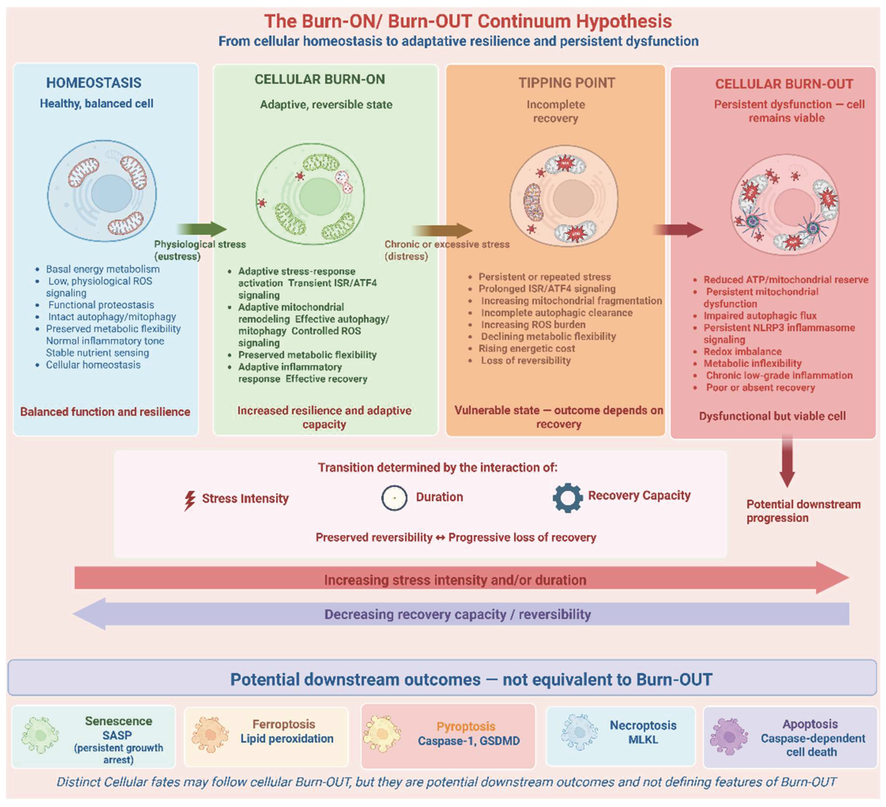

Table 1.
Proposed biological characteristics across the Burn-ON/Burn-OUT continuum.
| Domain | Burn-ON | Tipping Point | Burn-OUT | References |
| Sress response | Inducible and reversible | Delayed recovery | Persistent or dysregulated | [3,11,12] |
| ATP production | Preserved and responsive to demand | Declining energetic reserve | Reduced | [9] |
| Mitochondrial dynamics | Adaptive remodeling | Increasing fragmentation | Persistent fragmentation and dysfunction | [9,16,22] |
| Mitophagy | Effective quality control | Increased demand / incomplete clearance | Ineffective or dysregulated | [14,15] |
| Autophagy | Functional flux | Incomplete clearance | Impaired flux | [5,18,2]] |
| ROS | Controlled redox signaling | Increasing oxidative burden | Persistent oxidative stress | [1,9,31] |
| ISR/ATF4 | Adaptive and transient | Prolonged activation | Persistent or maladaptive | [2,3,11,12] |
| NLRP3 | Controlled and resolvable | Incomplete resolution | Chronic activation | [6,19,20] |
| Metabolic flexibility | Preserved | Declining | Impaired | [7,21] |
| Senescence | Absent or limited | Possible emergence | Increased | [8,28] |
| Recovery | Effective | Delayed or incomplete | Poor or absent | [3,64] |
| Cell death | Absent | Usually absent | Possible late consequence | [30,31] |
Note: The proposed characteristics integrate evidence from established stress-response pathways but do not represent validated diagnostic thresholds. The Burn-ON, tipping-point, and Burn-OUT classifications are components of the hypothesis-generating framework proposed in this review.
Disclaimer/Publisher’s Note: The statements, opinions and data contained in all publications are solely those of the individual author(s) and contributor(s) and not of MDPI and/or the editor(s). MDPI and/or the editor(s) disclaim responsibility for any injury to people or property resulting from any ideas, methods, instructions or products referred to in the content. |
© 2026 by the authors. Licensee MDPI, Basel, Switzerland. This article is an open access article distributed under the terms and conditions of the Creative Commons Attribution (CC BY) license (http://creativecommons.org/licenses/by/4.0/).
Copyright: This open access article is published under a Creative Commons CC BY 4.0 license, which permit the free download, distribution, and reuse, provided that the author and preprint are cited in any reuse.