

Submitted:
21 September 2026
Posted:
22 September 2026
You are already at the latest version
Abstract
Background: Hair loss is a prevalent but underaddressed consequence of the menopausal transition, reflecting the simultaneous decline of estrogen, progesterone, and the protective estrogen:androgen ratio that governs scalp follicular biology. Methods: Following SANRA guidelines, a systematic literature search was conducted across PubMed/MEDLINE and EMBASE (January 2000–September 2026) using MeSH and free-text terms encompassing female pattern hair loss (FPHL), menopause, sex steroids, hormone replacement therapy (HRT), 5-alpha-reductase, and dihydrotestosterone (DHT), among others.[25] Key Findings: Estrogen loss shortens anagen duration and amplifies follicular DHT accumulation through three converging mechanisms. Progesterone withdrawal removes an underappreciated anti-androgenic buffer via competitive 5-alpha-reductase inhibition. Testosterone, though gradually declining with age, becomes relatively more androgenic as estrogenic modulation of its conversion is lost. For eligible women, systemic transdermal estradiol combined with a hair-favorable progestogen — micronized progesterone or drospirenone — provides the most comprehensive follicular protection. Topical scalp estradiol constitutes a distinct hair-directed local modality. A structured hormonal panel including the testosterone:DHT ratio guides antiandrogen selection and treatment monitoring; serum DHT measured by LC-MS/MS functions as a pharmacodynamic marker, not a standalone diagnostic test for female pattern hair loss. Conclusions: Integrating hormonal hair assessment into routine gynecological-endocrinological practice — with attention to progestogen androgenicity, route of estrogen delivery, and the role of hormone replacement therapy as an adjunctive rather than primary hair indication — can meaningfully reduce the undertreatment of this prevalent dimension of menopausal women's health.
Keywords:
female pattern hair loss
; menopause
; estrogen
; progesterone
; testosterone
; dihydrotestosterone
; 5-alpha-reductase
; hormone replacement therapy
; SHBG
; trichology
Methods
This narrative review was conducted in accordance with the SANRA guidelines for the quality assessment of narrative review articles.[25] A systematic literature search was performed across PubMed/MEDLINE and EMBASE, covering publications from January 2000 to September 2026, supplemented by manual reference tracking of relevant primary studies identified in the bibliographies of included articles and guidelines. The following Medical Subject Headings (MeSH) and free-text terms were used individually and in Boolean combination: “female pattern hair loss,” “alopecia,” “menopause,” “estrogen,” “estradiol,” “progesterone,” “testosterone,” “dihydrotestosterone,” “5-alpha-reductase,” “SHBG,” “androgen receptor,” “aromatase,” “CYP19A1,” “SRD5A1,” “SRD5A2,” “hormone replacement therapy,” “progestogen,” “spironolactone,” “minoxidil,” and “trichology.” Inclusion criteria encompassed original research studies (including clinical trials and observational studies), systematic reviews, meta-analyses, and consensus guidelines published in English addressing the intersection of sex steroid hormones and scalp hair health in adult women. No date restriction was applied to seminal mechanistic papers predating 2000. Studies focused exclusively on male pattern hair loss, alopecia areata, or cicatricial alopecias without primary relevance to female hormonal hair loss were excluded. This article does not report original research; therefore, no ethical approval was required.
1. Introduction
Among the constellation of symptoms that define the menopausal transition, hair changes are among the most distressing and yet the least integrated into routine clinical gynecological care.[1,2] Cross-sectional surveys report that a significant proportion of perimenopausal and postmenopausal women experience increased shedding, reduced density, reduced shaft caliber, and altered texture, and that these changes carry a measurable negative impact on quality of life, psychological well-being, and self-image independent of other climacteric symptoms.[1] Despite this prevalence and impact, hair loss in midlife women is frequently addressed in clinical isolation from its hormonal context, leading to management strategies that treat the downstream trichological consequence without addressing the upstream endocrine drivers.
The reproductive endocrinologist or gynecologist who counsels a woman about HRT is ideally positioned to understand and integrate the follicular biology that explains why her hair is changing, what specific hormonal losses and imbalances are driving it, what happens to the follicle if those changes are not addressed, and how to construct a rational, individualized risk-benefit assessment that includes hair outcomes as one legitimate dimension of the overall clinical picture.[4,5] This review provides that integrated framework, grounded in the physiology of hormonal action on the hair follicle and extending to the practical clinical and laboratory tools available for evaluation and treatment.
2. Hormonal Physiology of the Hair Follicle: The Biological Foundation
2.1. The Hair Growth Cycle and Sex Steroid Receptors
Each scalp hair follicle operates as an autonomous mini-organ, cycling asynchronously through three principal phases: anagen (active growth, lasting 2 to 6 years under normal premenopausal conditions), catagen (apoptosis-driven involution, 2 to 3 weeks), and telogen (quiescence lasting 2 to 4 months), followed by shedding of the hair shaft during the exogen sub-phase (Figure 1).[3] Under normal reproductive-age conditions, approximately 85-90% of scalp follicles remain in anagen at any given moment; this ratio is the primary determinant of perceived hair density, and its progressive decline is the central pathophysiological event in menopausal hair loss.[1,3]
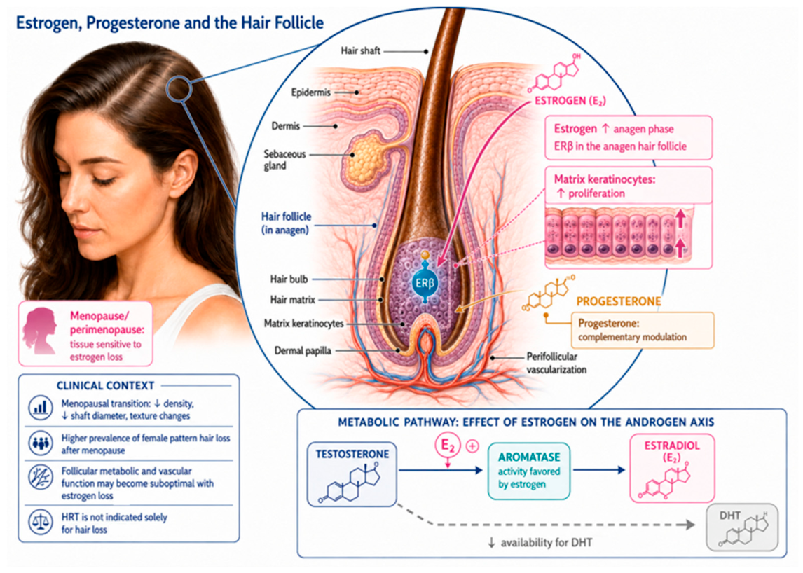
Sex steroid receptors are expressed throughout the follicle in a phase-specific and zone-specific manner. Estrogen receptors, particularly ER-beta, are strongly expressed in the outer root sheath and matrix keratinocytes during anagen, confirming estrogen's direct role in maintaining the growth phase.[3,9] Androgen receptors (AR) are concentrated in the dermal papilla, the mesenchymal signaling center that governs follicular cycling, across all phases.[10,11] Progesterone receptors have also been documented in the outer root sheath, consistent with evidence of direct sex steroid modulation of the hair follicle across all three major hormonal axes.[4,30] This receptor distribution means the follicle is simultaneously and continuously responsive to all three major sex steroids, and that changes in the relative balance of estrogenic, progestogenic, and androgenic signaling produce integrated rather than isolated follicular responses.[1,3]
Figure 1.
Estrogen, progesterone, and the hair follicle: molecular targets and effects during anagen. Estrogen acts via estrogen receptor-beta (β, ERβ), abundantly expressed in the hair bulb and matrix keratinocytes, stimulating keratinocyte proliferation and prolonging anagen duration [3,9]. Progesterone provides complementary anti-androgenic modulation at the follicular level [4,30]. The metabolic pathway panel illustrates how aromatase activity, favored by estrogen-rich conditions, converts testosterone to estradiol and reduces substrate availability for 5-alpha-reductase-mediated DHT synthesis [11]. The progressive loss of this estrogenic and progestogenic support during the menopausal transition underpins the accelerated follicular miniaturization that defines female pattern hair loss in postmenopausal women [1,2,4]. Author-created figure, based on [1,2,3,4,9].
Figure 1.
Estrogen, progesterone, and the hair follicle: molecular targets and effects during anagen. Estrogen acts via estrogen receptor-beta (β, ERβ), abundantly expressed in the hair bulb and matrix keratinocytes, stimulating keratinocyte proliferation and prolonging anagen duration [3,9]. Progesterone provides complementary anti-androgenic modulation at the follicular level [4,30]. The metabolic pathway panel illustrates how aromatase activity, favored by estrogen-rich conditions, converts testosterone to estradiol and reduces substrate availability for 5-alpha-reductase-mediated DHT synthesis [11]. The progressive loss of this estrogenic and progestogenic support during the menopausal transition underpins the accelerated follicular miniaturization that defines female pattern hair loss in postmenopausal women [1,2,4]. Author-created figure, based on [1,2,3,4,9].
2.2. Local Steroidogenesis: The Tissue-Level Determinant of Follicular Fate
The androgenic or estrogenic character of the follicular microenvironment is determined not primarily by circulating hormone levels, but by local enzymatic activity within follicular cells themselves (Figure 2).[11] Sebocytes and outer root sheath keratinocytes express both isoenzymes of 5-alpha-reductase (types 1 and 2), which convert testosterone to the highly potent androgen dihydrotestosterone (DHT), and aromatase (CYP19A1), which converts testosterone to estradiol.[9,10,11] The relative activity of these competing pathways, which varies by scalp region, individual genetics, and hormonal context, determines whether a given follicle operates in a predominantly estrogenic (anagen-favorable) or androgenic (miniaturization-prone) microenvironment.[10,11] This local steroidogenesis explains the classical “androgen paradox” of hair biology: the same systemic androgen load may produce miniaturization in the androgen-sensitive frontoparietal scalp while having no deleterious effect in the androgen-resistant occipital region, because of region-specific differences in AR density and 5-AR isoenzyme expression rather than differences in circulating hormone levels.[11]
A critical consequence of this local metabolism is that serum DHT concentration does not reliably predict the degree of androgenic activity within the follicle. DHT is synthesized intracellularly in the dermal papilla, acts locally through intracellular AR binding without substantial re-entry into the bloodstream, and circulating serum DHT reflects peripheral spillover from multiple tissue sources rather than follicular DHT concentration.[6] The clinical implications of this dissociation are discussed in detail in Section 5.3.
3. The Menopausal Transition and Its Consequences for Scalp Hair
3.1. The Estrogen Decline: Mechanisms and Follicular Consequences
The menopausal transition is defined endocrinologically by a progressive and ultimately near-total loss of ovarian follicular function, producing a steep decline in circulating estradiol from premenopausal concentrations of 100 to 400 pg/mL during the follicular phase, to postmenopausal concentrations typically below 15 to 20 pg/mL.[1,5] This estrogen loss affects the hair follicle through at least three distinct mechanisms that converge on the same outcome: shortening of anagen duration and acceleration of follicular miniaturization (Figure 3).[1]
First, the direct ER-beta-mediated signaling that prolongs anagen in the dermal papilla and matrix keratinocytes is reduced, shortening the growth phase of each successive hair cycle and producing finer, shorter hairs with each subsequent round.[3,9] Second, the estrogen-dependent upregulation of hepatic and follicular SHBG is lost; with declining SHBG, the free fraction of any remaining circulating testosterone rises, making more substrate available for 5-AR-mediated conversion to DHT within the follicle.[1,10] Third, estrogen's induction of local aromatase activity within follicular cells is withdrawn, reducing the capacity for intrafollicular testosterone-to-estradiol conversion and further shifting the local balance toward DHT accumulation.[9,11]
The net effect is a relative androgenization of the follicular microenvironment even in the absence of any absolute increase in circulating testosterone. This mechanism explains why the prevalence of female pattern hair loss (FPHL), defined by progressive thinning of the crown and frontal scalp through follicular miniaturization, increases from approximately 12% in premenopausal women to over 50% by age 70,[1] a trajectory that directly mirrors the progressive loss of the estrogenic protective buffer established during the reproductive years.[1,3]
In addition to these cycle-level changes, estrogen deprivation affects the hair fiber itself: postmenopausal women frequently report a change in texture (often described as coarser, more brittle, or drier), loss of shine (reflecting reduced sebaceous gland activity, which is estrogen-dependent), and increased mechanical fragility.[2] These textural changes reflect altered hair-shaft keratin composition and reduced hydration of the cortex, and are at least partially reversible with estrogen replacement, providing an additional trichological argument for HRT in appropriate candidates.[2,9]
Figure 3.
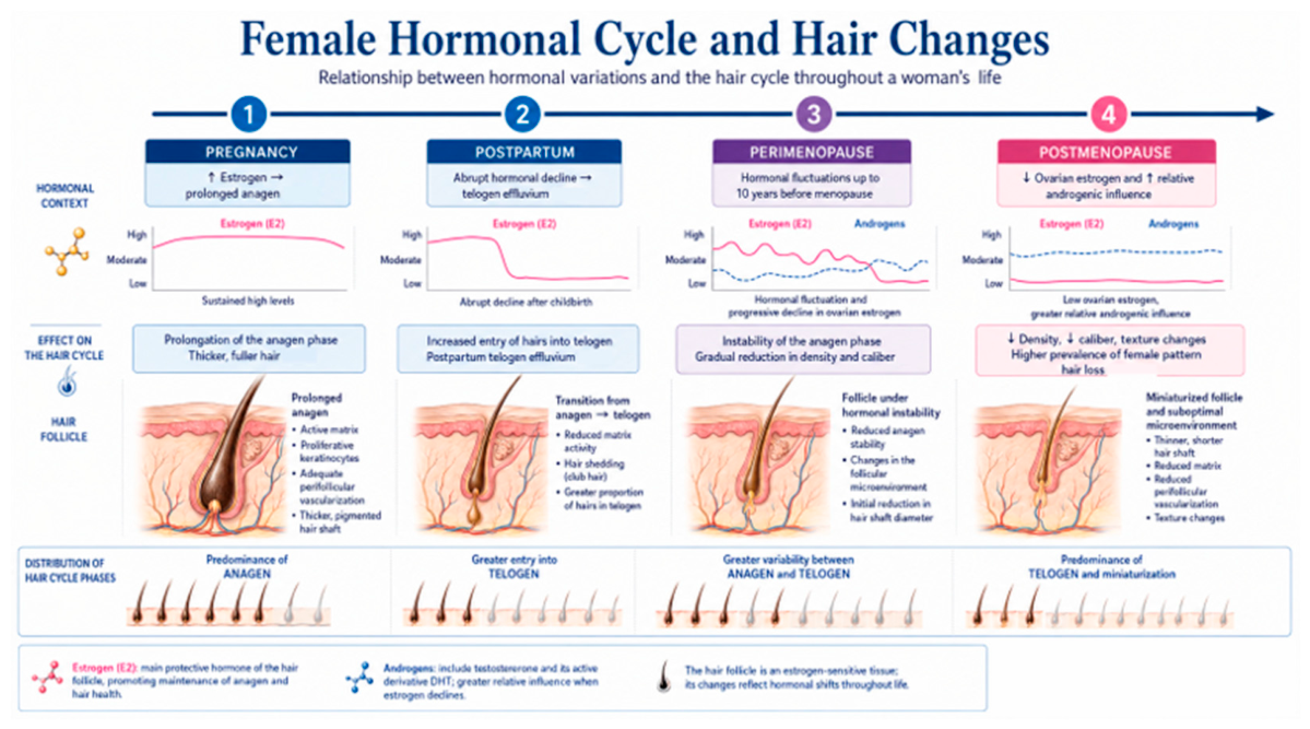
Hormonal variations and their effect on the hair cycle across a woman's reproductive life, organized by reproductive stage according to the STRAW+10 framework [26]. Each stage — pregnancy, postpartum, perimenopause, and postmenopause — is characterized by a specific hormonal context (upper row) that produces predictable changes in follicular biology and clinical hair quality (middle rows). During pregnancy, elevated estrogen (E₂) prolongs anagen and produces thicker, fuller hair [3,9]. The abrupt postpartum estrogen decline triggers synchronized follicular entry into telogen, manifesting as postpartum telogen effluvium [3,9]. Perimenopause introduces erratic hormonal fluctuations that destabilize anagen stability and gradually reduce hair density and caliber [1]. Postmenopause is characterized by persistently low ovarian estrogen and relative androgenic predominance, generating the highest population prevalence of FPHL and progressive follicular miniaturization [1]. Author-created figure, based on [1,3,9].
Figure 3.
Hormonal variations and their effect on the hair cycle across a woman's reproductive life, organized by reproductive stage according to the STRAW+10 framework [26]. Each stage — pregnancy, postpartum, perimenopause, and postmenopause — is characterized by a specific hormonal context (upper row) that produces predictable changes in follicular biology and clinical hair quality (middle rows). During pregnancy, elevated estrogen (E₂) prolongs anagen and produces thicker, fuller hair [3,9]. The abrupt postpartum estrogen decline triggers synchronized follicular entry into telogen, manifesting as postpartum telogen effluvium [3,9]. Perimenopause introduces erratic hormonal fluctuations that destabilize anagen stability and gradually reduce hair density and caliber [1]. Postmenopause is characterized by persistently low ovarian estrogen and relative androgenic predominance, generating the highest population prevalence of FPHL and progressive follicular miniaturization [1]. Author-created figure, based on [1,3,9].
3.2. Progesterone: The Overlooked Anti-Androgenic Buffer
Progesterone's contribution to hair health during the reproductive years has received considerably less attention than estrogen's, but its withdrawal at menopause removes an important and clinically underappreciated anti-androgenic protective mechanism. Consistent with pharmacological evidence from progestogen receptor-binding and enzyme-inhibition studies, the most significant action of progesterone at the follicular level is believed to be competitive inhibition of 5-alpha-reductase isoenzymes, reducing intrafollicular DHT synthesis from the available testosterone pool.[4,27,28] It should be noted that this evidence derives primarily from in vitro pharmacological studies and indirect clinical observations; direct proof of follicular 5-AR inhibition by progesterone in human hair follicle models in vivo is not yet available.[27,28] Progesterone also competes with DHT for androgen receptor binding at pharmacological concentrations, further attenuating androgenic follicular signaling.[4,27] These two mechanisms mean that adequate progesterone during the luteal phase of reproductive-age cycles likely serves as a biological buffer against androgenic hair loss, and the cyclic absence of progesterone beginning in perimenopause followed by its complete cessation postmenopausally removes this buffer entirely.[1,4]
The clinical relevance of this progesterone-mediated protection extends directly to the choice of progestogen in combined HRT regimens. Synthetic progestogens differ markedly in their androgen receptor-binding affinity and 5-AR inhibitory potential (Table 1).[4,27,28,31] For a woman with concurrent FPHL or a family history of androgenetic alopecia, the progestogen component of a combined HRT regimen is not interchangeable with respect to hair outcomes, and its selection should be guided by the androgenic risk profile of each molecule.[4,31] Micronized progesterone and drospirenone represent the most hair-favorable options currently available; androgenic 19-nortestosterone derivatives such as levonorgestrel and norethisterone should be avoided when alternatives are clinically feasible.[2,4]
3.3. Testosterone in Aging Women: Physiological Decline, Dual Follicular Role, and Individual Risk
Testosterone is the most complex hormonal variable in the context of menopausal hair loss because its trajectory in aging women is more nuanced than commonly described, and because its net effect on the hair follicle depends not on its circulating concentration but on the balance between two competing downstream enzymatic pathways: aromatization to estradiol or 5-AR-mediated conversion to DHT.[7,8]
Longitudinal data from the Study of Women's Health Across the Nation (SWAN), the Melbourne Women's Midlife Health Project, and recent Monash University analyses consistently indicate that total testosterone in women declines primarily as a function of chronological aging rather than of menopause specifically.[7,8] The decline averages approximately 1 to 2% per year from the third decade onward.[7,8] Natural menopause, in the absence of oophorectomy, does not typically produce an acute further decline in testosterone because the postmenopausal ovarian stroma retains androgen-secreting theca cell capacity, and adrenal DHEA-S continues its age-related decline independently of menopausal status.[7,8] Bilateral oophorectomy, however, removes approximately 50% of circulating testosterone acutely, and is consistently associated with an acceleration of FPHL in susceptible women.[7]
The dual role of testosterone at the hair follicle creates a clinically important paradox. Studies of testosterone supplementation in women with confirmed androgen deficiency following oophorectomy or adrenal insufficiency show improvements in scalp hair density and growth rate with restoration of physiological testosterone levels.[12] FPHL acceleration was not observed when doses remained within the normal female range.[12] On the other side of the paradox, when the estrogen-androgen ratio shifts toward androgenicity during the natural menopausal transition, the same testosterone that at physiological concentrations had an anabolic follicular effect now provides substrate for enhanced DHT production in the context of reduced aromatase induction and reduced SHBG, accelerating miniaturization in susceptible follicles.[1,8,11] The critical variables are therefore not the testosterone concentration itself, but: (a) SHBG, which governs free testosterone availability for 5-AR conversion; (b) 5-AR isoenzyme activity at the follicular level, which is genetically determined but also modulated by metabolic factors including insulin resistance; and (c) the absolute estrogen level, which determines aromatase induction and the competing pathway for testosterone disposal.[8,10,11]
The risk of testosterone supplementation accelerating FPHL in postmenopausal women is real but manageable. It is most significant in three scenarios: supraphysiological dosing (particularly with oral testosterone, which generates DHT peaks during intestinal and hepatic first-pass metabolism); pre-existing low SHBG from insulin resistance, obesity, or hypothyroidism; and genetic susceptibility to high follicular 5-AR activity or AR sensitivity.[8,12] When testosterone supplementation is clinically indicated (principally for hypoactive sexual desire disorder following oophorectomy), transdermal delivery at the lowest effective dose, close monitoring of total and free testosterone, DHT, and SHBG, and a low threshold for adding a 5-AR inhibitor in susceptible women, constitute a rational and manageable approach.[4,8]
4. Hormone Replacement Therapy: Hair Benefits and the Cost of Withholding Treatment
4.1. Evidence for Hair Benefit: Systemic Transdermal HRT Versus Topical Scalp Estradiol
4.1.1. Systemic Transdermal Hormone Replacement Therapy
Systemic HRT with estradiol is not currently indicated, approved, or recommended as a primary or stand-alone treatment for hair loss in menopausal women.[5,29] Any decision to initiate hormone therapy must be governed by the overall risk-benefit profile, principally driven by vasomotor and genitourinary symptom burden, bone health, and chronological distance from the final menstrual period.[2,5,29] For women who already meet clinical criteria for HRT on these grounds, ancillary trichological benefits constitute a legitimate and important component of informed, shared decision-making.[1,4]
The mechanisms by which systemic transdermal estradiol is expected to benefit scalp hair are threefold. First, circulating estradiol restores ER-beta-mediated anagen-prolonging signaling in the dermal papilla and matrix keratinocytes.[3,9] Second, estrogen re-establishes hepatic and follicular SHBG upregulation, reducing the free testosterone fraction available for 5-AR-mediated DHT conversion — an effect achieved most effectively via transdermal rather than oral estrogen, which avoids the first-pass hepatic metabolism that at some doses can paradoxically suppress SHBG.[5] Third, systemic estradiol partially restores follicular aromatase induction, shifting the local testosterone pool toward estradiol and away from DHT.[9,11] The available clinical evidence supporting these mechanisms consists principally of observational data showing better hair density and lower FPHL prevalence in women using systemic HRT compared with age-matched non-users, and of indirect physiological evidence from high-estrogen states such as pregnancy.[1,3,4,9] It must be explicitly acknowledged that adequately powered, hair-specific randomized controlled trials of systemic HRT are currently lacking; systemic HRT therefore remains an adjunctive consideration for hair, not a hair-directed therapeutic agent.
The choice of progestogen in combined HRT regimens determines whether the progesterone component adds hair protection — through 5-AR inhibition and anti-androgenic AR competition, as with micronized progesterone and drospirenone — or partially offsets the estrogenic benefit, as with androgenic 19-nortestosterone derivatives.[4,27,28,31] HRT combining transdermal estradiol with micronized progesterone or drospirenone is therefore the formulation most likely to confer net benefit on hair outcomes in women with concurrent FPHL.[4]
4.1.2. Topical Scalp Estradiol: A Hair-Directed Local Modality
Topical scalp estradiol — typically formulated as a 0.025% solution, gel, or compounded lotion applied directly to the scalp — is a conceptually and clinically distinct modality that must not be conflated with systemic HRT. Its rationale is the direct local activation of follicular ER-beta without generating clinically meaningful systemic estradiol concentrations, thereby providing anagen-prolonging and local aromatase-inducing effects at the target tissue while avoiding the systemic estrogen exposure that drives the thromboembolic, endometrial, and breast risk considerations governing HRT candidacy.[9,30] Absorption of estradiol through the scalp at the concentrations used in hair-directed formulations is low, and plasma estradiol levels following topical scalp application at standard doses typically remain within or close to postmenopausal reference ranges, distinguishing this approach pharmacokinetically from transdermal HRT patches or gels applied to non-scalp skin sites.
The evidence base for topical scalp estradiol consists of small uncontrolled and partially controlled studies reporting increases in the anagen hair fraction and improvements in hair density following regular scalp application, consistent with the ER-beta-mediated mechanism described in Section 2.1.[9,11] This evidence is limited by small sample sizes, heterogeneous formulation protocols, predominantly unblinded designs, and the absence of adequately powered randomized controlled trials. Topical scalp estradiol is not available as a licensed pharmaceutical product in most markets and is typically prepared by compounding pharmacies, which introduces additional variability in formulation quality, delivered dose, and vehicle penetration characteristics. Its use should therefore be supervised by a specialist with awareness of these evidence and quality limitations.
The practical clinical distinction between the two modalities can be summarized as follows: systemic transdermal HRT is appropriate for the menopausal woman who meets established HRT indications and stands to benefit from estradiol's systemic effects across multiple target organs simultaneously, with hair as an ancillary benefit; topical scalp estradiol is a hair-directed option for women who wish to target the follicular microenvironment locally without systemic hormonal exposure — including women who are not HRT candidates, who have already completed an HRT course, or who seek an adjunctive local treatment alongside systemic therapy.[1,4,9]
Figure 4.
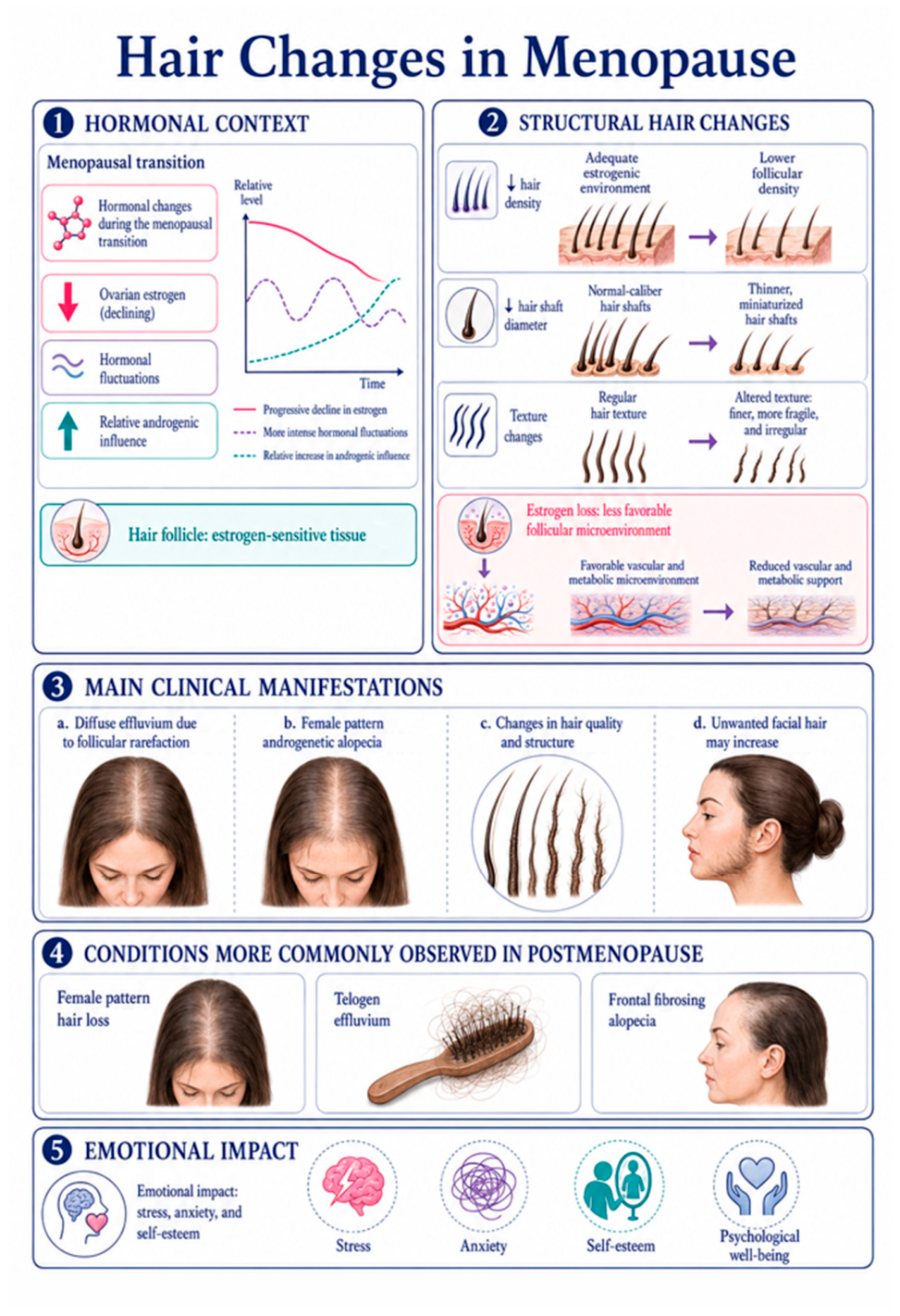
Structural, clinical, and psychosocial consequences of hair changes during the menopausal transition. Panel 1 (Hormonal context): the menopausal transition is characterized by progressive decline in ovarian estrogen, more intense hormonal oscillations, and relative increase in androgenic influence; the hair follicle constitutes an estrogen-sensitive target tissue [1]. Panel 2 (Structural hair changes): estrogen loss leads to measurable reductions in follicular density, hair shaft diameter, and fiber regularity, accompanied by a less favorable vascular and metabolic follicular microenvironment [1]. Panel 3 (Main clinical manifestations): principal presentations include diffuse effluvium due to follicular rarefaction, female pattern androgenetic alopecia, changes in hair fiber quality and structure, and paradoxical increase in unwanted facial hair [1,2]. Panel 4 (Conditions more commonly observed in postmenopause): female pattern hair loss, chronic telogen effluvium, and frontal fibrosing alopecia represent the predominant trichological diagnoses in this population [1,23]. Panel 5 (Emotional impact): hair changes carry a significant psychosocial burden, adversely affecting self-esteem, increasing anxiety, and impairing overall psychological well-being [1,2]. Author-created figure, based on [1,2,9].
Figure 4.
Structural, clinical, and psychosocial consequences of hair changes during the menopausal transition. Panel 1 (Hormonal context): the menopausal transition is characterized by progressive decline in ovarian estrogen, more intense hormonal oscillations, and relative increase in androgenic influence; the hair follicle constitutes an estrogen-sensitive target tissue [1]. Panel 2 (Structural hair changes): estrogen loss leads to measurable reductions in follicular density, hair shaft diameter, and fiber regularity, accompanied by a less favorable vascular and metabolic follicular microenvironment [1]. Panel 3 (Main clinical manifestations): principal presentations include diffuse effluvium due to follicular rarefaction, female pattern androgenetic alopecia, changes in hair fiber quality and structure, and paradoxical increase in unwanted facial hair [1,2]. Panel 4 (Conditions more commonly observed in postmenopause): female pattern hair loss, chronic telogen effluvium, and frontal fibrosing alopecia represent the predominant trichological diagnoses in this population [1,23]. Panel 5 (Emotional impact): hair changes carry a significant psychosocial burden, adversely affecting self-esteem, increasing anxiety, and impairing overall psychological well-being [1,2]. Author-created figure, based on [1,2,9].
4.2. The Trichological Cost of Withholding HRT
For a woman with progressive hair thinning as part of her climacteric symptom burden, the decision not to use HRT, when no absolute contraindication exists, carries a specific trichological cost that deserves explicit clinical communication during counseling. Without estrogen replacement, progressive anagen shortening continues unchecked: each successive hair cycle produces a shorter, finer hair, and over years this cumulative miniaturization produces a degree of follicular atrophy that may become irreversible.[1,2] Histopathological studies of scalp biopsies in androgenetic alopecia have documented the progressive replacement of follicular units by fibrous tracts; follicles that have undergone such fibrotic replacement of the dermal papilla are unlikely to regenerate even with subsequent hormonal or pharmacological treatment.[10,32]
By analogy with the well-established timing hypothesis in HRT for cardiovascular and bone protection, where therapy initiated in early postmenopause (within ten years of the final menstrual period) consistently demonstrates greater benefit than therapy started after prolonged estrogen deprivation has produced structural changes in target tissues,[5] it is biologically plausible that a comparable window of opportunity exists for follicular protection. While direct evidence from hair-specific longitudinal studies is currently lacking, the progressive and at least partially irreversible nature of follicular atrophy provides mechanistic support for early intervention as a clinical principle; the specific trichological benefit of the timing effect has not yet been formally quantified in prospective studies and awaits dedicated investigation.[1,4,5]
For women who are not candidates for systemic HRT or who decline it, this same principle of early action applies to non-hormonal hair-directed therapy: early initiation of topical minoxidil, spironolactone, or low-dose oral minoxidil provides greater trichological benefit when follicular miniaturization has not yet reached irreversible atrophy.[8,13,14] The hormonal context should therefore guide the urgency of treatment initiation, regardless of whether that treatment is hormonal or non-hormonal.
5. Risk-Benefit Framework for Hormonal Assessment and Treatment
5.1. Clinical Decision Steps
The risk-benefit assessment for HRT in a menopausal woman presenting with hair loss should follow the same evidence-based structure as for any other climacteric indication, with hair outcomes integrated explicitly rather than deferred to a separate consultation.[4,5] The assessment proceeds through four sequential steps:
- Step 1 — Establish menopausal stage and overall symptom burden. Document reproductive stage using the STRAW+10 framework,[26] severity of vasomotor, genitourinary, and musculoskeletal symptoms, bone mineral density status, and the chronological distance from the final menstrual period. A woman in early postmenopause with moderate-to-severe vasomotor symptoms plus progressive hair loss is a straightforward candidate for HRT; the hair benefit is additive to the primary indication.[1,5]
- Step 2 — Screen for absolute and relative HRT contraindications. These include personal history of hormone-sensitive breast cancer, unresolved unexplained genital bleeding, active thromboembolic disease, and severe hepatic dysfunction. Their absence, together with an appropriate benefit profile, justifies HRT initiation; their presence requires a documented and shared decision about alternative management.[2,5,29]
- Step 3 — Conduct a structured hormonal panel (Section 5.2) to characterize the androgenic environment driving hair loss, guide progestogen and formulation selection, and establish a monitoring baseline for therapeutic response assessment.[1,6]
- Step 4 — Select the HRT formulation guided by the androgenic risk profile. Prefer transdermal estradiol (to avoid first-pass hepatic effects that may suppress SHBG and paradoxically increase free androgen availability with some oral estrogen formulations)[5] combined with a progestogen of low or no androgenicity. In women with concurrent clinical or biochemical hyperandrogenism contributing to FPHL, consider adding a hair-directed antiandrogen (spironolactone 50-200 mg/day, or low-dose finasteride) to the HRT regimen.[4,13]
5.2. The Hormonal Panel: What to Measure
A structured hormonal assessment in a perimenopausal or postmenopausal woman with hair loss should include, at minimum: total testosterone, free testosterone or calculated free testosterone, SHBG, DHEA-S, estradiol, FSH, and TSH.[1,10] When clinical or biochemical signs of androgen excess are present (hirsutism, acne, virilization, or elevated free testosterone), androstenedione and 17-hydroxyprogesterone should be added to screen for late-onset congenital adrenal hyperplasia.[10] Table 2 summarizes the recommended panel with reference ranges and the specific clinical utility of each marker in the hair loss evaluation.
5.3. Serum DHT: Utility, Limitations, and the Right Clinical Question
Dihydrotestosterone (DHT), with an androgen receptor binding affinity approximately three to five times that of testosterone, is the androgen most directly responsible for follicular miniaturization via dermal papilla AR activation.[10,11] Despite this central mechanistic role, serum DHT should not function as a primary diagnostic test for FPHL. A diagnostic study measuring serum DHT in women with androgenetic alopecia versus controls found elevated concentrations in most FPHL cases but also in a proportion of controls without hair loss, demonstrating the poor specificity of serum DHT as a standalone diagnostic marker.[6] This aligns with the understanding that follicular sensitivity to DHT, governed by AR gene polymorphisms and local 5-AR isoenzyme expression density, is at least as important a determinant of FPHL as the absolute DHT concentration.[11,16]
On the analytical side, serum DHT concentrations in women are extremely low, typically below 0.10 ng/mL in premenopausal and below 0.08 ng/mL in postmenopausal women. This places them at or below the reliable detection threshold of conventional immunoassay platforms.[6] Liquid chromatography-tandem mass spectrometry (LC-MS/MS) is mandatory for clinically meaningful DHT measurement in women. Immunoassay results at these concentrations carry coefficients of variation that can encompass the entire clinically meaningful range, producing essentially uninterpretable quantitative data. Before ordering serum DHT, the clinician should confirm that the receiving laboratory uses LC-MS/MS methodology.
Given these limitations, serum DHT provides genuine value in the following specific contexts:
- Monitoring 5-alpha-reductase inhibitor therapy: finasteride at 1-5 mg/day should reduce serum DHT by 60-70%; dutasteride 0.5 mg/day by more than 90%.[11] Serum DHT measurement at 3 to 6 months after initiation confirms adequate pharmacological 5-AR suppression and permits dose titration. The testosterone:DHT ratio, which rises proportionally under effective 5-AR inhibition, is a more specific pharmacodynamic marker than DHT alone.[6]
- Identifying enhanced 5-AR activity in women with normal total testosterone but clinical androgen excess: an elevated serum DHT, particularly combined with a low testosterone:DHT ratio, corroborates the clinical picture and supports the use of a 5-AR inhibitor rather than, or in addition to, an androgen receptor antagonist such as spironolactone.[6,8]
- Calculating the testosterone:DHT ratio to guide 5-AR inhibitor selection: a low ratio (below 3:1) indicates dominant 5-AR type 2 activity and suggests the woman is likely to respond to finasteride. A normal or high ratio with persistent clinical androgen excess may point toward type 1 5-AR activity, supporting dutasteride over finasteride in refractory cases.[6]
- Baseline risk stratification before testosterone supplementation: in women for whom transdermal testosterone is being considered, a baseline serum DHT and testosterone:DHT ratio, combined with SHBG and metabolic status, allows identification of women with high-activity 5-AR conversion who may be at elevated risk of FPHL acceleration with even physiological testosterone supplementation, and can justify prophylactic use of a 5-AR inhibitor.[8,12]
In summary, serum DHT, measured by LC-MS/MS and interpreted alongside the full androgen panel and the testosterone:DHT ratio, contributes clinically meaningful information when the clinical question specifically pertains to 5-AR activity, 5-AR inhibitor monitoring, or testosterone supplementation safety. It should be included in the evaluation when the result is expected to change the therapeutic decision; it should not be used as a standalone screening test for hair loss.[6]
5.4. Genetic Analysis: Polymorphisms and Their Relationship with FPHL
Genetic evaluation and the study of polymorphisms are part of personalized medicine, including for patients with FPHL. Two key enzymes in steroidogenesis, CYP19A1 (aromatase) and SRD5A1/SRD5A2 (5-alpha-reductase), have been evaluated in studies on their relationship with FPHL.
CYP19A1: The CYP19A1 polymorphisms with reported association include rs4646, whose CC genotype was more frequent in women with FPHL in an Australian study, with an even stronger pattern in cases of onset before the age of 40.[17] This same finding was described in later reviews as one of the few repeatedly discussed positive findings for FPHL.[18] In a Chinese population, rs6493497 and rs7176005 showed significant differences in frequency between cases and controls, suggesting a population-specific association with FPHL.[19] In an Egyptian study, rs184895853 and rs116904472 were also associated with FPHL, with a higher frequency of the C allele in patients.[20] In early-onset FPHL, a Korean study found 49 significant SNPs around 5 candidate genes, including the CYP19A1 region, reinforcing that close variants of aromatase may participate at least in subgroups of women.[21]
SRD5A1 and SRD5A2: the largest case-control study specifically in women found no significant association of 21 selected variants with FPHL, neither in the global analysis nor in subgroups.[22] Reviews on FPHL genetics summarize this set as the absence of convincing evidence for genes of the steroid pathway, including SRD5A1/SRD5A2.[16]
In fact, the overall picture is mixed evidence: some CYP19A1 SNPs have been associated with female hair loss in Australian, Chinese, and Egyptian women, but European studies have not consistently confirmed these findings.[17,18,20] For 5-alpha-reductase, data specifically in women are weaker and tend to be negative, despite the biological plausibility of the androgen pathway and functional findings of enzyme expression in the follicle.[21,22] Thus, the polymorphisms most linked to hair loss in women in the available studies are those of CYP19A1 — especially rs4646, rs6493497, rs7176005, rs184895853, and rs116904472 — while variants of SRD5A1/SRD5A2 do not yet show a consistent association in women. It is probably too early to recommend routine genetic evaluation of CYP19A1 in clinical practice, given the lack of replication across ethnic populations and the absence of validated clinical utility thresholds.[16,18]
Table 3 provides a structured clinical reference summarizing recommended practices, common pitfalls, and emerging approaches for the management of scalp hair health in menopausal women, organized by clinical domain.
6. Discussion
The framework presented in this review reflects a fundamental reorientation of how hair loss in menopausal women should be understood and managed: not as a dermatological condition superimposed on the menopausal transition, but as an integral expression of the hormonal changes that define it. Estrogen, progesterone, and testosterone operate as an integrated follicular hormonal system, and the menopausal transition disrupts all three in ways that converge on progressive follicular miniaturization.[1,2,3]
The formulation decisions that flow from this framework have direct prescribing implications that extend beyond hairline management into the core of menopausal medicine. Transdermal estradiol outperforms oral routes for SHBG preservation; progestogen selection determines whether the combined regimen adds net hair protection or offsets estrogenic benefit; and the testosterone:DHT ratio provides the laboratory basis for personalizing antiandrogen therapy in women with inadequate response to single-drug interventions.[4,5,27,28,31] Collectively, these considerations transform an otherwise routine HRT consultation into an opportunity for comprehensive follicular protection — but only if hair outcomes are explicitly integrated into the shared decision-making process rather than deferred to a separate specialist referral.[1,4]
For women who are not HRT candidates or who decline systemic therapy, an evidence-graded non-hormonal approach provides a robust alternative. A large retrospective cohort demonstrated a favorable safety profile for low-dose oral minoxidil;[14] a 2024 blinded randomized clinical trial found that topical minoxidil combined with oral spironolactone produced superior hair density outcomes compared with topical minoxidil plus oral finasteride.[15] A systematic review and meta-analysis confirmed a pooled improvement rate of approximately 56.6% for oral spironolactone across published studies.[13] Frontal fibrosing alopecia, when present, should prompt scalp biopsy and dermatological referral given its potential for irreversible scarring.[23]
7. Conclusions
Hair loss in the menopausal transition is a multimechanistic consequence of the simultaneous decline of estrogen, progesterone, and the protective estrogen:androgen ratio that these hormones collectively maintain during reproductive life.[1,3] Estrogen loss removes both direct anagen-prolonging ER-beta signaling and indirect SHBG and aromatase-mediated protection from follicular DHT accumulation; progesterone withdrawal removes the competitive 5-alpha-reductase inhibitory buffer; and testosterone, while declining with age, becomes relatively more androgenic as estrogenic and progestogenic modulation of its local conversion is lost.[1,4,9,27] Hormone replacement therapy with transdermal estradiol and a hair-favorable progestogen represents the most comprehensive intervention available for eligible women, offering the greatest follicular protection when initiated early in the postmenopausal window.[4,5] For all women — whether or not they are HRT candidates — a structured hormonal panel including the testosterone:DHT ratio guides personalized antiandrogen selection, and serum DHT measured by LC-MS/MS provides pharmacodynamic monitoring rather than diagnosis.[6] Integrating these principles into routine gynecological-endocrinological practice will materially reduce the undertreatment of one of the most prevalent and impactful dimensions of women's midlife health.[1,2]
Author Contributions
Conceptualization, F.T.; methodology, F.T. and G.R.; formal analysis, F.T. and G.R.; investigation, F.T. and G.R.; resources, F.T. and G.R.; data curation, F.T. and G.R.; writing—original draft preparation, F.T.; writing—review and editing, F.T. and G.R.; visualization, F.T. All authors have read and agreed to the published version of the manuscript.
Funding
This research received no specific external funding.
Institutional Review Board Statement
Not applicable. This manuscript is a narrative review that does not involve original data collection from human participants or animal subjects.
Informed Consent Statement
Not applicable.
Data Availability Statement
No new data were created or analyzed in this study. Data sharing is not applicable to this article.
Conflicts of Interest
The authors declare no conflicts of interest relevant to the content of this manuscript. F.T. serves as Medical Director of an institution involved in hormonal therapeutics; this affiliation did not influence the selection, interpretation, or presentation of any evidence reviewed herein. G.R. declares no conflicts of interest.
References
- Gupta, A.K.; Economopoulos, V.; Mann, A.; Wang, T.; Mirmirani, P. Menopause and hair loss in women: exploring the hormonal transition. Maturitas 2025, 198, 108378. [Google Scholar] [CrossRef]
- Zouboulis, C.C.; Blume-Peytavi, U.; Kosmadaki, M.; et al. Skin, hair and beyond: the impact of menopause. Climacteric 2022, 25(5), 434–442. [Google Scholar] [CrossRef]
- Williams, R.; Pawlus, A.D.; Thornton, M.J. Getting under the skin of hair aging: the impact of the hair follicle environment. Exp. Dermatol. 2020, 29(7), 588–597. [Google Scholar] [CrossRef]
- Lee, J.; Krishnegowda, R.; Miteva, M.I. Hormone replacement therapy and hair: a review for trichologists treating menopausal women. Dermatology 2026, 242(3), 264–267. [Google Scholar] [CrossRef] [PubMed]
- Flores, V.A.; Pal, L.; Manson, J.E. Hormone therapy in menopause: concepts, controversies, and approach to treatment. Endocr. Rev. 2021, 42(6), 720–752. [Google Scholar] [CrossRef]
- Brzezińska-Wcisło, L.; Lis-święty, A.; Wcisło-Dziadecka, D.; Trzmiel, D. Assessment of the usefulness of dihydrotestosterone in the diagnostics of patients with androgenetic alopecia. Postep. Dermatol. Alergol. 2014, 31(4), 232–236. [Google Scholar] [CrossRef]
- Davison, S.L.; Bell, R.; Donath, S.; Montalto, J.G.; Davis, S.R. Androgen levels in adult females: changes with age, menopause, and oophorectomy. J. Clin. Endocrinol. Metab. 2005, 90(7), 3847–3853. [Google Scholar] [CrossRef] [PubMed]
- Davis, S.R.; Wahlin-Jacobsen, S. Testosterone in women: the clinical significance. Lancet Diabetes Endocrinol. 2015, 3(12), 980–992. [Google Scholar] [CrossRef] [PubMed]
- Verdier-Sévrain, S.; Bonté, F.; Gilchrest, B. Biology of estrogens in skin: implications for skin aging. Exp. Dermatol. 2006, 15(2), 83–94. [Google Scholar] [CrossRef] [PubMed]
- Ntshingila, S.; Oputu, O.; Arowolo, A.T.; Khumalo, N.P. Androgenetic alopecia: an update. JAAD Int. 2023, 13, 150–158. [Google Scholar] [CrossRef] [PubMed]
- Ustuner, E.T. Cause of androgenic alopecia: crux of the matter. Plast. Reconstr. Surg. Glob. Open 2013, 1(7), e64. [Google Scholar] [CrossRef] [PubMed]
- Gleeson, M.; Sheridan, C.; Dempsey, G.; et al. Improvement in scalp hair growth in androgen-deficient women treated with testosterone: a questionnaire study. Clin. Endocrinol. (Oxf) 2012, 76(3), 369–375. [Google Scholar] [CrossRef] [PubMed]
- Aleissa, M. The efficacy and safety of oral spironolactone in the treatment of female pattern hair loss: a systematic review and meta-analysis. Cureus 2023, 15(8), e43559. [Google Scholar] [CrossRef] [PubMed]
- Nohria, A.; Desai, D.; Sikora, M.; et al. Improving efficacy and maintaining safety in the treatment of alopecia with low-dose oral minoxidil and spironolactone combination therapy: a retrospective review. JAAD Int. 2024. [Google Scholar] [CrossRef]
- Sadeghzadeh Bazargan, A.; Tavana, Z.; Dehghani, A.; Jafarzadeh, A.; Tabavar, A.; Alavi Rad, E.; Goodarzi, A. The efficacy of the combination of topical minoxidil and oral spironolactone compared with topical minoxidil and oral finasteride in women with androgenic alopecia: a blinded randomized clinical trial. J. Cosmet. Dermatol. 2024, 23(2), 543–551. [Google Scholar] [CrossRef] [PubMed]
- Redler, S.; Messenger, A.G.; Betz, R.C. Genetics and other factors in the aetiology of female pattern hair loss. Exp. Dermatol. 2017, 26(6), 510–517. [Google Scholar] [CrossRef]
- Yip, L.; Zaloumis, S.; Irwin, D.; et al. Gene-wide association study between the aromatase gene (CYP19A1) and female pattern hair loss. Br. J. Dermatol. 2009, 161(2), 289–294. [Google Scholar] [CrossRef] [PubMed]
- Ho, C.Y.; Chen, J.Y.; Hsu, W.; et al. Female pattern hair loss: an overview with focus on the genetics. Genes. 2023, 14(7), 1326. [Google Scholar] [CrossRef] [PubMed]
- Rui, W.; Sheng, Y.; Hu, R.; et al. Association of single nucleotide polymorphisms in the CYP19A1 gene with female pattern hair loss in a Chinese population. Dermatology 2015, 231(3), 239–244. [Google Scholar] [CrossRef]
- Ahmed, N.E.; Shams, G.; Abdallah, O.; et al. Exploring the role of CYP19A1 single nucleotide polymorphisms in the pathogenesis of female pattern hair loss. Egypt. J. Dermatol. Venereol. 2025. [Google Scholar] [CrossRef] [PubMed]
- Ohn, J.; Son, H.Y.; Yu, D.A.; et al. Early onset female pattern hair loss: a case-control study for analyzing clinical features and genetic variants. J. Dermatol. Sci. 2022, 106(1), 15–23. [Google Scholar] [CrossRef] [PubMed]
- Redler, S.; Tazi-Ahnini, R.; Drichel, D.; et al. Selected variants of the steroid-5-alpha-reductase isoforms SRD5A1 and SRD5A2 and the sex steroid hormone receptors ESR1, ESR2 and PGR: no association with female pattern hair loss identified. Exp. Dermatol. 2012, 21(6), 483–485. [Google Scholar] [CrossRef] [PubMed]
- Sánchez, P.; Serrano-Falcón, C.; Torres, J.M.; Serrano, S.; Ortega, E. 5α-reductase isozymes and aromatase mRNA levels in plucked hair from young women with female pattern hair loss. Arch. Dermatol. Res. 2018, 310(1), 77–83. [Google Scholar] [CrossRef] [PubMed]
- Roman, A.M.; Petca, R.C.; Dumitraşcu, M.C.; et al. Frontal fibrosing alopecia and reproductive health: assessing the role of sex hormones in disease development. J. Pers. Med. 2024, 14(1), 72. [Google Scholar] [CrossRef] [PubMed]
- Baethge, C.; Goldbeck-Wood, S.; Mertens, S. SANRA — a scale for the quality assessment of narrative review articles. Res. Integr. Peer Rev. 2019, 4, 5. [Google Scholar] [CrossRef] [PubMed]
- Harlow, S.D.; Gass, M.; Hall, J.E.; et al. Executive summary of the Stages of Reproductive Aging Workshop + 10: addressing the unfinished agenda of staging reproductive aging. J. Clin. Endocrinol. Metab. 2012, 97(4), 1159–1168. [Google Scholar] [CrossRef] [PubMed]
- Raudrant, D.; Rabe, T. Progestogens with antiandrogenic properties. Drugs 2003, 63(5), 463–492. [Google Scholar] [CrossRef] [PubMed]
- Kuhl, H. Pharmacology of estrogens and progestogens: influence of different routes of administration. Climacteric 2005, 8 Suppl 1, 3–63. [Google Scholar] [CrossRef] [PubMed]
- The Menopause Society. The 2023 Menopause Society position statement. Menopause 2023, 30(6), 573–591. [Google Scholar] [CrossRef] [PubMed]
- Ohnemus, U.; Uenalan, M.; Inzunza, J.; Gustafsson, J.A.; Paus, R. The hair follicle as an estrogen target and source. Endocr. Rev. 2006, 27(6), 677–706. [Google Scholar] [CrossRef] [PubMed]
- Schindler, A.E.; Campagnoli, C.; Druckmann, R.; et al. Classification and pharmacology of progestins. Maturitas 2003, 46 Suppl 1, S7–16. [Google Scholar] [CrossRef] [PubMed]
- Whiting, D.A. Diagnostic and predictive value of horizontal sections of scalp biopsy specimens in male pattern androgenetic alopecia. J. Am. Acad. Dermatol. 1993, 28 5 Pt 1, 755–763. [Google Scholar] [CrossRef]
Figure 2.
Molecular mechanism of estrogen's protection of the follicular microenvironment during anagen. Estrogen acts through estrogen receptor-beta (ERβ), expressed in the outer root sheath and matrix keratinocytes, stimulating keratinocyte proliferation and maintenance of the anagen phase [3,9]. The inset metabolic pathway shows that testosterone is preferentially converted to estradiol by aromatase (CYP19A1) under estrogenic conditions, reducing substrate availability for 5-alpha-reductase and attenuating DHT accumulation in the dermal papilla [9]. The dashed arrow indicates the reduced DHT pathway when aromatase activity predominates. Author-created figure. Based on [9] Verdier-Sévrain S et al., Exp Dermatol. 2006 and [3] Williams R et al., Exp Dermatol. 2020.
Figure 2.
Molecular mechanism of estrogen's protection of the follicular microenvironment during anagen. Estrogen acts through estrogen receptor-beta (ERβ), expressed in the outer root sheath and matrix keratinocytes, stimulating keratinocyte proliferation and maintenance of the anagen phase [3,9]. The inset metabolic pathway shows that testosterone is preferentially converted to estradiol by aromatase (CYP19A1) under estrogenic conditions, reducing substrate availability for 5-alpha-reductase and attenuating DHT accumulation in the dermal papilla [9]. The dashed arrow indicates the reduced DHT pathway when aromatase activity predominates. Author-created figure. Based on [9] Verdier-Sévrain S et al., Exp Dermatol. 2006 and [3] Williams R et al., Exp Dermatol. 2020.
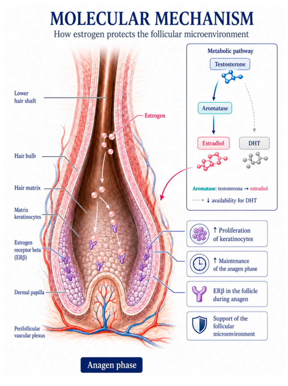

Table 1.
Androgenic and 5-alpha-reductase inhibitory profiles of progestogens used in menopausal hormone therapy.
Table 1.
Androgenic and 5-alpha-reductase inhibitory profiles of progestogens used in menopausal hormone therapy.
| Progestogen | Androgenicity | 5-AR Inhibition | Hair Safety Profile |
| Micronized progesterone (oral) | None / minimal | Moderate (via 5-alpha-reduced metabolites) | Preferred for hair-sensitive candidates; anti-androgenic indirect action through 5-AR inhibition [4] |
| Drospirenone | Anti-androgenic | Yes (also anti-mineralocorticoid) | Favorable; may add benefit in FPHL with concurrent fluid retention or hypertension [4] |
| Dienogest | Anti-androgenic (selective) | Yes (partial) | Emerging favorable profile; limited long-term hair-specific data in menopausal populations [4] |
| Medroxyprogesterone acetate | Mild androgenic activity | Minimal | Generally neutral for hair; used in major HRT trials without prominent hair-loss signal [5] |
| Norethisterone / levonorgestrel | Androgenic | None | Potentially unfavorable in women with FPHL susceptibility or low SHBG; avoid when alternatives are available [4] |
Table 1. Androgenicity classification based on androgen receptor binding affinity studies and clinical pharmacology data [4,27,28,31]. The selection of progestogen has direct clinical relevance for hair outcomes in women with pre-existing or family-history-positive FPHL. 5-AR: 5-alpha-reductase; WHI: Women's Health Initiative.
Table 2.
Recommended hormonal panel for menopausal women presenting with hair loss.
| Marker | Reference Range (Adult Women) | Role in the Androgenic Hair-Loss Workup |
| Total testosterone | 0.15–0.70 ng/mL | Identifies absolute androgen excess; normal result does not rule out androgen-driven FPHL, as follicular DHT activity depends on the free fraction and local 5-AR expression [10,11] |
| Free testosterone | 0.3–1.9 pg/mL | More informative than total testosterone when SHBG is suppressed or total T is borderline; preferred measurement is equilibrium dialysis or calculation from total T and SHBG (Vermeulen formula) [1,10] |
| SHBG | 18–144 nmol/L | Key regulator of free androgen bioavailability; suppressed by insulin resistance, obesity, hypothyroidism; low SHBG amplifies follicular DHT exposure even with normal total testosterone; rises with transdermal estrogens [1,2,4] |
| DHEA-S | 45–340 mcg/dL (declines >50% after age 50) | Reflects adrenal androgen reserve; elevated values suggest late-onset CAH or adrenal androgen excess; normal values still contribute to peripheral DHT synthesis via 3-HSD pathway in skin [7] |
| Androstenedione | 0.5–2.0 ng/mL | Intermediate androgen contributing to both testosterone and estrone pools; useful in atypical or mixed androgen/estrogen excess presentations [10] |
| Serum DHT (LC-MS/MS required) | < 0.10 ng/mL pre-menopausal; < 0.08 ng/mL postmenopausal | Not recommended as primary diagnostic marker for FPHL (elevated in some controls, low specificity) [6]; most useful for: confirming 5-AR inhibitor efficacy; identifying enhanced 5-AR activity when T is normal but androgen excess signs exist; baseline before testosterone supplementation [6] |
| Testosterone:DHT ratio | Typically 3:1 to 10:1 in women | Low ratio (<3:1) indicates enhanced 5-AR type 2 activity; helps select between finasteride (type 2-selective) and dutasteride (dual inhibitor); expected to rise under effective 5-AR inhibitor therapy [6] |
| Estradiol, FSH, LH | Varies with cycle/menopausal status | Confirms menopausal stage; permits direct assessment of estrogen:androgen ratio; essential when HRT candidacy is being evaluated concurrently [1,5] |
Table 2. LC-MS/MS: liquid chromatography-tandem mass spectrometry, the preferred analytical method for sex steroid quantification in women. CAH: congenital adrenal hyperplasia. SHBG: sex hormone-binding globulin. 5-AR: 5-alpha-reductase. FPHL: female pattern hair loss. Reference ranges and clinical guidance adapted from [1,6,7,8,10].
Table 3.
Clinical summary: what to do, what to avoid, and what is in the pipeline for scalp hair health in menopausal women.
Table 3.
Clinical summary: what to do, what to avoid, and what is in the pipeline for scalp hair health in menopausal women.
| What to Do✓ | What to Avoid✗ | What Is in the Pipeline◈ |
| 1. DIAGNOSIS | ||
| • Use trichoscopy as first-line non-invasive diagnostic tool to differentiate FPHL, telogen effluvium, and cicatricial processes • Refer for scalp biopsy when a cicatricial process (e.g., frontal fibrosing alopecia) is suspected — early biopsy prevents irreversible fibrosis |
• Defer specialist referral in suspected scarring alopecia — follicular fibrosis is irreversible once established [23] • Assume all postmenopausal hair loss is androgenetic without excluding reversible causes (TSH, ferritin, vitamin D) [1,10] |
• AI-assisted trichoscopy image analysis for automated FPHL grading and differential diagnosis (research phase) • Confocal reflectance microscopy for non-invasive in vivo follicular microstructure assessment |
| 2. LABORATORY ASSESSMENT | ||
| • Conduct structured hormonal panel before any hair-directed intervention: total T, free T (equilibrium dialysis or calculated), SHBG, DHEA-S, estradiol, FSH, TSH [1,10] • Add androstenedione and 17-OHP when clinical or biochemical androgen excess is present • Order serum DHT by LC-MS/MS (not immunoassay) specifically to: monitor 5-AR inhibitor efficacy; establish pre-testosterone-supplementation baseline; evaluate suspected enhanced 5-AR activity [6] • Compute testosterone:DHT ratio to guide 5-AR inhibitor selection |
• Use serum DHT as a standalone diagnostic for FPHL — low specificity, elevated in some controls [6] • Measure DHT by immunoassay — unreliable at female concentrations; insist on LC-MS/MS [6] • Treat a normal total testosterone result as ruling out androgen-driven FPHL — free T and 5-AR activity are the critical variables [8,10] |
• CYP19A1 polymorphism genotyping (especially rs4646) for personalized prediction of aromatase activity and treatment response [17,18] • Testosterone:DHT ratio as a validated pharmacodynamic biomarker for 5-AR inhibitor precision dosing (prospective validation ongoing) |
| 3. SYSTEMIC TRANSDERMAL HRT | ||
| • In eligible women: initiate transdermal HRT early (within the first decade after the FMP) for established indications (vasomotor symptoms, bone protection); discuss hair benefit explicitly as ancillary consideration [4,5,29] • Prefer transdermal estradiol — avoids first-pass hepatic SHBG suppression [5] • Combine with micronized progesterone or drospirenone in women with FPHL susceptibility [4,27,28,31] |
• Prescribe systemic HRT solely for hair loss — not an approved primary indication [5,7,29] • Use oral estrogen as preferred route in FPHL-susceptible women — may paradoxically reduce SHBG and increase free androgen availability [5] • Use androgenic progestogens (levonorgestrel, norethisterone, norgestrel) in women with FPHL or low SHBG [4,27,28,31] |
• Tissue-selective estrogen complexes (TSECs) and next-generation SERMs with preferential follicular ER-β activity (preclinical development) • Prospective RCTs of systemic HRT with scalp hair density as a primary endpoint — currently absent from the literature |
| 4. TOPICAL SCALP ESTRADIOL (HAIR-DIRECTED LOCAL MODALITY) | ||
| • Consider topical scalp estradiol 0.025% (solution or compounded lotion) in women who wish to target the follicular ER-β locally without systemic HRT exposure [9,30] • Use under specialist supervision; verify compounding pharmacy quality and formulation consistency • Combine with oral or topical antiandrogens for additive follicular protection |
• Conflate topical scalp estradiol with systemic HRT — different pharmacokinetics, indications, and risk profiles [9,30] • Use topical scalp estradiol as a substitute for systemic HRT in symptomatic menopausal women — insufficient systemic absorption to address vasomotor or bone endpoints |
• Nanoparticle and lipid vesicle delivery systems for topical scalp estradiol: controlled local release with validated minimal systemic absorption (early-phase development) • Microneedle patch scalp delivery of 17β-estradiol (preclinical) |
| 5. NON-HORMONAL & ANTIANDROGEN PHARMACOTHERAPY | ||
| • Topical minoxidil 2-5%: first-line, FDA-approved for FPHL [1,14] • Low-dose oral minoxidil (0.25-2.5 mg/day): effective alternative or adjunct; favorable safety profile confirmed in large retrospective cohorts [14] • Oral spironolactone (50-200 mg/day): add in androgen-driven FPHL; combine with minoxidil for superior outcomes [13,15] • Finasteride or dutasteride: use in non-reproductive-age women; choose between them using the testosterone:DHT ratio to infer 5-AR isoenzyme dominance [6,15] |
• Delay treatment initiation once FPHL is recognized — progressive follicular atrophy is at least partially irreversible [1,4] • Use finasteride or dutasteride in women of reproductive potential without verified, effective contraception (FDA Category X, teratogenic) [8,13] • Use oral testosterone or supraphysiological androgen doses — excessive DHT generation through first-pass hepatic conversion [8,13] • Substitute unvalidated supplements or 'hair vitamins' for evidence-based pharmacotherapy |
• Clascoterone (cortexolone 17α-propionate): topical androgen receptor antagonist; phase 2/3 trials for androgenetic alopecia; scalp-specific AR blockade with minimal systemic absorption • Topical dutasteride 0.5%: dual 5-AR type 1 and 2 inhibition at the follicular level; reduced systemic DHT suppression vs. oral route (early clinical evidence) • Prostaglandin D2 receptor antagonists (setipiprant): phase 2 AGA trials |
| 6. REGENERATIVE & PROCEDURAL ADJUNCTS | ||
| • Platelet-rich plasma (PRP) and microneedling as procedural adjuncts to pharmacological therapy, particularly in partial responders [1,4] • Low-level laser therapy (LLLT/photobiomodulation): FDA-cleared adjunct for AGA; consider as add-on in women with inadequate pharmacological response • Ensure standardized protocols for PRP (centrifugation speed, platelet concentration, injection interval) to maximize reproducibility |
• Recommend PRP or procedural treatments as monotherapy without concomitant pharmacological treatment — current evidence supports adjunctive, not standalone, use • Use PRP from non-standardized protocols — variable platelet concentration and growth factor content limit comparability and efficacy |
• Mesenchymal stem cell–derived exosome therapies: early-phase clinical evidence for follicular regeneration in AGA; potential future integration with hormonal protocols • Wnt/β-catenin pathway activators (SM04554, valproate derivatives): phase 2 trials for AGA targeting dermal papilla cell reactivation • JAK inhibitors (baricitinib, ritlecitinib): approved for alopecia areata; exploratory studies ongoing for androgenetic phenotypes |
Table 3. FPHL: female pattern hair loss; HRT: hormone replacement therapy; SHBG: sex hormone-binding globulin; 5-AR: 5-alpha-reductase; DHT: dihydrotestosterone; LC-MS/MS: liquid chromatography-tandem mass spectrometry; FMP: final menstrual period; PRP: platelet-rich plasma; LLLT: low-level laser therapy; AR: androgen receptor; ER-β: estrogen receptor-beta; SERM: selective estrogen receptor modulator; TSEC: tissue-selective estrogen complex. Reference numbers in square brackets refer to the manuscript reference list.
Disclaimer/Publisher’s Note: The statements, opinions and data contained in all publications are solely those of the individual author(s) and contributor(s) and not of MDPI and/or the editor(s). MDPI and/or the editor(s) disclaim responsibility for any injury to people or property resulting from any ideas, methods, instructions or products referred to in the content. |
© 2026 by the authors. Licensee MDPI, Basel, Switzerland. This article is an open access article distributed under the terms and conditions of the Creative Commons Attribution (CC BY) license (http://creativecommons.org/licenses/by/4.0/).
Copyright: This open access article is published under a Creative Commons CC BY 4.0 license, which permit the free download, distribution, and reuse, provided that the author and preprint are cited in any reuse.