
Submitted:
19 September 2026
Posted:
21 September 2026
You are already at the latest version
Abstract
Background/Objectives: To compare static pelvic floor MRI morphometry between women with symptom-defined urinary incontinence (UI) and continent women and to examine whether observed differences persisted after accounting for parity and other clinical covariates.
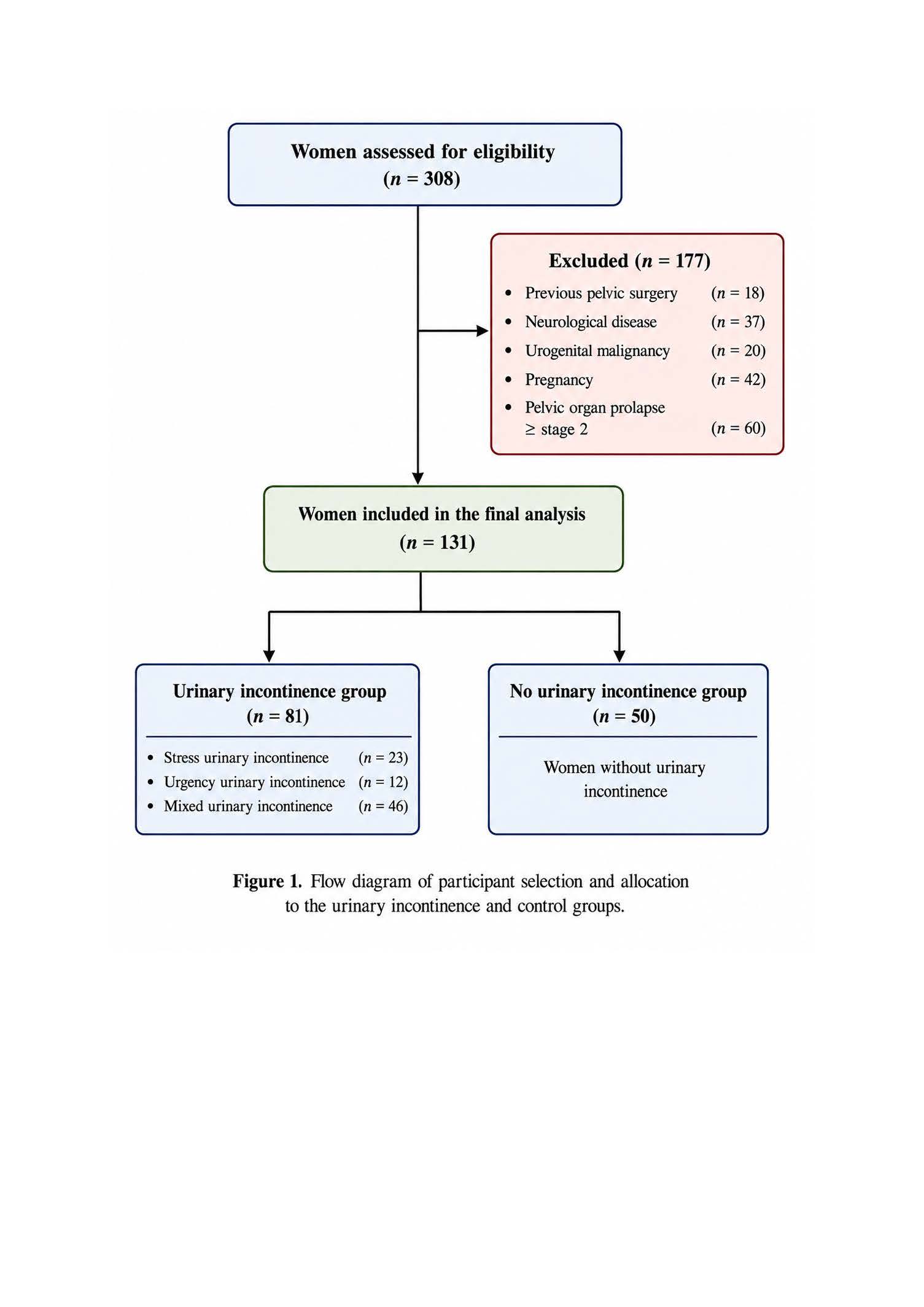
Methods: This retrospective cross-sectional comparative study included 131 women: 81 classified as having symptom-defined UI and 50 continent participants. UI classification was based on self-reported involuntary urinary leakage together with at least one positive response in the urinary distress domain of the PFDI-20; no urodynamic or objective continence test was used for study classification. Static 3.0-T pelvic MRI at rest was used to measure puborectalis thickness, genital hiatus dimensions, urethral morphology, obturator internus thickness, and rectus muscle thickness. Group comparisons, exploratory within-sample ROC analyses, and parsimonious logistic regression models were performed.
Results: Women with symptom-defined UI had lower bilateral puborectalis thickness (left: 5.54 ± 1.59 vs. 6.76 ± 1.33 mm; right: 5.53 ± 1.67 vs. 6.51 ± 1.46 mm; p ≤ 0.001) and larger genital hiatus width (105.54 ± 15.97 vs. 93.07 ± 15.07 mm; p < 0.001) and area (median 396.9 vs. 308.1 mm²; p < 0.001). Urethral diameter was also greater (12.34 ± 1.91 vs. 11.60 ± 1.45 mm; p = 0.021). Discrimination was moderate for genital hiatus width (AUC = 0.720) and left puborectalis thickness (AUC = 0.730). In separate parity-adjusted models, hiatus dimensions and bilateral puborectalis thickness remained associated with UI. None retained statistical significance when age, BMI, parity, and multiple correlated morphometric measures were entered together in broader models.
Conclusions: Women with symptom-defined UI showed differences in puborectalis thickness and genital hiatus dimensions on static MRI. These associations were evident in unadjusted and parity-adjusted analyses but were attenuated in broader multivariable models, indicating important confounding and shared variance with clinical factors. The exploratory thresholds were derived within this sample and should not be interpreted as diagnostic cut-offs. Static MRI morphometry may help characterize pelvic floor anatomy, but the present data do not support its use as a stand-alone diagnostic test for UI.
Keywords:
female urinary incontinence
; urogynecology
; pelvic floor
; magnetic resonance imaging
; puborectalis muscle
; genital hiatus
; morphometry
Copyright: This open access article is published under a Creative Commons CC BY 4.0 license, which permit the free download, distribution, and reuse, provided that the author and preprint are cited in any reuse.