
Submitted:
17 September 2026
Posted:
20 September 2026
You are already at the latest version
Abstract
Current concepts recognize that effective host defense depends on coordinated interactions among innate and adaptive immunity, inflammatory signaling, immunometabolic regulation, and multiple complementary adaptive biological processes. However, far less attention has focused on the regulatory systems that integrate these diverse responses across the evolving phases of the host response. This Review proposes that glucocorticoid receptor alpha (GRα) functions as a systems-level integrative regulator that coordinates immune signaling, antimicrobial competence, mitochondrial bioenergetics, inflammatory regulation, and other adaptive processes according to the temporal phase of homeostatic correction. A central premise is that these phase-specific actions are shaped by dynamic changes in chromatin accessibility and transcription-factor interactions, providing a molecular basis for differential GRα regulation throughout the adaptive response.
Rather than functioning solely as a transcriptional regulator of inflammation, GRα coordinates pathogen sensing, NF-κB/AP-1 signaling, immunometabolic adaptation, innate and adaptive immune responses, endothelial and epithelial barrier integrity, lymphatic function, tissue repair, and the transition from early host defense to inflammatory resolution and restoration of physiological homeostasis. These integrated biological processes are organized within the proposed Priming, Modulatory, and Restorative phases of homeostatic correction. By synthesizing evidence across multiple physiological systems, this Review provides a biologically plausible model for understanding how coordinated GRα-dependent regulation may influence immune effectiveness, disease tolerance, host-defense outcomes, and adaptive recovery during infection and critical illness. The proposed model generates testable hypotheses to guide future mechanistic investigation and the development of precision therapeutic strategies.
Keywords:
glucocorticoid receptor alpha (GRα)
; glucocorticoids
; host defense
; homeostatic correction
; immune regulation
; immunometabolism
; mitochondrial function
; systemic inflammation
Introduction
Effective host defense is increasingly recognized as a dynamic, systems-level process requiring coordinated interactions among pathogen recognition, innate and adaptive immune responses, inflammatory regulation, resistance mechanisms, disease tolerance, immunometabolic adaptation, and tissue-protective responses. [1,2] Rather than depending solely on efficient pathogen elimination, successful host defense requires an appropriate balance between resistance mechanisms that reduce pathogen burden and disease-tolerance mechanisms that preserve tissue integrity and physiological function during infection. [3,4] More recent studies further emphasize that these coordinated adaptive responses operate as integrated, systems-level biological networks rather than isolated immune pathways. [5,6]
Although these adaptive biological processes have been extensively studied individually, considerably less attention has been directed toward the integrative regulatory systems that coordinate their functional and temporal interactions throughout the host response to infection. [5,6] This Review proposes that glucocorticoid receptor alpha (GRα) functions as a systems-level integrative regulator that contributes to the coordination of these adaptive biological processes across the temporal phases of homeostatic correction, thereby influencing host-defense effectiveness, adaptation to infection, recovery, and restoration of physiological homeostasis. [7,8,9] Designation of GRα as a central integrative regulator does not imply that all components of host defense are GRα-dependent or that GRα exerts uniform or synchronous effects across tissues. Rather, integration emerges from context- and phase-dependent interactions of GC–GRα signaling with interdependent but partially autonomous immune, metabolic, vascular, neuroendocrine, barrier, lymphatic, mitochondrial, and regenerative networks whose adaptive responses may overlap and proceed asynchronously. Accordingly, this Review does not propose previously unrecognized functions of GRα but integrates evidence from multiple biological systems to examine whether its established context-, tissue-, and phase-dependent actions collectively support this systems-level regulatory role.
To systematically evaluate this proposed role of GRα in immune sensing (the early recognition of pathogens and danger signals), host defense, and immune effectiveness, a structured hypothesis-driven query framework was developed using artificial intelligence (AI)-assisted literature search and evidence retrieval. The framework prospectively organized the literature into predefined functional domains and phase-specific biological processes to support systematic evidence retrieval and synthesis. Table 1 [3,4,7,8,10,11,12,13,14,15,16,17,18,19,20,21] summarizes the framework used to guide evidence acquisition, organization, and synthesis throughout this Review and provides a transparent conceptual roadmap for the evidence discussed in the following sections.
Queries are organized across major immune domains, including pathogen recognition, innate immune activation, antimicrobial function, immunometabolism, inflammatory regulation, host defense outcomes, and resolution and repair. These domains are mapped onto the phases of homeostatic correction (Priming, Modulatory, and Restorative), reflecting the temporal evolution and functional organization of immune responses during host defense. Each query tests a specific aspect of GRα-mediated regulation, linking molecular signaling pathways (e.g., PRR activation, NF-κB/AP-1 signaling, mitochondrial function) to functional outcomes such as pathogen clearance, immune effectiveness, and restoration of homeostasis. The table includes representative references for each functional domain to support verification of the evidence base.
This structured approach enables systematic interrogation of whether GRα functions as a central integrator of immune sensing, signaling, and coordinated immune responses across different phases of host defense, rather than as a purely anti-inflammatory or immunosuppressive mediator. By integrating temporal phase dynamics with receptor-level signaling, transcriptional control, metabolic regulation, and effector outcomes, the framework provides a basis for evaluating how GRα signaling helps coordinate the transition between pathogen-clearing and pathogen-permissive states. Collectively, the table summarizes the evidence used to evaluate the proposed role of GRα as a systems-level integrator of host defense and phase-specific homeostatic correction.
Abbreviations: GRα, glucocorticoid receptor alpha; PRR, pattern-recognition receptor; TLR, Toll-like receptor; NLR, nucleotide-binding oligomerization domain–like receptor; NF-κB, nuclear factor kappa B; AP-1, activator protein-1.
1. Glucocorticoid Receptor Alpha: An Integrative Regulator of Host Defense
GRα is a ligand-activated transcription factor in the nuclear receptor superfamily, expressed in nearly all nucleated cells. [22] Primarily activated by endogenous glucocorticoids, GRα integrates neuroendocrine stress signals with immune, metabolic, vascular, and reparative pathways through tissue-specific, context-dependent gene regulation that varies by cell type, local microenvironment, and the temporal phase of the host response. [19,22] Beyond regulating inflammatory gene transcription, GRα also influences mitochondrial bioenergetics, cellular metabolism, endothelial and epithelial barrier integrity, circadian regulation, oxidative stress, and inter-organ communication, thereby contributing to organism-wide adaptation to physiological stress and infection. [7,18] GRα functions in both the nucleus and mitochondria, providing a mechanism for coordinating nuclear gene expression with mitochondrial bioenergetics and cellular metabolism. [16,18,23]
These broad biological actions position GRα as a plausible integrative regulator of the adaptive processes that collectively determine effective host defense. From an evolutionary perspective, this capacity to coordinate diverse adaptive processes supports the hypothesis that GRα functions as a core survival receptor that contributes to organism-wide adaptation during physiological stress, infection, and critical illness. [8]
Rather than functioning solely as an anti-inflammatory receptor, GRα participates in the coordinated regulation of pathogen sensing, inflammatory signaling, antimicrobial competence, immunometabolic adaptation, vascular stability, and immune resolution and recovery, in accordance with the host response's temporal and tissue-specific requirements. [12,20,24] These diverse biological actions are mediated by genomic and non-genomic signaling mechanisms that vary by cell type, tissue microenvironment, and the evolving phase of the host response. [19,25] The context dependence of GRα signaling is further shaped by chromatin architecture, which determines the accessibility of specific regulatory regions and thereby influences which transcriptional programs GRα can engage across different cells and biological states.
2.1. Chromatin Remodeling Provides the Molecular Basis for Phase-Specific GRα Regulation
The remarkable ability of GRα to coordinate diverse biological functions across multiple tissues cannot be explained solely by ligand binding or receptor abundance. A key determinant of this context-dependent regulation is chromatin—the dynamic organization of DNA and associated proteins that governs access to the genome. [26] Chromatin determines which genes are accessible for transcription and which remain inactive, thereby providing a molecular basis for selective gene regulation during physiological adaptation. Thus, although every nucleated cell contains essentially the same DNA, only selected genes are available for activation at any given time.
Chromatin is not static. During physiological stress and infection, chromatin architecture is continuously remodeled, exposing some regions of DNA while restricting access to others. Importantly, most GRα binding occurs at regulatory regions that are already accessible before glucocorticoid activation, indicating that baseline chromatin accessibility is a major determinant of tissue-specific GRα binding and the resulting transcriptional responses. [27,28] Changes in chromatin accessibility determine which transcription factors—including GRα, NF-κB, and AP-1—can bind specific regulatory regions and regulate gene expression. Consequently, the biological actions of GRα are determined not only by receptor activation but also by the dynamic chromatin landscape in which the receptor operates. [24,25,29]
During physiological stress and infection, activation of pattern-recognition receptors rapidly induces NF-κB, AP-1, and other stress-responsive transcription factors, which in turn recruit chromatin-remodeling complexes to thousands of regulatory regions across the genome. This process increases chromatin accessibility, enabling coordinated binding of GRα with other transcriptional regulators. In addition to recognizing accessible chromatin, GRα recruits chromatin-remodeling complexes that further modify local chromatin accessibility, reorganize nucleosomes, and facilitate the recruitment of additional transcriptional regulators, thereby shaping context- and phase-specific transcriptional programs. [30,31,32]
Rather than functioning independently, GRα, NF-κB, and AP-1 operate within a shared chromatin environment in which cooperative and antagonistic interactions shape the magnitude, timing, and tissue specificity of gene expression [24,25,33] As chromatin accessibility changes during the host response, distinct regulatory regions become accessible to transcription-factor binding, enabling GRα to engage in different transcriptional programs as physiological conditions evolve. [24,25,29]
This chromatin-based model provides a molecular framework for understanding how GRα's actions may evolve across the Priming, Modulatory, and Restorative Phases described in this Review. During the Priming Phase, stress-induced chromatin remodeling may facilitate GRα-dependent transcriptional programs that support pathogen recognition, innate immune activation, immunometabolic adaptation, and antimicrobial defense. [7,24,25,33] During the Modulatory and Restorative Phases, evolving chromatin accessibility may enable transcriptional programs that restrain excessive inflammation, preserve endothelial and mitochondrial function, promote tissue repair, and ultimately restore physiologic homeostasis. [7,8,29]
Accordingly, dynamic chromatin remodeling provides a plausible mechanism through which the same activated GRα receptor can engage distinct transcriptional programs as the host response progresses from pathogen recognition to inflammatory modulation and ultimately tissue repair and restoration of physiological homeostasis. Thus, the diverse biological functions of GRα reflect, in part, dynamic changes in chromatin accessibility and transcription-factor interactions that may contribute to the phase-specific regulation of homeostatic correction. [7,8,9]
2. Biological Basis for GRα-Mediated Integration of Host Defense
2.1. Ubiquitous Cellular Distribution Enables System-Wide Coordination of Host Defense
GRα is expressed in virtually all nucleated cells, including innate and adaptive immune cells, endothelial and epithelial cells, fibroblasts, neurons, hepatocytes, skeletal muscle, adipose tissue, and other parenchymal organs. [22] Unlike most immune regulatory molecules, whose actions are largely confined to specific cell populations or signaling pathways, the nearly ubiquitous distribution of GRα provides a biological basis for regulating adaptive responses across multiple tissues and organ systems, thereby enabling organism-wide integration of the host response. [8]
In addition to its well-established nuclear actions, GRα also localizes to mitochondria, where it directly influences mitochondrial gene expression, bioenergetics, oxidative phosphorylation, reactive oxygen species homeostasis, and cellular adaptation to physiological stress. [16,34] This dual nuclear-mitochondrial localization provides a mechanistic basis for coupling nuclear transcriptional programs with mitochondrial energy metabolism, thereby linking immune regulation to the bioenergetic requirements of effective host defense. [18]
This broad cellular distribution and dual subcellular localization enable neuroendocrine stress signals to be translated into coordinated transcriptional, metabolic, vascular, immunologic, and reparative responses throughout the organism. [7] Consequently, GRα is well positioned to help coordinate adaptive cellular responses across diverse tissues, thereby integrating immune regulation with metabolic, vascular, mitochondrial, neuroendocrine, and other physiological processes required for effective host defense during infection and other forms of biological stress. [8,18]
The following sections examine these major functional domains individually while emphasizing their coordinated integration during homeostatic correction. Table 2 [4,7,8,10,13,14,16,17,19,20,21,35] provides an overview of these interconnected biological programs and summarizes the evidence for GRα regulation of pathogen recognition, innate and adaptive immunity, inflammatory regulation, tissue repair, and restoration of physiological homeostasis across the temporal phases of the host response.
Pathogen recognition is mediated by pattern-recognition receptors (PRRs), including Toll-like receptors (TLRs), nucleotide-binding oligomerization domain–like receptors (NLRs), and RIG-I–like receptors (RLRs), which detect pathogen-associated molecular patterns (PAMPs) and damage-associated molecular patterns (DAMPs) and activate downstream signaling pathways involving nuclear factor kappa B (NF-κB), activator protein-1 (AP-1), and interferon regulatory factors (IRFs).
Within this integrated system, GRα does not directly mediate pathogen recognition but functions as a central regulator of immune coordination and response integration. GRα dynamically modulates PRR-driven signaling, cytokine production, immunometabolic adaptation, and downstream effector pathways in a tissue- and phase-specific manner, thereby coordinating immune sensing with antimicrobial effectiveness, tissue protection, and restoration of homeostasis.
Importantly, GRα signaling helps determine whether inflammatory responses remain pathogen-clearing or progress toward pathogen-permissive states. Appropriate GRα signaling may support mitochondrial function, phagolysosomal integrity, intracellular pathogen killing, and coordinated antimicrobial defense while constraining excessive NF-κB- and AP-1-driven inflammatory amplification. In contrast, impaired GRα signaling may contribute to dysregulated inflammatory responses that promote pathogen persistence, tissue injury, immune dysfunction, and impaired host defense.
Accordingly, effective immune regulation requires coordinated GRα-mediated integration of inflammatory control, antimicrobial competence, adaptive immune recalibration, and tissue repair, emphasizing that reduction of inflammatory cytokines alone does not necessarily indicate restoration of immune function or homeostatic resilience.
Abbreviations: PRR, pattern-recognition receptor; TLR, Toll-like receptor; NLR, nucleotide-binding oligomerization domain–like receptor; RLR, RIG-I–like receptor; PAMP, pathogen-associated molecular pattern; DAMP, damage-associated molecular pattern; NF-κB, nuclear factor kappa B; AP-1, activator protein-1; IRF, interferon regulatory factor; GRα, glucocorticoid receptor alpha.
2.2. GRα Modulates Early Host Defense Following Pathogen Recognition
Host defense begins when pattern-recognition receptors (PRRs), including Toll-like receptors (TLRs), NOD-like receptors (NLRs), RIG-I-like receptors (RLRs), and the cGAS-STING pathway, recognize pathogen-associated molecular patterns (PAMPs) and damage-associated molecular patterns (DAMPs). [10,11] Activation of these evolutionarily conserved sensing systems triggers intracellular signaling cascades that induce inflammatory cytokines, type I interferons, antimicrobial mediators, and other early defense mechanisms required for rapid pathogen containment, activation of innate immune effector mechanisms, and initiation of adaptive immunity. [36]
GRα does not function as a pathogen-recognition receptor; instead, it modulates the magnitude, duration, and biological consequences of PRR signaling by selectively regulating downstream transcriptional programs. [12,37] Following activation by endogenous glucocorticoids released through the hypothalamic-pituitary-adrenal (HPA) axis during physiological stress and infection, GRα interacts with multiple intracellular signaling pathways to shape the quality and coordination of the early host response while preserving the capacity for effective antimicrobial defense. [7,13] As discussed above, these interactions are influenced by the evolving chromatin landscape, which shapes the accessibility of inflammatory and adaptive regulatory elements and contributes to differential transcriptional responses.
Mechanistically, GRα modulates PRR signaling through multiple complementary pathways. These include transrepression of NF-κB- and AP-1-dependent inflammatory gene expression, [37] induction of endogenous inhibitory mediators such as glucocorticoid-induced leucine zipper (GILZ) and mitogen-activated protein kinase phosphatase-1 (MKP-1), [38] and dynamic interactions with transcriptional cofactors and chromatin remodeling complexes that collectively regulate inflammatory gene expression. [12,13]
Importantly, growing evidence indicates that GRα's biological effects during early host defense are highly context-dependent. [13] Although GRα generally restrains excessive inflammatory signaling, it may also maintain—or, under selected cellular and inflammatory conditions, enhance—specific innate immune functions required for effective pathogen control. [12,13] Consequently, GRα is better viewed not simply as an anti-inflammatory receptor but as an integrative regulator of early host defense that calibrates pathogen control and limits immune-mediated tissue injury according to the tissue microenvironment and the evolving host response. [3,4]
2.3. GRα Calibrates Innate Immune Effector Functions
Innate immune effector cells constitute the first cellular line of defense following pathogen recognition, rapidly containing invading microorganisms, producing inflammatory mediators, eliminating infected cells, and initiating adaptive immunity. [19] Effective host defense requires not only rapid activation of these effector mechanisms but also precise regulation of their magnitude, duration, and resolution to prevent excessive tissue injury. [39,40]
GRα regulates innate immune effector functions in a cell-, tissue-, and context-dependent manner. During the early Priming Phase of host defense, GRα may support innate immune mechanisms downstream of pathogen recognition, inflammatory signaling, and antimicrobial defense. [7] Early activation of NF-κB and AP-1 also remodels chromatin, creating accessible regulatory regions that influence subsequent GRα-dependent transcriptional responses. As the host response shifts toward modulation and recovery, GRα helps recalibrate these responses by limiting excessive inflammation, promoting disease tolerance, and facilitating inflammatory resolution. [13] Through this adaptive recalibration, GRα influences cytokine production, cellular activation, antimicrobial activity, metabolic adaptation, and inflammatory resolution, thereby helping preserve pathogen control while limiting immune-mediated tissue injury. [7,12]
Macrophages provide one of the clearest examples of GRα-mediated regulation during innate host defense. Rather than simply suppressing macrophage function, GRα regulates macrophage polarization, phagocytosis, cytokine production, and antimicrobial defense in ways that may support effective host defense while limiting excessive inflammatory tissue injury. [15,41] Recent evidence demonstrates that endogenous glucocorticoid signaling is required for appropriate macrophage activation during Helicobacter pylori infection, where loss of GR signaling results in aberrant chromatin remodeling, impaired induction of inflammatory genes, reduced T-cell recruitment, and diminished bacterial control. [14]
Natural killer (NK) cells provide a complementary example of GRα-mediated immune regulation. During viral infection, endogenous glucocorticoids induce tissue-specific PD-1 expression on splenic NK cells, limiting excessive interferon-γ production and preventing immunopathology without compromising viral clearance. [20] These findings illustrate that GRα promotes effective host defense not by simply suppressing innate immunity but by calibrating immune effector responses to preserve pathogen control while minimizing collateral tissue damage. [40]
Neutrophils provide another example of GRα-mediated regulation during innate host defense. GRα generally restrains excessive neutrophil recruitment and activation by suppressing endothelial adhesion, chemotactic signaling, NF-κB activation, and reactive oxygen species generation, thereby reducing collateral tissue injury. [42,43] However, its effects on phagocytosis, microbial killing, and neutrophil extracellular trap (NET) formation vary by pathogen, inflammatory milieu, and the timing and duration of glucocorticoid exposure, suggesting that GRα calibrates rather than uniformly suppresses neutrophil effector functions. [44,45]
Collectively, these observations support a broader role for GRα in regulating innate immune effector functions and influencing the subsequent development of adaptive immunity. [7,19] Because innate immune responses shape the quality, magnitude, and subsequent regulation of adaptive immune responses, GRα-mediated regulation of dendritic-cell function and antigen presentation may provide an important mechanistic link between innate and adaptive host defense.
2.4. GRα Regulates the Transition from Innate to Adaptive Immunity
Dendritic cells, specialized antigen-presenting cells that capture, process, and present microbial antigens to naïve T lymphocytes, occupy a pivotal position at the interface between innate and adaptive immunity by linking pathogen recognition to antigen presentation and T-cell activation. [46,47] Consequently, regulation of dendritic cell maturation and function provides an important mechanism through which GRα can influence the transition from innate to adaptive immune responses. [48]
GRα regulates dendritic-cell maturation and function throughout the evolving host response. Within the proposed framework, during the Priming Phase of host defense, dendritic cells preserve their capacity for pathogen sensing, antigen uptake, processing, migration to secondary lymphoid organs, and initiation of adaptive immune responses. [19] As the host response evolves toward modulation, GRα can recalibrate dendritic-cell function by limiting excessive maturation, reducing co-stimulatory molecule expression, modulating pro-inflammatory cytokine production, and restraining naïve T-lymphocyte activation, thereby promoting immune regulation and limiting excessive adaptive immune activation. [49,50] Rather than uniformly suppressing dendritic-cell function, GRα can modulate the magnitude, quality, and duration of adaptive immune activation according to biological context and the evolving host response. [19,48]
2.4.1. GRα Calibrates Adaptive T-Cell Responses
Adaptive T-cell responses are essential for pathogen clearance, immunologic memory, and long-term protection. [51] However, excessive or dysregulated T-cell activation can also be a major source of immune-mediated tissue injury. [19] Consequently, effective host defense requires not only robust adaptive immunity but also precise regulation of T-cell activation, differentiation, trafficking, and resolution. [19,51]
GRα regulates adaptive T-cell responses in a cell-, tissue-, and context-dependent manner. [19,51] Rather than uniformly suppressing T-cell immunity, GRα modulates T-cell activation and differentiation according to the biological context, limiting excessive inflammatory responses while supporting effective host defense. [51] Experimental studies show that loss of endogenous GR signaling results in exaggerated T-cell activation, excessive inflammatory cytokine production, and immune-mediated tissue injury, underscoring GRα's important role in maintaining adaptive immune homeostasis during infection. [40]
GRα also shapes the functional specialization and long-term memory of adaptive immunity by regulating helper T-cell differentiation, cytotoxic T-cell responses, and memory-cell development. [51] These effects depend on the T-cell activation state, local inflammatory signals, the tissue microenvironment, and circadian neuroendocrine regulation. [52,53] Consequently, GRα functions not merely as an inhibitor of adaptive immunity but as a regulator that helps balance effective antimicrobial defense with limitation of immune-mediated tissue injury. [19]
In addition to regulating effector T-cell activation and differentiation, GRα contributes to adaptive immune homeostasis by stabilizing regulatory T-cell (Treg) function and promoting immune tolerance during inflammation and recovery. [54,55] Experimental studies demonstrate that endogenous GRα signaling preserves the suppressive phenotype of Tregs during inflammation, thereby preventing excessive effector T-cell activation and limiting immune-mediated tissue injury. [54,55] Beyond its direct effects on Tregs, GRα also contributes to inflammatory resolution by regulating pro-resolving programs in macrophages, dendritic cells, and other immune cells, thereby facilitating restoration of immune homeostasis while preserving protective immunity. [7,48] Collectively, these findings further support the concept that GRα functions not as a generalized immunosuppressive receptor but as an adaptive regulator that helps calibrate immune responses to the evolving temporal and biological requirements of host defense. [7]
The regulation of cytotoxic CD8⁺ T cells further illustrates GRα's regulatory breadth in adaptive immunity. During infection, GRα modulates CD8⁺ T-cell activation, differentiation, and effector function, thereby contributing to the balance between antimicrobial immunity and limitation of immune-mediated tissue injury. [40,51] These effects vary according to activation state, inflammatory context, and glucocorticoid exposure. In addition, GRα influences the balance between terminal effector differentiation and memory-cell formation, thereby supporting the development of long-lived protective immunity under appropriate conditions. [56] Together, these observations illustrate how GRα can calibrate cytotoxic T-cell responses while contributing to durable protective immunity. [51,56]
2.4.2. GRα Regulates Humoral Immune Responses
B lymphocytes are the principal effector cells of humoral immunity, differentiating into antibody-producing plasma cells and memory B cells that confer long-term protection against recurrent infection. [57] Although GRα's effects on B-cell biology have been studied less extensively than its actions on innate immune cells and T cells, accumulating evidence indicates that GRα regulates B-cell activation, survival, trafficking, differentiation, antibody production, and memory-cell development in a context-dependent manner. [19,58]
Rather than functioning solely as a suppressor of antibody production, endogenous GRα signaling modulates B-cell receptor (BCR) and Toll-like receptor (TLR) signaling, regulates CXCR4-dependent trafficking, and contributes to specific antibody responses while maintaining immune homeostasis. [40,57] GRα also influences the balance between effective humoral immunity and immune regulation by modulating B-cell activation in response to the inflammatory microenvironment and the evolving host response. Collectively, these observations support a broader role for GRα in regulating humoral immunity and in linking B-cell responses to broader adaptive immune processes involved in host defense and immune homeostasis. [7,19]
3. GRα Coordinates Systemic Adaptive Responses
3.1. GRα Preserves Endothelial Function and Microvascular Homeostasis During Host Defense
The vascular endothelium, a dynamic monolayer of specialized cells lining the entire circulatory system, plays a central role in host defense by regulating vascular permeability, leukocyte trafficking, coagulation, vascular tone, and tissue perfusion. [59] During infection, coordinated endothelial responses are essential for delivering immune cells and antimicrobial mediators to sites of injury while preserving microvascular integrity and organ perfusion. [60] Conversely, endothelial dysfunction contributes to capillary leak, tissue edema, dysregulated inflammation, microvascular thrombosis, impaired oxygen delivery, and multiple-organ dysfunction, making preservation of endothelial homeostasis a critical determinant of clinical outcome. [18]
GRα is increasingly recognized as a key regulator of endothelial adaptation during infection. [7,59] Rather than functioning solely as an anti-inflammatory receptor, GRα regulates endothelial responses by modulating inflammatory signaling, barrier integrity, leukocyte-endothelial interactions, vascular permeability, and microvascular homeostasis. [7,8] Experimental studies demonstrate that endothelial-specific deletion of GR markedly increases susceptibility to endotoxemia and sepsis, providing direct evidence that endothelial GR signaling contributes to vascular stability and survival during severe inflammatory stress. [60]
At the cellular level, GRα supports endothelial barrier integrity through multiple complementary mechanisms. GRα suppresses NF-κB-dependent inflammatory activation and supports the expression and organization of adherens and tight junction proteins—including vascular endothelial (VE)-cadherin, occludin, claudin-5, and zonula occludens-1 (ZO-1). [59,61] GRα also helps preserve endothelial barrier function through mechanisms involving the endothelial glycocalyx and barrier-stabilizing pathways, including sphingosine kinase-1 (SphK1), sphingosine-1-phosphate (S1P), and angiopoietin-1 (Ang-1). [18] Collectively, these mechanisms help reduce vascular permeability, limit capillary leakage, and preserve tissue oxygenation during systemic inflammation. [18,59]
Beyond maintaining barrier integrity, GRα also regulates leukocyte trafficking by regulating endothelial activation rather than preventing immune-cell recruitment. [59] Through suppression of endothelial adhesion molecules, inflammatory cytokines, and chemokine expression, GRα limits excessive leukocyte adhesion and endothelial injury while preserving the controlled recruitment of immune cells required for effective antimicrobial defense. [60,62] In parallel, GRα contributes to maintenance of vascular tone, nitric oxide homeostasis, tissue perfusion, and microvascular blood flow, thereby linking endothelial regulation to broader systemic adaptation during host response to infection. [7,63]
Collectively, these observations support the concept that GRα functions as an integrative regulator of endothelial integrity and microvascular homeostasis. By regulating endothelial inflammatory signaling, barrier integrity, leukocyte-endothelial interactions, vascular permeability, and tissue perfusion, GRα influences multiple complementary mechanisms that contribute to vascular homeostasis as the host response evolves. [59,60] These coordinated actions help maintain effective antimicrobial defense while limiting endothelial injury, capillary leak, microvascular dysfunction, and progression to multiple-organ dysfunction. [18]
3.2. GRα Coordinates Immunometabolic Adaptation and Mitochondrial Bioenergetics During Host Defense
Effective host defense is an energetically demanding process that requires coordinated regulation of cellular metabolism to sustain immune activation, antimicrobial defense, tissue repair, and the restoration of physiological homeostasis. Successful adaptation to infection, therefore, depends not only on the activation of immune responses but also on the capacity of cells and tissues to generate, distribute, and efficiently utilize metabolic energy. Increasing evidence indicates that GRα links neuroendocrine stress responses with immunometabolic adaptation and mitochondrial bioenergetics, thereby contributing to the regulation of metabolic requirements during host defense across multiple tissues and organ systems. [7,8,29]
During acute infection, endogenous glucocorticoids acting through GRα promote the redistribution of metabolic substrates required to support the increased energetic demands of host defense. Through coordinated regulation of hepatic gluconeogenesis, lipolysis, proteolysis, and ketogenesis, GRα helps maintain glucose availability while providing alternative energy substrates for immune cells and metabolically active tissues during physiological stress. [29,64] These systemic metabolic adaptations help provide the energy substrates required to sustain immune and organ function during the evolving host response.
Beyond its well-established nuclear transcriptional actions, GRα is one of the few nuclear receptors that also localizes to mitochondria. Following glucocorticoid activation, cytoplasmic GRα translocates to mitochondria, where it can directly influence mitochondrial gene transcription and bioenergetic function. [16,23] Within mitochondria, GRα interacts with mitochondrial DNA and modulates components of the oxidative phosphorylation machinery and processes involved in ATP production, oxidative phosphorylation, reactive oxygen species homeostasis, and mitochondrial quality control. [16,18,65] The dual localization of functional GRα in both the nucleus and mitochondria provides a mechanistic basis for linking nuclear transcriptional programs with mitochondrial energy metabolism, thereby connecting immune activation to the bioenergetic requirements of effective host defense. [16,18,23]
GRα also regulates immunometabolic adaptation within immune cells. During the Priming Phase of host defense, immune cells undergo metabolic reprogramming to support rapid antimicrobial activity and inflammatory signaling. As the host response evolves toward modulation and recovery, GRα contributes to metabolic reprogramming from predominantly inflammatory glycolytic programs toward greater reliance on mitochondrial oxidative phosphorylation and tricarboxylic acid (TCA)-cycle activity, thereby limiting excessive inflammation and supporting disease tolerance, inflammatory resolution, and tissue repair. [17,66,67] In macrophages, these metabolic adaptations are essential for optimal glucocorticoid responsiveness and directly regulate inflammatory gene expression and antimicrobial defense. [17,66]
Collectively, these observations support the concept that GRα functions as an integrative regulator of immunometabolic adaptation rather than merely an anti-inflammatory transcription factor. By linking nuclear transcriptional regulation to mitochondrial bioenergetics, GRα couples endocrine stress signaling to the metabolic demands of immune activation. [16,23] By regulating systemic fuel allocation, mitochondrial bioenergetics, cellular metabolism, and immune-cell metabolic programming across multiple organs, GRα may help align energy availability with the shifting metabolic demands of host defense, disease tolerance, inflammatory resolution, tissue repair, and recovery. [7,8,18]
3.3. GRα Regulates Intestinal Barrier Integrity and Microbiome–Host Interactions During Host Defense
The intestinal barrier is a dynamic interface composed of epithelial cells, tight junctions, mucus, resident immune cells, and the gut microbiota, which collectively restrict microbial translocation while permitting nutrient absorption and regulated communication between the host and its microbial environment. [68,69] During infection and critical illness, disruption of this barrier can promote dysbiosis, increased intestinal permeability, bacterial and endotoxin translocation, systemic inflammation, and remote-organ injury. [18,69]
GRα regulates intestinal barrier integrity through its effects on epithelial inflammatory signaling, tight-junction integrity, mucosal renewal, and local immune response. [18,70] Intestinal GR signaling restrains STAT1- and TNF-dependent inflammatory injury, thereby helping preserve epithelial integrity during severe inflammation. [71] However, its effects are not uniformly barrier protective. In experimental models combining glucocorticoid exposure with ethanol-induced epithelial injury, intestinal epithelial GR was associated with tight-junction disruption, increased permeability, endotoxemia, and systemic inflammation, showing that GRα effects on barrier function vary by biological context and the nature of the associated tissue injury. [72]
GRα also participates in bidirectional communication among the intestinal epithelium, the gut microbiota, and the hypothalamic-pituitary-adrenal (HPA) axis. Glucocorticoid signaling can alter microbial composition. Conversely, microbial signals and metabolites—including short-chain fatty acids, bile-acid derivatives, and other immunometabolites—can influence neuroendocrine activity, local glucocorticoid availability, and downstream GR signaling. [18,73,74] Experimental studies further indicate that glucocorticoid-associated alterations in microbial communities can have divergent effects, including dysbiosis or modulation of inflammatory responses, depending on treatment duration, epithelial integrity, and the underlying inflammatory environment. [72,75]
Through these reciprocal interactions, GRα integrates epithelial barrier integrity, microbiome–host communication, mucosal immunity, immunometabolic adaptation, and systemic host defense. [18,70] Appropriate GRα signaling may help limit microbial translocation, systemic inflammatory amplification, and remote-organ injury while supporting epithelial repair and restoration of intestinal homeostasis. Conversely, excessive, prolonged, or biologically mismatched glucocorticoid signaling may disrupt epithelial and microbial equilibrium.
Barrier disruption may also create a self-amplifying interaction among microbial exposure, inflammation, and impaired host defense. Experimental studies demonstrate GR-dependent regulation of epithelial barrier integrity in both intestinal and respiratory tissues, whereas excessive or contextually inappropriate glucocorticoid signaling can alter microbial communities and antimicrobial defenses. [72,76,77,78] Once barrier integrity is compromised, microbial products or organisms may gain greater access to host tissues, amplifying inflammatory signaling and systemic exposure. Importantly, this interaction may be bidirectional: elevated concentrations of pro-inflammatory cytokines, including IL-1β, IL-6, and TNF-α, can enhance both extracellular and intracellular bacterial growth, suggesting that dysregulated inflammation may not only damage host tissues but also create conditions that favor microbial proliferation and persistence. [79,80,81] Consistent with this reciprocal host–microbe interaction, recent multi-omics analysis of patients with acute, severe lower respiratory tract infection identified distinct lower-respiratory microbial community states associated with divergent local immune responses and clinical outcomes, including pathogen-dominant states linked to heightened inflammatory signaling and poor prognosis. [82] In parallel, dysregulated inflammatory and metabolic responses may impair effective antimicrobial function and pathogen clearance. Although the complete sequence has not been demonstrated as a single GRα-dependent pathway in humans, these observations collectively support a broader role for GRα in coordinating epithelial integrity, inflammatory regulation, and host–microbe interactions across mucosal barrier surfaces, including the gastrointestinal and respiratory tracts. Thus, these diverse and sometimes divergent effects support the proposed role of GRα as an integrative regulator of mucosal barrier–microbiome interactions, rather than as a uniformly protective or suppressive influence. [18,83]
4. GRα Integrates Organ-System Communication During Homeostatic Correction
4.1. GRα Facilitates Lymphatic Function and Inflammatory Resolution During Host Defense
The lymphatic system is an essential component of host defense, serving as the principal conduit for tissue-fluid drainage, antigen transport, immune-cell trafficking, and communication between peripheral tissues and secondary lymphoid organs. Efficient lymphatic function promotes clearance of excess interstitial fluid, inflammatory mediators, microbial products, and cellular debris while facilitating dendritic-cell migration and adaptive immune activation. Impaired lymphatic function contributes to persistent tissue edema, prolonged inflammation, impaired immune surveillance, and delayed restoration of tissue homeostasis. [84,85]
Experimental evidence indicates that GR signaling directly regulates lymphatic endothelial barrier function. In inflamed airway lymphatics, dexamethasone reversed inflammation-induced remodeling of lymphatic endothelial junctions and restored button-like junctions. Dexamethasone also increased phosphorylated nuclear GR in lymphatic endothelial cells and promoted button formation in the absence of inflammation, supporting a direct receptor-mediated effect on lymphatic endothelial plasticity. [86] In primary lymphatic endothelial cells, glucocorticoid exposure altered junctional gene expression and increased barrier integrity, effects reversed by GR antagonism, supporting receptor-dependent regulation of lymphatic endothelial permeability. [18,87] Additional evidence indicates that GRα coordinates adaptive biological processes that influence lymphatic function, including endothelial inflammatory signaling, vascular barrier integrity, leukocyte–endothelial interactions, and tissue-fluid homeostasis. [60,88,89] However, GC–GR effects on lymphatic function are not uniformly protective, as glucocorticoid exposure has also been associated with reduced lymphatic endothelial proliferation and migration and impaired lymphatic vessel contractility. [87,90]
By preserving endothelial barrier integrity, suppressing excessive NF-κB-dependent inflammatory activation, limiting leukocyte-endothelial interactions, and maintaining tissue perfusion, GRα may create physiological conditions that support efficient lymphatic drainage and interstitial fluid homeostasis. [59,60]
GRα also contributes indirectly to lymphatic immune function by regulating macrophage activation, dendritic-cell maturation, antigen presentation, and leukocyte trafficking, all of which influence antigen delivery to draining lymph nodes and the initiation and resolution of adaptive immune responses. [19,40,48] During the Modulatory and Restorative Phases of the host response, these coordinated actions facilitate clearance of inflammatory mediators and cellular debris while promoting tissue repair and restoration of physiological homeostasis. [7,8]
Collectively, current evidence supports the concept that GRα contributes to lymphatic function primarily through emerging direct effects on lymphatic endothelial barrier biology and through broader, integrated regulation of vascular integrity, immune-cell trafficking, inflammatory resolution, and interstitial fluid homeostasis.
4.2. GRα Coordinates Neuroendocrine Regulation of Host Defense
Host defense is regulated not only by local immune responses but also by integrated neuroendocrine networks that coordinate systemic adaptation to infection. These networks include the hypothalamic-pituitary-adrenal (HPA) axis, autonomic nervous system, circadian regulatory pathways, and bidirectional neuroimmune communication. [19,91] Together they synchronize immune, metabolic, cardiovascular, and behavioral responses required for effective adaptation to both bacterial and viral infections. [18,19] Consistent with this systems-level interpretation, a recent independent model of sepsis proposes that HPA-axis and glucocorticoid signaling participate in centrally coordinated neuroendocrine–metabolic regulation of the immune response, emphasizing differential effects on innate and adaptive immunity rather than uniform immunosuppression. [92] GRα serves as the principal intracellular effector of glucocorticoid signaling and interacts with these broader neuroendocrine networks to generate tissue-specific transcriptional programs that coordinate these adaptive responses. [7,18]
Direct experimental evidence demonstrates that GR signaling is required for effective neuroendocrine feedback and host-defense regulation. Genetic disruption of GR impairs negative feedback within the stress axis and reduces survival during acute stress, while cell-specific GR deletion alters susceptibility to endotoxemia and viral infection by disrupting control of IFN-γ, IL-10-dependent tolerance, and PD-1-mediated restraint of immunopathology. [20,93,94] These findings indicate that effective neuroendocrine regulation depends on receptor competence rather than circulating glucocorticoid concentrations alone. Consistent with this concept, human and translational studies of critical illness demonstrate marked tissue- and cell-specific differences in GRα expression and signaling despite shared systemic glucocorticoid exposure, with suppression in circulating neutrophils but preservation or enhancement of signaling in other tissues and immune-cell populations. [95,96,97]
During bacterial and viral infections, activation of the HPA axis increases endogenous glucocorticoid production in response to inflammatory cytokines and physiological stress. [91] Following activation by endogenous glucocorticoids, GRα regulates phase-specific transcriptional programs that initially support innate immune activation and metabolic adaptation, subsequently limit excessive inflammatory injury, and ultimately promote tissue repair and restoration of physiological homeostasis. [7,18] Intact GRα signaling is equally important for negative feedback regulation of the HPA axis, [91] whereas impaired receptor abundance or function may contribute to reduced glucocorticoid responsiveness during critical illness and represents one component of the broader pathophysiology associated with critical illness-related corticosteroid insufficiency (CIRCI). [7]
GRα also interfaces neuroendocrine regulation with autonomic and circadian control of immunity. Circadian glucocorticoid secretion coordinates daily oscillations in leukocyte trafficking, cytokine production, lymphocyte maintenance, and immune responsiveness. [19,98] Autonomic neural pathways operate in parallel with and interact with HPA-axis/glucocorticoid signaling in regulating the timing and magnitude of host-defense responses. [19] Through these interacting neuroendocrine mechanisms, GRα-dependent glucocorticoid signaling helps synchronize immune activation with predictable physiological changes and environmental stressors, enhancing host defense efficiency while minimizing unnecessary inflammatory injury. [18,19]
Neuroendocrine regulation of host defense operates through continuous bidirectional communication between the immune and nervous systems. [91] Peripheral inflammatory signals activate central neuroendocrine pathways, whereas glucocorticoid feedback through GRα recalibrates immune and tissue responses as physiological conditions evolve. [91] GRα subsequently integrates these endocrine signals into coordinated, tissue-specific transcriptional programs that regulate innate and adaptive immunity, endothelial function, immunometabolic adaptation, inflammatory resolution, and tissue repair, thereby supporting effective host defense. [7,18,20,94] Conversely, chronic inflammation, hypoxia, altered receptor isoform balance, or glucocorticoid resistance can disrupt GRα signaling, impairing this adaptive neuroimmune communication and contributing to dysregulated host responses. [7,99]
Collectively, these observations support the concept that GRα serves as a major molecular interface through which glucocorticoid signals are translated into coordinated immune, metabolic, vascular, and reparative responses during host defense. [18] By operating within integrated HPA-axis, autonomic, circadian, and bidirectional neuroimmune networks, GRα helps synchronize organism-wide adaptation across the temporal phases of the host response to bacterial and viral infections, thereby promoting effective pathogen control while facilitating restoration of physiological homeostasis. [7,19]
4.3. GRα Coordinates Tissue Repair and Restoration of Organ Function During Host Defense
Successful host defense extends beyond pathogen elimination to include restoration of tissue integrity, recovery of organ function, and re-establishment of physiological homeostasis. As the initial infectious insult is brought under control, injured tissues must transition from inflammatory defense to coordinated programs of inflammatory resolution, cellular regeneration, extracellular matrix remodeling, angiogenesis, and functional recovery. Increasing evidence indicates that GRα serves as an integrative regulator of this transition by coordinating immune, stromal, metabolic, vascular, and reparative responses in a context- and phase-dependent manner. [7,8,18]
One of the principal mechanisms through which GRα contributes to tissue repair is by facilitating the transition from inflammatory effector responses toward pro-resolving immune programs. Rather than simply suppressing inflammation, GRα promotes macrophage reprogramming toward reparative phenotypes and induces endogenous pro-resolving mediators, including glucocorticoid-induced leucine zipper (GILZ) and Annexin A1, thereby facilitating inflammatory resolution and tissue repair. [7,100] Direct genetic evidence supports a critical role for macrophage GR in this transition: myeloid GR deletion following myocardial infarction disrupts macrophage reparative programming, impairs scar formation and angiogenesis, worsens ventricular remodeling and cardiac function, and increases mortality from cardiac rupture. [101] Experimental studies further demonstrate that glucocorticoid-induced macrophage reprogramming requires AMPKα1-FOXO3 signaling, linking GR-dependent transcriptional responses to metabolic programs required for regenerative inflammation. [21]
GRα effects on tissue repair are not uniformly regenerative and vary by cell type, tissue context, and injury phase. Endothelial GR loss accelerates fibrotic remodeling and disrupts metabolic and inflammatory programs, supporting a protective role for endothelial GR during tissue repair. [102] Conversely, glucocorticoid–GR signaling can restrain selected regenerative processes: glucocorticoid exposure improves epithelial barrier organization while delaying epithelial migration and wound closure, and myeloid GR deletion after spinal cord injury has been associated with improved neurological recovery. [103,104] These divergent effects emphasize that GRα regulates the balance among inflammatory resolution, structural stabilization, remodeling, and regeneration rather than functioning as a uniformly pro-reparative signal.
GRα also coordinates structural repair by regulating interactions among immune cells, fibroblasts, endothelial cells, epithelial cells, and the extracellular matrix. Direct receptor-specific studies demonstrate that GR signaling regulates multiple components of post-injury remodeling and regeneration, including scar formation, angiogenesis, fibroblast differentiation, cellular proliferation, and functional recovery. Importantly, these effects are strongly cell- and context-dependent: macrophage GR is required for effective scar formation, angiogenesis, and cardiac remodeling after myocardial infarction, whereas cardiomyocyte GR restrains proliferative regeneration, with GR deletion or antagonism enhancing cardiomyocyte cell-cycle re-entry and cardiac muscle regeneration. [101,105,106] These divergent effects indicate that GRα regulates the balance among inflammatory resolution, structural repair, and regenerative growth rather than functioning as a uniformly pro- or anti-regenerative signal.
Beyond structural repair, GRα supports restoration of organ function by coordinating tissue repair with mitochondrial recovery, vascular homeostasis, and cellular bioenergetics. Tissue-specific GRα signaling contributes to recovery of pulmonary gas exchange, cardiovascular integrity, intestinal barrier function, and other organ-specific adaptive programs required to restore physiological function following severe infection and critical illness. [18] Experimental and translational studies further indicate that preserving tissue-specific GRα signaling supports effective repair responses, whereas impaired GRα expression or function may delay recovery and contribute to persistent organ dysfunction during prolonged critical illness. [101,107]
Collectively, these observations support the concept that GRα functions as a context- and phase-dependent regulator of tissue repair rather than merely an anti-inflammatory receptor. By coordinating immune-cell reprogramming, stromal remodeling, extracellular matrix homeostasis, angiogenesis, barrier reconstruction, mitochondrial recovery, and tissue-specific regeneration, GRα orchestrates the transition from inflammatory injury to restoration of organ structure and physiological function. These integrated adaptive programs characterize the Restorative Phase of homeostatic correction and are essential for successful recovery from both bacterial and viral infections. [7,8,18]
4.4. GRα Integrates Inter-Organ Communication Networks During Host Defense
Host defense is increasingly recognized as a systems-level adaptive process that requires coordinated communication among immune, neuroendocrine, metabolic, vascular, lymphatic, epithelial, and stromal tissues, rather than the independent function of individual organ systems. Effective adaptation to bacterial and viral infections therefore depends on continuous bidirectional communication that synchronizes pathogen recognition, inflammatory responses, metabolic adaptation, tissue protection, and regenerative processes across the organism. GRα is well positioned to coordinate these interactions because it is expressed in virtually all nucleated cells and translates systemic glucocorticoid signals into tissue-specific transcriptional programs based on biological context, thereby contributing to organism-wide adaptive responses during host defense. [13,19,29]
Unlike most immune regulators, whose activity is largely confined to individual cell populations or signaling pathways, GRα regulates multiple interconnected biological systems through tissue-specific, context-dependent signaling across diverse cell populations and organ systems. Throughout this Review, GRα has been shown to coordinate pathogen sensing, innate and adaptive immunity, endothelial function, immunometabolic adaptation, mitochondrial bioenergetics, intestinal barrier integrity, lymphatic regulation, neuroendocrine communication, and tissue repair. These coordinated actions illustrate that host defense is best understood as an integrated network of interacting adaptive systems rather than as a collection of isolated biological responses. [13,19]
Integration across organ systems is achieved because glucocorticoid signals are interpreted differently within individual tissues shaped by local chromatin accessibility, GR isoform expression, intracellular coregulators, glucocorticoid metabolism, and the prevailing inflammatory microenvironment. [29,99,108] Emerging evidence further indicates that, in addition to regulation mediated by local chromatin accessibility, GRα also regulates gene expression through long-range three-dimensional chromatin interactions that coordinate enhancer-promoter communication across the genome, providing an additional mechanism by which tissue-specific transcriptional programs are integrated during homeostatic correction. [109,110] Consequently, a single endocrine signal can simultaneously promote innate immune activation in one tissue, preserve endothelial integrity in another, regulate mitochondrial bioenergetics elsewhere, and initiate tissue repair in damaged organs. This tissue-specific interpretation enables coordinated organism-wide adaptation while preserving the functional specialization of individual organs. [13,107]
As the host response progresses from pathogen recognition through inflammatory modulation to tissue repair, GRα orchestrates sequential communication among immune, vascular, metabolic, neuroendocrine, microbiome, and reparative pathways. Rather than functioning as a generalized anti-inflammatory receptor, GRα integrates these diverse adaptive networks into phase-specific biological programs that optimize pathogen control, limit collateral tissue injury, promote disease tolerance, and restore physiological homeostasis following bacterial and viral infections. [7,19,20]
Collectively, these observations support the concept that GRα functions as a systems-level integrator of host defense. By coordinating communication among multiple organ systems, GRα synchronizes immune defense, metabolic adaptation, vascular homeostasis, neuroendocrine regulation, barrier integrity, mitochondrial function, and tissue repair throughout the temporal phases of homeostatic correction. This systems-level perspective extends the traditional view of GRα beyond inflammatory regulation and provides a biologic framework for understanding organism-wide adaptation during infection, critical illness, and recovery. [18,20,29]
5. Unified Framework for GRα-Mediated Homeostatic Correction
5.1. GRα Coordinates Pathogen Control and Disease Tolerance During Host Defense
Successful host defense requires coordinated regulation of pathogen control, inflammatory responses, metabolic adaptation, tissue protection, and repair across the temporal phases of the host response. Growing evidence indicates that GRα orchestrates these adaptive processes in a phase-specific manner, thereby promoting effective antimicrobial defense, limiting collateral tissue injury, and supporting the restoration of physiological homeostasis. Importantly, GRα does not directly eliminate invading pathogens. Rather, it coordinates immune responses together with the complementary adaptive biological processes that collectively determine effective host defense, disease tolerance, and restoration of physiological homeostasis. Through context-dependent, tissue-specific, and phase-specific regulation of these integrated adaptive programs, GRα helps coordinate the host's capacity to control infection while limiting immune-mediated tissue injury and promoting successful homeostatic correction. [14,20]
Rather than functioning as a generalized immunosuppressive receptor, GRα calibrates immune responses to the biological context, pathogen, tissue, and temporal phase of infection. During the early stages of bacterial and viral infections, endogenous glucocorticoid signaling supports appropriate activation of innate immune defenses while mobilizing the metabolic resources required for effective antimicrobial responses. [8,12] As the host response progresses through the Modulatory and Restorative Phases, GRα increasingly restrains excessive inflammatory signaling by coordinating the regulation of NF-κB, AP-1, inflammatory cytokines, oxidative stress, and immune cell activation, thereby limiting immunopathology without necessarily compromising pathogen clearance. [19,111]
Experimental studies provide direct evidence that endogenous GRα signaling contributes to effective pathogen control with disease tolerance, thereby limiting immune-mediated tissue injury without compromising pathogen clearance. [14,20] During viral infection, glucocorticoid-dependent GRα signaling induces tissue-specific PD-1 expression on splenic natural killer cells, limiting excessive interferon-γ production and preventing immune-mediated tissue injury without impairing viral clearance. [20] Similarly, studies of bacterial infection demonstrate that endogenous GR signaling contributes to appropriate macrophage activation, chromatin remodeling, inflammatory gene regulation, and effective antimicrobial defense, whereas loss of GR signaling results in dysregulated inflammation and impaired control of bacteria. [14,112]
Beyond regulating immune-cell function, GRα promotes disease tolerance by preserving the biological infrastructure required for recovery. Throughout this Review, GRα has been shown to maintain endothelial integrity, support mitochondrial bioenergetics, preserve intestinal barrier function, regulate immunometabolic adaptation, and coordinate tissue repair. These integrated actions reduce collateral tissue injury, preserve organ function, and facilitate the restoration of physiological homeostasis despite ongoing infection. [7,8,18]
The balance between host resistance and disease tolerance is highly context- and phase-dependent. Physiological endogenous glucocorticoid signaling supports coordinated adaptive responses that balance host defense with preservation of physiological homeostasis, while appropriately timed therapeutic glucocorticoid administration may support these adaptive processes in selected clinical contexts. In contrast, prolonged, excessive, or biologically inappropriate glucocorticoid exposure may impair host defense and increase susceptibility to secondary infection. Likewise, impaired GRα expression or function, altered receptor isoform balance, chronic inflammation, oxidative stress, and glucocorticoid resistance disrupt this adaptive equilibrium, contributing to persistent inflammation, tissue injury, and organ dysfunction. [19,99,113]
Collectively, these observations support the proposed conceptual framework in which GRα orchestrates a dynamic sequence of phase-specific adaptive responses throughout homeostatic correction. Rather than functioning simply as an anti-inflammatory receptor, GRα integrates pathogen control, disease tolerance, immunometabolic adaptation, vascular homeostasis, tissue repair, and inter-organ communication into coordinated biological programs that optimize survival and recovery from both bacterial and viral infections. [7,8,20]
5.2. Integrated Phase-Specific Framework for Homeostatic Correction
The preceding sections demonstrate that GRα coordinates multiple adaptive biological processes spanning immune sensing and antimicrobial defense, inflammatory regulation, endothelial and epithelial barrier integrity, immunometabolism, mitochondrial bioenergetics, neuroendocrine adaptation, lymphatic function, microbiome–gut homeostasis, tissue repair and regeneration, and inter-organ communication. Collectively, these observations support the proposed conceptual framework in which GRα orchestrates the coordinated, phase-specific integration of these adaptive biological programs throughout homeostatic correction. Table 3 [4,7,8,10,13,14,16,17,19,20,21,35] integrates these concepts by synthesizing the principal adaptive biological programs within the Priming, Modulatory, and Restorative Phases of homeostatic correction, providing a systems-level overview of the coordinated biological processes discussed throughout this Review.
6. Conclusions
Effective host defense requires far more than eliminating invading pathogens. Successful recovery depends on the coordinated regulation of immune responses and multiple complementary adaptive biological processes that preserve organ function, limit collateral tissue injury, promote disease tolerance, and restore physiological homeostasis. Although these processes have traditionally been studied as largely independent biological systems, growing experimental and clinical evidence indicates that they function as an integrated adaptive network, and that their coordination is essential for successful host defense.
This Review presents a systems-level conceptual framework in which GRα functions as an integrative regulator of host defense by coordinating context-dependent, tissue-specific, and phase-specific adaptive responses throughout homeostatic correction. Rather than attributing previously unrecognized biological functions to GRα, this framework integrates a broad body of established experimental and clinical evidence into a unified, biologically testable model that explains how the diverse actions of GRα collectively support effective host defense. Within this framework, immune regulation is integrated with complementary adaptive biological processes—including endothelial and epithelial barrier integrity, immunometabolic adaptation, mitochondrial bioenergetics, neuroendocrine regulation, lymphatic function, microbiome–host interactions, tissue repair, and inter-organ communication—that collectively enable effective pathogen control, limit immune-mediated tissue injury, and restore physiological homeostasis.
The Priming, Modulatory, and Restorative Phases provide a biologically coherent framework for understanding how these adaptive processes evolve during the host response. Rather than representing independent biological events, these phases constitute a coordinated continuum in which immune activation, inflammatory regulation, metabolic adaptation, tissue protection, and regenerative repair are dynamically integrated according to biological context and the temporal stage of infection. Within this framework, GRα is proposed to contribute to the coordination of transitions among these adaptive programs, thereby supporting both effective antimicrobial defense and the homeostatic correction required for successful recovery.
This systems-level perspective has important implications for future mechanistic investigation and precision medicine. Rather than focusing exclusively on isolated biomarkers or individual signaling pathways, future approaches may benefit from assessing the integrated adaptive capacity of biological systems that collectively determine host defense, disease tolerance, and recovery. Such an approach may improve biological phenotyping, therapeutic monitoring, and the development of individualized interventions to restore adaptive capacity and physiological resilience across acute and chronic disease states.
The principal contribution of this Review is the integration of extensive experimental and clinical evidence into a unified, biologically testable framework for understanding how the diverse actions of GRα may coordinate effective host defense with the complementary adaptive processes required for successful homeostatic correction. If supported by future mechanistic, translational, and clinical studies, this framework may provide a foundation for understanding coordinated host defense and for guiding functional assessment and precision therapeutic strategies aimed at restoring adaptive capacity, physiological resilience, and homeostasis across acute and chronic disease.
Figures
Figure 1.
Three Phases of Homeostatic Correction and Phase-Specific GRα Regulation During Severe
Physiological Stress. Legend: This schematic illustrates the proposed framework for glucocorticoid receptor
alpha (GRα)-mediated homeostatic correction during severe physiological stress and critical illness. The
hypothalamic-pituitary-adrenal (HPA) axis is the principal neuroendocrine stress-response system.
Physiological stress and infection stimulate hypothalamic corticotropin-releasing hormone (CRH) release,
which induces pituitary adrenocorticotropic hormone (ACTH) secretion and subsequent adrenal
glucocorticoid production. Activated glucocorticoids bind GRα, which mediates adaptive responses
throughout the three phases of homeostatic correction while simultaneously providing negative feedback to
the hypothalamus and pituitary to regulate HPA-axis activity. The adaptive response unfolds in three
dynamic, partially overlapping phases: the Priming Phase, the Modulatory Phase, and the Restorative Phase. A
detailed summary of the principal GRα-mediated molecular, metabolic, immune, vascular, barrier-protective,
antioxidant, and reparative functions for each phase is provided in Table 3. The Priming Phase initiates early
host-defense and adaptive-readiness responses, including immune activation, immunometabolic adaptation,
mitochondrial activation, barrier defense, cardiovascular adaptation, antioxidant responses, and
neuroendocrine stress signaling. The Modulatory Phase calibrates inflammatory amplification and preserves
systemic stability by coordinating regulation of inflammatory signaling, endothelial and barrier integrity,
vascular homeostasis, redox balance, and integrated immune–metabolic adaptation. The Restorative Phase
promotes resolution of inflammation, tissue repair, extracellular matrix remodeling, adaptive immune
recalibration, metabolic recovery, and restoration of organ function and physiological resilience. GRα is active
throughout all three phases, although its predominant biological functions evolve according to the temporal stage of the host response. Across all phases, GRα functions as a systems-level integrator coordinating
immune, metabolic, vascular, neuroendocrine, barrier, and reparative responses during homeostatic
correction. The author conceptually developed this figure with assistance from ChatGPT (OpenAI) for
graphical generation, visual organization, and figure refinement. The author independently developed,
critically reviewed, and approved the biological framework, scientific content, interpretation, and
final figure design. Abbreviations: HPA, hypothalamic-pituitary-adrenal; CRH, corticotropin-releasing
hormone; ACTH, adrenocorticotropic hormone; GRα, glucocorticoid receptor alpha.
Figure 1.
Three Phases of Homeostatic Correction and Phase-Specific GRα Regulation During Severe
Physiological Stress. Legend: This schematic illustrates the proposed framework for glucocorticoid receptor
alpha (GRα)-mediated homeostatic correction during severe physiological stress and critical illness. The
hypothalamic-pituitary-adrenal (HPA) axis is the principal neuroendocrine stress-response system.
Physiological stress and infection stimulate hypothalamic corticotropin-releasing hormone (CRH) release,
which induces pituitary adrenocorticotropic hormone (ACTH) secretion and subsequent adrenal
glucocorticoid production. Activated glucocorticoids bind GRα, which mediates adaptive responses
throughout the three phases of homeostatic correction while simultaneously providing negative feedback to
the hypothalamus and pituitary to regulate HPA-axis activity. The adaptive response unfolds in three
dynamic, partially overlapping phases: the Priming Phase, the Modulatory Phase, and the Restorative Phase. A
detailed summary of the principal GRα-mediated molecular, metabolic, immune, vascular, barrier-protective,
antioxidant, and reparative functions for each phase is provided in Table 3. The Priming Phase initiates early
host-defense and adaptive-readiness responses, including immune activation, immunometabolic adaptation,
mitochondrial activation, barrier defense, cardiovascular adaptation, antioxidant responses, and
neuroendocrine stress signaling. The Modulatory Phase calibrates inflammatory amplification and preserves
systemic stability by coordinating regulation of inflammatory signaling, endothelial and barrier integrity,
vascular homeostasis, redox balance, and integrated immune–metabolic adaptation. The Restorative Phase
promotes resolution of inflammation, tissue repair, extracellular matrix remodeling, adaptive immune
recalibration, metabolic recovery, and restoration of organ function and physiological resilience. GRα is active
throughout all three phases, although its predominant biological functions evolve according to the temporal stage of the host response. Across all phases, GRα functions as a systems-level integrator coordinating
immune, metabolic, vascular, neuroendocrine, barrier, and reparative responses during homeostatic
correction. The author conceptually developed this figure with assistance from ChatGPT (OpenAI) for
graphical generation, visual organization, and figure refinement. The author independently developed,
critically reviewed, and approved the biological framework, scientific content, interpretation, and
final figure design. Abbreviations: HPA, hypothalamic-pituitary-adrenal; CRH, corticotropin-releasing
hormone; ACTH, adrenocorticotropic hormone; GRα, glucocorticoid receptor alpha.
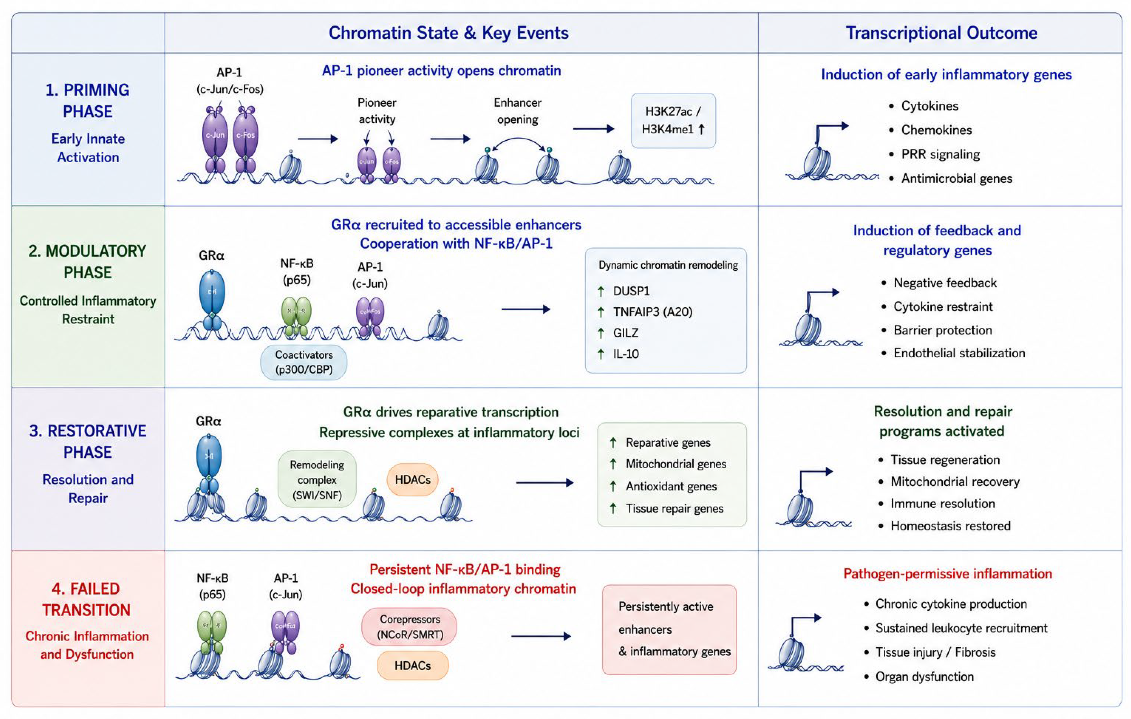
Figure 2.
Figure 2. Phase-Specific GRα Regulatory Programs Coordinate Successful and Failed Homeostatic Correction.
Subtitle: Mechanistic Framework of Phase-Specific GRα Regulation During Successful and Failed Homeostatic
Correction. Legend: This figure illustrates the proposed mechanistic framework by which glucocorticoid
receptor alpha (GRα) dynamically coordinates inflammatory signaling, metabolic adaptation, chromatin
remodeling, immune regulation, and tissue repair across the temporal phases of homeostatic correction. The
central GRα row summarizes the evolving phase-specific regulatory programs of GRα that orchestrate the
inflammatory, metabolic, transcriptional, vascular, barrier-protective, and reparative processes illustrated in
the surrounding functional domains. Rather than functioning as a generalized anti-inflammatory receptor,
GRα progressively shifts from facilitating adaptive host-defense responses during the Priming Phase to
calibrating inflammatory and metabolic homeostasis during the Modulatory Phase, and finally to promoting
reparative gene programs and restoring physiological homeostasis during the Restorative Phase. The three left
panels depict successful adaptive progression through the Priming, Modulatory, and Restorative phases,
whereas the right panel illustrates failed transition states associated with dysregulated GRα signaling,
glucocorticoid resistance, persistent inflammatory activation, metabolic dysfunction, impaired reparative
programming, and pathogen-permissive inflammation. During the Priming Phase, GRα functions permissively
and coordinatively to support responses downstream of pathogen recognition, leukocyte recruitment,
inflammatory activation, mitochondrial adaptation, glycolytic energy mobilization, chromatin accessibility,
and antimicrobial competence required for early host defense. During the Modulatory Phase, GRα increasingly
calibrates inflammatory amplification by coordinating the regulation of NF-κB/AP-1 signaling, chromatin
remodeling, endothelial stabilization, mitochondrial preservation, metabolic flexibility, and the induction of
negative-feedback and tissue-protective pathways. As homeostatic correction progresses into the Restorative
Phase, GRα promotes reparative transcriptional reprogramming, oxidative metabolism, mitochondrial
recovery, efferocytosis, tissue repair, extracellular matrix remodeling, and restoration of immune and
physiological homeostasis. The failed-transition pathway illustrates how impaired or dysregulated GRα
signaling may disrupt the coordinated progression toward resolution, resulting in persistent NF-κB/AP-1 activation, inflammasome amplification, mitochondrial dysfunction, defective chromatin remodeling, impaired
reparative responses, tissue injury, and chronic pathogen-permissive inflammatory states. This panel
emphasizes that failure of homeostatic correction reflects disruption of coordinated phase-specific GRα
regulatory programs rather than loss of a single anti-inflammatory mechanism. The author conceptually
developed this figure with assistance from ChatGPT (OpenAI) for graphical generation, visual organization,
and figure refinement. The author independently developed, critically reviewed, and
approved the biological framework, scientific content, interpretation, and final figure design.
Abbreviations: GRα, glucocorticoid receptor alpha; PAMPs, pathogen-associated molecular patterns; DAMPs,
damage-associated molecular patterns; PRR, pattern-recognition receptor; NF-κB, nuclear factor kappa B; AP-
1, activator protein-1; TNF, tumor necrosis factor; IL, interleukin; DUSP1, dual-specificity phosphatase 1
(MAPK phosphatase-1); TNFAIP3 (A20), tumor necrosis factor alpha-induced protein 3; GILZ, glucocorticoidinduced
leucine zipper; ROS, reactive oxygen species; ATP, adenosine triphosphate; PPP, pentose phosphate
pathway; OXPHOS, oxidative phosphorylation; Nrf2, nuclear factor erythroid 2-related factor 2; mTOR,
mechanistic target of rapamycin; TF, transcription factor; H3K27ac, histone H3 lysine 27 acetylation; H3K4me1,
histone H3 lysine 4 monomethylation; CBP, CREB-binding protein; ECM, extracellular matrix; ALXR, lipoxin
A4 receptor; TGF-β, transforming growth factor-beta; VEGF, vascular endothelial growth factor; KGF,
keratinocyte growth factor.
Figure 2.
Figure 2. Phase-Specific GRα Regulatory Programs Coordinate Successful and Failed Homeostatic Correction.
Subtitle: Mechanistic Framework of Phase-Specific GRα Regulation During Successful and Failed Homeostatic
Correction. Legend: This figure illustrates the proposed mechanistic framework by which glucocorticoid
receptor alpha (GRα) dynamically coordinates inflammatory signaling, metabolic adaptation, chromatin
remodeling, immune regulation, and tissue repair across the temporal phases of homeostatic correction. The
central GRα row summarizes the evolving phase-specific regulatory programs of GRα that orchestrate the
inflammatory, metabolic, transcriptional, vascular, barrier-protective, and reparative processes illustrated in
the surrounding functional domains. Rather than functioning as a generalized anti-inflammatory receptor,
GRα progressively shifts from facilitating adaptive host-defense responses during the Priming Phase to
calibrating inflammatory and metabolic homeostasis during the Modulatory Phase, and finally to promoting
reparative gene programs and restoring physiological homeostasis during the Restorative Phase. The three left
panels depict successful adaptive progression through the Priming, Modulatory, and Restorative phases,
whereas the right panel illustrates failed transition states associated with dysregulated GRα signaling,
glucocorticoid resistance, persistent inflammatory activation, metabolic dysfunction, impaired reparative
programming, and pathogen-permissive inflammation. During the Priming Phase, GRα functions permissively
and coordinatively to support responses downstream of pathogen recognition, leukocyte recruitment,
inflammatory activation, mitochondrial adaptation, glycolytic energy mobilization, chromatin accessibility,
and antimicrobial competence required for early host defense. During the Modulatory Phase, GRα increasingly
calibrates inflammatory amplification by coordinating the regulation of NF-κB/AP-1 signaling, chromatin
remodeling, endothelial stabilization, mitochondrial preservation, metabolic flexibility, and the induction of
negative-feedback and tissue-protective pathways. As homeostatic correction progresses into the Restorative
Phase, GRα promotes reparative transcriptional reprogramming, oxidative metabolism, mitochondrial
recovery, efferocytosis, tissue repair, extracellular matrix remodeling, and restoration of immune and
physiological homeostasis. The failed-transition pathway illustrates how impaired or dysregulated GRα
signaling may disrupt the coordinated progression toward resolution, resulting in persistent NF-κB/AP-1 activation, inflammasome amplification, mitochondrial dysfunction, defective chromatin remodeling, impaired
reparative responses, tissue injury, and chronic pathogen-permissive inflammatory states. This panel
emphasizes that failure of homeostatic correction reflects disruption of coordinated phase-specific GRα
regulatory programs rather than loss of a single anti-inflammatory mechanism. The author conceptually
developed this figure with assistance from ChatGPT (OpenAI) for graphical generation, visual organization,
and figure refinement. The author independently developed, critically reviewed, and
approved the biological framework, scientific content, interpretation, and final figure design.
Abbreviations: GRα, glucocorticoid receptor alpha; PAMPs, pathogen-associated molecular patterns; DAMPs,
damage-associated molecular patterns; PRR, pattern-recognition receptor; NF-κB, nuclear factor kappa B; AP-
1, activator protein-1; TNF, tumor necrosis factor; IL, interleukin; DUSP1, dual-specificity phosphatase 1
(MAPK phosphatase-1); TNFAIP3 (A20), tumor necrosis factor alpha-induced protein 3; GILZ, glucocorticoidinduced
leucine zipper; ROS, reactive oxygen species; ATP, adenosine triphosphate; PPP, pentose phosphate
pathway; OXPHOS, oxidative phosphorylation; Nrf2, nuclear factor erythroid 2-related factor 2; mTOR,
mechanistic target of rapamycin; TF, transcription factor; H3K27ac, histone H3 lysine 27 acetylation; H3K4me1,
histone H3 lysine 4 monomethylation; CBP, CREB-binding protein; ECM, extracellular matrix; ALXR, lipoxin
A4 receptor; TGF-β, transforming growth factor-beta; VEGF, vascular endothelial growth factor; KGF,
keratinocyte growth factor.
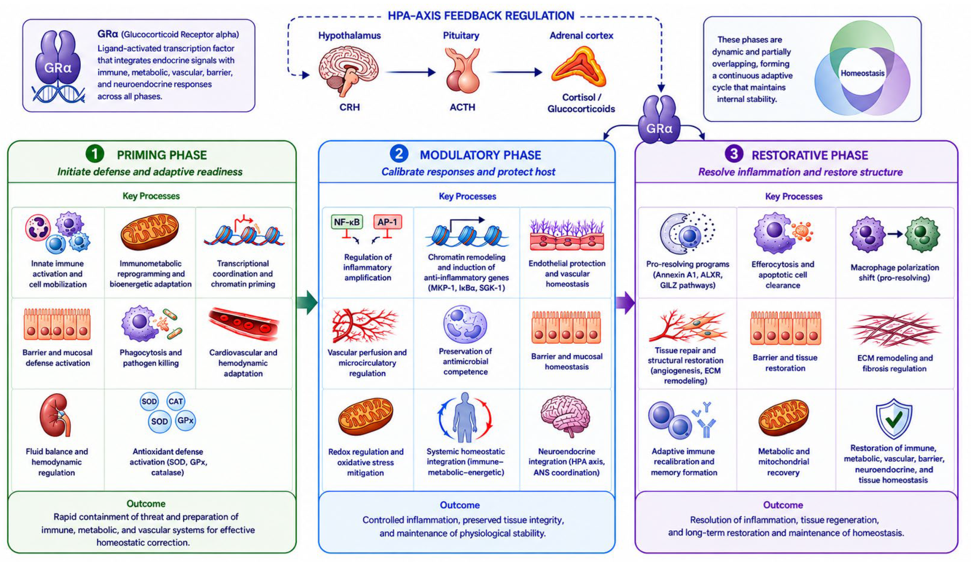
Figure 3.
Figure 3. Phase-Specific GRα Regulation of Chromatin Remodeling and Transcriptional Reprogramming
During Homeostatic Correction. Legend: This figure illustrates the proposed molecular framework by which
glucocorticoid receptor alpha (GRα) contributes to phase-specific changes in chromatin accessibility,
transcriptional regulation, and gene-expression programs throughout homeostatic correction. The central
GRα regulatory-program column summarizes the evolving phase-specific actions of GRα that coordinate the
chromatin remodeling, transcription-factor interactions, and gene-expression changes illustrated in the
adjacent columns. During the Priming Phase, GRα promotes adaptive transcriptional activation by facilitating
chromatin accessibility, inflammatory gene expression, immunometabolic adaptation, and antimicrobial
competence. During the Modulatory Phase, GRα recalibrates inflammatory signaling through coordinated
chromatin remodeling, induction of negative-feedback pathways, endothelial protection, and metabolic
flexibility. During the Restorative Phase, GRα promotes reparative transcriptional programs that support
mitochondrial recovery, inflammatory resolution, extracellular matrix remodeling, tissue repair, and restoration of physiological homeostasis. The failed-transition pathway illustrates how disruption of
coordinated GRα regulatory programs may contribute to persistent inflammatory chromatin states, defective
reparative transcription, metabolic dysfunction, glucocorticoid resistance, and failure of homeostatic
correction. This figure emphasizes that successful adaptation reflects dynamic, phase-specific transcriptional
reprogramming rather than static inflammatory suppression, thereby providing a molecular framework
linking GRα signaling to systems-level homeostatic correction. The author conceptually developed
this figure with assistance from ChatGPT (OpenAI) for graphical generation, visual organization, and figure
refinement. The author independently developed, critically reviewed, and
approved the biological framework, scientific content, interpretation, and final figure design.
Figure 3.
Figure 3. Phase-Specific GRα Regulation of Chromatin Remodeling and Transcriptional Reprogramming
During Homeostatic Correction. Legend: This figure illustrates the proposed molecular framework by which
glucocorticoid receptor alpha (GRα) contributes to phase-specific changes in chromatin accessibility,
transcriptional regulation, and gene-expression programs throughout homeostatic correction. The central
GRα regulatory-program column summarizes the evolving phase-specific actions of GRα that coordinate the
chromatin remodeling, transcription-factor interactions, and gene-expression changes illustrated in the
adjacent columns. During the Priming Phase, GRα promotes adaptive transcriptional activation by facilitating
chromatin accessibility, inflammatory gene expression, immunometabolic adaptation, and antimicrobial
competence. During the Modulatory Phase, GRα recalibrates inflammatory signaling through coordinated
chromatin remodeling, induction of negative-feedback pathways, endothelial protection, and metabolic
flexibility. During the Restorative Phase, GRα promotes reparative transcriptional programs that support
mitochondrial recovery, inflammatory resolution, extracellular matrix remodeling, tissue repair, and restoration of physiological homeostasis. The failed-transition pathway illustrates how disruption of
coordinated GRα regulatory programs may contribute to persistent inflammatory chromatin states, defective
reparative transcription, metabolic dysfunction, glucocorticoid resistance, and failure of homeostatic
correction. This figure emphasizes that successful adaptation reflects dynamic, phase-specific transcriptional
reprogramming rather than static inflammatory suppression, thereby providing a molecular framework
linking GRα signaling to systems-level homeostatic correction. The author conceptually developed
this figure with assistance from ChatGPT (OpenAI) for graphical generation, visual organization, and figure
refinement. The author independently developed, critically reviewed, and
approved the biological framework, scientific content, interpretation, and final figure design.
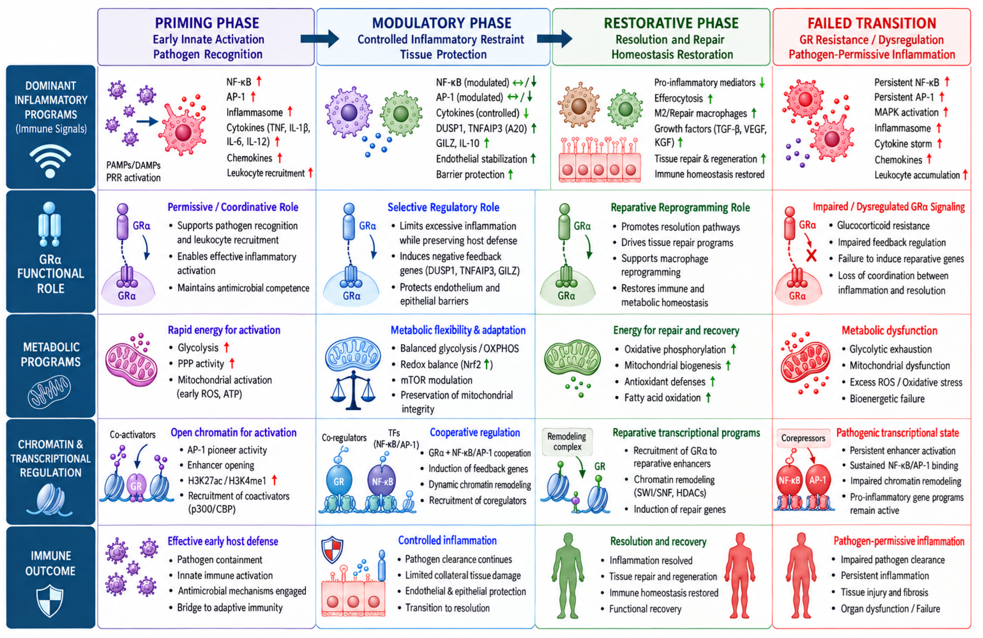
References
- Medzhitov, R. Recognition of microorganisms and activation of the immune response. Nature 2007, 449, 819–26. [Google Scholar] [CrossRef] [PubMed]
- Iwasaki; Medzhitov, R. Regulation of adaptive immunity by the innate immune system. Science 2010, 327, 291–5. [Google Scholar] [CrossRef] [PubMed]
- Medzhitov, R.; Schneider, D.S.; Soares, M.P. Disease tolerance as a defense strategy. Science 2012, 335, 936–41. [Google Scholar] [CrossRef] [PubMed]
- Soares, M.P.; Teixeira, L.; Moita, L.F. Disease tolerance and immunity in host protection against infection. Nat. Rev. Immunol. 2017, 17, 83–96. [Google Scholar] [CrossRef] [PubMed]
- Paludan, S.R.; Pradeu, T.; Pichlmair, A.; Wray, K.B.; Mikkelsen, J.G.; Olagnier, D.; Mogensen, T.H. Early host defense against virus infections. Cell Rep. 2024, 43, 115070. [Google Scholar] [CrossRef] [PubMed]
- Zhao, X.; Du, R.; Rong, L. Balancing Host Defense and Viral Tolerance for the Development of Next-Generation Broad-Spectrum Antiviral Agents. Pathogens 2025, 14. [Google Scholar] [CrossRef] [PubMed]
- Meduri, G.U.; Chrousos, G.P. General Adaptation in Critical Illness: Glucocorticoid Receptor-alpha Master Regulator of Homeostatic Corrections. Front. Endocrinol. 2020, 11, 161. [Google Scholar] [CrossRef] [PubMed]
- G. Meduri, Glucocorticoid receptor alpha: origins and functions of the master regulator of homeostatic corrections in health and critical illness. Explor Endocr. Metab. Dis. 2025, 2, 101426. [CrossRef]
- Meduri, G.U. Glucocorticoids and GRα Signaling in Critical Illness: Phase-Specific Homeostatic Corrections Across Systems. Semin. Respir. Crit. Care Med. 2026, 47, 3–31. [Google Scholar] [CrossRef] [PubMed]
- Takeuchi; Akira, S. Pattern recognition receptors and inflammation. Cell 2010, 140, 805–20. [Google Scholar] [CrossRef] [PubMed]
- Kawai, T.; Akira, S. The role of pattern-recognition receptors in innate immunity: update on Toll-like receptors. Nat. Immunol. 2010, 11, 373–84. [Google Scholar] [CrossRef] [PubMed]
- Xavier, A.M.; Anunciato, A.K.; Rosenstock, T.R.; Glezer, I. Gene Expression Control by Glucocorticoid Receptors during Innate Immune Responses. Front. Endocrinol. 2016, 7 31. [Google Scholar] [CrossRef] [PubMed]
- Quatrini, L.; Ugolini, S. New insights into the cell- and tissue-specificity of glucocorticoid actions. Cell. Mol. Immunol. 2021, 18, 269–278. [Google Scholar] [CrossRef] [PubMed]
- Khadka, S.; Dziadowicz, S.A.; Xu, X.; Wang, L.; Hu, G.; Carrero, J.A.; DiPaolo, R.J.; Busada, J.T. Endogenous glucocorticoids are required for normal macrophage activation and gastric Helicobacter pylori immunity. Am. J. Physiol. Gastrointest. Liver Physiol. 2024, 327, G531–g544. [Google Scholar] [CrossRef] [PubMed]
- Ehrchen, J.M.; Roth, J.; Barczyk-Kahlert, K. More than suppression: glucocorticoid action on monocytes and macrophages. Front. Immunol. 2019, 10, 2028. [Google Scholar] [CrossRef] [PubMed]
- Psarra, A.M.; Sekeris, C.E. Glucocorticoids induce mitochondrial gene transcription in HepG2 cells: role of the mitochondrial glucocorticoid receptor. Biochim. Biophys. Acta 2011, 1813, 1814–21. [Google Scholar] [CrossRef] [PubMed]
- Stifel, U.; Wolfschmitt, E.M.; Vogt, J.; Wachter, U.; Vettorazzi, S.; Tews, D.; Hogg, M.; Zink, F.; Koll, N.M.; Winning, S.; Mounier, R.; Chazaud, B.; Radermacher, P.; Fischer-Posovszky, P.; Caratti, G.; Tuckermann, J. Glucocorticoids coordinate macrophage metabolism through the regulation of the tricarboxylic acid cycle. Mol. Metab. 2022, 57, 101424. [Google Scholar] [CrossRef] [PubMed]
- Meduri, G.U.; Psarra, Anna-Maria G. The Glucocorticoid System: A Multifaceted Regulator of Mitochondrial Function, Endothelial Homeostasis, and Intestinal Barrier Integrity. Semin. Respir. Crit. Care Med. 2026, 47, 32–46. [Google Scholar] [CrossRef] [PubMed]
- Shimba; Ikuta, K. Control of immunity by glucocorticoids in health and disease. Semin. Immunopathol. 2020, 42, 669–680. [Google Scholar] [CrossRef] [PubMed]
- Quatrini, L.; Wieduwild, E.; Escaliere, B.; Filtjens, J.; Chasson, L.; Laprie, C.; Vivier, E.; Ugolini, S. Endogenous glucocorticoids control host resistance to viral infection through the tissue-specific regulation of PD-1 expression on NK cells. Nat. Immunol. 2018, 19, 954–962. [Google Scholar] [CrossRef] [PubMed]
- Caratti, G.; Desgeorges, T.; Juban, G.; Stifel, U.; Fessard, A.; Koenen, M.; Caratti, B.; Théret, M.; Skurk, C.; Chazaud, B.; Tuckermann, J.P.; Mounier, R. Macrophagic AMPKα1 orchestrates regenerative inflammation induced by glucocorticoids. EMBO Rep. 2023, 24, e55363. [Google Scholar] [CrossRef] [PubMed]
- Cain, D.W.; Cidlowski, J.A. Immune regulation by glucocorticoids. Nat. Rev. Immunol. 2017, 17, 233–247. [Google Scholar] [CrossRef] [PubMed]
- Kokkinopoulou; Moutsatsou, P. Mitochondrial Glucocorticoid Receptors and Their Actions. Int. J. Mol. Sci. 2021, 22, 6054. [Google Scholar] [CrossRef] [PubMed]
- Newton, R.; Shah, S.; Altonsy, M.O.; Gerber, A.N. Glucocorticoid and cytokine crosstalk: Feedback, feedforward, and co-regulatory interactions determine repression or resistance. J. Biol. Chem. 2017, 292, 7163–7172. [Google Scholar] [CrossRef] [PubMed]
- Escoter-Torres, L.; Caratti, G.; Mechtidou, A.; Tuckermann, J.; Uhlenhaut, N.H.; Vettorazzi, S. Fighting the Fire: Mechanisms of Inflammatory Gene Regulation by the Glucocorticoid Receptor. Front. Immunol. 2019, 10, 1859. [Google Scholar] [CrossRef] [PubMed]
- Jenuwein, T.; Allis, C.D. Translating the histone code. Science 2001, 293, 1074–80. [Google Scholar] [CrossRef] [PubMed]
- John, S.; Sabo, P.J.; Thurman, R.E.; Sung, M.-H.; Biddie, S.C.; Johnson, T.A.; Hager, G.L.; Stamatoyannopoulos, J.A. Chromatin accessibility pre-determines glucocorticoid receptor binding patterns. Nat. Genet. 2011, 43, 264–268. [Google Scholar] [CrossRef] [PubMed]
- Biddie, S.C.; John, S.; Sabo, P.J.; Thurman, R.E.; Johnson, T.A.; Schiltz, R.L.; Miranda, T.B.; Sung, M.-H.; Trump, S.; Lightman, S.L. Transcription factor AP1 potentiates chromatin accessibility and glucocorticoid receptor binding. Mol. Cell 2011, 43, 145–155. [Google Scholar] [CrossRef] [PubMed]
- Fadel, L.; Dacic, M.; Fonda, V.; Sokolsky, B.A.; Quagliarini, F.; Rogatsky, I.; Uhlenhaut, N.H. Modulating glucocorticoid receptor actions in physiology and pathology: Insights from coregulators. Pharmacol. Ther. 2023, 251, 108531. [Google Scholar] [CrossRef] [PubMed]
- Deroo, B.J.; Archer, T.K. Glucocorticoid receptor-mediated chromatin remodeling in vivo. Oncogene 2001, 20, 3039–46. [Google Scholar] [CrossRef] [PubMed]
- Hoffman, J.A.; Trotter, K.W.; Ward, J.M.; Archer, T.K. BRG1 governs glucocorticoid receptor interactions with chromatin and pioneer factors across the genome. Elife 2018, 7. [Google Scholar] [CrossRef] [PubMed]
- Johnson, T.A.; Chereji, R.V.; Stavreva, D.A.; Morris, S.A.; Hager, G.L.; Clark, D.J. Conventional and pioneer modes of glucocorticoid receptor interaction with enhancer chromatin in vivo. Nucleic Acids Res. 2018, 46, 203–214. [Google Scholar] [CrossRef] [PubMed]
- Uhlenhaut, N.H.; Barish, G.D.; Ruth, T.Y.; Downes, M.; Karunasiri, M.; Liddle, C.; Schwalie, P.; Hübner, N.; Evans, R.M. Insights into negative regulation by the glucocorticoid receptor from genome-wide profiling of inflammatory cistromes. Mol. Cell 2013, 49, 158–171. [Google Scholar] [CrossRef] [PubMed]
- Psarra, A.-M.G.; Sekeris, C.E. Nuclear receptors and other nuclear transcription factors in mitochondria: regulatory molecules in a new environment. Biochim. Et. Biophys. Acta (BBA) -Mol. Cell Res. 2008, 1783, 1–11. [Google Scholar] [CrossRef] [PubMed]
- Oakley, R.H.; Ren, R.; Cruz-Topete, D.; Bird, G.S.; Myers, P.H.; Boyle, M.C.; Schneider, M.D.; Willis, M.S.; Cidlowski, J.A. Essential role of stress hormone signaling in cardiomyocytes for the prevention of heart disease. Proc. Natl. Acad. Sci. U S A 2013, 110, 17035–40. [Google Scholar] [CrossRef] [PubMed]
- Fitzgerald, K.A.; Kagan, J.C. Toll-like Receptors and the Control of Immunity. Cell 2020, 180, 1044–1066. [Google Scholar] [CrossRef] [PubMed]
- Chinenov, Y.; Rogatsky, I. Glucocorticoids and the innate immune system: crosstalk with the toll-like receptor signaling network. Mol. Cell. Endocrinol. 2007, 275, 30–42. [Google Scholar] [CrossRef] [PubMed]
- Hermoso, M.A.; Matsuguchi, T.; Smoak, K.; Cidlowski, J.A. Glucocorticoids and tumor necrosis factor alpha cooperatively regulate toll-like receptor 2 gene expression. Mol. Cell. Biol. 2004, 24, 4743–56. [Google Scholar] [CrossRef] [PubMed]
- Strehl, C.; Ehlers, L.; Gaber, T.; Buttgereit, F. Glucocorticoids-All-Rounders Tackling the Versatile Players of the Immune System. Front. Immunol. 2019, 10, 1744. [Google Scholar] [CrossRef] [PubMed]
- Rocamora-Reverte, L.; Villunger, A.; Wiegers, G.J. Cell-Specific Immune Regulation by Glucocorticoids in Murine Models of Infection and Inflammation. Cells 2022, 11. [Google Scholar] [CrossRef] [PubMed]
- Ansari, S.A.; Dantoft, W.; Ruiz-Orera, J.; Syed, A.P.; Blachut, S.; van Heesch, S.; Hübner, N.; Uhlenhaut, N.H. Integrative analysis of macrophage ribo-Seq and RNA-Seq data define glucocorticoid receptor regulated inflammatory response genes into distinct regulatory classes. Comput Struct. Biotechnol. J. 2022, 20, 5622–5638. [Google Scholar] [CrossRef] [PubMed]
- Ronchetti, S.; Ricci, E.; Migliorati, G.; Gentili, M.; Riccardi, C. How Glucocorticoids Affect the Neutrophil Life. Int. J. Mol. Sci. 2018, 19. [Google Scholar] [CrossRef] [PubMed]
- Sun, K.; Xie, Z.; Li, Y.; Li, Y.; Song, J.; Meng, Z. Glucocorticoids Regulate the Activation of Neutrophils and Inhibit the Formation of Pulmonary Embolism. Iran. J. Immunol. 2020, 17, 303–312. [Google Scholar] [CrossRef] [PubMed]
- Steffensen, N.; Imker, R.; Lassnig, S.; Fulde, M.; Rieder, J.C.; de Buhr, N. Methylprednisolone Induces Extracellular Trap Formation and Enhances Bactericidal Effect of Canine Neutrophils. Int. J. Mol. Sci. 2021, 22. [Google Scholar] [CrossRef] [PubMed]
- Thrikawala, S.U.; Anderson, M.H.; Rosowski, E.E. Glucocorticoids Suppress NF-κB-Mediated Neutrophil Control of Aspergillus fumigatus Hyphal Growth. J. Immunol. 2024, 213, 971–987. [Google Scholar] [CrossRef] [PubMed]
- Piemonti, L.; Monti, P.; Allavena, P.; Sironi, M.; Soldini, L.; Leone, B.E.; Socci, C.; Di Carlo, V. Glucocorticoids affect human dendritic cell differentiation and maturation. J. Immunol. 1999, 162, 6473–81. [Google Scholar] [CrossRef]
- Moser, M.; De Smedt, T.; Sornasse, T.; Tielemans, F.; Chentoufi, A.A.; Muraille, E.; Van Mechelen, M.; Urbain, J.; Leo, O. Glucocorticoids down-regulate dendritic cell function in vitro and in vivo. Eur. J. Immunol. 1995, 25, 2818–24. [Google Scholar] [CrossRef] [PubMed]
- Liberman, A.C.; Budziñski, M.L.; Sokn, C.; Gobbini, R.P.; Steininger, A.; Arzt, E. Regulatory and Mechanistic Actions of Glucocorticoids on T and Inflammatory Cells. Front. Endocrinol. 2018, 9, 235. [Google Scholar] [CrossRef] [PubMed]
- Elftman, M.D.; Norbury, C.C.; Bonneau, R.H.; Truckenmiller, M.E. Corticosterone impairs dendritic cell maturation and function. Immunology 2007, 122, 279–90. [Google Scholar] [CrossRef] [PubMed]
- Rozkova, D.; Horvath, R.; Bartunkova, J.; Spisek, R. Glucocorticoids severely impair differentiation and antigen presenting function of dendritic cells despite upregulation of Toll-like receptors. Clin. Immunol. 2006, 120, 260–71. [Google Scholar] [CrossRef] [PubMed]
- Taves, M.; Ashwell, J. Glucocorticoids in T cell development, differentiation and function. Nat. Rev. Immunol. 2020, 21, 233–243. [Google Scholar] [CrossRef] [PubMed]
- Shimba; Cui, G.; Tani-Ichi, S.; Ogawa, M.; Abe, S.; Okazaki, F.; Kitano, S.; Miyachi, H.; Yamada, H.; Hara, T.; Yoshikai, Y.; Nagasawa, T.; Schütz, G.; Ikuta, K. Glucocorticoids Drive Diurnal Oscillations in T Cell Distribution and Responses by Inducing Interleukin-7 Receptor and CXCR4. Immunity 2018, 48, 286. [Google Scholar] [CrossRef] [PubMed]
- Mittelstadt, P.; Monteiro, J.; Ashwell, J. Thymocyte responsiveness to endogenous glucocorticoids is required for immunological fitness. J. Clin. Invest 2012, 122 7, 2384–2394. [Google Scholar] [CrossRef] [PubMed]
- Kim, D.; Nguyen, Q.; Lee, J.; Lee, S.H.; Janocha, A.; Kim, S.; Le, H.; Dvorina, N.; Weiss, K.; Cameron, M.; Asosingh, K.; Erzurum, S.; Baldwin, W.; Lee, J.-S.; Min, B. Anti-inflammatory Roles of Glucocorticoids Are Mediated by Foxp3+ Regulatory T Cells via a miR-342-Dependent Mechanism. Immunity 2020, 53, 581–596. [Google Scholar] [CrossRef] [PubMed]
- Rocamora-Reverte, L.; Tuzlak, S.; Von Raffay, L.; Tisch, M.; Fiegl, H.; Drach, M.; Reichardt, H.; Villunger, A.; Tischner, D.; Wiegers, G. Glucocorticoid Receptor-Deficient Foxp3+ Regulatory T Cells Fail to Control Experimental Inflammatory Bowel Disease. Front. Immunol. 2019, 10. [Google Scholar] [CrossRef] [PubMed]
- Tehseen; Kumar, D.; Dubey, A.; Sarkar, R.; Singh, S.; Sehrawat, S. Glucocorticoid-mediated Suppression of Effector Programming Assists the Memory Transition of Virus-specific CD8+ T Cells. J. Immunol. 2024, 213, 1170–1186. [Google Scholar] [CrossRef] [PubMed]
- Cain, D.; Bortner, C.; Díaz-Jiménez, D.; Petrillo, M.; Gruver-Yates, A.; Cidlowski, J. Murine Glucocorticoid Receptors Orchestrate B-cell Migration Selectively between Bone Marrow and Blood. J. Immunol. 2020, 205, 619–629. [Google Scholar] [CrossRef] [PubMed]
- Franco, L.; Gadkari, M.; Howe, K.; Sun, J.; Kardava, L.; Kumar, P.; Kumari, S.; Hu, Z.; Fraser, I.; Moir, S.; Tsang, J.; Germain, R. Immune regulation by glucocorticoids can be linked to cell type–dependent transcriptional responses. J. Exp. Med. 2019, 216, 384–406. [Google Scholar] [CrossRef] [PubMed]
- Zielinska, K.A.; Van Moortel, L.; Opdenakker, G.; De Bosscher, K.; Van den Steen, P.E. Endothelial Response to Glucocorticoids in Inflammatory Diseases. Front. Immunol. 2016, 7, 592. [Google Scholar] [CrossRef] [PubMed]
- Goodwin, J.E.; Feng, Y.; Velazquez, H.; Sessa, W.C. Endothelial glucocorticoid receptor is required for protection against sepsis. Proc. Natl. Acad. Sci. USA 2013, 110, 306–11. [Google Scholar] [CrossRef] [PubMed]
- Förster, C.; Waschke, J.; Burek, M.; Leers, J.; Drenckhahn, D. Glucocorticoid effects on mouse microvascular endothelial barrier permeability are brain specific. J. Physiol. 2006, 573. [Google Scholar] [CrossRef] [PubMed]
- Mehta, S.; Srivastava, S.; Grabińska, K.; Zhang, X.; Wong, C.; Hedayat, A.; Perrotta, P.; Fernández-Hernando, C.; Sessa, W.; Goodwin, J. Endothelial cell-glucocorticoid receptor interactions and regulation of Wnt signaling. JCI Insight 2020, 5 3. [Google Scholar]
- Akhter, M.S.; Goodwin, J. Endothelial Dysfunction in Cardiorenal Conditions: Implications of Endothelial Glucocorticoid Receptor-Wnt Signaling. Int. J. Mol. Sci. 2023, 24. [Google Scholar] [CrossRef] [PubMed]
- Loft; Schmidt, S.F.; Caratti, G.; Stifel, U.; Havelund, J.; Sekar, R.; Kwon, Y.; Sulaj, A.; Chow, K.K.; Alfaro, A.J.; Schwarzmayr, T.; Rittig, N.; Svart, M.; Tsokanos, F.F.; Maida, A.; Blutke, A.; Feuchtinger, A.; Møller, N.; Blüher, M.; Nawroth, P.; Szendrödi, J.; Færgeman, N.J.; Zeigerer, A.; Tuckermann, J.; Herzig, S. A macrophage-hepatocyte glucocorticoid receptor axis coordinates fasting ketogenesis. Cell Metab. 2022, 34, 473–486.e9. [Google Scholar] [CrossRef] [PubMed]
- Karra, A.G.; Sioutopoulou, A.; Gorgogietas, V.; Samiotaki, M.; Panayotou, G.; Psarra, A.-M.G. Proteomic analysis of the mitochondrial glucocorticoid receptor interacting proteins reveals pyruvate dehydrogenase and mitochondrial 60 kDa heat shock protein as potent binding partners. J. Proteom. 2022, 257, 104509. [Google Scholar] [CrossRef] [PubMed]
- Auger, J.P.; Zimmermann, M.; Faas, M.; Stifel, U.; Chambers, D.; Krishnacoumar, B.; Taudte, R.V.; Grund, C.; Erdmann, G.; Scholtysek, C.; Uderhardt, S.; Ben Brahim, O.; Pascual Maté, M.; Stoll, C.; Böttcher, M.; Palumbo-Zerr, K.; Mangan, M.S.J.; Dzamukova, M.; Kieler, M.; Hofmann, M.; Blüml, S.; Schabbauer, G.; Mougiakakos, D.; Sonnewald, U.; Hartmann, F.; Simon, D.; Kleyer, A.; Grüneboom, A.; Finotto, S.; Latz, E.; Hofmann, J.; Schett, G.; Tuckermann, J.; Krönke, G. Metabolic rewiring promotes anti-inflammatory effects of glucocorticoids. Nature 2024, 629, 184–192. [Google Scholar] [CrossRef] [PubMed]
- Lu, Y.; Liu, H.; Bi, Y.; Yang, H.-T.; Li, Y.; Wang, J.; Zhang, Z.; Wang, Y.; Li, C.; Jia, A.; Han, L.; Hu, Y.; Zhao, Y.; Wang, R.; Liu, G. Glucocorticoid receptor promotes the function of myeloid-derived suppressor cells by suppressing HIF1α-dependent glycolysis. Cell. Mol. Immunol. 2017, 15, 618–629. [Google Scholar] [CrossRef] [PubMed]
- De Vos, W.; Tilg, H.; Van Hul, M.; Cani, P. Gut microbiome and health: mechanistic insights. Gut 2022, 71, 1020–1032. [Google Scholar] [CrossRef] [PubMed]
- Di Vincenzo, F.; Del Gaudio, A.; Petito, V.; Lopetuso, L.R.; Scaldaferri, F. Gut microbiota, intestinal permeability, and systemic inflammation: a narrative review. Intern. Emerg. Med. 2024, 19, 275–293. [Google Scholar] [CrossRef] [PubMed]
- Merk, V.M.; Phan, T.S.; Brunner, T. Regulation of Tissue Immune Responses by Local Glucocorticoids at Epithelial Barriers and Their Impact on Interorgan Crosstalk. Front. Immunol. 2021, 12, 1639. [Google Scholar] [CrossRef] [PubMed]
- Ballegeer, M.; Vandewalle, J.; Eggermont, M.; Van Isterdael, G.; Dejager, L.; De Bus, L.; Decruyenaere, J.; Vandenbroucke, R.E.; Libert, C. Overexpression of Gilz Protects Mice Against Lethal Septic Peritonitis. Shock 2019, 52, 208–214. [Google Scholar] [CrossRef] [PubMed]
- Shukla, P.K.; Meena, A.S.; Pierre, J.F.; Rao, R. Central role of intestinal epithelial glucocorticoid receptor in alcohol- and corticosterone-induced gut permeability and systemic response. Faseb J. 2022, 36, e22061. [Google Scholar] [CrossRef] [PubMed]
- Vagnerová, K.; Vodička, M.; Hermanová, P.; Ergang, P.; Šrůtková, D.; Klusoňová, P.; Balounová, K.; Hudcovic, T.; Pácha, J. Interactions Between Gut Microbiota and Acute Restraint Stress in Peripheral Structures of the Hypothalamic-Pituitary-Adrenal Axis and the Intestine of Male Mice. Front. Immunol. 2019, 10, 2655. [Google Scholar] [CrossRef] [PubMed]
- Rizzetto, L.; Fava, F.; Tuohy, K.M.; Selmi, C. Connecting the immune system, systemic chronic inflammation and the gut microbiome: The role of sex. J. Autoimmun. 2018, 92, 12–34. [Google Scholar] [CrossRef] [PubMed]
- Huang, E.Y.; Inoue, T.; Leone, V.A.; Dalal, S.; Touw, K.; Wang, Y.; Musch, M.W.; Theriault, B.; Higuchi, K.; Donovan, S.; Gilbert, J.; Chang, E.B. Using Corticosteroids to Reshape the Gut Microbiome: Implications for Inflammatory Bowel Diseases. Inflamm. Bowel Dis. 2015, 21, 963–972. [Google Scholar] [PubMed]
- Kutsuzawa, N.; Ito, Y.; Kagawa, S.; Kohno, C.; Takiguchi, H.; Asano, K. Dexamethasone restores TNFα-induced epithelial barrier dysfunction in primary rat alveolar epithelial cells. PLoS ONE 2023, 18. [Google Scholar] [CrossRef] [PubMed]
- Tena-Garitaonaindia, M.; Arredondo-Amador, M.; Mascaraque, C.; Asensio, M.; Marin, J.J.G.; Martínez-Augustin, O.; Sánchez de Medina, F. Modulation of intestinal barrier function by glucocorticoids: Lessons from preclinical models. Pharmacol. Res. 2022, 177, 106056. [Google Scholar] [CrossRef] [PubMed]
- Singh, S.; Pragman, A.A.; Segal, L.N. Balancing Benefits and Risks: Do Inhaled Corticosteroids Modify the Lung Microbiome? Am. J. Respir. Crit. Care Med. 2021, 204, 1117–1119. [Google Scholar] [CrossRef] [PubMed]
- Meduri, G.U.; Kanangat, S.; Stefan, J.; Tolley, E.; Schaberg, D. Cytokines IL-1beta, IL-6, and TNF-alpha enhance in vitro growth of bacteria. Am. J. Respir. Crit. Care Med. 1999, 160, 961–7. [Google Scholar] [CrossRef] [PubMed]
- Kanangat, S.; Meduri, G.U.; Tolley, E.A.; Patterson, D.R.; Meduri, C.U.; Pak, C.; Griffin, J.P.; Bronze, M.S.; Schaberg, D.R. Effects of cytokines and endotoxin on the intracellular growth of bacteria. Infect. Immun. 1999, 67, 2834–40. [Google Scholar] [CrossRef] [PubMed]
- Meduri, G.U. A paradigm shift: The bidirectional effect of inflammation on bacterial growth. In Sepsis and Organ Dysfunction: The Challenge Continues; Baue, A.E., Berlot, G., Gullo, A., Vincent, J.-L., Eds.; Springer-Verlag: Milano, Italy, 1999; pp. 145–154. [Google Scholar]
- Zhan, M.; Chen, H.; Li, Z.; Liu, S.; Lu, B.; Wang, Z.; Wang, H. Available at SSRN 5200008; Lower Respiratory Microbiome Dysbiosis is Associated with Poor Prognosis in Acute Severe Lower Respiratory Tract Infection.
- Meduri, G.U. Factors Influencing Glucocorticoid Treatment Response: Mechanism-Based Strategies to Overcome Glucocorticoid Resistance and Restore GRα Function. Semin. Respir. Crit. Care Med. 2026, 47, 47–65. [Google Scholar] [CrossRef] [PubMed]
- Oliver, G.; Kipnis, J.; Randolph, G.J.; Harvey, N.L. The Lymphatic Vasculature in the 21(st) Century: Novel Functional Roles in Homeostasis and Disease. Cell 2020, 182, 270–296. [Google Scholar] [CrossRef] [PubMed]
- Petrova, T.V.; Koh, G.Y. Organ-specific lymphatic vasculature: From development to pathophysiology. J. Exp. Med. 2018, 215, 35–49. [Google Scholar] [CrossRef] [PubMed]
- Yao, L.C.; Baluk, P.; Srinivasan, R.S.; Oliver, G.; McDonald, D.M. Plasticity of button-like junctions in the endothelium of airway lymphatics in development and inflammation. Am. J. Pathol. 2012, 180, 2561–75. [Google Scholar] [CrossRef] [PubMed]
- Zhong, J.; Yang, H.; Shelton, E.L.; Kon, V. Lymphatic Dysfunction and Perirenal Adiposity: Role of Glucocorticoids: SA-PO717. J. Am. Soc. Nephrol. 2024, 35 10, 1681. [Google Scholar] [CrossRef]
- Zhou, H.; Mehta, S.; Srivastava, S.P.; Grabinska, K.; Zhang, X.; Wong, C.; Hedayat, A.; Perrotta, P.; Fernández-Hernando, C.; Sessa, W.C. Endothelial cell–glucocorticoid receptor interactions and regulation of Wnt signaling. JCI Insight 2020, 5, e131384. [Google Scholar] [CrossRef] [PubMed]
- Liu, B.; Zhou, H.; Zhang, T.; Gao, X.; Tao, B.; Xing, H.; Zhuang, Z.; Dardik, A.; Kyriakides, T.R.; Goodwin, J.E. Loss of endothelial glucocorticoid receptor promotes angiogenesis via upregulation of Wnt/β-catenin pathway. Angiogenesis 2021, 24, 631–645. [Google Scholar] [CrossRef] [PubMed]
- Zhong, J.; Yang, H.; Shelton, E.L.; Kon, V. Glucocorticoid Treatment Induces Lymphatic Dysfunction via ATP-Sensitive Potassium Channel: TH-PO942. J. Am. Soc. Nephrol. 2023, 34, 355–355. [Google Scholar] [CrossRef]
- Silverman, M.N.; Sternberg, E.M. Glucocorticoid regulation of inflammation and its functional correlates: from HPA axis to glucocorticoid receptor dysfunction. Ann. N. Y. Acad. Sci. 2012, 1261, 55–63. [Google Scholar] [CrossRef] [PubMed]
- Yurtsever, B.; Demirkol, D. Sepsis as CNS-Governed Metabolic Triage. Front. Immunol. 2026, 17, 1842915. [Google Scholar] [CrossRef] [PubMed]
- Paul, B.; Sterner, Z.; Bhawal, R.; Anderson, E.; Zhang, S.; Buchholz, D. Impaired negative feedback and death following acute stress in glucocorticoid receptor knockout Xenopus tropicalis tadpoles. Gen. Comp. Endocrinol. 2022, 114072. [Google Scholar] [CrossRef] [PubMed]
- Quatrini, L.; Wieduwild, E.; Guia, S.; Bernat, C.; Glaichenhaus, N.; Vivier, E.; Ugolini, S. Host resistance to endotoxic shock requires the neuroendocrine regulation of group 1 innate lymphoid cells. J. Exp. Med. 2017, 214, 3531–3541. [Google Scholar] [CrossRef] [PubMed]
- Téblick; Van Dyck, L.; Van Aerde, N.; Vander Perre, S.; Pauwels, L.; Derese, I.; Debaveye, Y.; Wouters, P.J.; Vanhorebeek, I.; Langouche, L. OR26-4 Glucocorticoid Receptor Expression and Signaling During Critical Illness, in Relation to the Duration of Illness and the Systemic Glucocorticoid Availability: A Prospective, Observational, Cross-Sectional Human and a Translational Mouse Study. J. Endocr. Soc. 2022, 6, A726. [Google Scholar] [CrossRef]
- Poupouzas, G.; Lotsios, N.S.; Vrettou, C.S.; Issaris, V.; Keskinidou, C.; Papavassiliou, K.A.; Halioti, A.; Botoula, E.; Tzanela, M.; Papavassiliou, A.G.; Kotanidou, A.; Vassiliadi, D.A.; Vassiliou, A.G.; Dimopoulou, I. Cell-specific expression and signaling of glucocorticoid receptor isoforms over time in critically ill patients with a low inflammatory response. Crit. Care 2025, 29, 390. [Google Scholar] [CrossRef] [PubMed]
- Langouche, L.; Téblick, A.; Gunst, J.; Van Den Berghe, G. The Hypothalamus-pituitary-adrenocortical Response to Critical Illness: A Concept in Need of Revision. Endocr. Rev. 2023, 44, 1096–1106. [Google Scholar] [CrossRef] [PubMed]
- Shimba; Ikuta, K. Glucocorticoids Regulate Circadian Rhythm of Innate and Adaptive Immunity. Front. Immunol. 2020, 11, 2143. [Google Scholar] [CrossRef] [PubMed]
- Ramos-Ramírez, P.; Tliba, O. Glucocorticoid receptor β (GRβ): beyond its dominant-negative function. Int. J. Mol. Sci. 2021, 22, 3649. [Google Scholar] [CrossRef] [PubMed]
- Vago, J.P.; Tavares, L.P.; Riccardi, C.; Teixeira, M.M.; Sousa, L.P. Exploiting the pro-resolving actions of glucocorticoid-induced proteins Annexin A1 and GILZ in infectious diseases. Biomed. Pharmacother. 2021, 133, 111033. [Google Scholar] [CrossRef] [PubMed]
- Galuppo, P.; Vettorazzi, S.; Hövelmann, J.; Scholz, C.J.; Tuckermann, J.P.; Bauersachs, J.; Fraccarollo, D. The glucocorticoid receptor in monocyte-derived macrophages is critical for cardiac infarct repair and remodeling. Faseb J. 2017, 31, 5122–5132. [Google Scholar] [CrossRef] [PubMed]
- Srivastava, S.; Setia, O.; Dardik, A.; Fernández-Hernando, C.; Goodwin, J. Loss of endothelial glucocorticoid receptor accelerates diabetic nephropathy. Nat. Commun. 2021, 12. [Google Scholar] [CrossRef] [PubMed]
- Kadmiel, M.; Janoshazi, A.; Xu, X.; Cidlowski, J.A. Glucocorticoid action in human corneal epithelial cells establishes roles for corticosteroids in wound healing and barrier function of the eye. Exp. Eye Res. 2016, 152, 10–33. [Google Scholar] [CrossRef] [PubMed]
- Madalena, K.M.; Brennan, F.H.; Popovich, P.G. Genetic deletion of the glucocorticoid receptor in Cx3cr1+ myeloid cells is neuroprotective and improves motor recovery after spinal cord injury. Exp. Neurol. 2022, 355, 114114. [Google Scholar] [CrossRef] [PubMed]
- Pianca, N.; Sacchi, F.; Umansky, K.B.; Chirivì, M.; Iommarini, L.; Da Pra, S.; Papa, V.; Bongiovanni, C.; Miano, C.; Pontis, F. Glucocorticoid receptor antagonization propels endogenous cardiomyocyte proliferation and cardiac regeneration. Nat. Cardiovasc. Res. 2022, 1, 617–633. [Google Scholar] [CrossRef] [PubMed]
- Da Pra, S.; Boriati, S.; Miano, C.; Sacchi, F.; Batho, C.; Bongiovanni, C.; Del Bono, I.; Aharonov, A.; Pianca, N.; Tassinari, R. Harnessing glucocorticoid receptor antagonism to enhance the efficacy of cardiac regenerative growth factors and cytokines. Nat. Cardiovasc. Res. 2026, 5, 118–137. [Google Scholar] [CrossRef] [PubMed]
- Téblick; Van Dyck, L.; Van Aerde, N.; Van der Perre, S.; Pauwels, L.; Derese, I.; Debaveye, Y.; Wouters, P.J.; Vanhorebeek, I.; Langouche, L. Impact of duration of critical illness and level of systemic glucocorticoid availability on tissue-specific glucocorticoid receptor expression and actions: A prospective, observational, cross-sectional human and two translational mouse studies. EBioMedicine 2022, 80. [Google Scholar] [CrossRef] [PubMed]
- Marchi, D.; van Eeden, F.J.M. Homeostatic Regulation of Glucocorticoid Receptor Activity by Hypoxia-Inducible Factor 1: From Physiology to Clinic. Cells 2021, 10. [Google Scholar] [CrossRef] [PubMed]
- Rinaldi, L.; Fettweis, G.; Kim, S.; Garcia, D.A.; Fujiwara, S.; Johnson, T.A.; Tettey, T.T.; Ozbun, L.; Pegoraro, G.; Puglia, M.; Blagoev, B.; Upadhyaya, A.; Stavreva, D.A.; Hager, G.L. The glucocorticoid receptor associates with the cohesin loader NIPBL to promote long-range gene regulation. Sci. Adv. 2022, 8, eabj8360. [Google Scholar] [CrossRef] [PubMed]
- D'Ippolito, A.M.; McDowell, I.C.; Barrera, A.; Hong, L.K.; Leichter, S.M.; Bartelt, L.C.; Vockley, C.M.; Majoros, W.H.; Safi, A.; Song, L.; Gersbach, C.A.; Crawford, G.E.; Reddy, T.E. Pre-established Chromatin Interactions Mediate the Genomic Response to Glucocorticoids. Cell Syst. 2018, 7, 146–160.e7. [Google Scholar] [CrossRef] [PubMed]
- Petta; Dejager, L.; Ballegeer, M.; Lievens, S.; Tavernier, J.; De Bosscher, K.; Libert, C. The Interactome of the Glucocorticoid Receptor and Its Influence on the Actions of Glucocorticoids in Combatting Inflammatory and Infectious Diseases. Microbiol. Mol. Biol. Rev. 2016, 80, 495–522. [Google Scholar] [CrossRef] [PubMed]
- Bai, X.; Bai, A.; Tomasicchio, M.; Hagman, J.R.; Buckle, A.M.; Gupta, A.; Kadiyala, V.; Bevers, S.; Serban, K.A.; Kim, K.; Feng, Z.; Spendier, K.; Hagen, G.; Fornis, L.; Griffith, D.E.; Dzieciatkowska, M.; Sandhaus, R.A.; Gerber, A.N.; Chan, E.D. α1-Antitrypsin Binds to the Glucocorticoid Receptor with Anti-Inflammatory and Antimycobacterial Significance in Macrophages. J. Immunol. 2022, 209, 1746–1759. [Google Scholar] [CrossRef] [PubMed]
- Spies, L.-M.L.; Verhoog, N.J.; Louw, A. Acquired glucocorticoid resistance due to homologous glucocorticoid receptor downregulation: a modern look at an age-old problem. Cells 2021, 10, 2529. [Google Scholar] [CrossRef] [PubMed]

Table 1.
Structured, Hypothesis-Driven AI-Assisted Query Framework for Evaluating GRα Regulation of Immune Sensing, Antimicrobial Defense, and Immune Effectiveness.
Table 1.
Structured, Hypothesis-Driven AI-Assisted Query Framework for Evaluating GRα Regulation of Immune Sensing, Antimicrobial Defense, and Immune Effectiveness.
| Functional domain | Evidence domain | Consensus query | Scientific Objective | Phase of Homeostatic Correction |
| Pathogen recognition | PRR signaling (TLR, NLR, inflammasome) | Does glucocorticoid receptor alpha (GRα) signaling modulate Toll-like receptor and inflammasome activation, signaling thresholds, and coordinated host-defense responses during infection? | Assess GRα coordination of pathogen recognition, host-defense signaling, and immune effectiveness. [10,11,12] |
Priming Phase |
| Innate immunity | Inflammatory signaling | How does GRα coordinate NF-κB– and AP-1–dependent inflammatory and transcriptional programs during host defense and phase-specific homeostatic correction? | Assess GRα regulation of inflammatory signaling and preservation of effective host defense. [7,12,13] | Priming Phase |
| Innate immunity | Antimicrobial function | Does GRα signaling coordinate macrophage antimicrobial competence, phagocytic activity, and intracellular pathogen clearance during host defense? | Assess GRα regulation of antimicrobial competence and pathogen clearance. [7,14,15] | Priming Phase |
| Innate immunity | Immunometabolism | Does GRα signaling coordinate mitochondrial function, immunometabolic adaptation, and bioenergetic regulation during host defense responses? | Assess GRα regulation of immunometabolic adaptation and mitochondrial bioenergetics. [16,17,18] | Priming → Modulatory Phase |
| Inflammation regulation | Cytokine and chemokine regulation | Does GRα regulate the magnitude, duration, coordination, and phase-specific transition of cytokine and chemokine networks during host defense and homeostatic correction? | Assess GRα regulation of cytokine networks during phase-specific host defense. [7,13,19] | Modulatory Phase |
| Host defense outcome | Pathogen clearance vs persistence | Does GRα help coordinate the transition between pathogen-clearing and pathogen-permissive immune states during infection and homeostatic correction? | Assess GRα regulation of pathogen clearance and recovery-oriented immune responses [3,4,20] | Modulatory → Organ Functional Recovery: Restorative Phase |
| Resolution and repair | Immune reprogramming and resolution | How does GRα regulate immune reprogramming, resolution pathways, and restoration of immune homeostasis following pathogen clearance? | Assess GRα regulation of inflammatory resolution, tissue repair, and restoration of physiological homeostasis. [7,8,21] | Restorative Phase |
Legend: This table presents a structured, hypothesis-driven query framework developed to systematically evaluate the role of glucocorticoid receptor alpha (GRα) in immune sensing, host defense, and immune effectiveness. Artificial intelligence (Consensus AI) was used exclusively to generate structured, domain-specific literature queries and facilitate evidence retrieval. The scientific framework, interpretation of the evidence, synthesis of the literature, table organization, and all scientific conclusions were independently developed, critically reviewed, and approved by the author.
Table 2.
Major Functional Domains of Immune Responses Relevant to GRα Regulation.
| Immune Response Domain | Principal Components | Primary Function | Relevance to the GRα Conceptual Framework |
|---|---|---|---|
| Barrier immunity | Skin, airway, gut epithelium, mucus, antimicrobial peptides, microbiome | Maintains host–environment barrier integrity, regulates microbial interactions, and preserves tissue-integrated host defense | Highlights epithelial and mucosal interfaces as dynamic regulators of microbiome–immune–GRα interactions and host–environment homeostasis. [7,8] |
| Pathogen recognition and sensing | Pattern-recognition receptors (TLRs, NLRs, RLRs), inflammasomes | Detects microbial and danger signals and initiates coordinated innate immune signaling and host-defense responses | Highlights the role of GRα in coordinating pathogen-sensing and integrated host-defense signaling programs that shape immune effectiveness and adaptive response outcomes. [10,11,12] |
| Innate immunity | Macrophages, neutrophils, dendritic cells, NK cells | Integrates early antimicrobial defense, innate immune signaling, immunometabolic adaptation, and tissue-protective host-defense responses | Defines the role of GRα in coordinating inflammatory signaling, antimicrobial competence, immunometabolic adaptation, and tissue-protective innate host-defense responses across homeostatic phases, antimicrobial competence, immunometabolic adaptation, and phase-specific innate immune responses. [7,13,14,15,16,17,18] |
| Inflammasome-mediated immunity | NLRP3 inflammasome, caspase-1, IL-1β, IL-18 | Regulates inflammasome-dependent signaling and innate host-defense responses during tissue stress and infection | Highlights the phase-dependent role of GRα in coordinating inflammasome priming, activation, and resolution across host defense and homeostatic correction responses. [7,12,13,19] |
| Complement-mediated immunity | Complement proteins, opsonins, membrane attack complex | Supports complement-mediated opsonization, antimicrobial signaling, and inflammatory responses during innate immune defense | Highlights GRα-regulated integration of complement-mediated host defense with inflammatory coordination, tissue protection, and repair responses. [7,13] |
| Humoral immunity | B cells, plasma cells, antibodies | Supports antibody-mediated host defense and long-term immune adaptation following microbial exposure | Highlights the role of GRα in regulating B-cell function, antibody responses, and adaptive immune recalibration during host defense and recovery. [4,20] |
| Cell-mediated immunity | CD4 T cells, CD8 T cells, NK cells | Regulates adaptive cellular host-defense responses, immune recalibration, and tissue-specific regulation during infection and recovery | Highlights the role of GRα in adaptive host-defense regulation, immune recalibration, and tissue-specific immune responses during infection and recovery. [3,4,20] |
| Tissue-resident immunity | Tissue-resident macrophages, memory T cells, local immune cells | Supports tissue-specific immune adaptation, local host-defense responses, and microenvironmental homeostasis within organ systems | Highlights tissue-specific and microenvironment-dependent GRα regulation of local immune adaptation, repair responses, and organ-system homeostasis. [8,15,21] |
| Systemic immunity | Circulating leukocytes, antibodies, cytokines, lymphoid organs | Supports systemic immune signaling, host-defense responses, and inter-organ immune integration during physiological stress and recovery | Highlights GRα-mediated integration of immune, endocrine, vascular, metabolic, and circadian regulatory networks involved in systemic host defense and adaptive homeostatic coordination. [7,16,17,18] |
| Resolution and repair immunity | Macrophages (M2-like), regulatory T cells, stromal and epithelial cells, pro-resolving mediators | Promotes coordinated inflammatory resolution, tissue repair, and restoration of physiological homeostasis | Directly aligns with GRα-mediated restorative mechanisms involved in inflammation resolution, tissue repair, and return to homeostasis. [7,8,21] |
| Immune memory | Memory T cells, memory B cells, long-lived plasma cells | Supports adaptive immune recalibration, long-term host-defense readiness, and maintenance of physiological homeostasis following microbial exposure | Highlights the role of GRα in long-term immune adaptation, coordinated physiological recalibration, and maintenance of systemic homeostasis and resilience following infection. [4,8,20] |
Legend: This table summarizes the major functional domains of immune responses and their relevance to glucocorticoid receptor alpha (GRα)-mediated regulation within the framework of homeostatic correction. Immune responses are organized according to their roles in pathogen recognition, innate and adaptive host defense, inflammatory resolution and tissue repair, and long-term adaptation and maintenance of physiological homeostasis.
Table 3.
GRα-Mediated Functions During Homeostatic Correction.
| Phase | Key Processes | Functions |
| Priming Phase | HPA Axis Activation and Early GRα-Inducible Transcriptional Response [7,19,35] |
|
| Priming Phase | Activating Innate Immunity, Immunometabolic Adaptation, and Cardiovascular Response [10,14,16,17] |
|
| Modulatory Phase | Repressing Inflammation, Mitigating Oxidative Stress, and Restoring Vascular Integrity [7,13,21] |
|
| Restorative Phase | Resolving Inflammation, Facilitating Tissue Repair, Restoring Normal Structure, and Activating Adaptive Immunity [4,8,20,21] |
|
Legend: This table summarizes the principal glucorticoid receptor alpha (GRα)-mediated functions involved in homeostatic correction, as previously reviewed, [8] and integrates these functions within the Priming, Modulatory, and Restorative phases of critical illness developed in the present Review. The phase-specific assignments incorporate molecular, physiological, translational, and clinical evidence discussed throughout the manuscript. Each phase represents a coordinated set of GRα-mediated responses that are tailored to the host's evolving physiological demands. The Priming Phase initiates immune activation, metabolic adaptation, antioxidant defense, and cardiovascular responsiveness. The Modulatory Phase focuses on coordinating inflammatory regulation while mitigating oxidative stress, restoring vascular integrity, and preserving systemic homeostasis. The Restorative Phase supports inflammation resolution, tissue repair, adaptive immune recovery, and restoration of normal organ structure and function. This integrated progression enables phase-specific, coordinated, and proportionate adaptive responses to severe physiological stress, promoting the recovery of systemic homeostasis and physiological resilience. Abbreviations: ACTH (adrenocorticotropic hormone), AP-1 (activator protein-1), CRH (corticotropin-releasing hormone), DUSP1 (dual specificity phosphatase 1), ERK (extracellular signal-regulated kinase), GC(glucocorticoid), GILZ (glucocorticoid-induced leucine zipper), GR (glucocorticoid receptor), GRα (glucocorticoid receptor alpha isoform), HPA axis (hypothalamic-pituitary-adrenal axis), JNK (c-Jun N-terminal kinase), MAPK (mitogen-activated protein kinase), NF-κB (nuclear factor kappa-light-chain-enhancer of activated B cells).
Disclaimer/Publisher’s Note: The statements, opinions and data contained in all publications are solely those of the individual author(s) and contributor(s) and not of MDPI and/or the editor(s). MDPI and/or the editor(s) disclaim responsibility for any injury to people or property resulting from any ideas, methods, instructions or products referred to in the content. |
© 2026 by the authors. Licensee MDPI, Basel, Switzerland. This article is an open access article distributed under the terms and conditions of the Creative Commons Attribution (CC BY) license (http://creativecommons.org/licenses/by/4.0/).
Copyright: This open access article is published under a Creative Commons CC BY 4.0 license, which permit the free download, distribution, and reuse, provided that the author and preprint are cited in any reuse.