
Submitted:
17 September 2026
Posted:
18 September 2026
You are already at the latest version
Abstract
Male factor infertility contributes to around half of all cases of couple infertility, and semen analysis remains the main first-line investigation of male fertility. However, conventional semen parameters do not always provide a complete picture of sperm quality, and there is growing interest in additional tests such as sperm DNA fragmentation analysis. A retrospective observational study was conducted using anonymised clinical data collected from men undergoing routine semen analysis between January 2024 and May 2026. Semen analyses were performed according to the World Health Organization laboratory manual for the examination and processing of human semen using computer-assisted semen analysis (CASA). Macroscopic and microscopic semen parameters were recorded alongside morphology, laboratory assessment and referral for sperm DNA fragmentation testing using the Halosperm sperm chromatin dispersion assay. We retrospectively analysed data from 387 men who underwent routine semen analysis at Marylebone Diagnostic Centre, London, between January 2024 and May 2026. Samples were assessed according to WHO 6th Edition recommendations using computer-assisted semen analysis (CASA). Macroscopic and microscopic semen parameters were recorded, together with clinical classification and subsequent sperm DNA fragmentation testing using the Halosperm® sperm chromatin dispersion assay where indicated. The mean age of the cohort was 36.6 years (range 21–66 years). Although average semen parameters were generally above WHO lower reference limits, approximately 39–43% of men fell below the reference limits for key measures including sperm concentration, total sperm number, motility and morphology. Following review by the attending doctor, 61.0% of samples were classified as good, 30.0% as poor and 9.0% as very poor. Among the 170 men with available DNA fragmentation results, 25.6% of those classified as good were positive for increased DNA fragmentation, compared with 64.6% of the poor group and 82.1% of the very poor group. These findings show that conventional semen analysis remains valuable for identifying abnormalities and guiding further investigation. Importantly, sperm DNA fragmentation was also identified in a proportion of men whose routine semen assessment was considered satisfactory, suggesting that selective DNA fragmentation testing may add useful information to the conventional diagnostic pathway.
Keywords:
male infertility
; semen analysis
; computer-assisted semen analysis
; sperm DNA fragmentation
; fertility assessment
; retrospective study
1. Introduction
Infertility is a significant global health problem, affecting approximately one in six people during their reproductive lifetime [1,2]. Male factors contribute either alone or in combination with female factors to a substantial proportion of infertility cases [3,4]. Despite major advances in reproductive medicine and assisted reproductive technologies, the initial assessment of male fertility still relies heavily on clinical history, physical examination and semen analysis [5].
Semen analysis remains the cornerstone of laboratory assessment of male reproductive function. Over several decades, the World Health Organization (WHO) laboratory manual has provided an increasingly standardised framework for semen collection, processing and analysis. The sixth edition of the WHO Laboratory manual for the examination and processing of Human Semen, published in 2021, represents the current international standard and places particular emphasis on standardised methodology, quality assurance and appropriate interpretation of semen characteristics [6]. Importantly, WHO reference limits should not be regarded as absolute boundaries between fertility and infertility. Rather, semen quality exists across a biological continuum, and laboratory findings need to be interpreted alongside the wider clinical picture [6,7,8].
Conventional semen analysis examines both macroscopic and microscopic characteristics of the ejaculate. Macroscopic assessment includes semen volume, pH, liquefaction, viscosity and appearance, while microscopic analysis evaluates sperm concentration, total sperm number, motility and morphology [6]. Together, these measurements provide important information about male reproductive function. However, semen characteristics can vary considerably between individuals and between samples from the same individual, making careful standardisation, quality control and clinical interpretation particularly important [6,8,9].
Computer-assisted semen analysis (CASA) has further developed the assessment of semen by providing objective measurement of sperm concentration, motility and kinetic characteristics. Modern CASA systems can improve standardisation and reduce some of the observer-dependent variability associated with conventional manual assessment. However, their performance remains dependent on appropriate sample preparation, instrument configuration, quality control and laboratory oversight [6,10]. CASA should therefore be considered part of a standardised laboratory workflow rather than a replacement for appropriate clinical and laboratory interpretation.
Despite its importance, conventional semen analysis has recognised limitations in predicting fertility. Men with semen parameters within WHO reference limits may still experience infertility, while conception may occur in men with one or more parameters below these limits [7,11]. Conventional semen analysis primarily describes the number, movement and morphology of spermatozoa and does not directly assess several functional or molecular characteristics that may influence fertilisation, embryo development and reproductive outcome. This has encouraged investigation of additional markers of sperm quality.
One of the most extensively investigated is sperm DNA fragmentation (SDF) [12]. Damage to sperm DNA may arise through several mechanisms, including abnormal chromatin packaging, oxidative stress and apoptotic processes, and increased SDF has been associated with adverse reproductive outcomes in a number of studies [13,14]. Several laboratory methods are available for assessing sperm DNA integrity, including the sperm chromatin dispersion (SCD) assay, terminal deoxynucleotidyl transferase dUTP nick-end labelling (TUNEL), sperm chromatin structure assay (SCSA) and Comet assay [13,14,15,16]. The SCD method used in the present study identifies differences in chromatin dispersion following controlled DNA denaturation and protein removal, producing characteristic halo patterns according to DNA integrity [15].
The clinical role of SDF testing nevertheless remains debated. Associations have been reported between increased SDF and reproductive outcomes, but differences between testing methods, thresholds and patient populations have limited universal clinical application [13,14,17]. Current AUA/ASRM (American Urological Association / American Society for Reproductive Medicine) guidance does not recommend SDF analysis as part of the routine initial evaluation of the infertile couple, although testing is recommended in particular clinical circumstances such as recurrent pregnancy loss [17]. This distinction supports a more selective approach to SDF assessment rather than its use as a universal replacement for conventional semen analysis.
This uncertainty raises an important clinical question: does SDF testing provide additional diagnostic information when used selectively alongside conventional semen analysis? Of particular interest are men whose conventional semen assessment appears satisfactory but who may nevertheless demonstrate increased SDF. Conversely, in men with markedly abnormal conventional semen findings, the additional information provided by SDF testing may be different.
Real-world clinical data provide an opportunity to examine this question because decisions regarding additional investigation are rarely based on a single semen parameter. Clinical assessment may incorporate the overall semen profile together with reproductive history, fertility concerns and individual circumstances. Understanding how conventional semen analysis and selective SDF testing interact within such a pathway may therefore help clarify the potential role of SDF assessment in routine male fertility investigation.
The present study retrospectively examined semen analysis data from 387 men attending Marylebone Diagnostic Centre in London UK between January 2024 and May 2026. Semen samples were assessed using WHO 6th Edition laboratory recommendations and CASA with selected patients undergoing additional SDF testing using the Halosperm® SCD assay.
The primary aim was to characterise conventional semen quality within this real-world cohort and examine how these findings related to subsequent clinical classification and selection for SDF testing. A further aim was to explore whether SDF assessment provided additional information beyond conventional semen parameters, particularly among selected men whose routine semen assessment was otherwise considered satisfactory. In doing so, the study sought to examine the potential role of selective SDF testing within an integrated diagnostic pathway for male fertility assessment.
2. Materials and Methods
2.1. Study Design, Setting and Population
This retrospective observational study analysed anonymised clinical data from men who underwent routine semen analysis at Marylebone Diagnostic Centre in London, United Kingdom, between 1 January 2024 and 31 May 2026. The study was designed to evaluate semen quality in a real-world private diagnostic setting and to describe the distribution of conventional semen parameters along with consultant-directed referrals for SDF testing. All semen analyses were carried out as part of standard clinical care. No extra tests or interventions were performed specifically for the study. Data were extracted retrospectively after anonymisation and analysed following established principles for observational research.
Eligible participants were consenting male patients aged 18 years or older who attended the clinic for fertility assessment and underwent routine semen analysis during the study period. Additional requirements were the availability of a complete laboratory dataset and the sample meeting quality levels. Patients were excluded from the study if the semen specimen was compromised during collection or transport, insufficient laboratory data were available or duplicate samples were identified. A total of 387 consecutive patients fulfilled the eligibility criteria and were included in the final analysis.
The study utilised anonymised routinely collected clinical data obtained during standard diagnostic practice. All patients provided informed consent for laboratory testing and the anonymised use of their data for research, audit and service evaluation. The study was conducted in accordance with the ethical principles of the Declaration of Helsinki and relevant UK data governance regulations. No patient-identifiable information was accessible during data analysis.
2.2. Semen Collection
Patients received standardised written and verbal pre-test instructions before sample collection. Individuals were advised to maintain 2–5 days of sexual abstinence, avoid excessive alcohol consumption, minimise exposure to elevated temperatures, and postpone testing during acute illness where possible. Samples were produced by masturbation into sterile containers either within dedicated private collection facilities at the clinic or externally, provided transportation to the laboratory occurred within 60 minutes while maintaining samples close to body temperature. The exact time of ejaculation and receipt within the laboratory was documented for every specimen.
2.3. Sample Processing and Macroscopic Analysis
Following receipt, specimens were incubated at 37 °C and allowed to liquefy naturally before analysis. Liquefaction was monitored for approximately 15–30 minutes according to laboratory protocol. Macroscopic examination parameters included volume, visual appearance, pH, viscosity and liquefaction status. Measurements were performed according to recommendations contained within the WHO laboratory manual for the examination and processing of human semen (6th Edition, 2021) [6].
2.4. Computer-Assisted Semen Analysis (CASA)
Microscopic assessment of sperm within semen specimens was performed using validated CASA platforms LensHooke® X1 Pro Semen Quality Analyzer (Bonraybio Co., Ltd., Taiwan) and CEROS II CASA System (Hamilton Thorne Inc., Beverly, Massachusetts, USA). These systems provide automated assessment of sperm concentration, motility and kinetic characteristics plus morphology whilst reducing operator-dependent variability associated with manual microscopy. Where necessary, automated findings were verified manually by experienced laboratory personnel to ensure consistency with internal laboratory quality standards.
Microscopic variables including sperm concentration, total sperm number, total motility, progressive motility, rapid progressive motility, slow progressive motility, non-progressive motility, immotile sperm, and morphology were determined. The morphology assessment was performed according to recommendations contained within the WHO 6th Edition laboratory manual [6].
2.5. Clinical Laboratory Assessment and Referral for Sperm DNA Fragmentation Testing
Following semen macroscopic and microscopic analyses routine clinical interpretation was performed to classify semen quality as good, poor or very poor, according to according to the centre’s routine clinical reporting practice. Classification was based on the overall pattern of conventional semen findings, including sperm concentration, total sperm number, motility and morphology, together with relevant macroscopic findings, rather than any single parameter in isolation. The number, degree and combination of abnormalities relative to WHO reference values were considered when assigning the overall classification. These categories were used to support clinical interpretation, discussion with the patient and, where appropriate, consideration of further investigation. As such, they were routine clinical reporting categories and were not derived from a formally validated scoring system.
Following review of conventional semen analysis, consultant clinicians discussed potential additional testing with individual patients where it was considered clinically appropriate. Referral decisions incorporated conventional semen findings, particularly reduced progressive motility, reproductive history, duration of infertility, previous fertility treatment where applicable, and patient preference following informed discussion regarding the potential benefits and recognised limitations of additional testing.
SDF analysis was performed using the Halosperm® SCD assay , an established method for evaluating sperm DNA integrity through controlled chromatin dispersion following acid denaturation.
2.6. Quality Assurance
Marylebone Diagnostic Centre operates an internal quality management framework based upon the Marylebone High Standard, incorporating routine equipment maintenance, calibration and quality assurance procedures. Quality control measures included daily instrument calibration, internal quality control procedures, manual verification of selected CASA results and regular competency assessment of laboratory personnel. All laboratory analyses were undertaken by appropriately trained staff who followed standard operating procedures.
2.7. Data Management and Statistical Analyses
Prior to analysis, all patient identifiers were removed, and each participant assigned a unique study identification number. The anonymised dataset was stored securely in accordance with UK General Data Protection Regulation (UK GDPR) requirements and accessed only by authorised investigators.
Statistical analyses were performed using GraphPad Prism version 10.6.1 (GraphPad Software LLC, Boston, MA, USA). Continuous variables were assessed for normality using both the Shapiro–Wilk test and D’Agostino & Pearson test. Data are reported as mean ± standard deviation (SD), whereas discrete variables are summarised as frequencies and percentages.
Comparisons between groups was performed using unpaired t-test with Welch’s correction (discrete variables) depending on data distribution. Associations between continuous variables were identified using Pearson’s correlation coefficients.
Multivariable regression analyses were undertaken where appropriate to identify independent predictors of abnormal semen quality and referral for SDF testing.
3. Results
3.1. Demographics of the Study Population and Macroscopic Semen Characteristics
A total of 387 men underwent routine semen analysis at Marylebone Diagnostic Centre between January 2024 and May 2026 and met the study inclusion criteria. All participants provided semen samples after the recommended 2–5 days of sexual abstinence, which was confirmed through the standardised pre-analytical consent process. Samples that did not meet this abstinence requirement were excluded before analysis.
The mean age of the study population was 36.6 (SD ± 9.4) years, a median age of 36 years with a range of 21–66 years (Table 1). The macroscopic semen characteristics are also summarised in Table 1. The mean semen volume was 2.71 ± 1.45 mL, while the mean semen pH was 7.60 ± 0.35. Figure 1A & 1B shows these individual data of the entire cohort, demonstrating the spread of values within and outside the WHO reference levels (red dashed lines). Reduced semen volume corelates with increased age (Figure 1A) whereas age does not affect semen pH in this cohort (Figure 1B). It can easily be seen that the great majority of specimens fall within the accepted WHO reference levels.
Complete liquefaction within 30 minutes was observed in 68.5% of specimens whereas delayed liquefaction occurred in 19.4% of specimens and 12.1% demonstrated incomplete liquefaction (Table 1). Graphical representation of these data according to patient age revealed that delayed and incomplete liquefaction was correlated with increased patient age (Figure 1C). Mean age of those with complete liquefaction was 34.55 years, those with delayed liquefaction was 38.32 years, and those with incomplete liquefaction was 42.66 years. Both delayed and incomplete liquefaction groups showed statistically significant differences from the complete liquefaction group.
Normal semen viscosity was found in 70.0% of specimens while increased viscosity and highly viscous samples accounted for 19.4% and 10.6% of the cohort, respectively (Table 1). Graphical representation of these data alongside age of patient showed that increased viscosity and highly viscous specimens correlated with increased patient age (Figure 1D). The mean age of those providing semen specimens with normal viscosity was 34.69 years, those with increased viscosity was 39.4 years, and those with highly viscous semen was 40.95 years. Both increased viscosity and highly viscous groups showed statistically significant differences from the complete liquefaction group.
Semen visual appearance varied across the study population and could be grouped into 7 different appearances. The most specimens displayed normal appearance and was recorded in 34.4% of the cohort, corresponding to 133 specimens (Table 1). The next frequent appearance was grey-opalescent (n = 83, 21.4%), followed by opalescent (n = 61, 15.8%), low opacity (n = 46, 11.9), and the remaining categories slightly yellow, watery and yellowish contributing fewer specimens and less than 10% of the cohort each. When graphically representing these varied semen appearances alongside patient age, it can be seen that increased age correlates significantly with semen samples appearance classified as low opacity and yellowish (Figure 1E). The normal appearance group had an average age of 35.21 years whereas the low opacity group was 40.07 years and the yellowish group was 41.77 years. Other appearance groups showed no significant differences according to patient age.
3.2. Microscopic Semen Characteristics of the Cohort
Microscopic semen characteristics are presented in Table 2. The mean sperm concentration was 38.5 ± 30.3 million/mL, with a median value of 36.6 million/mL. The mean total sperm number per ejaculate was 129.3 ± 123.9 million. The large standard deviations represent the large variability within a cohort of this size and are also reflected in the ranges shown within the table (sperm concentration 0–97.4 million/mL; total sperm number per ejaculate 0–521.4 million). These data are also represented graphically in Figure 2 as a function of patient age. The majority of specimens had a sperm concentration (n = 236, 61%) and total sperm number (n = 234, 60.5%) above the WHO reference limit. Increased age was significantly associated with reduced sperm concentration and reduced total number (Figure 2A & Figure 2B) although all patient ages had specimens with reductions in both parameters below the reference limit.
Mean total motility was 42.6 ± 19.7%, very close to the WHO reference limit of 42% (Table 2) and specimens below the limit were significantly associated with increased age (Figure 2C). The ability of sperm to swim forwards, progressive motility, ranged from 0 to 71% within the cohort, averaging 33.4 ± 18.7%, greater than the WHO reference limit of 30% (Table 2) with increased age again significantly resulting in reduced ability (Figure 2D). Parameters that do not have an associated WHO reference limit (rapid progressive motility, slow progressive motility, non-progressive motility) were observed to contribute a relatively minor component of specimens (Table 2). These motility parameters however showed increased prevalence in specimens from older patients (Figs 2E-G). The proportion of immotile sperm was found to be 57.4 ± 19.7% and ranged from 22–100% (Table 2) and increased age was associated with higher proportions of immotile sperm (Figure 2H). The % of normal sperm morphology in specimens ranged from 0 to 18% with a mean of 6.5 ± 4.9%, well above the WHO lower reference limit of 4% (Table 2).
3.3. Specimen Classification According to WHO Reference Limits and Clinical Interpretations
Classifying specimens according to the WHO lower reference limits (Table 3) demonstrated that 39.0% of participants had sperm concentrations below the recommended threshold. Reduced total sperm number was observed in 39.5%, reduced total motility in 42.9%, reduced progressive motility in 39.5%, and normal morphology below the WHO lower reference limit in 39.0% of participants. These deficits were observed across the entire age range of patients but tended to be more prevalent those with age > 50 years.
Following completion of routine semen analysis, all laboratory findings were reviewed by the attending doctor. Each patient’s semen profile was interpreted in the context of the complete laboratory assessment and assigned to one of three routine clinical reporting categories used within Marylebone Diagnostic Centre. Numbers assigned to the various categories below are shown in Figure 3A and 3B.
Good (n = 236, 61%)—overall semen quality considered satisfactory based on conventional WHO semen parameters, with no immediate indication for further investigation arising from the laboratory findings.
Poor (n = 116, 30%)—one or more clinically relevant abnormalities identified during routine semen analysis, requiring further discussion with the patient and consideration of additional investigation or follow-up.
Very Poor (n = 35, 9%)—marked abnormalities affecting one or more semen parameters, suggesting significantly impaired semen quality and warranting further clinical assessment and investigation.
A proportion of all specimens (43.9%), derived from each of the clinical categories were subsequently referred for SDF analysis (Figure 3B), Thus, 170 of the 387 semen specimens, consisting of 43 that were classified as good, 99 classified as poor, and 28 classified as very poor were tested. Of those that were referred for additional testing, a small proportion (n = 11, 25.6%) of good specimens, a greater proportion (n = 64, 64.6%) of poor specimens and a large proportion (n = 23, 82.1%) of the very poor specimens were identified as being positive for SDF. Age did not influence SDF status (Figure 3C). When considering the entire cohort of semen specimens, SDF positivity identified just 4.7% that fell into the good category, 55.2% that belonged to the poor category, and 65.7% of the very poor category (Figure 3B). However, less than half of the cohort underwent SDF analysis so these numbers likely do not indicate the full picture. However, these data do show that SDF analysis can identify additional sperm abnormalities to microscopic and macroscopic clinical interpretation, providing an additional layer of diagnostic security, and one that can be integrated into the diagnostic pathway shown in Figure 4.
4. Discussion
This real-world observational study examined conventional semen characteristics and subsequent SDF testing in 387 men attending a private diagnostic centre for fertility assessment. Several findings are of particular interest—although the mean sperm concentration, total sperm number, progressive motility and morphology were above the WHO lower reference limits, approximately 39–43% of men fell below the corresponding reference threshold for individual conventional semen parameters. Following integrated clinical review, 61% of samples were classified as good, 30% as poor and 9% as very poor. Among the 170 men who subsequently underwent SDF testing, the proportion with increased DNA fragmentation showed a marked gradient across these clinical groups: 25.6% in those classified as good, 64.6% in the poor group and 82.1% in the very poor group. Age was not significantly associated with SDF status in this cohort whereas macroscopic (volume, liquefaction, viscosity) and microscopic (sperm count and motility) semen parameters did show typical age-related effects. Taken together, these findings support the continued value of conventional semen analysis while suggesting that selective SDF testing may provide additional information that is not completely captured by routine semen parameters.
An important observation was the difference between population-level averages and individual patient findings. Mean semen parameters across the cohort were generally within WHO reference limits, yet a substantial proportion of individual men had values below those thresholds. This reinforces the principle that WHO lower reference limits should not be interpreted as absolute boundaries separating fertile from infertile men [11,18,19]. Semen characteristics are continuous biological variables, and conventional semen analysis has recognised limitations when used in isolation to predict reproductive potential. Previous studies have similarly demonstrated that men with apparently normal conventional semen parameters may experience infertility, while abnormal parameters do not necessarily preclude natural conception [20]. This distinction is important in clinical practice, where interpretation of semen analysis should extend beyond comparison with individual numerical cut-offs.
Macroscopic semen characteristics were largely within the expected ranges, although delayed liquefaction and increased viscosity were observed in a proportion of samples. In the present cohort, both abnormal liquefaction and increased viscosity were associated with greater patient age, consistent with prior studies [21]. These findings are likely to be clinically relevant because seminal liquefaction and viscosity influence the physical environment through which spermatozoa must move following ejaculation. Abnormal seminal viscosity has previously been associated with impaired sperm motility and altered semen quality [22], although its relationship with fertility is complex and may reflect abnormalities in accessory gland function, inflammation or other biological factors [23].
The most clinically relevant finding of the study concerned SDF. Among men undergoing this additional testing, 57.6% showed increased DNA fragmentation within spermatozoa, with positivity becoming progressively more frequent from the clinically defined good through poor and very poor clinical categories. This pattern is consistent with previous evidence demonstrating relationships between increased SDF and impaired conventional semen characteristics, particularly reduced sperm motility, and ultimately to fertility [24]. Large observational datasets have reported inverse associations between DNA fragmentation and sperm concentration, total motility, progressive motility and normal morphology, although the strength of these relationships varies between studies and testing methodologies [25,26,27].
Importantly, however, the relationship between conventional semen quality and sperm DNA integrity is not absolute. In the present study, 11 of 43 selected men (25.6%) whose semen was clinically classified as good nevertheless had increased SDF. Previous studies have similarly identified increased sperm DNA damage among men with otherwise normal conventional semen characteristics [28]. In a cohort of 350 men from infertile couples, elevated DNA fragmentation was reported in a proportion of men despite normal conventional semen parameters [29], while studies of unexplained infertility have also demonstrated clinically elevated DNA fragmentation among men for whom routine semen assessment did not identify an obvious male factor [30]. This supports the concept that SDF and conventional semen analysis assess overlapping but distinct aspects of sperm quality. The finding in the good group may therefore be particularly relevant to the diagnostic pathway examined here. SDF testing should not be regarded as a replacement for conventional semen analysis, nor do the present data support universal testing. Rather, the results suggest that selective SDF assessment may reveal additional abnormalities in some men whose conventional semen profile is otherwise reassuring. This distinction is clinically important. Conventional semen analysis principally assesses sperm number, movement and morphology, whereas SDF assays examine the integrity of sperm nuclear DNA. Consequently, a satisfactory conventional semen profile does not necessarily establish normal sperm DNA integrity. Studies examining natural conception and reproductive outcomes have similarly suggested that measures of sperm DNA integrity can provide information beyond conventional semen parameters in selected populations [16].
At the other end of the clinical spectrum, 82.1% of tested men that were classified with very poor semen were identified as SDF-positive, compared with 64.6% of those classified as poor. This progressive increase suggests substantial overlap between worsening conventional semen quality and sperm DNA damage. However, the fact that not all men in the very poor group were SDF-positive is equally informative. Conventional semen abnormalities and sperm DNA fragmentation should not therefore be considered interchangeable measures of the same biological process. The diagnostic value of SDF testing may therefore differ according to the underlying semen phenotype and clinical indication [31].
This interpretation is supported by the wider literature, although findings are not entirely consistent. Some studies have demonstrated significant relationships between SDF and conventional semen parameters [32], while others have found weaker or absent associations [26]. A recent cross-sectional study, for example, found no statistically significant association between individual conventional semen abnormalities and high SDF [33], reinforcing the possibility that DNA fragmentation may behave as a partially independent marker of sperm quality. Differences in patient selection, SDF methodology, thresholds and clinical setting are likely to contribute to the variation between published studies. This heterogeneity is one reason why the role of SDF testing within routine male infertility assessment remains debated.
Interestingly, age was not significantly associated with SDF positivity in the present cohort, despite some previous studies reporting increasing DNA fragmentation with paternal age [34,35]. A large systematic review and meta-analysis identified paternal age, particularly above 50 years, among factors associated with increased SDF [36], while other large cohorts have also demonstrated relatively modest positive associations between age and DNA fragmentation [37]. The absence of a significant association in our cohort may reflect the smaller number of men undergoing SDF testing, the age distribution of the population, differences in assay methodology or other unmeasured factors. It should not therefore be interpreted as evidence that paternal age has no relationship with sperm DNA integrity. Therefore, the relatively minor age-related associations of semen volume, viscosity, liquefaction and motility observed in the present study, should be interpreted cautiously, particularly given the multiple semen characteristics examined and the observational nature of the analysis.
One strength of the present study is that it reflects routine clinical practice rather than a tightly selected experimental population. Semen samples were assessed using WHO 6th Edition laboratory principles and CASA, and additional SDF testing was performed following clinical review within the cohort. This provides insight into how conventional and advanced sperm testing may interact within a real-world diagnostic pathway. The study also included a relatively large overall semen-analysis cohort and captured a broad range of semen phenotypes.
There are, however, important limitations. Firstly, this was a retrospective, single-centre study of men attending a private diagnostic service, and the findings should not be interpreted as representative of the general male population. Secondly, SDF testing was selective rather than universal. The 170 men with available SDF results therefore represent a clinically selected subgroup, introducing referral and verification bias. This is particularly important when interpreting the 25.6% positivity observed among men with semen classified as good. This figure represents selected men within the good category who underwent SDF testing and cannot be interpreted as the prevalence of increased SDF among all 236 men classified as good. Third, detailed information on potentially relevant confounders such as BMI, smoking, alcohol consumption, varicocele, comorbidities and reproductive history was not consistently available in the current dataset and therefore not used. Lifestyle, environmental and health-related factors have been associated with increased SDF in previous research [36], but their contribution to the findings of the present cohort cannot be determined. Finally, pregnancy, miscarriage, live birth and assisted reproductive treatment outcomes were not available, preventing assessment of whether the observed laboratory findings translated into differences in reproductive outcome.
These limitations also define the next stage of investigation. Prospective studies in which SDF testing is performed according to predefined clinical criteria—or across an unselected cohort—would allow the incremental diagnostic value of SDF to be quantified more accurately. Incorporating reproductive history, lifestyle factors, hormonal profiles and subsequent pregnancy or assisted reproduction outcomes would further clarify which patients are most likely to benefit from additional testing. Multivariable modelling may also help determine whether individual conventional semen parameters, combinations of abnormalities, or integrated clinical assessment can identify men at increased probability of abnormal SDF.
Overall, the present findings support an integrated rather than replacement-based approach to male fertility assessment. Conventional semen analysis remains the essential first-line investigation and provides substantial information about semen quality. SDF testing appears to assess an additional dimension of sperm health and, when used selectively, may identify abnormalities that are not evident from conventional semen assessment alone. The observation of increased SDF in a subset of selected men with otherwise satisfactory semen profiles is particularly relevant and warrants further prospective investigation.
Author Contributions
Conceptualization, T.F.; methodology, T.F.; formal analysis, T.F, S.H., G.G., G.R.M.; investigation, T.F.; data curation, T.F. and G.R.M.; writing—original draft preparation, T.F. and G.R.M.; writing—review and editing, T.F., S.H., G.G., G.R.M.; project administration, T.F. All authors have read and agreed to the published version of the manuscript.
Funding
This research received no external funding.
Institutional Review Board Statement
Ethical review and approval were not required for this retrospective observational study because it involved the secondary analysis of anonymised data collected during routine clinical care, with no additional research-specific intervention, procedure or biological sample collection. All data were anonymised prior to research analysis, and no patient-identifiable information was available to the investigators. Participants had provided consent for the anonymised use of routinely generated clinical data for research and publication. This approach is consistent with UK Health Research Authority governance principles concerning research using anonymised information previously collected during routine clinical care.
Informed Consent Statement
Informed consent was obtained from all participants for the clinical investigations performed and for the anonymised use of routinely generated clinical data for research, statistical analysis and publication.
Data Availability Statement
The de-identified data supporting the findings of this study are available from the corresponding author upon reasonable request, subject to appropriate data-governance and confidentiality requirements.
Acknowledgments
The authors thank the clinical and laboratory staff at Marylebone Diagnostic Centre for their contribution to routine sample processing, laboratory analysis and clinical service delivery during the study period.
Conflicts of Interest
T.F. is a director and shareholder of Marylebone Diagnostic Centre (MDC); G.R.M. is a director of PicNic Scientific Solutions that performs research and scientific writing services.:
Abbreviations
The following abbreviations are used in this manuscript:
| CASA | Computer assisted semen analysis |
| WHO | World Heath Organisation |
| SD | Standard deviation |
| DNA | Deoxyribonucleic acid |
| SDF | Sperm DNA fragmentation |
| SCD | Sperm chromatin dispersion |
References
- Skakkebæk, N.E.; Lindahl-Jacobsen, R.; Levine, H.; Andersson, A.M.; Jørgensen, N.; Main, K.M.; Lidegaard, Ø.; Priskorn, L.; Holmboe, S.A.; Bräuner, E.V.; et al. Environmental factors in declining human fertility. Nat. Rev. Endocrinol. 2022, 18, 139–157. [Google Scholar] [CrossRef] [PubMed]
- Zeng, G.; Liu, L.; Wang, Y.; Yu, J.; Wang, H.; Li, F. Global, regional, and national burden and trends of reproductive-aged male and female infertility from 1990-2021. Front. Endocrinol. 2025, 16, 1506229. [Google Scholar] [CrossRef] [PubMed]
- Agarwal, A.; Baskaran, S.; Parekh, N.; Cho, C.L.; Henkel, R.; Vij, S.; Arafa, M.; Panner Selvam, M.K.; Shah, R. Male infertility. Lancet 2021, 397, 319–333. [Google Scholar] [CrossRef] [PubMed]
- Women’s, N.C.C.f.; Health, C.s. National Institute for Health and Clinical Excellence: Guidance. In Fertility: Assessment and Treatment for People with Fertility Problems; Royal College of Obstetricians & Gynaecologists Copyright © 2013, National Collaborating Centre for Women’s and Children’s Health.: London, 2013. [Google Scholar]
- Barratt, C.L.R.; Björndahl, L.; De Jonge, C.J.; Lamb, D.J.; Osorio Martini, F.; McLachlan, R.; Oates, R.D.; van der Poel, S.; St John, B.; Sigman, M.; et al. The diagnosis of male infertility: an analysis of the evidence to support the development of global WHO guidance-challenges and future research opportunities. Hum. Reprod. Update 2017, 23, 660–680. [Google Scholar] [CrossRef] [PubMed]
- Björndahl, L.; Kirkman Brown, J. The sixth edition of the WHO Laboratory Manual for the Examination and Processing of Human Semen: ensuring quality and standardization in basic examination of human ejaculates. Fertil. Steril. 2022, 117, 246–251. [Google Scholar] [CrossRef] [PubMed]
- Cooper, T.G.; Noonan, E.; von Eckardstein, S.; Auger, J.; Baker, H.W.; Behre, H.M.; Haugen, T.B.; Kruger, T.; Wang, C.; Mbizvo, M.T.; et al. World Health Organization reference values for human semen characteristics. Hum. Reprod. Update 2010, 16, 231–245. [Google Scholar] [CrossRef] [PubMed]
- Björndahl, L.; Esteves, S.C.; Ferlin, A.; Jørgensen, N.; O’Flaherty, C. Improving standard practices in studies using results from basic human semen examination. Andrology 2023, 11, 1225–1231. [Google Scholar] [CrossRef] [PubMed]
- Roldan, E.R.S. Assessments of sperm quality integrating morphology, swimming patterns, bioenergetics and cell signalling. Theriogenology 2020, 150, 388–395. [Google Scholar] [CrossRef] [PubMed]
- Finelli, R.; Leisegang, K.; Tumallapalli, S.; Henkel, R.; Agarwal, A. The validity and reliability of computer-aided semen analyzers in performing semen analysis: a systematic review. Transl. Androl. Urol. 2021, 10, 3069–3079. [Google Scholar] [CrossRef] [PubMed]
- Patel, A.S.; Leong, J.Y.; Ramasamy, R. Prediction of male infertility by the World Health Organization laboratory manual for assessment of semen analysis: A systematic review. Arab J. Urol. 2018, 16, 96–102. [Google Scholar] [CrossRef] [PubMed]
- Esteves, S.C.; Humaidan, P. Sperm DNA fragmentation: how to test, when to test, and what to do with abnormal results-a pragmatic mini-review for clinical practice. Hum. Reprod. 2026, 41, 1024–1039. [Google Scholar] [CrossRef] [PubMed]
- Agarwal, A.; Majzoub, A.; Baskaran, S.; Panner Selvam, M.K.; Cho, C.L.; Henkel, R.; Finelli, R.; Leisegang, K.; Sengupta, P.; Barbarosie, C.; et al. Sperm DNA Fragmentation: A New Guideline for Clinicians. World J. Men’s Health 2020, 38, 412–471. [Google Scholar] [CrossRef] [PubMed]
- Deng, C.; Li, T.; Xie, Y.; Guo, Y.; Yang, Q.Y.; Liang, X.; Deng, C.H.; Liu, G.H. Sperm DNA fragmentation index influences assisted reproductive technology outcome: A systematic review and meta-analysis combined with a retrospective cohort study. Andrologia 2019, 51, e13263. [Google Scholar] [CrossRef] [PubMed]
- Fernández, J.L.; Muriel, L.; Rivero, M.T.; Goyanes, V.; Vazquez, R.; Alvarez, J.G. The sperm chromatin dispersion test: a simple method for the determination of sperm DNA fragmentation. J. Androl. 2003, 24, 59–66. [Google Scholar] [CrossRef]
- Vitthala, S.; Nedelcu, S.; El-Toukhy, T.; Maheshwari, A. Value of sperm DNA fragmentation (SDF) in predicting reproductive outcomes - a systematic review and meta-analysis. Hum. Fertil. 2026, 29, 2618228. [Google Scholar] [CrossRef] [PubMed]
- Halpern, J.A.; Davis, A.M.; Brannigan, R.E. Diagnosis and Treatment of Infertility in Men. Jama 2022, 328, 2056–2057. [Google Scholar] [CrossRef] [PubMed]
- Punjabi, U.; Goovaerts, I.; Peeters, K.; De Neubourg, D. Semen Thresholds of Normality Established by the WHO Do Not Reveal Genome Instability-A Potential Occult Male Factor. Genes 2023, 14. [Google Scholar] [CrossRef] [PubMed]
- Agarwal, A.; Sharma, R.; Gupta, S.; Finelli, R.; Parekh, N.; Selvam, M.K.P.; Pompeu, C.P.; Madani, S.; Belo, A.; Darbandi, M.; et al. Standardized Laboratory Procedures, Quality Control and Quality Assurance Are Key Requirements for Accurate Semen Analysis in the Evaluation of Infertile Male. World J. Men’s Health 2022, 40, 52–65. [Google Scholar] [CrossRef] [PubMed]
- Mburu, G.; Santesso, N.; Brignardello-Petersen, R.; Kennedy, R.; Farquhar, C.; Boivin, J.; Pennings, G.; Giudice, L.C.; Rebar, R.W.; Gianaroli, L.; et al. Recommendations from the WHO guideline for the prevention, diagnosis, and treatment of infertility†. Hum. Reprod. 2026, 41, 25–38. [Google Scholar] [CrossRef] [PubMed]
- Bhasin, S.; Valderrábano, R.J.; Gagliano-Jucá, T. Age-Related Changes in the Male Reproductive System. In Endotext; Feingold, K.R., Adler, R.A., Ahmed, S.F., Anawalt, B., Blackman, M.R., Chrousos, G., Corpas, E., de Herder, W.W., Dhatariya, K., Dungan, K., et al., Eds.; MDText.com, Inc. Copyright © 2000-2026, MDText.com, Inc.: South Dartmouth (MA), 2000. [Google Scholar]
- Qin, J.; Cheng, Q.; Wu, Y.; Long, S.; Zeng, Y.; Zhao, W.; Yu, L.; Li, F. Seminal hyperviscosity is associated with poor sperm quality and function. J. Assist. Reprod. Genet. 2025, 42, 3967–3978. [Google Scholar] [CrossRef] [PubMed]
- Du Plessis, S.S.; Gokul, S.; Agarwal, A. Semen hyperviscosity: causes, consequences, and cures. Front. Biosci. (Elite edition) 2013, 5, 224–231. [Google Scholar] [CrossRef] [PubMed]
- Dai, Y.; Liu, J.; Yuan, E.; Li, Y.; Shi, Y.; Zhang, L. Relationship Among Traditional Semen Parameters, Sperm DNA Fragmentation, and Unexplained Recurrent Miscarriage: A Systematic Review and Meta-Analysis. Front. Endocrinol. 2021, 12, 802632. [Google Scholar] [CrossRef] [PubMed]
- Rotar, I.C.; Buda, R.; Marcu, A.; Naghi, P.; Sachelarie, L.; Buzlea, D.C.; Huniadi, A.; Sandor, M.I. Sperm DNA Fragmentation Is Associated with Impaired Directional Motility and Kinematic Efficiency: A CASA-Based Study. Medicina 2026, 62. [Google Scholar] [CrossRef] [PubMed]
- Le, M.T.; Nguyen, T.A.T.; Nguyen, H.T.T.; Nguyen, T.T.T.; Nguyen, V.T.; Le, D.D.; Nguyen, V.Q.H.; Cao, N.T. Does sperm DNA fragmentation correlate with semen parameters? Reprod. Med. Biol. 2019, 18, 390–396. [Google Scholar] [CrossRef] [PubMed]
- Liu, K.; Mao, X.; Pan, F.; Chen, Y.; An, R. Correlation analysis of sperm DNA fragmentation index with semen parameters and the effect of sperm DFI on outcomes of ART. Sci. Rep. 2023, 13, 2717. [Google Scholar] [CrossRef] [PubMed]
- Aydos, O.S.; Yükselten, Y.; Kaplan, F.; Sunguroğlu, A.; Aydos, K. Analysis of the correlation between sperm DNA integrity and conventional semen parameters in infertile men. Turk. J. Urol. 2015, 41, 191–197. [Google Scholar] [CrossRef] [PubMed]
- Maher Hassan Al Ghedan, H.A.M.; Al Sufyan, Hamad; Al-Tannir, Mohamad. Sperm DNA Fragmentation in Saudi Infertile Men with Normal Standard Semen Parameters. Androl. Open Access 2015, 4. [Google Scholar]
- Andrabi, S.W.; Ara, A.; Saharan, A.; Jaffar, M.; Gugnani, N.; Esteves, S.C. Sperm DNA Fragmentation: causes, evaluation and management in male infertility. JBRA Assist. Reprod. 2024, 28, 306–319. [Google Scholar] [CrossRef] [PubMed]
- Agarwal, A.; Majzoub, A.; Esteves, S.C.; Ko, E.; Ramasamy, R.; Zini, A. Clinical utility of sperm DNA fragmentation testing: practice recommendations based on clinical scenarios. Transl. Androl. Urol. 2016, 5, 935–950. [Google Scholar] [CrossRef] [PubMed]
- Ebrahimi, M.; Akbari Asbagh, F.; Tavakoli, M.; Eshraghi, N.; Poormand, N.; Ghaemi, M. Correlation of Sperm DNA Fragmentation Index With Semen Parameters, Lifestyle and Clinical Pregnancy Outcome After Intracytoplasmic Injection. J. Fam. Reprod. Health 2024, 18, 122–128. [Google Scholar] [CrossRef] [PubMed]
- Chamanmalik, S.I.; Nerli, R.B.; Umarane, P. Lifestyle and hormonal factors affecting semen quality and sperm DNA integrity: A cross-sectional study. Oncoscience 2025, 12, 115–129. [Google Scholar] [CrossRef] [PubMed]
- Sharma, R.; Agarwal, A.; Rohra, V.K.; Assidi, M.; Abu-Elmagd, M.; Turki, R.F. Effects of increased paternal age on sperm quality, reproductive outcome and associated epigenetic risks to offspring. Reprod. Biol. Endocrinol. RB&E 2015, 13, 35. [Google Scholar] [CrossRef] [PubMed]
- Rosiak-Gill, A.; Gill, K.; Jakubik, J.; Fraczek, M.; Patorski, L.; Gaczarzewicz, D.; Kurzawa, R.; Kurpisz, M.; Piasecka, M. Age-related changes in human sperm DNA integrity. Aging 2019, 11, 5399–5411. [Google Scholar] [CrossRef] [PubMed]
- Szabó, A.; Váncsa, S.; Hegyi, P.; Váradi, A.; Forintos, A.; Filipov, T.; Ács, J.; Ács, N.; Szarvas, T.; Nyirády, P.; et al. Lifestyle-, environmental-, and additional health factors associated with an increased sperm DNA fragmentation: a systematic review and meta-analysis. Reprod. Biol. Endocrinol. RB&E 2023, 21, 5. [Google Scholar] [CrossRef] [PubMed]
- Gonzalez, D.C.; Ory, J.; Blachman-Braun, R.; Nackeeran, S.; Best, J.C.; Ramasamy, R. Advanced Paternal Age and Sperm DNA Fragmentation: A Systematic Review. World J. Men’s Health 2022, 40, 104–115. [Google Scholar] [CrossRef] [PubMed]
Figure 1.
Macroscopic analysis of semen samples. Shown are semen volume (A) and semen pH (B) provided as a function of patient age with the WHO upper and lower limits displayed as red dashed lines. Semen volume is significantly associated with patient age by Spearman’s coefficient (p<0.0001). Semen pH and age is not significant. Semen appearance violin plots are shown as liquefaction status (C), viscosity (D), and visual appearance (E). Significant statistical analyses by unpaired t-test and Welch’s correction are indicated by asterisk (*p<0.05; **p<0.01; ***p<0.001; ****p<0.0001).
Figure 1.
Macroscopic analysis of semen samples. Shown are semen volume (A) and semen pH (B) provided as a function of patient age with the WHO upper and lower limits displayed as red dashed lines. Semen volume is significantly associated with patient age by Spearman’s coefficient (p<0.0001). Semen pH and age is not significant. Semen appearance violin plots are shown as liquefaction status (C), viscosity (D), and visual appearance (E). Significant statistical analyses by unpaired t-test and Welch’s correction are indicated by asterisk (*p<0.05; **p<0.01; ***p<0.001; ****p<0.0001).
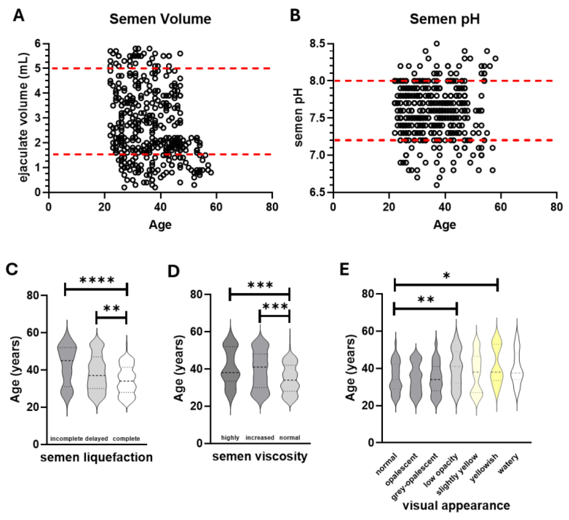
Figure 2.
Microscopic analysis of sperm quality in terms of numbers and motility. Shown are the total sperm count (A), sperm concentration (B), % total motility (C), % progressive motility, % repaid progressive motility (E), 5 slow progressive motility (F), 5 non progressive motility (G) and % immotile sperm (H) plotted in relation to patient age. WHO upper and lower limits where available, are displayed as red dashed lines. Sperm total numbers, concentration and motility, excluding % non progressive, is significantly associated with patient age by Spearman’s coefficient (p<0.0001).
Figure 2.
Microscopic analysis of sperm quality in terms of numbers and motility. Shown are the total sperm count (A), sperm concentration (B), % total motility (C), % progressive motility, % repaid progressive motility (E), 5 slow progressive motility (F), 5 non progressive motility (G) and % immotile sperm (H) plotted in relation to patient age. WHO upper and lower limits where available, are displayed as red dashed lines. Sperm total numbers, concentration and motility, excluding % non progressive, is significantly associated with patient age by Spearman’s coefficient (p<0.0001).
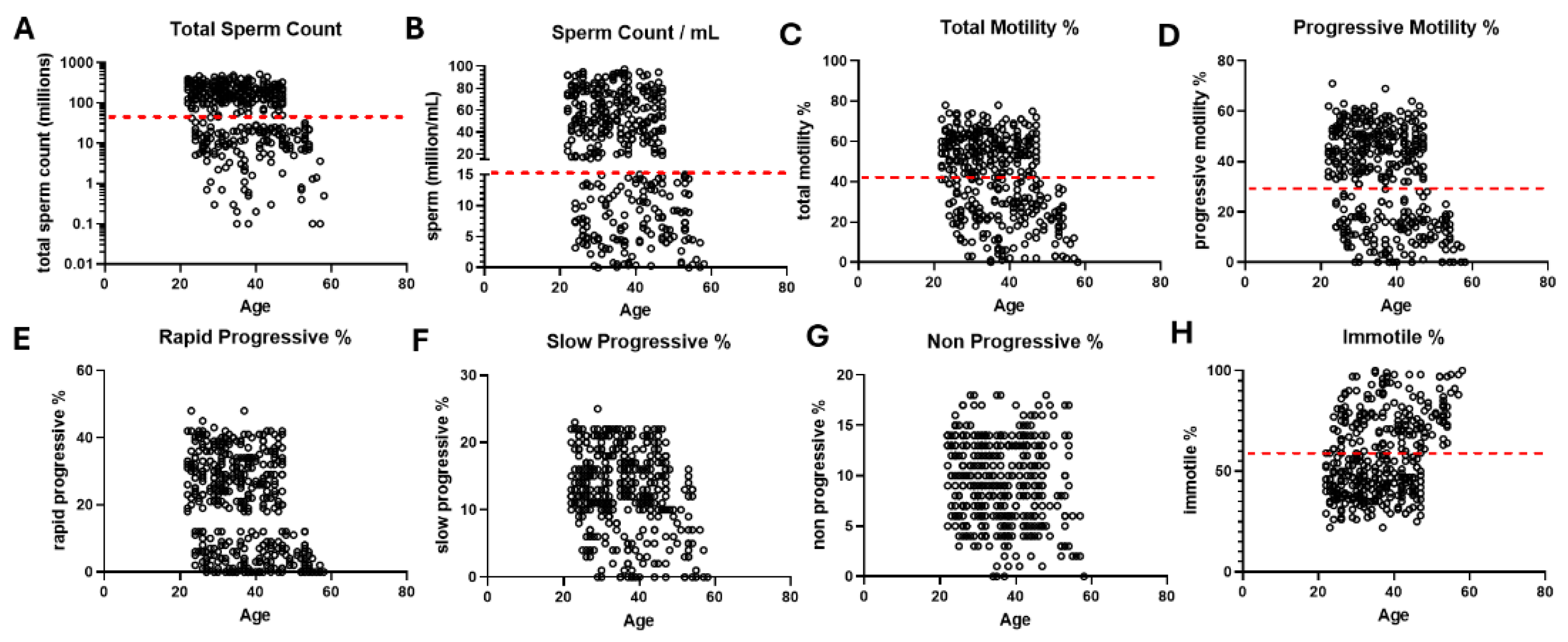
Figure 3.
Summary of semen classification and sperm quality based on DNA fragmentation (SDF). Semen was classified into good, poor or very poor groups according to the macroscopic and microscopic data alongside patient clinical history (A). These do not represent internationally validated diagnostic classifications. Those within each category and referred for SDF analysis and a summary of the findings are shown in the data table (B). Graphical representation of SDF positivity according to patient age is shown in (C). SDF positivity showed no significant association with patient age.
Figure 3.
Summary of semen classification and sperm quality based on DNA fragmentation (SDF). Semen was classified into good, poor or very poor groups according to the macroscopic and microscopic data alongside patient clinical history (A). These do not represent internationally validated diagnostic classifications. Those within each category and referred for SDF analysis and a summary of the findings are shown in the data table (B). Graphical representation of SDF positivity according to patient age is shown in (C). SDF positivity showed no significant association with patient age.
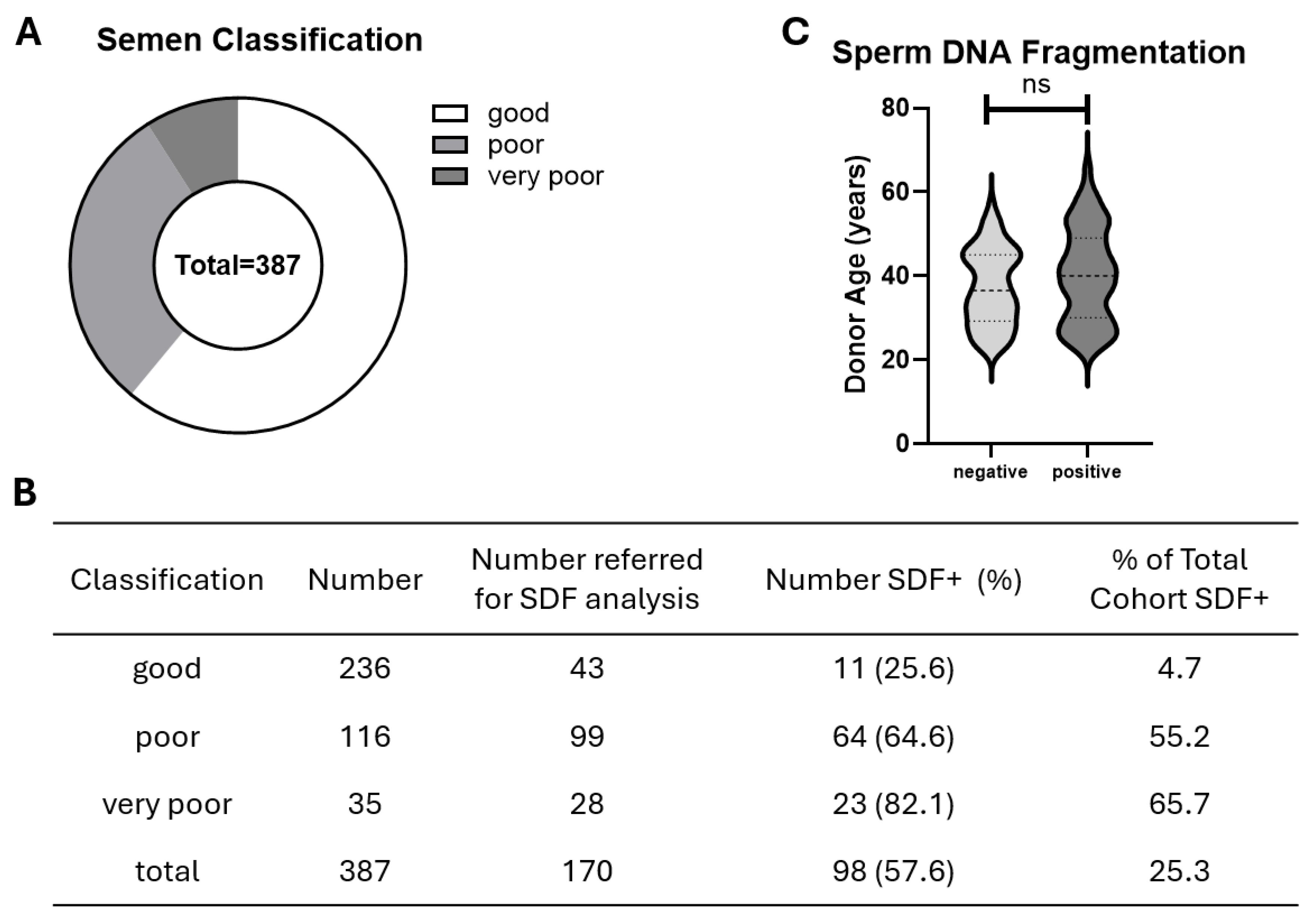
Figure 4.
Integrated sperm/semen diagnostic assessment pathway. Traditional macroscopic and microscopic analyses of semen according to WHO parameters define sperm and semen quality in conjunction with physician assessment. Selected sperm DNA fragmentation (SDF) testing is applied following definition of sperm/semen quality assessment (QA). The positivity frequency depends upon sperm/semen QA. As identified in this study, adoption of SDF in conjunction with sperm/semen QA by traditional methods and WHO standardisation will likely provide additional diagnostic security within all groups.
Figure 4.
Integrated sperm/semen diagnostic assessment pathway. Traditional macroscopic and microscopic analyses of semen according to WHO parameters define sperm and semen quality in conjunction with physician assessment. Selected sperm DNA fragmentation (SDF) testing is applied following definition of sperm/semen quality assessment (QA). The positivity frequency depends upon sperm/semen QA. As identified in this study, adoption of SDF in conjunction with sperm/semen QA by traditional methods and WHO standardisation will likely provide additional diagnostic security within all groups.
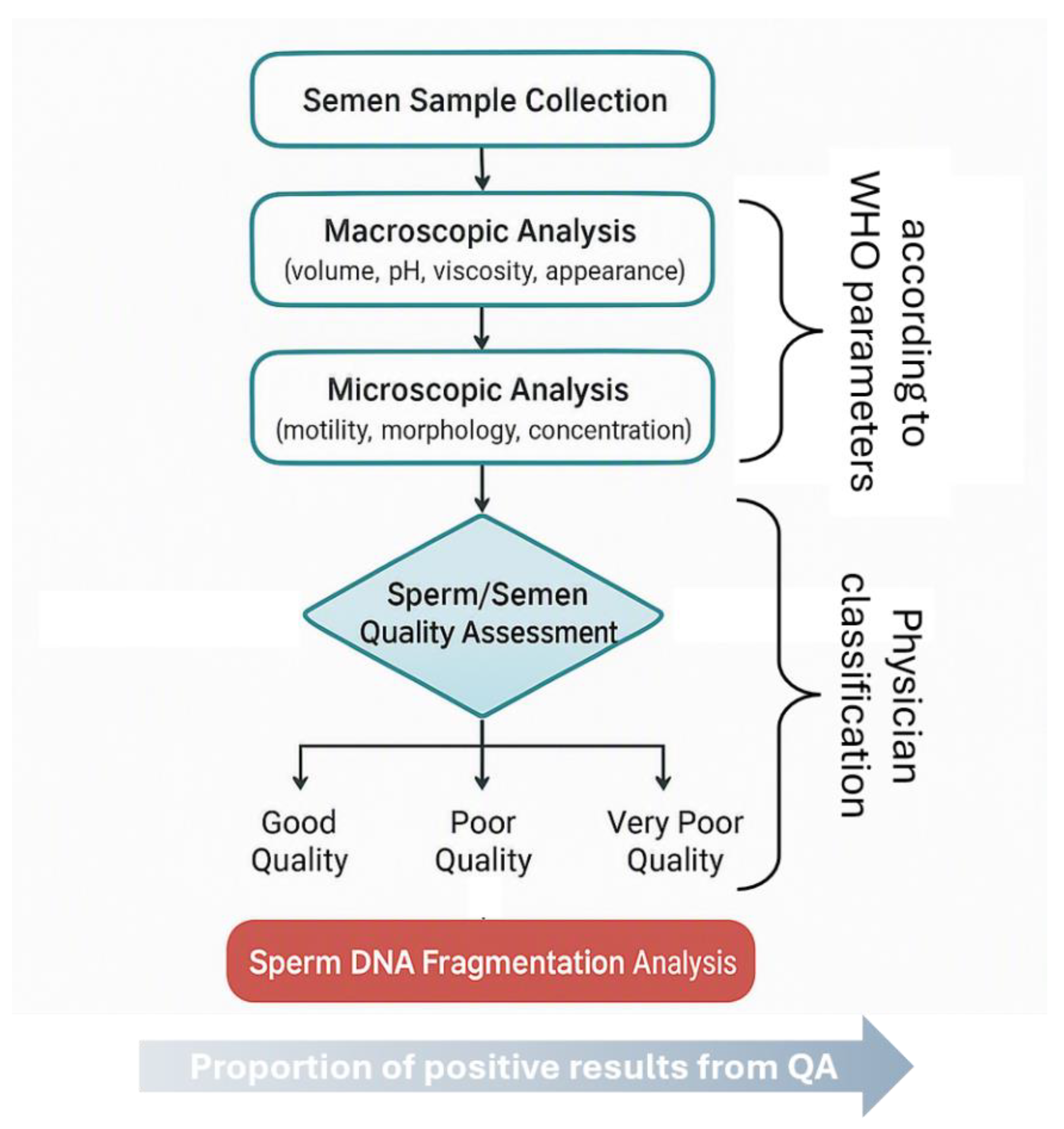

Table 1.
Demographic characteristics of the study population and macroscopic semen features of entire cohort.
Table 1.
Demographic characteristics of the study population and macroscopic semen features of entire cohort.
| Characteristic | Values |
|---|---|
| Number of participants | 387 |
| Study period | Jan 2024–May 2026 |
| Participant Age (years) (mean, SD, range) | 36.6, 9.4, 21–66 |
| Semen volume (mL) (mean, SD, range) | 2.71, 1.45, 0.3–8.5 |
| Semen pH (mean, SD, range) | 7.60, 0.35, 6.8–8.5 |
| Liquefaction (number and % complete in 30mins) | 265, 68.5 |
| Normal viscosity (number and %) | 271, 70.0 |
| Normal appearance (number and %) | 133, 34.4 |
Data are summarised from the entire cohort. Continuous variables are presented as mean and standard deviation (SD), alongside the cohort range.
Table 2.
Microscopic semen characteristics of the study population and WHO lower reference limit.
| Parameter | Mean (SD) | Range | WHO Lower Reference Limit |
|---|---|---|---|
| Sperm concentration (million/mL) | 38.5 (30.3) | 0.0–97.4 | 16 |
| Total sperm number (million/ejaculate) | 129.3 (123.9) | 0.0–521.4 | 39 |
| Total motility (%) | 42.6 (19.7) | 0–78 | 42 |
| Progressive motility (%) | 33.4 (18.7) | 0–71 | 30 |
| Rapid progressive motility (%) | 20.5 (14.1) | 0–48 | - |
| Slow progressive motility (%) | 12.9 (6.0) | 0–25 | - |
| Non-progressive motility (%) | 9.2 (4.1) | 0–18 | - |
| Immotile sperm (%) | 57.4 (19.7) | 22–100 | - |
| Normal morphology (%) | 6.5 (4.9) | 0–18 | 4 |
Continuous variables are presented as mean and standard deviation (SD), with the cohort range. Categorical variables are presented as mean and SD of frequency (percentage). WHO lower reference limits are based on the WHO Laboratory Manual for the Examination and Processing of Human Semen (6th Edition, 2021). Rapid, slow and non- progressive motility are descriptive subcategories and are not assigned independent WHO lower reference limits.
Table 3.
Cohort sperm characteristics based on WHO reference criteria.
| Parameter | Above WHO Limit (%) | Below WHO limit (%) |
|---|---|---|
| Sperm concentration | 236 (61.0) | 151 (39.0) |
| Total sperm number | 234 (60.5) | 153 (39.5) |
| Total motility | 221 (57.1) | 166 (42.9) |
| Progressive motility | 234 (60.5) | 153 (39.5) |
| Normal morphology | 236 (61.0) | 151 (39.0) |
Disclaimer/Publisher’s Note: The statements, opinions and data contained in all publications are solely those of the individual author(s) and contributor(s) and not of MDPI and/or the editor(s). MDPI and/or the editor(s) disclaim responsibility for any injury to people or property resulting from any ideas, methods, instructions or products referred to in the content. |
© 2026 by the authors. Licensee MDPI, Basel, Switzerland. This article is an open access article distributed under the terms and conditions of the Creative Commons Attribution (CC BY) license (http://creativecommons.org/licenses/by/4.0/).
Copyright: This open access article is published under a Creative Commons CC BY 4.0 license, which permit the free download, distribution, and reuse, provided that the author and preprint are cited in any reuse.