

Submitted:
17 September 2026
Posted:
18 September 2026
You are already at the latest version
Abstract
Anaemia is almost universal during the neonatal course of extremely preterm infants and red blood cell transfusion (BT) remains one of the most frequently used treatments in neonatal intensive care. The fall in haemoglobin after preterm birth reflects developmental physiology compounded by shortened RBC survival, impaired erythropoietin response, rapid somatic growth, iron availability and iatrogenic blood loss. Although haemoglobin (Hb) concentration remains the principal laboratory measure used to guide transfusion, it provides an incomplete assessment of oxygen delivery and erythropoietic reserve. Recent evidence has therefore renewed interest in complementary measures including fetal haemoglobin (HbF), absolute reticulocyte count and reticulocyte haemoglobin content. High risk infants such as those with fetal growth restriction (FGR) may represent a distinct haematological phenotype because chronic placental insufficiency alters fetal erythropoiesis and iron utilisation before birth. Large randomised trials and a subsequent international guideline now support restrictive haemoglobin-based transfusion thresholds in very preterm infants, but substantial variation in practice persists. Furthermore, transfusion with adult donor RBCs changes more than haemoglobin concentration: it replaces HbF-rich neonatal cells with HbA-rich adult cells, altering oxygen affinity and endogenous erythropoiesis. This narrative review examines the developmental physiology and contemporary management of neonatal anaemia, with particular focus on HbF, reticulocyte parameters, FGR, transfusion thresholds and emerging approaches to individualised transfusion. Future strategies should move beyond haemoglobin concentration alone towards integration of haematological and physiological markers of oxygen delivery.
Keywords:
anaemia of prematurity
; fetal haemoglobin
; reticulocytes
; red blood cell transfusion
; fetal growth restriction
; preterm infant
; iron
; tissue oxygenation
1. Introduction
Anaemia is one of the most frequent haematological problems encountered in neonatal intensive care. All infants experience a physiological fall in haemoglobin (Hb) concentration after birth, but this decline occurs earlier and is more pronounced in preterm infants. In extremely preterm infants, developmental suppression of erythropoiesis is compounded by shortened red blood cell (RBC) lifespan, rapid growth, inadequate iron availability, illness and repeated phlebotomy, resulting in the characteristic anaemia of prematurity (AOP) [1].
Anaemia is conventionally defined as a haemoglobin or haematocrit below the age-specific normal reference range. Christensen et al. advocate using values below the 5th percentile for gestational and postnatal age to define neonatal anaemia [2]. This statistical definition, however, does not establish when anaemia becomes physiologically important. The capacity of blood to deliver oxygen is dependent not only on Hb concentration but also on cardiac output, arterial oxygen saturation, regional perfusion and the affinity of haemoglobin for oxygen [3]. Consequently, infants with apparently similar Hb concentrations may have different capacities to maintain adequate tissue oxygenation.
Haemoglobin nevertheless remains the principal biomarker used to decide whether a blood transfusion (BT) is required. Higher Hb concentrations at birth have been associated with short-term outcomes and survival in preterm infants [4], and transfusion guidelines continue to use Hb thresholds modified according to postnatal age and respiratory support. The recent Transfusion of Prematures (TOP) [5] and Effects of Transfusion Thresholds on Neurocognitive Outcome of Extremely Low Birth-Weight Infants (ETTNO) [6] trials demonstrated that maintaining higher haemoglobin transfusion thresholds did not reduce death or neurodevelopmental impairment at approximately 2 years compared with lower transfusion thresholds. These findings support the safety of a more restrictive transfusion strategy in extremely preterm/ELBW infants. However, both trials used Hb-based transfusion algorithms and did not establish whether Hb concentration alone is the optimal physiological marker of transfusion.
There is therefore increasing interest in factors that provide information beyond total Hb concentration. These include the proportion of fetal haemoglobin (HbF), endogenous marrow response measured using reticulocyte indices, functional iron availability and direct measures of tissue oxygenation. These variables may help distinguish infants with similar Hb concentrations but differing capacities for oxygen delivery and erythropoietic recovery. They may be particularly relevant to infants exposed to placental insufficiency FGR, in whom erythropoiesis and iron utilisation have already been altered before birth.
While previous reviews have comprehensively described the pathophysiology and management of anaemia of prematurity [7], this narrative review focuses specifically on factors beyond total Hb concentration that may influence the physiological significance of neonatal anaemia. Particular emphasis is placed on HbF, reticulocyte indices, functional iron availability and high-risk infants, alongside contemporary approaches to prevention, assessment and treatment. The review also considers whether integrating these haematological and physiological markers with established Hb-based transfusion thresholds could contribute to a more individualised assessment of transfusion need.
2. Developmental Physiology of Neonatal Anaemia
2.1. The Physiological Fall in Haemoglobin After Birth
Haemoglobin concentration is relatively high at birth, reflecting the intrauterine environment and placental contribution to circulating blood volume. Following delivery, arterial oxygen tension rises abruptly. The resulting increase in tissue oxygen availability suppresses erythropoietin production and erythropoiesis. In healthy term infants, Hb typically reaches a physiological nadir of approximately 9–11 g/dL at 8–12 weeks of age. This process is usually asymptomatic and is often termed physiological anaemia of infancy [1].
The response is exaggerated following preterm birth, with the Hb nadir occurring earlier and often falling to approximately 7–10 g/dL, particularly in the most immature infants [7,8]. Preterm infants have an inappropriately low erythropoietin response for the degree of anaemia, despite preservation of the intrinsic responsiveness of erythroid progenitor cells to erythropoietin [9]. In addition, neonatal RBCs have a shorter lifespan than adult RBCs, while rapid postnatal growth increases circulating blood volume and erythropoietic and iron requirements [7,10]. Together, these developmental factors contribute to the pathophysiology of anaemia of prematurity.
In modern neonatal intensive care, however, AOP is rarely a purely physiological phenomenon. Extremely preterm infants undergo repeated phlebotomy during a period when their total circulating blood volume may be only 68–70 mL/kg [11] and these losses may therefore remove a clinically important proportion of the circulating RBC mass. Common complications of prematurity, including sepsis, nutritional deficiency, haemorrhage and haemolysis, may also contribute to anaemia [12]. In addition, inflammation may contribute through hepcidin-mediated restriction of iron availability and suppression of erythropoiesis [13], while significant renal dysfunction may further impair erythropoietin production [14]. AOP should consequently be considered a multifactorial developmental and iatrogenic disorder rather than simply an exaggerated physiological Hb nadir.
2.2. Oxygen Delivery is Not Determined by Haemoglobin Concentration Alone
The clinical importance of anaemia derives primarily from reduced oxygen-carrying capacity. Systemic oxygen delivery is determined by cardiac output and arterial oxygen content, with the latter predominantly determined by Hb concentration and oxygen saturation [15]:
and approximately:
Oxygen delivery = cardiac output × arterial oxygen content
Arterial oxygen content = (1.34 × Hb × arterial oxygen saturation) + dissolved oxygen.
The contribution of dissolved oxygen (0.003 × PaO₂) is small under normal physiological conditions; consequently, arterial oxygen content is predominantly determined by haemoglobin concentration and arterial oxygen saturation [15,16].
A fall in Hb can therefore be compensated for through changes in cardiac output, oxygen extraction and regional blood flow. The capacity for such adaptation is likely to differ substantially between a clinically stable growing preterm infant and an infant with severe respiratory or cardiovascular disease.
Clinical manifestations attributed to anaemia include tachycardia, increasing oxygen requirement, recurrent apnoea or desaturation, poor weight gain and reduced activity. However, none is specific to anaemia. Apnoea and desaturation, for example, may reflect lung disease, infection, gastro-oesophageal reflux, immature respiratory control or other pathology. Clinical response to transfusion is also variable. This creates an important limitation for strategies that define “symptomatic anaemia” without objective physiological measures [15].
Because Hb concentration represents only one component of systemic oxygen delivery, assessment of anaemia based on Hb alone may not reflect whether tissue oxygenation is adequate. This has prompted interest in physiological measures such as near-infrared spectroscopy (NIRS), which estimates regional tissue oxygen saturation and oxygen extraction and may therefore provide information on the balance between oxygen delivery and consumption. Studies in anaemic preterm infants have demonstrated changes in cerebral and splanchnic oxygenation following RBC transfusion [17,18,19], although NIRS measurements are influenced by gestational and postnatal age [20], measurement site [21], device and sensor [22]. No validated NIRS threshold for transfusion currently exists, and its role remains predominantly investigational.
3. Fetal Haemoglobin: An Important but Overlooked Component of Oxygen Delivery
3.1. Developmental Transition from HbF to HbA
Fetal haemoglobin is the predominant haemoglobin during fetal life. Its greater affinity for oxygen compared with adult haemoglobin (HbA) facilitates placental oxygen transfer by shifting the oxygen–haemoglobin dissociation curve to the left. HbF concentration and proportion are highest at lower gestational ages and decline progressively with advancing gestation and postnatal age as globin production switches towards HbA [10,23].
Preterm birth interrupts this transition. Extremely preterm infants therefore enter postnatal life with a high proportion of HbF while simultaneously experiencing an abrupt change in oxygen environment. Their circulating RBC population is biologically different from that of adults, not only in haemoglobin composition but also in cellular metabolism, membrane properties, deformability and lifespan [10].
The clinical importance of HbF is conceptually straightforward. Total Hb describes how much haemoglobin is present, whereas Hb type influences how readily the oxygen carried by that haemoglobin is released to tissues. An infant with a high HbF fraction has a more left-shifted oxygen dissociation curve than an infant with the same total Hb but a greater proportion of HbA. Thus, Hb concentration and HbF fraction represent related but distinct components of oxygen transport.
3.2. RBC Transfusion Changes HbF as Well as Total Hb
Standard neonatal RBC transfusions use adult donor blood containing predominantly HbA. Transfusion therefore has two simultaneous effects: it increases total circulating Hb and replaces part of the infant's HbF-rich RBC population with adult HbA-rich cells. Repeated transfusions can markedly accelerate the postnatal reduction in measured HbF [3,5].
This distinction matters because the physiological response to transfusion cannot be attributed solely to the increase in total Hb. Introduction of HbA shifts the oxygen dissociation characteristics of circulating blood and may facilitate tissue oxygen unloading. Earlier physiological studies demonstrated relationships between HbF and fractional tissue oxygen extraction, suggesting that total Hb alone does not fully describe tissue oxygen availability.
Recent work has renewed interest in this concept. Yazdanbakhsh and colleagues demonstrated that mixing preterm cord blood (n=19) with adult donor RBCs produces measurable changes in haemoglobin composition and oxygen affinity [24]. Bachman and colleagues subsequently analysed 3452 blood gas observations from 2464 infants and demonstrated clinically measurable alterations in the relationship between oxygen saturation and partial pressure of oxygen according to HbF and previous adult RBC transfusion exposure [25]. These observations are important but cannot yet be interpreted as evidence for changing routine oxygen saturation targets according to transfusion history. Rather, they demonstrate that transfusion changes oxygen physiology in a way that is not captured by total Hb concentration alone.
3.3. Could Preservation of HbF Matter?
A provocative extension of this concept is whether maintaining a more developmentally appropriate RBC phenotype could improve neonatal outcomes. Cord-blood-derived RBCs provide cells with fetal characteristics and preserve HbF to a greater extent than adult donor RBCs.
The BORN multicentre randomised trial investigated whether transfusing 112 extremely preterm infants with HbF-rich cord-blood-derived RBCs, rather than standard adult donor RBCs, could reduce severe ROP. In the intention-to-treat analysis, cord-blood transfusion did not significantly reduce severe ROP. However, almost half of infants allocated to cord blood also received adult donor RBCs, limiting separation between the treatment groups. Although severe ROP was lower among infants who received exclusively cord-blood RBCs in a per-protocol analysis, these findings require confirmation in further trials. Nevertheless, BORN demonstrates that cord-blood-derived RBC transfusion is feasible and provides proof of concept for strategies aimed at preserving HbF in extremely preterm infants. [26].
At present, adult donor RBC remains standard care. HbF should therefore be viewed as a potentially important physiological variable rather than a transfusion target.
4. Reticulocytes: Measuring the Infant's Erythropoietic Response
4.1. Absolute Reticulocyte Count
Reticulocytes are newly released RBCs and provide a direct indicator of recent erythropoietic activity. During AOP, Hb concentration falls while the reticulocyte response is often inappropriately low because of reduced erythropoietin stimulation. A low reticulocyte count in an anaemic infant therefore suggests limited endogenous replacement of RBCs, whereas a rising count indicates marrow recovery.
Reticulocytes may be expressed as a percentage of total RBCs or as an absolute reticulocyte count. Percentage values can be misleading during anaemia because the denominator (the total RBC count) is reduced. The absolute reticulocyte count more directly reflects the number of newly produced RBCs and is generally preferable when assessing marrow activity [27]. The timing of reticulocyte measurement in relation to RBC transfusion is important. Schwarz et al. studied 39 preterm infants with anaemia of prematurity using weekly and post-transfusion measurements of haemoglobin, reticulocyte count and erythropoietin. Both reticulocyte count and erythropoietin concentrations fell significantly 7 days after transfusion but had returned to baseline by 14 days, demonstrating transient suppression of endogenous erythropoiesis following RBC transfusion [28]. A later single-centre randomised pilot study of 20 very-low-birth-weight infants receiving a late transfusion (>14 days after birth) similarly found a significant fall in reticulocyte count 7–10 days after transfusion in infants receiving transfusion alone; the mean reduction was 85 ± 62 × 10³/µL. This suppression was prevented in infants randomised to receive concomitant darbepoetin [29]. Reticulocyte counts obtained during the first 1–2 weeks after transfusion should therefore be interpreted cautiously when assessing endogenous erythropoietic activity.
Current transfusion guidelines do not use reticulocyte counts as independent transfusion triggers. A very low count in a progressively anaemic, otherwise stable infant may nevertheless provide useful complementary information. Conversely, an infant approaching a conventional transfusion threshold with a strong reticulocyte response may be biologically different from an infant with the same Hb but almost absent erythropoiesis.
4.2. Reticulocyte Haemoglobin Content
Reticulocyte haemoglobin content, reported as CHr or Ret-He depending on the analyser, provides a different type of information. Because reticulocytes circulate for only a short period, their haemoglobin content reflects recent iron availability to the developing erythron rather than historical iron stores.
Ferritin is widely used to assess iron status but is an acute-phase reactant and can be difficult to interpret in clinically unwell preterm infants. Reticulocyte haemoglobin therefore offers an attractive functional measure of iron-restricted erythropoiesis. Lorenz and colleagues found that a reticulocyte haemoglobin content of approximately 29 pg had useful diagnostic performance for iron deficiency in very preterm or very-low-birth-weight infants [30]. Subsequent studies have similarly used a threshold around 29 pg, although assay-specific and gestation-specific reference ranges remain important [31].
Interest in Ret-He has expanded beyond haematological endpoints. In a 2026 retrospective cohort of 381 preterm infants, 52% had at least one Ret-He below 29 pg. Lower average and minimum Ret-He and longer exposure to low Ret-He were associated with measures of abnormal neurodevelopment, although associations varied according to gestation, postnatal age and developmental outcome [32]. These observational data do not establish causality or a treatment threshold, but they reinforce the potential importance of detecting functional iron deficiency before overt anaemia develops.
4.3. Combining Hb and Reticulocytes
Hb and reticulocyte parameters answer different clinical questions. Table 1 summarises the principal information provided by each marker, together with its potential clinical utility and important limitations. The logical next step is not to replace Hb with a single novel biomarker, but to determine whether combinations of these variables identify clinically important anaemia more accurately than Hb alone.
5. Fetal Growth Restriction and Other High-Risk Groups
FGR deserves particular consideration because placental insufficiency and chronic fetal hypoxaemia may alter erythropoiesis before preterm birth. Increased fetal erythropoietic drive may result in elevated nucleated RBC counts and polycythaemia, while simultaneously increasing iron utilisation; impaired placental iron transfer and premature interruption of third-trimester iron accretion may further limit iron availability [33]. However, increased fetal erythropoiesis may not translate into effective postnatal RBC production. Reibel et al. reported increased nucleated RBC counts and greater transfusion exposure in extremely preterm infants with growth restriction [34], while Takahashi et al. found lower early reticulocyte counts in SGA compared with AGA preterm infants [35,36]. More recently, Hulsbergen-Veelken et al. described enhanced nucleated RBC production but ineffective reticulocyte differentiation in preterm growth-restricted infants [36]. HbF trajectories are also poorly characterise, Dani et al. found a similar overall postnatal decline in HbF in very preterm SGA (n=39) and AGA (n=60) infants, but differing associations between transfusion and HbF in the two groups [37]. Together, these findings suggest that FGR preterm infants may have a distinct erythropoietic and iron phenotype that warrants consideration when interpreting Hb, reticulocytes and HbF.
6. Deciding When to Transfuse
6.1. What Have the Large Randomised Trials Established?
For decades, neonatal transfusion practice varied substantially because studies used different Hb thresholds, respiratory definitions and clinical triggers. Two large randomised trials published in 2020 have substantially clarified the safety of restrictive transfusion strategies.
The TOP trial compared higher and lower Hb transfusion thresholds in extremely-low-birth-weight infants and found that maintaining higher Hb thresholds did not improve survival without neurodevelopmental impairment at 22–26 months [5]. Similarly, the ETTNO trial found no significant improvement in death or neurocognitive disability with a liberal transfusion strategy in extremely-low-birth-weight infants [6].
These findings informed the 2024 international clinical practice guideline from the Neonatal Transfusion Network. Based on a systematic review of six randomised trials including 3483 infants, the guideline recommends a restrictive RBC transfusion strategy for preterm neonates born at <30 weeks' gestation [40].
6.2. Recommended Restrictive Thresholds
The 2024 guideline recommends the following Hb thresholds Table 2:
Respiratory support in the guideline includes positive airway pressure or nasal cannula flow ≥1 L/min [40].
These thresholds provide an important evidence-based framework and reduce unnecessary exposure to transfusion. They should not, however, be interpreted as absolute physiological boundaries. The trials compare strategies at a population level; they do not demonstrate that an Hb of 91 g/L represents adequate oxygen delivery in every infant receiving respiratory support, or that an Hb of 89 g/L necessarily requires transfusion.
Clinical context remains important, particularly in infants with acute haemorrhage, haemodynamic instability, substantial oxygen requirement or other circumstances inadequately represented in randomised trials.
6.3. The Evidence/Practice Gap
Despite increasingly robust trial evidence, transfusion practice remains variable. A prospective study of 64 neonatal intensive care units in 22 European countries include 1143 preterm infants during 2022-2023 found substantial variation in RBC transfusion use and transfusion thresholds [41]. By 28 days, more than one-third of included extremely preterm infants had received an RBC transfusion, and many transfusions were administered at Hb concentrations above restrictive trial thresholds.
This discrepancy suggests that clinician concern about individual physiology continues to influence transfusion decisions. It also emphasises the need to distinguish appropriate clinical individualisation from unnecessary transfusion.
7. Can Physiological Biomarkers Improve Transfusion Decisions?
Hb concentration remains attractive because it is objective, rapidly available and supported by randomised trial evidence. However, several investigators have explored biomarkers that might better identify inadequate tissue oxygen delivery.
NIRS enables continuous, non-invasive estimation of regional tissue oxygen saturation and fractional tissue oxygen extraction. In 33 preterm infants, van Hoften et al. found that pre-transfusion cerebral tissue oxygenation and extraction correlated with Hb, and that cerebral oxygen saturation increased while oxygen extraction decreased following RBC transfusion [19]. Banerjee et al. similarly demonstrated increased splanchnic tissue oxygenation and reduced splanchnic fractional oxygen extraction following transfusion without a corresponding change in mesenteric blood flow velocity [42]. However, these relationships are not consistent. Balegar et al. found no significant correlation between pre-transfusion Hb and either cerebral or splanchnic fractional tissue oxygen extraction in 29 anaemic preterm infants [3]
These findings suggest that NIRS may provide physiological information that is not captured by Hb concentration alone, but it is not currently validated as a routine transfusion trigger. Other proposed indicators include lactate, heart rate, oxygen requirement, frequency of apnoea and indices of cardiac output. None currently has evidence sufficient to replace Hb-based thresholds.
A future approach may instead integrate multiple domains; Hb concentration, postnatal age, respiratory support, reticulocyte response, HbF fraction and a physiological marker of oxygen delivery. Such a model would require prospective validation against clinically meaningful outcomes before implementation.
The principal haematological, physiological and clinical factors that may contribute to assessment of transfusion need are summarised in Figure 1.
8. Anaemia, RBC Transfusion and Necrotising Enterocolitis
The relationship between anaemia, transfusion and necrotising enterocolitis (NEC) has generated considerable concern. Earlier observational studies described clusters of NEC occurring within 48–72 h of RBC transfusion, leading to the concept of “transfusion-associated NEC”. However, infants receiving transfusion are intrinsically different from untransfused infants: they are often more premature, more unwell and more severely anaemic. Separating the effects of transfusion from the indication for transfusion is therefore difficult.
Patel and colleagues found that severe anaemia was associated with an increased risk of NEC, while the independent contribution of transfusion was less clear [43]. More recently, a secondary analysis of 1690 extremely-low-birth-weight infants enrolled in the TOP trial examined 4947 post-transfusion hazard periods. RBC transfusion was not temporally associated with a significantly greater risk of NEC during the subsequent 72 h within the Hb ranges studied in TOP [44].
These findings challenge a simple causal model in which transfusion itself precipitates NEC. A more plausible framework is that anaemia, intestinal oxygen delivery, feeding, developmental immaturity and transfusion interact within a susceptible infant [45].
Whether enteral feeds should routinely be withheld around RBC transfusion also remains uncertain. Observational evidence has been inconsistent and adequately powered randomised evidence is awaited. The WHEAT trial is designed specifically to address whether withholding enteral feeds around transfusion reduces NEC [46]. Until definitive results are available, local practice is likely to remain variable.
9. Prevention of Anaemia and Reduction of Transfusion Exposure
9.1. Placental Transfusion
Prevention begins at birth. Delayed umbilical cord clamping increases placental transfusion and neonatal circulating blood volume. An individual participant data meta-analysis of 48 randomised trials involving more than 6000 preterm infants found high-certainty evidence that delayed compared with immediate cord clamping reduced death before hospital discharge [47]. Delayed cord clamping should therefore be considered part of neonatal blood conservation rather than simply a delivery-room intervention.
Umbilical cord milking should not be considered equivalent to delayed clamping. Concerns regarding severe intraventricular haemorrhage in extremely preterm infants mean that gestational age and technique must be considered when interpreting cord-milking evidence [47].
9.2. Reducing Diagnostic Blood Loss
Iatrogenic phlebotomy remains a major and modifiable contributor to AOP. This is particularly important in the first postnatal weeks, when laboratory investigations are most frequent and circulating blood volume is smallest.
Contemporary multicentre data demonstrate the magnitude of the problem. In a 2026 prospective study across 64 neonatal intensive care units in 22 European countries, cumulative diagnostic blood loss by day 28 represented approximately 50% of initial circulating blood volume in infants born at 24 weeks' gestation [41]. Blood loss varied markedly between centres. Units using smaller-volume laboratory analysers had substantially lower cumulative diagnostic blood loss.
Practical patient blood management measures therefore include using cord blood for admission investigations when feasible, minimising unnecessary investigations, combining blood tests, using microsampling and point-of-care techniques, and avoiding discard volumes from indwelling lines whenever possible.
9.3. Iron
Iron is essential not only for erythropoiesis but also for rapidly developing neural tissue where it contributes to processes including myelination, neurotransmitter synthesis and cellular energy metabolism [33,48]. Preterm infants begin life with smaller total iron stores, experience greater postnatal growth requirements and lose iron directly through repeated blood sampling [33]. Conversely, repeated RBC transfusion introduces substantial exogenous iron and may increase ferritin and tissue iron stores; both iron deficiency and excess are therefore undesirable in preterm infants [33].
Iron supplementation should therefore be considered in relation to gestational age, diet, growth, transfusion exposure and biochemical evidence of iron status rather than Hb concentration alone. Reticulocyte haemoglobin content (Ret-He/CHr) reflects recent functional iron availability for erythropoiesis and may be less affected by inflammation than ferritin, which is an acute-phase reactant [30,48]. However, neonatal target values and evidence for using Ret-He to individualise iron supplementation remain limited
9.4. Erythropoiesis-Stimulating Agents
The role of erythropoietin and other erythropoiesis-stimulating agents (ESAs) has evolved. Earlier evidence showed that ESAs stimulate erythropoiesis and can reduce transfusion exposure, but uncertainties surrounding clinical benefit, iron requirements, ROP and neurodevelopment limited widespread adoption.
The 2025 Neonatal Research Network darbepoetin trial provides important contemporary evidence [49]. In 650 infants born at 23–28 weeks' gestation, weekly darbepoetin increased haematocrit and red cell mass, reduced transfusion and donor exposure, and increased the proportion of infants who remained transfusion-free. However, cognitive outcomes at 22–26 months were similar to placebo. ROP was not increased, and a reduction in bronchopulmonary dysplasia was observed as a secondary outcome.
Thus, it is no longer accurate to conclude simply that ESAs are ineffective in AOP. They have a clear erythropoietic effect and reduce transfusion exposure, but evidence that routine use improves long-term neurodevelopment is lacking. Optimal patient selection, dosing and iron supplementation remain areas for further study.
10. What Does an RBC Transfusion Actually Treat?
The traditional concept is that transfusion “corrects the Hb”. Physiologically, however, neonatal RBC transfusion does considerably more:
- it increases circulating RBC mass and total Hb;
- it increases arterial oxygen-carrying capacity;
- it alters the HbF:HbA ratio;
- it changes oxygen affinity and potentially tissue oxygen unloading;
- it suppresses endogenous erythropoiesis and reticulocyte production;
- it introduces donor iron;
- it changes RBC age, metabolism and membrane characteristics; and
- it expands intravascular volume.
The response measured solely as the post-transfusion rise in Hb therefore captures only one component of the intervention.
Donor RBC characteristics have also attracted attention. For infants requiring repeated small-volume transfusions, RBC units can be divided into multiple paediatric aliquots from a single donor, reducing exposure to multiple donors. Although sequential use of these aliquots may result in transfusion of progressively older RBCs [50]. The ARIPI randomised 307 preterm infants to receive transfusion of RBCs stored 7 days or less (n = 188) vs standard-issue RBCs in accordance with standard blood bank practice (n = 189). They found no clinical advantage from transfusing fresher rather than standard-issue RBCs to premature very-low-birth-weight infants [51]. These findings suggest that chronological storage age alone is unlikely to be a major determinant of transfusion outcome.
11. From Restrictive to Individualised Transfusion
The evidence supporting restrictive transfusion thresholds represents an important advance. It demonstrates that routinely maintaining higher Hb concentrations exposes infants to additional transfusions without improving major outcomes [5,6,40]. The next stage should not be a return to liberal transfusion but refinement of restrictive practice. A potential framework for integrating these additional measures with established Hb-based thresholds is illustrated in Figure 2.
An individualised strategy could potentially distinguish three situations:
1. Low Hb with adequate compensation and active erythropoiesis.A clinically stable infant with improving respiratory status, a rising absolute reticulocyte count and satisfactory growth may tolerate a low Hb while recovering without transfusion.
2. Low Hb with limited endogenous response.An infant with falling Hb, very low absolute reticulocyte count and evidence of iron-restricted erythropoiesis has little immediate capacity to replace RBCs and may have a different trajectory even at the same Hb concentration.
3. Low Hb with evidence of impaired oxygen delivery.An infant with significant cardiorespiratory disease or reproducible physiological evidence of inadequate tissue oxygenation may have a greater transfusion requirement than a stable infant at an identical Hb.
These phenotypes remain conceptual and should not currently override evidence-based Hb thresholds. They provide, however, a framework for prospective studies designed to identify which infants benefit from transfusion.
12. Priorities for Future Research
Several important questions remain.
First, longitudinal reference trajectories for Hb, HbF, absolute reticulocyte count and Ret-He need to be defined simultaneously in contemporary very and extremely preterm populations. Transfusion exposure must be incorporated explicitly because adult donor RBCs alter both HbF and endogenous erythropoiesis.
Second, FGR infants require dedicated study. Current evidence indicates altered prenatal and postnatal erythropoiesis, but FGR and SGA are often used interchangeably and antenatal Doppler phenotypes are rarely characterised in neonatal haematology studies. Studies separating constitutionally small infants from infants with placental insufficiency are needed.
Third, the physiological significance of HbF loss following adult RBC transfusion warrants further investigation. HbF preservation should not yet be assumed to be beneficial: the higher oxygen affinity of HbF facilitates placental uptake but reduces peripheral oxygen unloading, while HbA may improve tissue oxygen release. The optimal postnatal balance may differ according to gestational age, oxygen exposure and organ system.
Fourth, future transfusion trials should evaluate whether combinations of Hb concentration, reticulocyte response, HbF and measures of tissue oxygenation can improve prediction of transfusion benefit. The relevant outcome is not simply whether Hb rises, but whether transfusion improves oxygen delivery and clinically meaningful neonatal outcomes.
Finally, prevention should remain central. The 2024 demonstration of striking diagnostic blood loss in extremely preterm infants [41] indicates that reducing unnecessary phlebotomy may prevent anaemia more effectively and safely than refining the treatment of anaemia once it is established.
13. Conclusions
Anaemia of prematurity results from the interaction of developmental erythropoietic physiology with rapid growth, limited iron availability, illness and substantial iatrogenic blood loss. Large randomised trials and international guidance now provide reassuring evidence that restrictive Hb-based transfusion thresholds are appropriate for most very preterm infants.
However, haemoglobin concentration is an incomplete description of neonatal oxygen transport. HbF affects oxygen affinity, reticulocytes quantify endogenous erythropoietic response, Ret-He provides information on functional iron availability, and physiological measures can assess the balance between oxygen delivery and utilisation. Transfusion itself changes each of these systems.
The future of neonatal transfusion practice is therefore unlikely to involve identifying a single superior Hb threshold. Rather, the challenge is to retain the safety and simplicity of restrictive transfusion practice while developing reliable approaches that identify the individual infant in whom anaemia has become physiologically significant. Moving beyond haemoglobin concentration towards a developmental and physiological assessment of neonatal anaemia offers a promising route towards more precise transfusion medicine.
Author Contributions
Conceptualization, C.M.; writing, original draft preparation, C.M.; review and editing, C.M., J.B. and N.A. All authors have read and agreed to the published version of the manuscript.
Funding
This research received no external funding.
Conflicts of Interest
The authors declare no conflicts of interest.
References
- Strauss, R.G. Anaemia of prematurity: pathophysiology and treatment. Blood Rev. 2010, 24(6), 221–5. [Google Scholar] [CrossRef] [PubMed]
- Christensen, R.D.; Bahr, T.M.; Tweddell, S.M.; Ohls, R.K.; Henry, E. Diagnosing Anemia in Neonates: An Evidence-Based Approach. Neoreviews 2023, 24(6), e343–e55. [Google Scholar] [CrossRef] [PubMed]
- Balegar, V.K.; Low, G.K.; Nanan, R.K. Regional tissue oxygenation and conventional indicators of red blood cell transfusion in anaemic preterm infants. EClinicalMedicine 2022, 46, 101365. [Google Scholar] [CrossRef] [PubMed]
- Banerjee, J.; Asamoah, F.K.; Singhvi, D.; Kwan, A.W.G.; Morris, J.K.; Aladangady, N. Haemoglobin level at birth is associated with short term outcomes and mortality in preterm infants. BMC Med. 2015, 13(16). [Google Scholar] [CrossRef] [PubMed]
- Kirpalani, H.; Bell, E.F.; Hintz, S.R.; Tan, S.; Schmidt, B.; Chaudhary, A.S.; et al. Higher or Lower Hemoglobin Transfusion Thresholds for Preterm Infants. N. Engl. J. Med. 2020, 383(27), 2639–51. [Google Scholar] [CrossRef] [PubMed]
- Franz, A.R.; Engel, C.; Bassler, D.; Rüdiger, M.; Thome, U.H.; Maier, R.F.; et al. Effects of Liberal vs Restrictive Transfusion Thresholds on Survival and Neurocognitive Outcomes in Extremely Low-Birth-Weight Infants: The ETTNO Randomized Clinical Trial. JAMA 2020, 324(6), 560–70. [Google Scholar] [PubMed]
- Cibulskis, C.C.; Maheshwari, A.; Rao, R.; Mathur, A.M. Anemia of prematurity: how low is too low? J. Perinatol. 2021, 41(6), 1244–57. [Google Scholar] [CrossRef] [PubMed]
- Holzapfel, L.F.; Rysavy, M.A.; Bell, E.F. Red Blood Cell Transfusion Thresholds for Anemia of Prematurity. NeoReviews 2023, 24(6), e370–e6. [Google Scholar] [CrossRef] [PubMed]
- Shannon, K.M.; Naylor, G.S.; Torkildson, J.C.; Clemons, G.K.; Schaffner, V.; Goldman, S.L.; et al. Circulating Erythroid Progenitors in the Anemia of Prematurity; New England Journal of Medicine, 1987; Volume 317, 12, pp. 728–33. [Google Scholar]
- Pellegrino, C.; Stone, E.F.; Valentini, C.G.; Teofili, L. Fetal Red Blood Cells: A Comprehensive Review of Biological Properties and Implications for Neonatal Transfusion. Cells 2024, 13(22). [Google Scholar] [CrossRef] [PubMed]
- Hellström, W.; Forssell, L.; Morsing, E.; Sävman, K.; Ley, D. Neonatal clinical blood sampling led to major blood loss and was associated with bronchopulmonary dysplasia. Acta Paediatr. 2020, 109(4), 679–87. [Google Scholar] [CrossRef] [PubMed]
- Widness, J.A. Pathophysiology of Anemia During the Neonatal Period, Including Anemia of Prematurity. Neoreviews 2008, 9(11), e520. [Google Scholar] [CrossRef] [PubMed]
- Müller, K.F.; Lorenz, L.; Poets, C.F.; Westerman, M.; Franz, A.R. Hepcidin concentrations in serum and urine correlate with iron homeostasis in preterm infants. J. Pediatr. 2012, 160(6), 949–53.e2. [Google Scholar] [CrossRef] [PubMed]
- Atkinson, M.A.; Warady, B.A. Anemia in chronic kidney disease. Pediatr. Nephrol. 2018, 33(2), 227–38. [Google Scholar] [CrossRef] [PubMed]
- Vrancken, S.L.; van Heijst, A.F.; de Boode, W.P. Neonatal Hemodynamics: From Developmental Physiology to Comprehensive Monitoring. In Frontiers in Pediatrics; 2018; Volume 6 - 2018. [Google Scholar]
- Nguyen, T.C.; Madappa, R.; Siefkes, H.M.; Lim, M.J.; Siddegowda, K.M.; Lakshminrusimha, S. Oxygen saturation targets in neonatal care: A narrative review. Early Hum. Dev. 2024, 199, 106134. [Google Scholar] [CrossRef] [PubMed]
- Banerjee, J.; Leung, T.S.; Aladangady, N. Effect of blood transfusion on intestinal blood flow and oxygenation in extremely preterm infants during first week of life. Transfusion 2016, 56(4), 808–15. [Google Scholar] [CrossRef] [PubMed]
- Banerjee, J.; Leung, T.S.; Aladangady, N. Cerebral blood flow and oximetry response to blood transfusion in relation to chronological age in preterm infants. Early Hum. Dev. 2016, 97, 1–8. [Google Scholar] [CrossRef] [PubMed]
- van Hoften, J.C.; Verhagen, E.A.; Keating, P.; ter Horst, H.J.; Bos, A.F. Cerebral tissue oxygen saturation and extraction in preterm infants before and after blood transfusion. Arch. Dis. Child Fetal Neonatal Ed. 2010, 95(5), F352–8. [Google Scholar] [CrossRef] [PubMed]
- Mohamed, M.A.; Frasketi, M.J.; Aly, S.; El-Dib, M.; Hoffman, H.J.; Aly, H. Changes in cerebral tissue oxygenation and fractional oxygen extraction with gestational age and postnatal maturation in preterm infants. J. Perinatol. 2021, 41(4), 836–42. [Google Scholar] [CrossRef] [PubMed]
- Howarth, C.N.; Leung, T.S.; Banerjee, J.; Eaton, S.; Morris, J.K.; Aladangady, N. Regional cerebral and splanchnic tissue oxygen saturation in preterm infants - Longitudinal normative measurements. Early Hum. Dev. 2022, 165, 105540. [Google Scholar] [CrossRef] [PubMed]
- Dix, L.M.; van Bel, F.; Baerts, W.; Lemmers, P.M. Comparing near-infrared spectroscopy devices and their sensors for monitoring regional cerebral oxygen saturation in the neonate. Pediatr. Res. 2013, 74(5), 557–63. [Google Scholar] [CrossRef] [PubMed]
- Terrenato, L.; Bertilaccio, C.; Spinelli, P.; Colombo, B. The Switch from Haemoglobin F to A: the Time Course of Qualitative and Quantitative Variations of Haemoglobins after Birth. Br. J. Haematol. 1981, 47(1), 31–41. [Google Scholar] [CrossRef] [PubMed]
- Yazdanbakhsh, M.; Eid, H.; Acker, J.P.; Bar Am, N.; Cheung, P.-Y.; Dotchin, S.A.; et al. Hemoglobin-oxygen affinity changes in neonatal blood transfusions: RBC selection insights. Pediatr. Res. 2025, 97(6), 2090–6. [Google Scholar] [CrossRef] [PubMed]
- Bachman, T.E.; Nguyen, T.A.; Tejkl, L.; Plavka, R. Changes in Fetal Hemoglobin Associated with Erythrocyte Transfusions Are Clinically Relevant in SpO2 Targeting: A Retrospective Cohort Observational Study. Neonatology 2025, 122(4), 407–13. [Google Scholar] [CrossRef] [PubMed]
- Teofili, L.; Papacci, P.; Pellegrino, C.; Dani, C.; Cresi, F.; Remaschi, G.; et al. Cord red blood cell transfusions for severe retinopathy in preterm neonates in Italy: a multicenter randomized controlled trial. eClinicalMedicine 2025, 87. [Google Scholar] [CrossRef] [PubMed]
- Parodi, E.; Romano, F.; Ramenghi, U. How We Use Reticulocyte Parameters in Workup and Management of Pediatric Hematologic Diseases. Front Pediatr. 2020, 8, 588617. [Google Scholar] [CrossRef] [PubMed]
- Schwarz, K.B.; Dear, P.R.F.; Gill, A.B.; Newell, S.J.; Richards, M. EFFECTS OF TRANSFUSION IN ANEMIA OF PREMATURITY. Pediatr. Hematol. Oncol. 2005, 22(7), 551–9. [Google Scholar] [CrossRef] [PubMed]
- Warwood, T.L.; Lambert, D.K.; Henry, E.; Christensen, R.D. Very low birth weight infants qualifying for a 'late' erythrocyte transfusion: does giving darbepoetin along with the transfusion counteract the transfusion's erythropoietic suppression? J. Perinatol. 2011, 31 Suppl 1, S17–21. [Google Scholar] [CrossRef] [PubMed]
- Lorenz, L.; Arand, J.; Büchner, K.; Wacker-Gussmann, A.; Peter, A.; Poets, C.F.; et al. Reticulocyte haemoglobin content as a marker of iron deficiency. Arch. Dis. Child Fetal Neonatal Ed. 2015, 100(3), F198–202. [Google Scholar] [CrossRef] [PubMed]
- Sriranjan, J.; Kalata, C.; Fusch, G.; Thomas, K.; Goswami, I. Prevalence and Implications of Low Reticulocyte-Hemoglobin Levels among Extreme Preterm Neonates: A Single-Center Retrospective Study. Nutrients 2022, 14(24). [Google Scholar] [CrossRef] [PubMed]
- Mueller, G.K.; Yitayew, M.; Robinson, A.; Sabo, R.; Hendricks-Muñoz, K.D. Association between reticulocyte hemoglobin content (RetHe) and neurodevelopmental outcomes in preterm infants. J. Perinatol. 2026. [Google Scholar] [CrossRef] [PubMed]
- Rao, R.; Georgieff, M.K. Iron in fetal and neonatal nutrition. Semin. Fetal Neonatal Med. 2007, 12(1), 54–63. [Google Scholar] [CrossRef] [PubMed]
- Reibel, N.J.; Dame, C.; Bührer, C.; Muehlbacher, T. Aberrant Hematopoiesis and Morbidity in Extremely Preterm Infants With Intrauterine Growth Restriction. In Frontiers in Pediatrics; 2021; pp. 9–2021. [Google Scholar]
- Takahashi, Y.; Kanai, Y.; Chishiki, M.; Goto, A.; Imamura, T. Neonatal reticulocytes among preterm infants of small for gestational age. Pediatr. Neonatol. 2022, 63(5), 462–7. [Google Scholar] [CrossRef] [PubMed]
- Hulsbergen-Veelken, C.A.R.; Chon, H.; Nikkels, P.G.J.; Huisman, A.; van Wijk, R.; van Solinge, W.W.; et al. Enhanced Nucleated Red Blood Cell Production but Ineffective Reticulocyte Differentiation in Preterm Growth-Restricted Infants. Int. J. Lab. Hematol. 2026, 48(4), 875–8. [Google Scholar] [CrossRef] [PubMed]
- Dani, C.; Cipriani, F.; Ciavotta, M.; Remaschi, G. Changes in Fetal Hemoglobin in Very Preterm Infants Born Small for Gestational Age: A Retrospective Observational Study. Children 2026, 13(1), 117. [Google Scholar] [CrossRef] [PubMed]
- Kitaoka, H.; Shitara, Y.; Kashima, K.; Ochiai, S.; Chikai, H.; Watanabe, K.; et al. Risk factors for anemia of prematurity among 30-35-week preterm infants. Fukushima J. Med. Sci. 2023, 69(2), 115–23. [Google Scholar] [CrossRef] [PubMed]
- MacQueen, B.C.; Baer, V.L.; Scott, D.M.; Ling, C.Y.; O'Brien, E.A.; Boyer, C.; et al. Iron Supplements for Infants at Risk for Iron Deficiency. Glob. Pediatr. Health 2017, 4, 2333794x17703836. [Google Scholar] [CrossRef] [PubMed]
- Deschmann, E.; Dame, C.; Sola-Visner, M.C.; Fustolo-Gunnink, S.F.; Guyatt, G.H.; Patel, R.M.; et al. Clinical Practice Guideline for Red Blood Cell Transfusion Thresholds in Very Preterm Neonates. JAMA Netw. Open 2024, 7(6), e2417431–e. [Google Scholar] [CrossRef] [PubMed]
- Houben, N.A.M.; Fustolo-Gunnink, S.; Fijnvandraat, K.; Caram-Deelder, C.; Carrascosa, M.A.; Beuchée, A.; et al. Red Blood Cell Transfusion in European Neonatal Intensive Care Units, 2022 to 2023. JAMA Netw. Open 2024, 7(9), e2434077–e. [Google Scholar] [CrossRef] [PubMed]
- Banerjee, J.; Leung, T.S.; Aladangady, N.A. Blood transfusion in preterm infants improves intestinal tissue oxygenation without alteration in blood flow. Vox Sang. 2016, 111(4), 399–408. [Google Scholar] [CrossRef] [PubMed]
- Patel, R.M.; Knezevic, A.; Shenvi, N.; Hinkes, M.; Keene, S.; Roback, J.D.; et al. Association of Red Blood Cell Transfusion, Anemia, and Necrotizing Enterocolitis in Very Low-Birth-Weight Infants. JAMA 2016, 315(9), 889–97. [Google Scholar] [CrossRef] [PubMed]
- Salas, A.A.; Gunn, E.; Carlo, W.A.; Bell, E.F.; Das, A.; Josephson, C.D.; et al. Timing of Red Blood Cell Transfusions and Occurrence of Necrotizing Enterocolitis: A Secondary Analysis of a Randomized Clinical Trial. JAMA Netw. Open 2024, 7(5), e249643–e. [Google Scholar] [PubMed]
- Salem, A.; Patel, R.M. Red Blood Cell Transfusion, Anemia, Feeding, and the Risk of Necrotizing Enterocolitis. Clin. Perinatol. 2023, 50(3), 669–81. [Google Scholar] [CrossRef] [PubMed]
- Gale, C.; Modi, N.; Jawad, S.; Culshaw, L.; Dorling, J.; Bowler, U.; et al. The WHEAT Pilot Trial—WithHolding Enteral Feeds Around Packed Red Cell Transfusion to Prevent Necrotising Enterocolitis in Preterm Neonates: A Multicentre, Electronic Patient Record (EPR), Randomised Controlled Point-of-Care Pilot Trial. BMJ Open 2019, 9(9), e033543. [Google Scholar] [CrossRef] [PubMed]
- Seidler, A.L.; Aberoumand, M.; Hunter, K.E.; Barba, A.; Libesman, S.; Williams, J.G.; et al. Deferred cord clamping, cord milking, and immediate cord clamping at preterm birth: a systematic review and individual participant data meta-analysis. The Lancet 2023, 402(10418), 2209–22. [Google Scholar] [CrossRef] [PubMed]
- German, K.R.; Juul, S.E. Iron and Neurodevelopment in Preterm Infants: A Narrative Review. Nutrients 2021, 13(11), 3737. [Google Scholar] [CrossRef] [PubMed]
- Ohls, R.K.; Das, A.; Tan, S.; Lowe, J.R.; Schibler, K.; Beauman, S.S.; et al. Darbepoetin, Red Cell Mass, and Neuroprotection in Preterm Infants: A Randomized Clinical Trial. JAMA Pediatr. 2025, 179(8), 836–45. [Google Scholar] [PubMed]
- New, H.V.; Berryman, J.; Bolton-Maggs, P.H.B.; Cantwell, C.; Chalmers, E.A.; Davies, T.; et al. Guidelines on transfusion for fetuses, neonates and older children. Br. J. Haematol. 2016, 175(5), 784–828. [Google Scholar] [CrossRef] [PubMed]
- Fergusson, D.A.; Hébert, P.; Hogan, D.L.; LeBel, L.; Rouvinez-Bouali, N.; Smyth, J.A.; et al. Effect of Fresh Red Blood Cell Transfusions on Clinical Outcomes in Premature, Very Low-Birth-Weight Infants: The ARIPI Randomized Trial. JAMA 2012, 308(14), 1443–51. [Google Scholar] [CrossRef] [PubMed]
Figure 1.
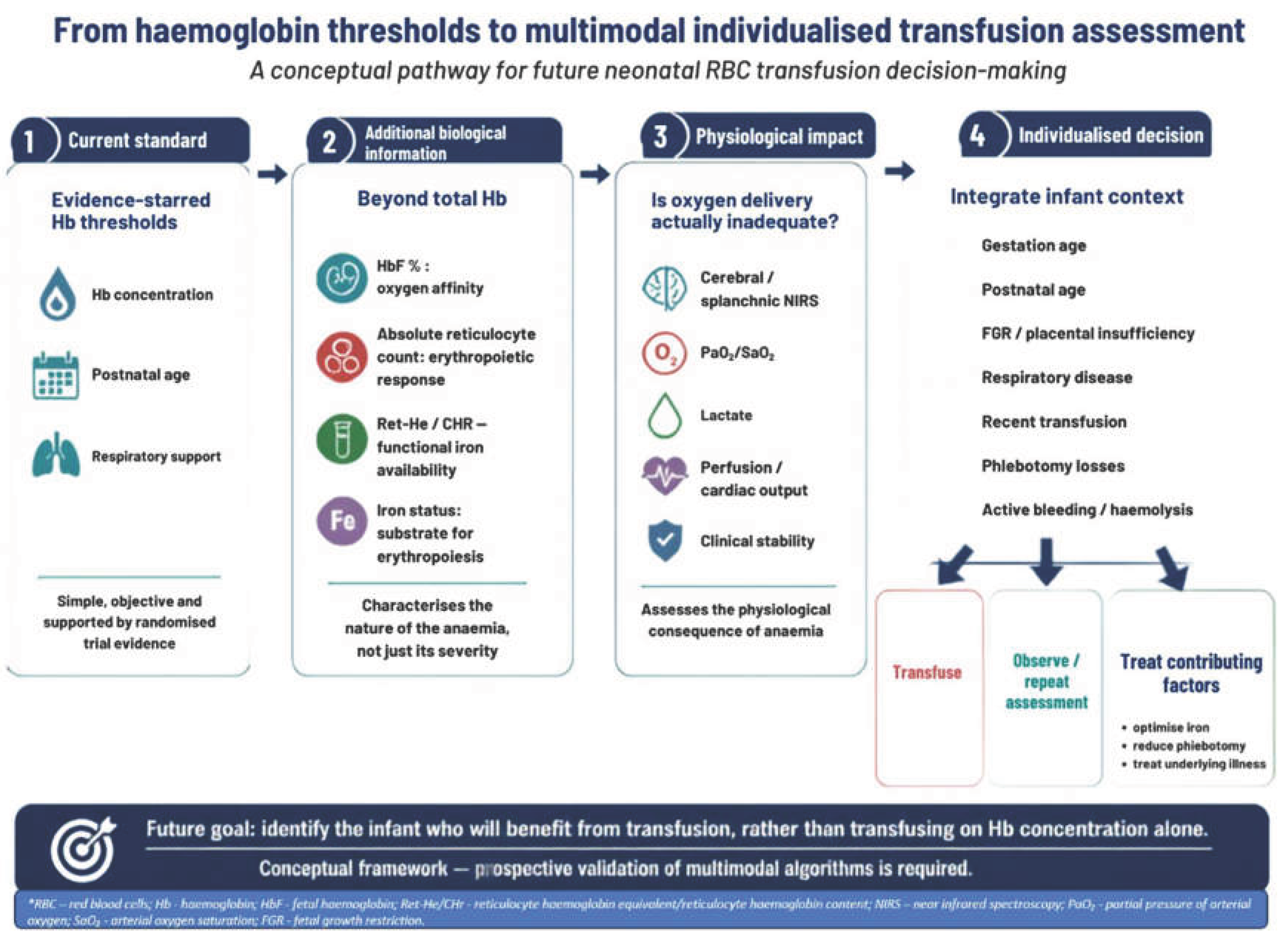

Table 1.
Principal information, potential clinical utility and limitations of haemoglobin, reticulocyte indices and markers of tissue oxygenation.
Table 1.
Principal information, potential clinical utility and limitations of haemoglobin, reticulocyte indices and markers of tissue oxygenation.
| Marker | Principal information | Potential advantage | Important limitation |
|---|---|---|---|
| Total Hb | Current oxygen-carrying capacity | Widely available; established transfusion thresholds | Does not measure marrow response or Hb oxygen affinity |
| Absolute reticulocyte count | Current endogenous RBC production | Indicates erythropoietic recovery or suppression | Suppressed following transfusion; gestation- and age-dependent |
| Reticulocyte % | Relative reticulocyte production | Routinely reported | Can be misleading when RBC count is low |
| Ret-He/CHr | Recent iron availability for erythropoiesis | Early functional marker of iron restriction | Assay-dependent; neonatal thresholds incompletely validated |
| HbF fraction | Haemoglobin phenotype and oxygen affinity | May better characterise oxygen transport | Not routinely measured serially; no validated treatment threshold |
| Ferritin | Stored iron | Familiar and widely available | Influenced by inflammation, transfusion and illness |
| NIRS-derived tissue oxygenation | Balance of regional oxygen delivery and consumption | Provides physiological information about regional tissue oxygenation | Device- and site-dependent; not validated as a routine transfusion trigger |
Table 2.
Hb thresholds for transfusion based on respiratory support and postnatal age.
| Postnatal age | Respiratory support | No/minimal respiratory support |
|---|---|---|
| Week 1 | 110 g/L (11 g/dL) | 100 g/L (10 g/dL) |
| Week 2 | 100 g/L (10 g/dL) | 85 g/L (8.5 g/dL) |
| Week ≥3 | 90 g/L (9 g/dL) | 70 g/L (7 g/dL) |
Disclaimer/Publisher’s Note: The statements, opinions and data contained in all publications are solely those of the individual author(s) and contributor(s) and not of MDPI and/or the editor(s). MDPI and/or the editor(s) disclaim responsibility for any injury to people or property resulting from any ideas, methods, instructions or products referred to in the content. |
© 2026 by the authors. Licensee MDPI, Basel, Switzerland. This article is an open access article distributed under the terms and conditions of the Creative Commons Attribution (CC BY) license (http://creativecommons.org/licenses/by/4.0/).
Copyright: This open access article is published under a Creative Commons CC BY 4.0 license, which permit the free download, distribution, and reuse, provided that the author and preprint are cited in any reuse.