

Submitted:
16 September 2026
Posted:
17 September 2026
You are already at the latest version
Abstract
Tuberculosis (TB) remains a major global public health threat. Driven by unhealthy lifestyles, the prevalence of concomitant glucose and lipid metabolism disorders, primarily diabetes mellitus (DM) and dyslipidemia, continues to rise and overlaps substantially with high TB-burden regions, posing significant challenges to clinical management. Both DM and dyslipidemia independently increase the risk of pulmonary tuberculosis (PTB) caused by Mycobacterium tuberculosis and contribute to poor clinical outcomes; however, research on the combined associations of these comorbid conditions on PTB outcomes, as well as the underlying host–pathogen interaction mechanisms and host immunity, remains limited. Existing evidence indicates that DM and dyslipidemia mutually exacerbate one another through insulin resistance, impairing macrophage function. Recent studies further suggest that DM-associated chronic hyperglycemia can induce T-cell memory exhaustion, thereby adversely affecting clinical outcomes in PTB patients. Based on this evidence, we propose the working hypothesis that concomitant dysglycemia and dyslipidemia may exert a synergistic or additive detrimental effect on PTB outcomes through the interplay between insulin resistance and T-cell exhaustion, and that combined interventions integrating glucose-lowering, lipid-lowering, and anti-tuberculosis therapies may yield greater clinical benefits than single-pathway approaches. This review examines the associations between dysglycemia, dyslipidemia and clinical outcomes in patients with PTB, elucidates the host–pathogen interaction mechanisms, and proposes a working hypothesis to inform the optimization of precision treatment strategies.
Keywords:
pulmonary tuberculosis
; diabetes mellitus
; dyslipidemia
; clinical outcomes
; mechanisms
; concomitant dysglycemia and dyslipidemia
1. Introduction
Tuberculosis (TB), caused by Mycobacterium tuberculosis, is a major global health threat that primarily affects the lungs [1,2]. In 2024, approximately 8.3 million TB cases were newly diagnosed and reported worldwide, of which 84% were pulmonary, against an estimated total of 10.7 million incident cases [3]. Pulmonary tuberculosis (PTB), the predominant form of the disease, is the primary focus of this review. Dysglycemia — comprising diabetes mellitus (DM) and its prediabetic states (impaired fasting glucose and impaired glucose tolerance) — is characterized by chronic hyperglycemia with disturbances in carbohydrate, lipid, and protein metabolism[4]. Dyslipidemia, characterized by an abnormal lipid profile comprising elevated cholesterol and/or triglycerides and/or reduced high-density lipoprotein cholesterol, is a major modifiable risk factor for cardiovascular disease[5]. Accumulating evidence indicates that unhealthy lifestyles — including high-calorie diets, sedentary behavior, and obesity — contribute to the rising prevalence of these metabolic disorders.
In 2024, the global prevalence of DM among adults aged 20–79 years reached 11.1%, affecting approximately 589 million people, over 80% of whom live in low- and middle-income countries, where 95.29% of new cases are projected to occur by 2050 [6]. A parallel epidemic of dyslipidemia is also evident: in the general adult population, prevalence varies by lipid parameter, at 28.77% for hypertriglyceridemia, 24.09% for hypercholesterolemia, 38.43% for low high-density lipoprotein cholesterol (HDL-C), and 18.93% for elevated low-density lipoprotein cholesterol (LDL-C)[7]. Among patients with DM, the prevalence of comorbid dyslipidemia is markedly higher, ranging from 60% to 65.68% across meta-analyses, although most of the underlying studies were rated as low or very low quality[8]. Notably, these metabolic epidemics are not geographically independent of TB: nearly two decades ago, eight of the 10 countries with the highest DM prevalence were also classified as high-TB-burden countries [9], and this overlap persists, with six of the current top 10 countries for diabetes remaining on the WHO high-TB-burden list [3,6].
Dysglycemia and dyslipidemia are closely interlinked, with insulin resistance as the shared core mechanism [10]. Substantial evidence indicates that DM increases the risk of developing PTB (hazard ratio [HR] 1.90, 95% confidence interval [CI] 1.51–2.40) [11], and within tuberculosis–diabetes mellitus (TB-DM) patients, uncontrolled hyperglycemia further raises the risk of treatment failure (relative risk [RR] 1.91) and sputum positivity at 3 months (RR 2.97), albeit with low certainty of evidence[12]. These metabolic disturbances may act in concert to compromise anti-tuberculosis immunity. Beyond the immune defects attributable to hyperglycemia, dyslipidemia independently impairs M. tuberculosis-specific T helper 1 (Th1) cytokine responses during latent tuberculosis infection (LTBI) [13]. Conversely, in patients with PTB, comorbid uncontrolled DM further dysregulates macrophage effector functions—reducing phagocytic capacity relative to non-diabetic PTB controls and altering bactericidal effector molecules (diminished nitric oxide [NO] and disproportionately elevated reactive oxygen species [ROS]), with phagocytic capacity declining in parallel with increasing disease severity in both groups[14]. Such immune impairment is paralleled by poorer clinical outcomes: compared with TB patients alone, those with TB-DM have higher odds of death (odds ratio [OR] 1.88), relapse (OR 1.64), and multidrug-resistant tuberculosis (MDR-TB) (OR 1.98), while delayed sputum culture conversion and treatment failure have also been evaluated in this setting[15].
Despite the high prevalence of comorbid dyslipidemia in DM patients and its established role in impairing M. tuberculosis-specific Th1 immunity during LTBI [8,13], the synergistic impact of dysglycemia and dyslipidemia on PTB clinical outcomes remains insufficiently synthesized. These synergistic deleterious effects underscore the clinical importance of a systematic synthesis of the evidence in this field. This review therefore aims to summarize the impact of dysglycemia, dyslipidemia, and their co-occurrence on clinical outcomes in PTB patients, along with the underlying mechanisms. Based on current evidence, we propose the working hypothesis that concomitant dysglycemia and dyslipidemia may synergistically worsen PTB outcomes, potentially through the interplay between insulin resistance and T-cell exhaustion. By elucidating these mechanisms, this review seeks to inform the optimization of precision diagnostic and therapeutic strategies in TB-DM care.
2. Association of Dysglycemia with Clinical Outcomes of PTB and Potential Mechanisms
Dysglycemia, particularly DM, is a major host factor shaping both the occurrence and the clinical course of TB. Globally, an estimated 0.93 million incident TB cases in 2024 were attributable to diabetes—the second leading attributable risk factor after undernutrition [3]. The World Health Organization has recognised diabetes as an important yet neglected risk factor for TB and, as one of the non-communicable disease determinants that must be addressed, regards it as a barrier to achieving the End TB Strategy target of a 90% reduction in TB incidence by 2035[16], particularly in low- and middle-income countries, where the diabetes burden is growing most rapidly [6,16]. Beyond increasing the risk of developing TB [12], diabetes also worsens clinical outcomes among patients with PTB, including higher odds of death, relapse, and multidrug-resistant disease [15]. Against this background, this review synthesises recent original studies to delineate the impact of DM on clinical outcomes in PTB and the underlying mechanisms.
2.1. Association of Dysglycemia with PTB Clinical Outcomes
Dysglycemia encompasses DM and prediabetes. Among PTB comorbid populations, DM is the best-studied and most influential factor affecting clinical outcomes. This section uses DM as a model to discuss its associations with clinical symptoms, radiological changes, and treatment outcomes.
2.1.1. Association of DM with Clinical Symptoms and Pulmonary Imaging Findings in PTB Patients
The influence of DM on clinical symptoms manifests primarily as a spectrum ranging from non-specific symptoms to exacerbated typical symptoms, a trajectory that, where the level of glycemic control has been examined, appears closely tied to it (Table 1). In patients with well-controlled glycemia, constitutional symptoms such as weight loss may persist even in the absence of more specific signs such as night sweats and lower-zone lesions [17]. Patients with DM and PTB frequently present with non-specific symptoms such as fatigue, weight loss, and mild anemia[18], while hyperglycemic status is significantly associated with a greater frequency of typical symptoms and higher symptom-based TB severity scores[19]. Glycemic indicators show positive associations with multiple markers of disease severity, with the most pronounced abnormalities observed in individuals with poor glycemic control[20].
The association of DM with imaging findings is even more pronounced. Computer-aided analysis of chest radiographs reveals more extensive radiographic abnormalities in patients with diabetes, together with a trend toward cavitary disease outside the upper lung zones[21]. Patients with TB-DM exhibit significantly more frequent cavitary disease[22,23] and more severe consolidation, bronchiectasis, and multilobar involvement than those without DM [23]. Notably, these severe imaging abnormalities are frequently not paralleled by significant differences in clinical symptoms [23]. However, as summarized in Table 1, the available evidence is dominated by non-longitudinal designs; although most studies performed multivariable adjustment for demographic and lifestyle factors, none accounted for diabetes duration, use of glucose-lowering agents, or dyslipidemia—variables that may independently influence pulmonary imaging manifestations. Therefore, active chest imaging screening should be considered for TB-DM patients with poorly controlled glycemia, even when respiratory symptoms are atypical.

Table 1.
Summary of studies on the impact of diabetes mellitus on clinical symptoms and imaging findings in pulmonary tuberculosis, ordered from symptom-related to imaging-related evidence.
Table 1.
Summary of studies on the impact of diabetes mellitus on clinical symptoms and imaging findings in pulmonary tuberculosis, ordered from symptom-related to imaging-related evidence.
| Study (Ref.) | Study design | Sample size | Key findings |
|---|---|---|---|
| Saalai et al. [17] | Cross-sectional | 50 | Well-controlled group (HbA1c≤7%): 80% had weight loss; no night sweats; no lower-zone lesions |
| Tong et al. [18] | Multicenter cross-sectional | 1417 | Fatigue (58.3% vs 47.5%, P = 0.001), weight loss (8.21 ± 6.2 vs 5.74 ± 4.0 kg, P < 0.001), and mild anemia (88.9% vs 77.6%, P = 0.021) more frequent in TB-DM |
| Xu et al. [19] | Prospective cohort | 791 | Hyperglycemia (FPG ≥ 6.1 mmol/L) associated with elevated TB score (OR 1.569, P = 0.032), cough (OR 1.332, P = 0.018), and night sweats (OR 1.694, P = 0.001) |
| Meng et al. [20] | Retrospective cohort | 3393 | FPG and HbA1c positively correlated with cavity formation, sputum positivity, and intrapulmonary lesions (all P < 0.001); worst in poor glycemic control group |
| Geric et al. [21] | Cross-sectional (CAD analysis) | 272 | More extensive radiographic abnormalities; trend toward non-upper-zone cavitary disease (17% vs 7.8%, P = 0.09) |
| Wang et al. [22] | Multicenter observational | 8421 | DM group had the highest cavity rate (54.8%); AOR 2.88 (95% CI 2.42–3.43) |
| Yang et al. [23] | Retrospective cross-sectional (age- and sex-matched) | 299 | More consolidation (79.22% vs 52.41%), cavitary lesions (85.06% vs 59.31%), bronchiectasis (71.43% vs 31.03%), and more lung lobes involved (all P < 0.0001); no difference in clinical symptoms |
1 Abbreviations: TB, tuberculosis; DM, diabetes mellitus; PTB, pulmonary tuberculosis; TB-DM, tuberculosis with diabetes mellitus; FPG, fasting plasma glucose; HbA1c, glycated hemoglobin; CAD, computer-aided detection; OR, odds ratio; AOR, adjusted odds ratio; CI, confidence interval.
2.1.2. Association of DM with Treatment Outcomes in Patients with PTB
Accumulating evidence indicates that DM, particularly when poorly controlled, adversely affects treatment outcomes in adults with PTB, encompassing delayed bacterial clearance, elevated risks of treatment failure and post-treatment recurrence, and—in observational studies of MDR-TB—poorer prognoses. Conversely, optimal glycemic control appears to improve selected outcomes, although its protective role against recurrence and in MDR-TB remains to be validated (Table 2).
Bacterial clearance is consistently delayed in patients with DM. Two independent cohorts employing different monitoring modalities—sputum culture and the TB molecular bacterial load assay—both demonstrated prolonged time to negativity, with a delay of approximately 5–8 days [24,25]; in the cohort monitored by Tuberculosis Molecular Bacterial Load Assay (TB-MBLA), this difference persisted throughout the intensive treatment phase [25]. Notably, this delay occurred despite a lower baseline bacterial burden in the DM group, suggesting impaired clearance kinetics rather than a higher initial bacillary load as the predominant mechanism [25]—a paradox that remains mechanistically incompletely understood. Poor glycemic control (hemoglobin A1c [HbA1c] ≥7%) amplifies these risks, roughly doubling the risk of treatment failure and tripling that of sputum positivity at three months, whereas optimal glycemic control confers measurable benefits across treatment outcomes, sputum positivity, and cavitary lesion resolution [12,26]. Regarding recurrence, DM has been identified as an independent risk factor for relapse, with a two- to threefold increase in hazard[27]. It should be noted, however, that the glycemic thresholds defining “poor control” varied across these studies, and few accounted for diabetes duration or concurrent glucose-lowering therapy, rendering direct comparison tenuous.
The picture is more nuanced in MDR-TB. Trial data from the Standardised Treatment Regimen of Anti-tuberculosis Drugs for Patients with Multidrug-resistant Tuberculosis (STREAM) cohort indicate that comorbid DM substantially increases the burden of serious adverse events—affecting over 40% of patients versus roughly 20% of those without DM—while efficacy outcomes remain comparable[28]. Nonetheless, pooled observational analyses indicate that DM increases the risk of unfavorable outcomes in drug-resistant tuberculosis (DR-TB) and MDR-TB by approximately 60%[29]—a discrepancy with trial data that likely reflects confounding by comorbidity burden in real-world cohorts rather than a true biological difference. Older patients appear particularly vulnerable, exhibiting higher sputum culture positivity, prolonged treatment duration, and lower cure rates when DM coexists[30].
Nevertheless, these conclusions derive predominantly from observational studies with modest sample sizes and marked heterogeneity in glycemic assessment (HbA1c vs. fasting glucose), outcome definitions, and follow-up duration. The mechanistic link between delayed clearance and adverse outcomes remains largely inferential, and the apparent protective effect of optimal glycemic control may be partly attributable to confounding by health-seeking behavior and access to care. Causal inferences therefore warrant confirmation through high-quality prospective cohort studies with standardized glycemic monitoring and adequately powered trials in MDR-TB.
Table 2.
Summary of studies on the association of DM with treatment outcomes in PTB patients.
| Category | Key finding | Primary result | Definition/criteria | Ref. |
|---|---|---|---|---|
| Bacterial clearance | Delayed sputum culture conversion | 60.0 vs. 52.0 days (P < 0.001); aHR 0.721 (95% CI 0.528–0.986) | Two consecutive negative cultures | [24] |
| Delayed TB-MBLA negativity | 62 vs. 57 days (P = 0.022); day 56 load 1.41 vs. 0.73 log₁₀ eCFU/mL (P = 0.028) | Cq > 30 (<1.0 log₁₀ eCFU/mL) = negative | [25] | |
| Treatment failure | Poor glycemic control ↑ treatment failure | RR 1.91 (95% CI 1.18–3.07), P = 0.008 (low certainty) | WHO: sputum positivity at end of treatment | [12] |
| Poor glycemic control ↑ 3-month sputum positivity | RR 2.97 (95% CI 1.10–8.07), P = 0.03 (low certainty) | Sputum smear positivity at month 3 | [12] | |
| Glycemic control (protective) | Optimal glycemic control improved multiple outcomes | Improved outcomes RR 1.13 (1.02–1.25); sputum positivity RR 0.23 (0.09–0.61); cavitary lesions RR 0.59 (0.51–0.68) | OGC definitions varied (mainly HbA1c < 7% or FPG < 7.0 mmol/L) | [26] |
| Recurrence | DM as independent risk factor for relapse | 20.21 vs. 6.28 per 1000 person-years; HR 2.40 (1.68–3.45), P < 0.001 | Active TB ≥ 6 months after successful treatment | [27] |
| MDR-TB | ↑ SAEs, comparable efficacy | SAE 41% vs. 22% (P < 0.001); FoR 8.3% vs. 5.9% (P = 0.38); adjusted HR 1.38 (0.51–3.73) | DAIDS criteria (AE/SAE grading); STREAM trial–defined Definite/Probable FoR | [28] |
| ↑ Unfavorable outcomes in DR-/MDR-TB | DR-TB OR 1.56 (1.24–1.96); MDR-TB OR 1.57 (1.20–2.04) | WHO criteria | [29] | |
| Special populations | Worse prognosis in elderly with PTB-DM | Sputum culture positivity 81.05% vs. 62.67% (P = 0.001); treatment duration 298 vs. 223 days (P < 0.001); cure rate 64.21% vs. 75.56% (P = 0.039); cavity rate 31.58% vs. 15.11% (P = 0.001) | Chinese WS 288-2017 (diagnosis); IUATLD criteria (outcomes) | [30] |
1 Abbreviations: ↑, increased; ↓, decreased; PTB, pulmonary tuberculosis; DM, diabetes mellitus; aHR, adjusted hazard ratio; HR, hazard ratio; OR, odds ratio; RR, risk ratio; CI, confidence interval; SAE, serious adverse event; FoR, failure or recurrence; DR-TB, drug-resistant tuberculosis; MDR-TB, multidrug-resistant tuberculosis; FPG, fasting plasma glucose; OGC, optimal glycemic control; DAIDS, Division of AIDS, National Institute of Allergy and Infectious Diseases; TB-MBLA, TB molecular bacterial load assay; Cq, quantification cycle; eCFU, equivalent colony-forming units. The reference group in each study was non-DM patients or patients with good glycemic control. For the association between poor glycemic control and treatment failure [30], the 95% CI is given as 1.18–3.07, following the forest plot of the source publication; the abstract and results text of that publication print 1.81–3.07, which is inconsistent with the reported test statistic (Z = 2.65, P = 0.008) and appears to be a typographical error.
The adverse prognostic impact of dysglycemia may extend below the diagnostic threshold for DM. A meta-analysis of eight prospective cohort studies (3001 patients, 752 with prediabetes) found prediabetes to be associated with a 41% increase in unfavourable treatment outcomes (RR 1.41, 95% CI 1.02–1.96; I² = 56%), although not with all-cause mortality (RR 1.59, 95% CI 0.75–3.38)[31]. Evidence on glycemic trajectories during treatment is more tentative: in a prospective cohort from Pune, India, transient hyperglycemia was associated with unfavourable outcomes (adjusted incidence rate ratio [aIRR] 2.07, 95% CI 1.04–4.15) whereas persistent hyperglycemia was not (aIRR 1.64, 95% CI 0.71–3.79), but this analysis included only 26 patients with transient hyperglycemia and diabetes treatment was itself strongly protective (aIRR 0.38, 95% CI 0.15–0.95)[32]; in the same cohort, DM predicted early mortality (adjusted hazard ratio [aHR] 4.36, 95% CI 1.62–11.76) but not the composite unfavourable outcome (adjusted relative risk [aRR] 1.13, 95% CI 0.75–1.70)[33]. Definitive assessment requires serial glycemic monitoring with standardized diabetes treatment protocols.
The above clinical evidence indicates that dysglycemia is associated with adverse PTB treatment outcomes across multiple clinical dimensions, with poor glycemic control and older patients being disproportionately affected. Nevertheless, the mechanisms by which dysglycemia impairs anti-TB immune responses remain incompletely elucidated—in particular, how chronic hyperglycemia modulates innate immunity (especially macrophage function) and M. tuberculosis-specific T-cell responses—and warrant further investigation.
2.2. Potential Mechanisms Underlying the Impact of Dysglycemia on PTB Clinical Outcomes
Defects in adaptive immune responses, particularly T-cell dysfunction, are key factors contributing to persistent TB, treatment failure, and relapse. Recent studies have demonstrated that DM can adversely affect clinical outcomes in PTB through T-cell memory exhaustion. The core mechanism lies in the fact that chronic hyperglycemia—the primary pathological feature of DM—can directly impair anti-TB immunity. Specifically, hyperglycemia induces T-cell memory exhaustion, thereby compromising the host's long-term immune control of M. tuberculosis. This finding not only reveals the direct immunotoxicity of hyperglycemia but also provides an important mechanistic framework for understanding how a broader spectrum of glucose metabolism disorders, including prediabetes and DM, affect clinical outcomes in PTB.
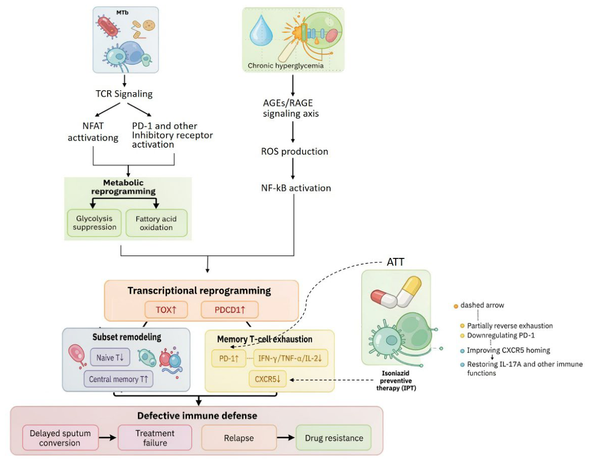
Persistent M. tuberculosis antigen exposure activates two parallel T cell receptor (TCR)-dependent branches: one engaging nuclear factor of activated T cells (NFAT) signaling, and the other inducing programmed cell death protein-1 (PD-1) and inhibitory receptor expression. These branches cooperatively drive metabolic reprogramming in exhausted T cells, characterized by suppressed glycolysis and augmented fatty acid oxidation (FAO) in defined subsets[34].Concurrently, chronic hyperglycemia activates the advanced glycation end products (AGEs)/ receptor for advanced glycation end products (RAGE) axis primarily within antigen-presenting cells (APCs), triggering ROS production and nuclear factor kappa B (NF–κB) activation—a process highlighted within the TB-DM research agenda[35,36].
Synthesizing evidence from chronic infection and oncology, we posit that persistent TCR signaling drives transcriptional reprogramming marked by thymocyte selection–associated high mobility group box protein (TOX) and programmed cell death 1 gene (PDCD1) upregulation[34], a process exacerbated by the AGEs–RAGE-derived pro-inflammatory milieu. This unified program precipitates two interrelated sequelae:
• Memory T cell exhaustion, defined by elevated PD-1[37,38], impaired polyfunctionality (interferon–γ [IFN–γ]), tumor necrosis factor–α [TNF–α], interleukin–2 [IL–2]) [13,39] dynamic C–X–C chemokine receptor type 5 (CXCR5) modulation restored upon preventive therapy [38]
• T cell subset remodeling, characterized by naïve contraction and central memory T cells (Tcm) expansion[40]
Collectively, these perturbations undermine antimycobacterial immunity and correlate with adverse outcomes, including delayed sputum conversion, treatment failure, and relapse[41].
Critically, AGEs–RAGE–driven inflammation represents a putative driver of the immunometabolic dysregulation underlying these poor trajectories [35] (Figure 1).
M. tuberculosis–induced memory T cell exhaustion exhibits partial reversibility. In PTB, standard antituberculosis therapy (ATT) downregulates PD-1 on CD4⁺ Tresp and Teff cells[42]. In latent TB infection with DM (LTBI–DM), isoniazid preventive therapy (IPT) upregulates CXCR5 expression, restores interleukin-17A (IL-17A) production, and downregulates PD-1, normalizing immune parameters [38]. These data substantiate treatment-induced plasticity, indicating partial phenotypic reversal; however, definitive evidence for complete reversion requires further mechanistic interrogation [38].
Figure 1.
Schematic Overview of T Cell Dysfunction and Therapeutic Nodes in Tuberculosis–Diabetes Mellitus. Persistent Mycobacterium tuberculosis (Mtb) antigens engage the T cell receptor (TCR), activating nuclear factor of activated T cells (NFAT) signaling and programmed cell death protein-1 (PD-1)/inhibitory receptor expression, while hyperglycemia activates the advanced glycation end-products/receptor for AGEs (AGE-RAGE) axis in antigen-presenting cells (APCs). TCR signaling drives thymocyte selection-associated HMG-box protein (TOX) and programmed cell death 1 gene (PDCD1) transcriptional programs and metabolic rewiring (glycolysis↓, fatty acid oxidation [FAO]↑), exacerbated by RAGE-derived inflammation. These processes converge into memory T-cell exhaustion (PD-1↑, polyfunctionality↓, C-X-C chemokine receptor type 5 [CXCR5] dynamics) and subset remodeling (naïve↓, central memory T cells [Tcm]↑), which map to adverse clinical endpoints, including delayed sputum conversion, treatment failure, and relapse. Dashed arrows denote interventions: antituberculosis therapy (ATT) downregulates PD-1; isoniazid preventive therapy (IPT) restores CXCR5/interleukin-17A (IL-17A) and downregulates PD-1. Upward and downward arrows (↑/↓) indicate relative increase and decrease, respectively. Abbreviations: Mtb, Mycobacterium tuberculosis; TCR, T cell receptor; NFAT, nuclear factor of activated T cells; PD-1, programmed cell death protein-1; AGE-RAGE, advanced glycation end-products/receptor for AGEs; APC, antigen-presenting cell; TOX, thymocyte selection-associated HMG-box protein; PDCD1, programmed cell death 1 gene; FAO, fatty acid oxidation; CXCR5, C-X-C chemokine receptor type 5; Tcm, central memory T cell; ATT, antituberculosis therapy; IPT, isoniazid preventive therapy; IL-17A, interleukin-17A.
Figure 1.
Schematic Overview of T Cell Dysfunction and Therapeutic Nodes in Tuberculosis–Diabetes Mellitus. Persistent Mycobacterium tuberculosis (Mtb) antigens engage the T cell receptor (TCR), activating nuclear factor of activated T cells (NFAT) signaling and programmed cell death protein-1 (PD-1)/inhibitory receptor expression, while hyperglycemia activates the advanced glycation end-products/receptor for AGEs (AGE-RAGE) axis in antigen-presenting cells (APCs). TCR signaling drives thymocyte selection-associated HMG-box protein (TOX) and programmed cell death 1 gene (PDCD1) transcriptional programs and metabolic rewiring (glycolysis↓, fatty acid oxidation [FAO]↑), exacerbated by RAGE-derived inflammation. These processes converge into memory T-cell exhaustion (PD-1↑, polyfunctionality↓, C-X-C chemokine receptor type 5 [CXCR5] dynamics) and subset remodeling (naïve↓, central memory T cells [Tcm]↑), which map to adverse clinical endpoints, including delayed sputum conversion, treatment failure, and relapse. Dashed arrows denote interventions: antituberculosis therapy (ATT) downregulates PD-1; isoniazid preventive therapy (IPT) restores CXCR5/interleukin-17A (IL-17A) and downregulates PD-1. Upward and downward arrows (↑/↓) indicate relative increase and decrease, respectively. Abbreviations: Mtb, Mycobacterium tuberculosis; TCR, T cell receptor; NFAT, nuclear factor of activated T cells; PD-1, programmed cell death protein-1; AGE-RAGE, advanced glycation end-products/receptor for AGEs; APC, antigen-presenting cell; TOX, thymocyte selection-associated HMG-box protein; PDCD1, programmed cell death 1 gene; FAO, fatty acid oxidation; CXCR5, C-X-C chemokine receptor type 5; Tcm, central memory T cell; ATT, antituberculosis therapy; IPT, isoniazid preventive therapy; IL-17A, interleukin-17A.
In conclusion, the TCR-dependent exhaustion program and AGEs-RAGE axis do not operate in isolation; instead, they forge a vicious cycle dictating antimicrobial immunity. This framework explains the heightened failure/relapse risk in DM and provides a lens to understand glucocentric immune remodeling. Current evidence remains cross-sectional, necessitating longitudinal studies integrating multi-omics and pharmacokinetic monitoring. Future efforts must identify predictive immune-metabolic signatures and evaluate host-directed therapies (HDTs) targeting RAGE or reversing T cell exhaustion to break this comorbidity cycle.
3. Association of Dyslipidemia with Clinical Outcomes of PTB and Potential Mechanisms
The epidemiological burden of dyslipidemia in PTB remains poorly characterized. The scarcity of large-scale, population-based surveys precludes reliable prevalence estimates, as existing evidence is derived predominantly from small case–control or cross-sectional studies with limited comparability. For instance, a single-center cross-sectional study in Uganda reported hypocholesterolemia (low total cholesterol [TC]) in 57.3% of treatment-naïve PTB patients[43]. Complementing these baseline observations, a nationwide Korean cohort study (n ≈ 5,000,000; median follow-up 8.2 years) identified low TC as an independent predictor of incident PTB, with the lowest TC quartile demonstrating an adjusted HR of 1.35 (95% CI 1.31–1.39) relative to the highest quartile[44]. This inter-study heterogeneity likely arises from population stratification, variable definitions of dyslipidemia, and divergent statistical methodologies. Given the bidirectional interplay between host lipids and M. tuberculosis— coupled with the inherent limitations of observational designs — it remains unclear whether low TC represents a causal precursor or a consequence of PTB. Furthermore, whether dyslipidemia modulates treatment response, sputum culture conversion, and relapse risk through immunometabolic mechanisms — such as altered macrophage cholesterol homeostasis and impaired T cell receptor signaling — warrants rigorous investigation.
Despite the availability of glycemic management guidelines for PTB patients with comorbid DM issued by several international bodies, including China[45], dedicated lipid management guidelines specifically tailored to PTB patients without comorbid DM remain largely unaddressed. Elucidating how dyslipidemia modulates T cell and macrophage function to shape clinical outcomes would establish a critical theoretical framework for implementing evidence based lipid management strategies in PTB.
3.1. Association of Dyslipidemia with PTB Clinical Outcomes
3.1.1. Hypolipidemia, Disease Severity, and Treatment Failure Risk
The association between dyslipidemia and clinical outcomes of PTB was initially characterized in cross-sectional studies. In DR-TB, serum total cholesterol (TC), HDL-C, and LDL-C levels were significantly negatively correlated with radiological severity scores (TC: r = −0.546, p = 0.001; HDL-C: r = −0.479, p = 0.005; LDL-C: r = −0.431, p = 0.012), such that lower lipid levels tracked with more extensive parenchymal involvement[46]. Concordant observations in drug-sensitive PTB demonstrated significantly reduced TC, HDL-C, and LDL-C levels relative to healthy controls. HDL-C correlated inversely with with the degree of radiological extent of disease (DRED) (r = −0.60, p < 0.001), and all three lipid parameters correlated negatively with sputum smear positivity grade[47]. Collectively, these cross-sectional data indicate that hypolipidemia parallels greater radiographic disease extent and higher semi-quantitative sputum bacillary burden.
Crucially, the interplay between hypolipidemia and inflammation in PTB should not be oversimplified. While low TC, HDL-C, and LDL-C are consistent findings[47,48], and cross-sectional studies report inverse correlations with inflammatory markers, causality is difficult to infer from observational data alone. Thus, the notion that hypolipidemia directly fuels inflammation lacks mechanistic substantiation in the observational TB lipid literature and may risk confounding cause with consequence[47,48].
Regarding treatment outcomes, a drug-susceptible pulmonary tuberculosis (DS-PTB) cohort in North India (72 patients with dyslipidemia versus 72 without) reported higher treatment failure (5.56% versus 0%) and mortality (22.22% versus 8.33%) in the dyslipidemia group[49]. The authors' reported RR of 1.11 for "unfavorable outcomes" appears inconsistent with the raw event rates, which imply an RR of approximately 2.67 for mortality alone and approximately 3.33 for the composite of failure or death upon recomputation. The lack of adjustment for baseline imbalances in age, sex, and nutritional status further limits causal interpretability. Complementing these findings, a larger retrospective cohort (National Taiwan University Hospital; DS-PTB, n = 514, median follow-up nine months) showed that the highest tertiles of baseline TC and HDL-C independently predicted lower all-cause mortality (aHR 0.30, 95% CI 0.14–0.65 for TC; HR 0.17, 95% CI 0.07–0.44 for HDL-C) and infection-related mortality, while higher tertiles were also associated with corresponding reductions in C-reactive protein (CRP) and neutrophil-to-lymphocyte ratio (NLR)[50]. A pilot randomized controlled trial further demonstrated that supplementing standard ATT with a cholesterol-enriched diet (800 mg/day) accelerated sputum culture conversion (80% versus 9% culture-negative by week 2; median 14 versus 28 days) in newly diagnosed PTB[51], suggesting that correcting hypocholesterolemia may shorten the infectious period, albeit pending confirmation in adequately powered trials.
3.1.2. Lipidomic Profiles and Treatment Failure
A prospective nested case–control study leveraging untargeted lipidomics characterized baseline plasma lipidomes in relation to treatment outcomes among 192 patients with PTB[52]. This analysis identified 32 baseline lipids differing significantly by outcome: failures exhibited lower cholesteryl esters (CE) and oxylipins (e.g., 15,16-dihydroxyoctadecadienoic acid [15,16-DiHODE]), alongside elevated ceramides and select triacylglycerol (TG) species. A two-lipid classifier comprising two CEs achieved a test-set area under the curve (AUC) of 0.79 (95% CI 0.65–0.93) for predicting failure, suggesting that baseline lipidomic signatures may serve as potential predictors of adverse outcomes. Nevertheless, given the single-country design, generalizability warrants external validation across diverse populations.
In summary, dyslipidemia is closely linked to clinical severity and poor prognosis in PTB. Beyond correlating with extensive pulmonary lesions and heightened systemic inflammation, low TC and HDL-C levels are independently associated with increased risks of treatment failure and mortality. While comprehensive lipidomics offers a powerful research tool for risk stratification, routine clinical monitoring of standard lipid panels is pragmatically warranted. A critical distinction must be made between acute, infection-driven consumptive hypolipidemia and pre-existing metabolic dyslipidemia, as these entities may require divergent host-directed therapeutic approaches.
3.2. Potential Mechanisms Linking Dyslipidemia to PTB Clinical Outcomes
Most mechanistic studies of dyslipidemia without concurrent DM on PTB outcomes derive from in vitro experiments and animal models and lack large-scale population evidence. Some mechanisms-e.g., lipid droplet and foam cell formation-are triggered by M. tuberculosis infection, but elevated circulating lipids provide substrates that may worsen them, thus representing indirect effects of dyslipidemia. Dyslipidemia may affect anti-TB immunity through the following pathways.
3.2.1. Macrophage Function and Cholesterol Homeostasis:
In vitro studies have shown that physiological levels of TC are essential for M. tuberculosis to maintain immune evasion[53]. Although hypocholesterolemia can temporarily relieve the M. tuberculosis -induced blockade of phagosome maturation, it impairs macrophage membrane fluidity and signal transduction, thereby weakening the host's ability to clear intracellular pathogens. Furthermore, M. tuberculosis infection or exposure directly disrupts intracellular TC trafficking in macrophages, leading to TC accumulation in lysosomes. This causes major histocompatibility complex class II (MHC-II) molecules to become trapped in lysosomes, thereby inhibiting antigen presentation and CD4⁺ T cell activation[54]. These findings reveal a key mechanism by which TC metabolic disorder compromises adaptive immunity.
At the cellular level—where circulating lipids serve only as upstream substrates—cholesterol homeostasis within macrophages dictates phagosome fate, regardless of whether the host presents with metabolic dyslipidemia or infection-driven hypocholesterolemia. In vitro studies in closely related pathogenic mycobacteria have established that membrane cholesterol is required to maintain the close apposition between the phagosomal membrane and the bacterial surface, thereby blocking phagosome–lysosome fusion and enabling bacterial survival and eventual rescue from autophagic phagolysosomes. This cholesterol-dependent mechanism is conserved in M. tuberculosis, in which physiological membrane cholesterol similarly sustains phagosomal arrest and promotes persistence [53]. Extending these observations, M. tuberculosisinfection—or exposure to mycobacterial lipids such as mycolic acids—perturbs endogenous cholesterol trafficking, culminating in the aberrant sequestration of cholesterol within lysosomes. This sequestration physically entraps MHC-II molecules, thereby impairing antigen presentation and attenuating the activation of anti-mycobacterial CD4⁺ T cell responses [54]. Collectively, these data delineate a pivotal mechanism whereby M. tuberculosis-induced disruption of cholesterol homeostasis subverts adaptive immunity.
3.2.2. Foam Cell Formation and Lipid Droplet Functions in Mycobacterial Persistence
Foam macrophage formation in tuberculous granulomas is actively driven by M. tuberculosis infection. Oxygenated mycolic acids (OMAs) in the bacterial cell wall directly trigger macrophage conversion into lipid droplet–rich foam cells, which constitute a nutrient-rich reservoir for bacterial persistence[55]. These lipid droplets act not only as storage depots for TG and cholesterol but can also be induced via the Toll-like receptor 2 (TLR2)–peroxisome proliferator-activated receptor γ (PPARγ) pathway [56]. Functionally, lipid droplets serve as platforms for prostaglandin E₂ (PGE₂) synthesis, which suppresses Th1 immune responses [56]. Rab7 (Ras-related protein Rab-7a) and its effector RILP (Rab-interacting lysosomal protein) are recruited to lipid droplets, where they mediate lipid droplet–phagosome interactions that promote mycobacterial survival[56]. This local, infection-driven lipid accumulation is distinct from host systemic dyslipidemia. However, when circulating lipid levels are elevated under certain metabolic conditions (e.g., well-controlled dyslipidemia in DM), these lipids may serve as additional exogenous substrates that augment the intra-granulomatous lipid pool, thereby promoting caseous necrosis and bacterial persistence.
3.2.3. Ceramide and Nucleotide-Binding Oligomerization Domain-Like Receptor Family, Pyrin Domain-Containing 3 (NLRP3) Inflammasome Activation
Ceramide, an intermediate of lipid metabolism, activates the NLRP3 inflammasome in macrophages via the acid sphingomyelinase (ASM)–ceramide (Cer)–thioredoxin-interacting protein (TXNIP) signaling axis, driving interleukin–1β (IL–1β) and interleukin–18 (IL–18) secretion[57]. This pathway has been validated in lipopolysaccharide (LPS)/ATP-stimulated macrophage models [57]; however, whether and how this axis operates during M. tuberculosis infection remains to be elucidated.
3.2.4. Dyslipidemia Exerts Dual Effects on T Cell Responses in TB
Dyslipidemia also directly impacts T cell responses. Among conventional T cells, T helper 17 (Th17) cell differentiation is particularly sensitive to the lipid microenvironment: a high-fat diet induces acetyl-CoA carboxylase 1 (ACC1)–dependent reprogramming of fatty acid synthesis, which modulates retinoic acid receptor–related orphan receptor γt (RORγt) function and thereby promotes Th17 differentiation. A positive correlation between interleukin–17A (IL–17A)–producing CD45RO⁺ memory CD4⁺ T cells and acetyl-CoA carboxylase alpha (ACACA; encoding ACC1) expression was observed in obese individuals[58].
This "double-edged sword" effect extends beyond conventional T cells. A 2025 study using a humanized double-transgenic model expressing group-1 CD1 and a mycolic acid (MA)-specific, CD1b-restricted T cell receptor (DN1Tg) demonstrated that diet-induced dyslipidemia enhances TCR signaling and glycolysis, leading to enhanced IFN-γ production by MA-specific CD1b-restricted T cells and improved mycobacterial control in vitro. However, dyslipidemia concurrently promotes apoptosis of these T cells, resulting in a marked reduction in DN1 T cell numbers and increased pulmonary bacterial burden in an in vivo M. tuberculosis infection model, ultimately impairing lipid antigen-specific T cell-mediated protective immunity[59].
Most of these findings derive from in vitro or animal models. Whether they translate to PTB patients with uncomplicated dyslipidemia warrants prospective cohort studies complemented by mechanistic investigations. Future animal models should recapitulate clinical hypocholesterolemia or specific lipid profiles to identify causal targets.
4. Association of Concomitant Dysglycemia and Dyslipidemia with Clinical Outcomes of PTB and Potential Mechanisms
Concomitant dysglycemia and dyslipidemia remain a neglected yet critical aspect of TB-DM management. Dyslipidemia accompanies 60–90% of DM cases (up to 88.9% in T2DM)[60], and DM raises PTB incidence 1.5–2.4-fold [11]; once PTB is established, coexisting DM increases death risk approximately 1.9-fold (aOR 1.88, 95% CI 1.59–2.21) [15]. However, how concurrent glucose and lipid abnormalities jointly shape PTB treatment outcomes remains poorly understood. The following subsection (4.1) examines the clinical outcome evidence in established PTB, and Section 4.2 integrates glucose–lipid interaction pathways into a conceptual framework linking metabolic perturbation to treatment course.
4.1. Association of Concomitant Dysglycemia and Dyslipidemia with PTB Outcomes
Dysglycemia and dyslipidemia frequently co-occur in patients with PTB; however, their combined impact on disease course remains incompletely understood. The following subsections examine the associations between concomitant glucose–lipid disturbances and pulmonary severity, extra-pulmonary adverse outcomes, and pharmacological interventions, before addressing the methodological limitations of current evidence.
4.1.1. Concomitant Dysglycemia and Dyslipidemia Amplify Systemic and Pulmonary Disease Severity in PTB
Converging cross-sectional and cohort evidence indicates that PTB patients with concomitant dysglycemia and dyslipidemia exhibit more extensive pulmonary involvement and heightened systemic inflammation than those with either metabolic disturbance alone. Specifically, the highest versus lowest triglyceride–glucose (TyG) index quartile has been linked to a markedly increased odds of multiple cavities (aOR 7.1, 95% CI 1.7–32.0) and thick-walled cavities (aOR 7.8, 95% CI 1.9–34.7), with each 1-unit TyG increment associated with a 4.10-fold higher odds of multiple cavities (95% CI 1.26–13.31)[61]. Similarly, in a cross-sectional study of 132 PTB patients, prediabetes and diabetes were associated with significantly elevated odds of cavitary, infiltrative, and fibrotic radiographic lesions (P = 0.003 and P < 0.01, respectively)[62]. Elevated TyG index and related composite indices—which jointly capture glucose and lipid perturbations—consistently correlate with higher sputum smear grades, larger cavitary lesions, and advanced radiographic scores at diagnosis[61,63]. Importantly, the pro-atherogenic lipid profile observed in TB–DM comorbidity appears to persist beyond the acute treatment phase [63], underscoring its clinical relevance. While these observations imply a synergistic exacerbation of pulmonary pathology by combined glucose–lipid abnormalities, the predominance of observational designs precludes causal inference, and the overall evidence base remains associative rather than causal. Prospective longitudinal data directly linking baseline glucose–lipid profiles to subsequent radiographic progression are still scarce, leaving unresolved whether these associations denote a direct pathogenic mechanism or merely reflect a more advanced disease phenotype at presentation.
4.1.2. Other Adverse Outcomes: Liver Injury and Cardiovascular Risk
Beyond pulmonary manifestations, concomitant dysglycemia and dyslipidemia are associated with an increased risk of extra-pulmonary adverse outcomes during PTB treatment, particularly drug-induced liver injury (DILI) and cardiovascular events. In a cohort defined by metabolic abnormalities, the risk of DILI was 2.85-fold higher compared with metabolically healthy individuals (aHR 2.85, 95% CI 1.01–8.07)[64], while the persistence of a pro-atherogenic lipid profile post-treatment further implicates long-term cardiovascular vulnerability [63]. Patients with pre-existing dyslipidemia, especially those with low HDL-C and elevated LDL-C, appear to be at heightened risk for anti-tuberculosis drug-induced hepatotoxicity. In a case–control study, low LDL-C was associated with enlarged granulomatous caseous necrosis (aOR 0.421, 95% CI 0.183–0.969), whereas low HDL-C showed a trend toward association with severe perinecrotic fibro-encapsulation (aOR 0.165, P = 0.054) [65]. Importantly, in a cohort of 514 drug-sensitive TB patients, Chidambaram et al. [50] demonstrated that higher baseline HDL-C and total cholesterol levels were associated with lower risks of all-cause and infection-related mortality, independent of body mass index—a finding that underscores the clinical relevance of lipid abnormalities as outcome determinants. Nevertheless, this evidence should be interpreted cautiously: reverse causality cannot be excluded, as active TB itself induces catabolic consumption that lowers lipid levels, and many studies fail to account for potential confounding by statin use or nutritional status. Consequently, while the data point to a deleterious role of dyslipidemia in extra-pulmonary outcomes, objectively measured endpoints directly attributable to lipid disturbances remain lacking, and prospective longitudinal studies are warranted.
4.1.3. Potential Benefits of Metformin and Statin Use in PTB-DM Patients
Pharmacological interventions targeting glucose–lipid pathways have emerged as a promising adjunctive strategy in PTB management. Beyond its glucose-lowering effect, metformin exhibits immunomodulatory and antimicrobial properties that may improve treatment outcomes in PTB-DM patients. In a retrospective cohort of 2,416 patients, metformin use was associated with a 44% reduction in the risk of death during TB treatment (HR 0.56, 95% CI 0.39–0.82)[66]. Concurrently, statins—primarily used to correct dyslipidemia—have been associated with reduced inflammatory markers, improved lipid profiles, and potentially lower mortality in observational studies. A nested case–control study of 2,047 patients revealed a significant additive interaction between statin use and glycemic control (relative excess risk due to interaction [RERI] 0.388, 95% CI 0.165–0.669), with the lowest risk of unfavorable treatment outcomes observed in patients with good glycemic control who used statins (OR 0.394, 95% CI 0.264–0.521)[67]. Du et al.[68], using Mendelian randomization, highlighted a critical distinction in this context: whereas elevated HDL-C and diabetes were identified as direct causal factors for PTB incidence, in multivariable Mendelian randomization (MVMR), LDL and TG retained positive total associations with PTB, whereas their direct effects were attenuated after conditioning on HDL-C and DM, suggesting confounding or mediation via those pathways. This divergence underscores a broader causal hierarchy issue—interventions that mitigate disease progression may not necessarily alter initial infection risk. Bridging this incidence–outcome divide is essential for translating glucose–lipid modulation into effective TB control measures, and provides a foundation for examining causal susceptibility versus disease-state physiological compensation in subsequent sections.
4.1.4. Summary
Current evidence regarding concomitant dysglycemia and dyslipidemia in PTB is limited by three methodological shortcomings that collectively compromise causal inference. First, the predominance of cross-sectional designs precludes the establishment of temporal relationships, leaving unresolved whether reduced lipid levels represent a cause or a consequence of infection-driven catabolic consumption (i.e., reverse causation). Second, inconsistent definitions of dyslipidemia—ranging from arbitrary thresholds to composite indices—impede cross-study comparisons and obscure the independent contributions of specific lipid fractions. Third, uncontrolled confounding remains widespread: statin use, dietary intake, obesity, and glycemic control are seldom modeled jointly, such that observed lipid–outcome associations may merely reflect broader metabolic or pharmacological confounding.
These methodological limitations—particularly the frequent coexistence of hyperglycemia and heterogeneous dyslipidemia phenotypes and the paucity of experimental models capable of disentangling these disturbances—directly account for the persistent uncertainty regarding the relative contributions of glucose versus lipid abnormalities to PTB susceptibility and outcomes[69]. As systematically reviewed by Ngo et al. [69], chronic hyperglycemia impairs macrophage effector functions and promotes sustained M. tuberculosis carriage, whereas the effects of lipid perturbations are lipid-species dependent: elevated cholesterol is generally protective, whereas hypertriglyceridemia is consistently associated with treatment failure. Crucially, because these metabolic disturbances almost invariably coexist in humans, their independent and interactive effects on TB susceptibility and outcomes remain difficult to dissect [69].
Although individual associations between dysglycemia or dyslipidemia and PTB severity are increasingly reported, evidence concerning their combined impact on clinically meaningful endpoints—including treatment failure, relapse, and mortality—remains sparse. Prospective cohort data specifically evaluating whether PTB patients with concomitant diabetes and dyslipidemia experience worse outcomes than those with either disturbance alone are lacking. This gap is even more pronounced in non-diabetic populations, in which the independent effect of dyslipidemia on PTB outcomes is largely unexamined.
In summary, concomitant dysglycemia and dyslipidemia are consistently associated with more extensive radiographic lesions, a higher risk of drug-induced liver injury, persistent atherogenic lipid profiles, and adverse treatment outcomes in PTB patients. Both metformin and statin interventions may confer additive benefits beyond single-dimensional metabolic control. Nevertheless, the existing evidence base is predominantly cross-sectional or retrospective, and prospective data directly assessing the joint effects of concomitant gluco-lipid disturbances remain scarce. Future longitudinal cohorts should prioritize integrated metabolic profiling to disentangle these intertwined pathways, as emphasized in recent comprehensive reviews [69].
4.2. Synergistic Effects of Concomitant Dysglycemia and Dyslipidemia on PTB Outcomes: A Working Hypothesis
Does the immune defect in DM patients with dyslipidemia represent an additive effect of two independent metabolic abnormalities, or does a synergistic amplification (“1+1>2”) exist? No direct evidence currently answers this question. As discussed earlier, hyperglycemia induces T cell memory exhaustion through PD-1 upregulation and impaired homing, while dyslipidemia impairs anti-TB immunity through cholesterol accumulation (leading to MHC-II retention) and lipid droplet formation (resulting in PGE₂-mediated Th1 suppression). T2DM commonly presents with both hyperglycemia and dyslipidemia. Notably, Brake et al. [63] found that the pro-atherogenic lipid profile in TB-DM patients persisted even after two months of anti-tuberculosis treatment, suggesting that glycemic control alone may not fully reverse the metabolic disturbances driving adverse outcomes. This observation points to possible additive or synergistic effects of concomitant glucose and lipid abnormalities beyond mere summation. Based on the above evidence, we propose the following working hypothesis to guide future investigations into the joint effects of dysglycemia and dyslipidemia on PTB outcomes.
4.2.1. Potential Pathways for Additive or Synergistic Effects
- Dual Hit: Impaired MHC-Restricted T Cell Memory and Dysregulated CD1-Restricted T Cell Responses;
Within the context of M. tuberculosis infection, hyperglycemia compromises memory formation in MHC-restricted T cells (CD4⁺/CD8⁺). Dyslipidemia likewise disrupts CD1-restricted T cell responses, promoting apoptosis and functional impairment via p53 upregulation [69], while concurrently undermining macrophage antigen presentation—a pivotal link between innate and adaptive immunity—through MHC-II retention and lipid droplet–mediated blockade of phagosome maturation[70]. By targeting distinct arms of the host immune response in M. tuberculosis-infected hosts, these two metabolic disturbances may synergize to produce a more profound impairment in T cell–mediated anti-TB immunity than either alone.
- Synergy Between the Lipid Droplet–PGE₂–Th1 Suppression Axis and Hyperglycemia;
Dyslipidemia drives lipid droplet formation in macrophages via the TLR2–PPARγ pathway, wherein lipid droplet–derived PGE₂ suppresses Th1 responses and impairs phagosome maturation[71]. In the setting of M. tuberculosis infection, hyperglycemia-induced ROS further promotes macrophage lipid droplet accumulation—either directly under atherogenic conditions (high glucose suppresses LIPG, LPL, and ABCG1, facilitating triglyceride deposition into lipid droplets)[72] or during M. tuberculosis infection, where ROS downregulate PPARγ to favor foam cell formation[73]. Whether this ROS–lipid droplet axis (which may involve PPARγ modulation during M. tuberculosis infection [73]) amplifies TLR2-driven droplet biogenesis within TB granulomas remains untested and is advanced here as a speculative model. If such pathway crosstalk is confirmed experimentally, hyperglycemia-driven MHC-restricted T cell exhaustion and PGE₂-mediated Th1 suppression could act synergistically to further compromise anti-TB host defense.
- Oxidative Stress–Driven Positive Feedback Loop of Lipid Damage.
In the context of chronic M. tuberculosis infection, hyperglycemia-induced ROS can directly activate the NLRP3 inflammasome and oxidize LDL to oxLDL—the latter serving as a well-established NLRP3 trigger via ROS-dependent and lysosomal-disruptive mechanisms in atherogenesis [74,75]—thereby propagating inflammatory cascades. oxLDL additionally impairs lysosomal function and fosters cholesterol accumulation in macrophages, generating a permissive niche for M. tuberculosis persistence and hindering bacterial clearance. Concurrently, M. tuberculosis-derived antigens or TLR2 signaling may induce ceramide (putatively) to activate NLRP3 through the acid sphingomyelinase (ASM)–ceramide–thioredoxin-interacting protein (TXNIP) pathway [57]; hyperglycemia-induced ROS may similarly putatively enhance ceramide synthesis, thereby amplifying ROS-driven NLRP3 activation and intensifying inflammatory responses. Moreover, hypercholesterolemia supplies substrate for M. tuberculosis-induced cholesterol accumulation, further exacerbating MHC-II retention [70]. These interconnected mechanisms converge on the NLRP3 inflammasome, establishing a putative pathological feedback loop of “hyperglycemia → ROS → lipid damage → more ROS” that may yield cumulative and synergistic effects. This NLRP3-driven inflammatory loop likely impairs bacterial control while simultaneously exacerbating granulomatous inflammation and tissue destruction, potentially accounting for the higher cavitation rates observed in DM-TB patients. Consistent with this hypothesis, TB-DM patients exhibit a pro-atherogenic lipid profile characterized by elevated very low-density lipoprotein (VLDL) and apolipoprotein B (ApoB) [63]; notably, IL-17A/C—but not VLDL or ApoB—predicted treatment failure in the same cohort [69], underscoring the need for further causal validation.
The proposed feedback loop is further supported by preliminary clinical epidemiological evidence. In a nested case–control study, Meng et al. demonstrated an additive interaction between statin use and glycemic control (RERI = 0.388, 95% CI 0.165–0.669), with the lowest risk of unfavorable treatment outcomes observed when both interventions were combined (OR = 0.394, 95% CI 0.264–0.521) [67]. These findings suggest a potential synergistic benefit of combined lipid modulation and glycemic control, warranting prospective longitudinal validation.
5. Conclusions and Perspectives
Dysglycemia (including DM and prediabetes) and dyslipidemia are major host-related risk factors that adversely affect the onset, progression, and treatment outcomes of PTB. Dysglycemia primarily drives adverse clinical outcomes through immune-mediated mechanisms, whereas dyslipidemia is likewise associated with poor prognosis. When these two metabolic disorders co-occur, they may interact via shared metabolic–inflammatory pathways, potentially amplifying immune impairment and further worsening both PTB outcomes and long-term cardiovascular prognosis.
Based on a systematic review of current evidence regarding the impact and underlying mechanisms of dysglycemia and dyslipidemia in PTB, we hypothesize that these metabolic disturbances may exert additive or potentially synergistic effects. Hyperglycemia and dyslipidemia could impair host immunity through multiple, partially overlapping pathways, collectively disrupting key components of anti-tuberculosis immune responses and ultimately compromising infection control. This conceptual framework provides a theoretical basis for developing integrated intervention strategies that simultaneously target glycemic and lipid abnormalities in patients with PTB.
Future research should prioritize several directions. First, early screening and integrated clinical management of comorbid dysglycemia and dyslipidemia in PTB patients must be strengthened. Second, basic studies are needed to elucidate the molecular mechanisms by which hyperglycemia and dyslipidemia cooperatively undermine immune defense. Third, prospective longitudinal cohort studies should be conducted to confirm the independent and combined effects of these metabolic disorders on PTB outcomes. Fourth, randomized controlled trials (RCTs) are warranted to evaluate the clinical efficacy and safety of combined metformin and statin therapy in this population [66,67]. In addition, host-directed therapies (HDT) and multi-omics integrative analyses may offer novel insights for precision medicine in comorbid PTB. Ultimately, high-quality evidence is essential to drive clinical translation and improve the prognosis of PTB patients with concomitant metabolic disorders.
Author Contributions
Conceptualization: Z.-J.W. and X.-W.D.; Data curation: Z.-J.W.; Writing—original draft preparation: Z.-J.W.; Writing—review & editing: J.-M.W.; Supervision: X.-W.D.; Project administration: X.-W.D.; Funding acquisition: J.-M.W. All authors have read and agreed to the published version of the manuscript.
Funding
This research was funded by Funding: Prevention and Control of Emerging and Major Infectious Diseases-National Science and Technology Major Project , grant number 2025ZD01907900.
Institutional Review Board Statement
Not applicable.
Informed Consent Statement
Not applicable.
Data Availability Statement
The data used in this study are derived from the peer-reviewed literature cited in the reference list. No new data were collected, generated, or analyzed in this study. Therefore, data sharing is not applicable to this article.
Acknowledgments
During the preparation of this manuscript, Zi-Jing Wu used DeepSeek (Version as of May 2026) for the purposes of literature summarization, figure generation, and text translation. The authors have reviewed and edited all AI-generated output and take full responsibility for the content of this publication.
Conflicts of Interest
The authors declare no conflicts of interest.
Abbreviations
The following abbreviations are used in this manuscript:
| 15,16-DiHODE | 15,16-dihydroxyoctadecadienoic acid |
| ACACA | acetyl-CoA carboxylase alpha |
| ACC1 | acetyl-CoA carboxylase 1 |
| AGEs | advanced glycation end products |
| aHR | adjusted hazard ratio |
| aIRR | adjusted incidence rate ratio |
| APCs | antigen-presenting cells |
| ApoB | apolipoprotein B |
| aRR | adjusted relative risk |
| ASM | acid sphingomyelinase |
| ASM | acid sphingomyelinase |
| ATT | antituberculosis therapy |
| AUC | area under the curve |
| CE | cholesteryl esters |
| Cer | ceramide |
| CI | confidence interval |
| CRP | C-reactive protein |
| CXCR5 | C–X–C chemokine receptor type 5 |
| DILI | drug-induced liver injury |
| DM | diabetes mellitus |
| DN1Tg | T cell receptor |
| DRED | degree of radiological extent of disease |
| DR-TB | drug-resistant tuberculosis |
| DS-PTB | drug-susceptible pulmonary tuberculosis |
| FAO | fatty acid oxidation |
| HbA1c | hemoglobin A1c |
| HDL-C | high-density lipoprotein cholesterol |
| HDT | host-directed therapies |
| HDTs | host-directed therapies |
| HR | hazard ratio |
| IFN–γ | interferon–γ |
| IL–17A | interleukin–17A |
| IL–18 | interleukin–18 |
| IL–1β | interleukin–1β |
| IL–2 | interleukin–2 |
| LDL-C | low-density lipoprotein cholesterol |
| LPS | lipopolysaccharide |
| LTBI | latent tuberculosis infection |
| LTBI–DM | In latent TB infection with DM |
| M. tuberculosis | Mycobacterium tuberculosis |
| MA | mycolic acid |
| MDR-TB | multidrug-resistant tuberculosis |
| MHC-II | major histocompatibility complex class II |
| MVMR | multivariable Mendelian randomization |
| NFAT | nuclear factor of activated T cells |
| NF–κB | nuclear factor kappa B |
| NLR | neutrophil-to-lymphocyte ratio |
| NLRP3 | nucleotide-binding oligomerization domain-like receptor family, pyrin domain-containing 3 |
| NO | nitric oxide |
| OMAs | Oxygenated mycolic acids |
| OR | odds ratio |
| PD-1 | programmed cell death protein-1 |
| PDCD1 | programmed cell death 1 gene |
| PGE₂ | prostaglandin E₂ |
| PPARγ | peroxisome proliferator-activated receptor γ |
| PTB | Pulmonary tuberculosis |
| Rab7 | Ras-related protein Rab-7a |
| RAGE | receptor for advanced glycation end products |
| RCTs | randomized controlled trials |
| RERI | relative excess risk due to interaction |
| RILP | Rab-interacting lysosomal protein |
| RORγt | related orphan receptor γt |
| ROS | elevated reactive oxygen species |
| RR | relative risk |
| STREAM | the Standardised Treatment Regimen of Anti-tuberculosis Drugs for Patients with Multidrug-resistant Tuberculosis (STREAM) |
| TB | Tuberculosis |
| TB-DM | tuberculosis–diabetes mellitus |
| TB-MBLA | Tuberculosis Molecular Bacterial Load Assay |
| TC | total cholesterol |
| Tcm | central memory T cells |
| TCR | T cell receptor |
| Th1 | T helper 1 |
| Th17 | T helper 17 |
| TLR2 | Toll-like receptor 2 |
| TNF–α | tumor necrosis factor–α |
| TOX | thymocyte selection–associated high mobility group box protein |
| TXNIP | thioredoxin-interacting protein |
| TXNIP | thioredoxin-interacting protein |
| TyG | triglyceride–glucose |
| VLDL | very low-density lipoprotein |
References
- World Health Organization. Tuberculosis (TB). Available online: Https://Www.Who.Int/News-Room/Fact-Sheets/Detail/Tuberculosis (accessed on 3 June 2026).
- Miggiano, R.; Rizzi, M.; Ferraris, D.M. Mycobacterium Tuberculosis Pathogenesis, Infection Prevention and Treatment. Pathogens 2020, 9, 385. [Google Scholar] [CrossRef] [PubMed]
- World Health Organization. Global Tuberculosis Report 2025; World Health Organization: Geneva, Switzerland, 2025. [Google Scholar]
- Cosentino, F.; Grant, P.J.; Aboyans, V.; Bailey, C.J.; Ceriello, A.; Delgado, V.; Federici, M.; Filippatos, G.; Grobbee, D.E.; Hansen, T.B.; et al. 2019 ESC Guidelines on Diabetes, Pre-Diabetes, and Cardiovascular Diseases Developed in Collaboration with the EASD. Eur. Heart J. 2020, 41, 255–323. [Google Scholar] [CrossRef] [PubMed]
- Third Report of the National Cholesterol Education Program (NCEP) Expert Panel on Detection, Evaluation, and Treatment of High Blood Cholesterol in Adults (Adult Treatment Panel III) Final Report. Circulation 2002, 106, 3143–3143. [CrossRef]
- Genitsaridi, I.; Salpea, P.; Salim, A.; Sajjadi, S.F.; Tomic, D.; James, S.; Thirunavukkarasu, S.; Issaka, A.; Chen, L.; Basit, A.; et al. 11th Edition of the IDF Diabetes Atlas: Global, Regional, and National Diabetes Prevalence Estimates for 2024 and Projections for 2050. Lancet Diabetes Endocrinol. 2026, 14, 149–156. [Google Scholar] [CrossRef] [PubMed]
- Ballena-Caicedo, J.; Zuzunaga-Montoya, F.E.; Loayza-Castro, J.A.; Vásquez-Romero, L.E.M.; Tapia-Limonchi, R.; De Carrillo, C.I.G.; Vera-Ponce, V.J. Global Prevalence of Dyslipidemias in the General Adult Population: A Systematic Review and Meta-Analysis. J. Health Popul Nutr. 2025, 44, 308. [Google Scholar] [CrossRef] [PubMed]
- Hashempour, Z.; Esmaeili, F.; Tabatabaei-Malazy, O.; Mosallanejad, A.; Panahi, G. Worldwide Prevalence of Dyslipidemia in Diabetes: An Umbrella Overview of the Meta-Analysis Studies. EMIDDT 2025, 25. [Google Scholar] [CrossRef] [PubMed]
- Restrepo, B.I. Convergence of the Tuberculosis and Diabetes Epidemics: Renewal of Old Acquaintances. Clin. Infect. Dis. 2007, 45, 436–438. [Google Scholar] [CrossRef] [PubMed]
- Yu, L.; Qian, J.; Li, X.; Tian, M.; Bai, X.; Yang, J.; Deng, R.; Lu, C.; He, X.; Lu, A.; et al. Insulin Resistance: Mechanisms and Therapeutic Interventions. Mol. BioMed 2026, 7, 12. [Google Scholar] [CrossRef] [PubMed]
- Franco, J.V.; Bongaerts, B.; Metzendorf, M.-I.; Risso, A.; Guo, Y.; Peña Silva, L.; Boeckmann, M.; Schlesinger, S.; Damen, J.A.; Richter, B.; et al. Diabetes as a Risk Factor for Tuberculosis Disease. Cochrane Database Syst. Rev. 2024, 2025. [Google Scholar] [CrossRef] [PubMed]
- Zahid, M.; Afaq, S.; Shafique, K.; Qazi, F.K.; Ashfaq, U.; Asim, M.; Nooreen, S.; Shehzad, S. Effect of Glycemic Control on Tuberculosis Treatment Outcomes among Patients with Tuberculosis and Diabetes Mellitus: A Systematic Review and Meta-analysis. Trop. Med. Int. Health 2025, 30, 749–762. [Google Scholar] [CrossRef] [PubMed]
- Ssekamatte, P.; Sitenda, D.; Nabatanzi, R.; Nkurunungi, G.; Nakibuule, M.; Kibirige, D.; Kyazze, A.P.; Kateete, D.P.; Bagaya, B.S.; Sande, O.J.; et al. Metabolic Dysfunction Impairs Mycobacterium Tuberculosis-Specific Cytokine and Chemokine Responses in Latent Tuberculosis and Type 2 Diabetes Mellitus. Sci. Rep. 2025, 15, 30474. [Google Scholar] [CrossRef] [PubMed]
- Panda, S.; Seelan, D.M.; Faisal, S.; Arora, A.; Luthra, K.; Palanichamy, J.K.; Mohan, A.; Vikram, N.K.; Gupta, N.K.; Ramakrishnan, L.; et al. Chronic Hyperglycemia Drives Alterations in Macrophage Effector Function in Pulmonary Tuberculosis. Eur. J. Immunol. 2022, 52, 1595–1609. [Google Scholar] [CrossRef] [PubMed]
- Huangfu, P.; Ugarte-Gil, C.; Golub, J.; Pearson, F.; Critchley, J. The Effects of Diabetes on Tuberculosis Treatment Outcomes: An Updated Systematic Review and Meta-Analysis. Int. J. Tuberc. Lung Dis. 2019, 23, 783–796. [Google Scholar] [CrossRef] [PubMed]
- Kerama, C.; Horne, D.; Ong’ang’o, J.; Anzala, O. Rethinking the Syndemic of Tuberculosis and Dysglycaemia: A Kenyan Perspective on Dysglycaemia as a Neglected Risk Factor for Tuberculosis. Bull. Natl. Res. Cent. 2023, 47, 53. [Google Scholar] [CrossRef] [PubMed]
- Saalai, K.M.; Mohanty, A. The Effect of Glycemic Control on Clinico-Radiological Manifestations of Pulmonary Tuberculosis in Patients with Diabetes Mellitus. Int. J. Mycobacteriology 2021, 10, 268–270. [Google Scholar] [CrossRef] [PubMed]
- Tong, X.; Wang, D.; Wang, H.; Liao, Y.; Song, Y.; Li, Y.; Zhang, Y.; Fan, G.; Zhong, X.; Ju, Y.; et al. Clinical Features in Pulmonary Tuberculosis Patients Combined with Diabetes Mellitus in China: An Observational Study. Clin. Respir. J. 2021, 15, 1012–1018. [Google Scholar] [CrossRef] [PubMed]
- Yanqiu, X.; Yang, Y.; Xiaoqing, W.; Zhixuan, L.; Kuan, Z.; Xin, G.; Bo, Z.; Jinyu, W.; Jing, C.; Yan, M.; et al. Impact of Hyperglycemia on Tuberculosis Treatment Outcomes: A Cohort Study. Sci. Rep. 2024, 14, 13586. [Google Scholar] [CrossRef] [PubMed]
- Meng, F.; Lan, L.; Wu, G.; Ren, X.; Yuan, X.; Yang, M.; Chen, Q.; Peng, X.; Liu, D. Impact of Diabetes Itself and Glycemic Control Status on Tuberculosis. Front. Endocrinol. 2023, 14, 1250001. [Google Scholar] [CrossRef] [PubMed]
- Geric, C.; Majidulla, A.; Tavaziva, G.; Nazish, A.; Saeed, S.; Benedetti, A.; Khan, A.J.; Ahmad Khan, F. Artificial Intelligence-Reported Chest X-Ray Findings of Culture-Confirmed Pulmonary Tuberculosis in People with and without Diabetes. J. Clin. Tuberc. Other Mycobact. Dis. 2023, 31, 100365. [Google Scholar] [CrossRef] [PubMed]
- Wang, W.; Wang, X.; Chen, S.; Li, J.; Cheng, Q.; Zhang, Y.; Wu, Q.; Liu, K.; Jiang, X.; Chen, B. Prevalence and Clinical Profile of Comorbidity among Newly Diagnosed Pulmonary Tuberculosis Patients: A Multi-Center Observational Study in Eastern China. Front. Med. 2025, 12, 1446835. [Google Scholar] [CrossRef] [PubMed]
- Yang, Q.; Zhang, R.; Gao, Y.; Zhou, C.; Kong, W.; Tao, W.; Zhang, G.; Shang, L. Computed Tomography Findings in Patients with Pulmonary Tuberculosis and Diabetes at an Infectious Disease Hospital in China: A Retrospective Cross-Sectional Study. BMC Infect. Dis. 2023, 23, 436. [Google Scholar] [CrossRef] [PubMed]
- Shi, C.; Shen, X.; Liu, J.; Huang, L.; Ni, H.; Tang, P.; Feng, Y.; Wu, M.; Zhang, J. Influence of Type 2 Diabetes Mellitus on the Clinical Outcomes in Hospitalized Patients with Active Pulmonary Tuberculosis: A Retrospective, Single-Center, Real-World Study in China. IDR 2025, Volume 18, 2415–2425. [Google Scholar] [CrossRef] [PubMed]
- Danso, E.K.; Asare, P.; Osei-Wusu, S.; Tetteh, P.; Tetteh, A.Y.; Boadu, A.A.; Lamptey, I.N.K.; Sylverken, A.A.; Obiri-Danso, K.; Afriyie Mensah, J.; et al. Tuberculosis Patients with Diabetes Co-Morbidity Experience Reduced Mycobacterium Tuberculosis Complex Clearance. Heliyon 2024, 10, e35670. [Google Scholar] [CrossRef] [PubMed]
- Zhao, L.; Gao, F.; Zheng, C.; Sun, X. The Impact of Optimal Glycemic Control on Tuberculosis Treatment Outcomes in Patients With Diabetes Mellitus: Systematic Review and Meta-Analysis. JMIR Public Health Surveill. 2024, 10, e53948. [Google Scholar] [CrossRef] [PubMed]
- Wang, Y.; Shi, J.; Yin, X.; Tao, B.; Shi, X.; Mao, X.; Wen, Q.; Xue, Y.; Wang, J. The Impact of Diabetes Mellitus on Tuberculosis Recurrence in Eastern China: A Retrospective Cohort Study. BMC Public Health 2024, 24, 2534. [Google Scholar] [CrossRef] [PubMed]
- Gurumurthy, M.; Gopalan, N.; Patel, L.; Davis, A.; Srinivasalu, V.A.; Rajaram, S.; Goodall, R.; Bronson, G. STREAM Trial Collaboration Treatment Outcomes in People with Diabetes and Multidrug-Resistant Tuberculosis (MDR TB) Enrolled in the STREAM Clinical Trial. PLoS Glob. Public Health 2025, 5, e0004259. [Google Scholar] [CrossRef] [PubMed]
- Xu, G.; Hu, X.; Lian, Y.; Li, X. Diabetes Mellitus Affects the Treatment Outcomes of Drug-Resistant Tuberculosis: A Systematic Review and Meta-Analysis. BMC Infect. Dis. 2023, 23, 813. [Google Scholar] [CrossRef] [PubMed]
- Wang, X.; Song, Y.; Li, N.; Huo, J.; Wang, B.; Jiang, X.; Zhang, Y. Elderly Patients with Tuberculosis Combined with Diabetes Mellitus: A Comprehensive Analysis of Lymphocyte Subpopulation Dynamics, Clinical Features, Drug Resistance and Disease Regression. IJGM 2025, Volume 18, 1271–1282. [Google Scholar] [CrossRef] [PubMed]
- Liang, L.; Su, Q. Prediabetes and the Treatment Outcome of Tuberculosis: A Meta-analysis. Trop. Med. Int. Health 2024, 29, 757–767. [Google Scholar] [CrossRef] [PubMed]
- Pardeshi, G.; Mave, V.; Gaikwad, S.; Kadam, D.; Barthwal, M.; Sahasrabudhe, T.; Kakrani, A.; Gupte, N.; Atre, S.; Deshmukh, S.; et al. Glycated Hemoglobin Trajectories and Their Association With Treatment Outcomes Among Patients With Pulmonary TB in India. CHEST 2024, 165, 278–287. [Google Scholar] [CrossRef] [PubMed]
- Mave, V.; Gaikwad, S.; Barthwal, M.; Chandanwale, A.; Lokhande, R.; Kadam, D.; Dharmshale, S.; Bharadwaj, R.; Kagal, A.; Pradhan, N.; et al. Diabetes Mellitus and Tuberculosis Treatment Outcomes in Pune, India. Open Forum Infect. Dis. 2021, 8, ofab097. [Google Scholar] [CrossRef] [PubMed]
- Seo, W.; Jerin, C.; Nishikawa, H. Transcriptional Regulatory Network for the Establishment of CD8+ T Cell Exhaustion. Exp. Mol. Med. 2021, 53, 202–209. [Google Scholar] [CrossRef] [PubMed]
- Alexander, M.; Cho, E.; Gliozheni, E.; Salem, Y.; Cheung, J.; Ichii, H. Pathology of Diabetes-Induced Immune Dysfunction. IJMS 2024, 25, 7105. [Google Scholar] [CrossRef] [PubMed]
- Thong, P.M.; Wong, Y.H.; Kornfeld, H.; Goletti, D.; Ong, C.W.M. Immune Dysregulation of Diabetes in Tuberculosis. Semin. Immunol. 2025, 78, 101959. [Google Scholar] [CrossRef] [PubMed]
- Ssekamatte, P.; Nabatanzi, R.; Sitenda, D.; Nakibuule, M.; Bagaya, B.S.; Kibirige, D.; Kyazze, A.P.; Kateete, D.P.; Sande, O.J.; Crevel, R.V.; et al. Impaired Mycobacterium Tuberculosis-Specific T-Cell Memory Phenotypes and Functional Profiles among Adults with Type 2 Diabetes Mellitus in Uganda. Front. Immunol. 2024, 15, 1480739. [Google Scholar] [CrossRef] [PubMed]
- Ssekamatte, P.; Sitenda, D.; Nabatanzi, R.; Nakibuule, M.; Kibirige, D.; Kyazze, A.P.; Kateete, D.P.; Bagaya, B.S.; Sande, O.J.; Van Crevel, R.; et al. Isoniazid Preventive Therapy Modulates Mycobacterium Tuberculosis-Specific T-Cell Responses in Individuals with Latent Tuberculosis and Type 2 Diabetes. Sci. Rep. 2025, 15, 10423. [Google Scholar] [CrossRef] [PubMed]
- Kumar, N.P.; Moideen, K.; George, P.J.; Dolla, C.; Kumaran, P.; Babu, S. Coincident Diabetes Mellitus Modulates Th1-, Th2-, and Th17-cell Responses in Latent Tuberculosis in an IL-10- and TGF-β-dependent Manner. Eur. J. Immunol. 2016, 46, 390–399. [Google Scholar] [CrossRef] [PubMed]
- Kumar, N.P.; Moideen, K.; Viswanathan, V.; Kornfeld, H.; Babu, S. Effect of Standard Tuberculosis Treatment on Naive, Memory and Regulatory T-cell Homeostasis in Tuberculosis–Diabetes Co-morbidity. Immunology 2016, 149, 87–97. [Google Scholar] [CrossRef] [PubMed]
- Ye, Z.; Li, L.; Yang, L.; Zhuang, L.; Aspatwar, A.; Wang, L.; Gong, W. Impact of Diabetes Mellitus on Tuberculosis Prevention, Diagnosis, and Treatment from an Immunologic Perspective. Exploration 2024, 4, 20230138. [Google Scholar] [CrossRef] [PubMed]
- Shen, L.; Shi, H.; Gao, Y.; Ou, Q.; Liu, Q.; Liu, Y.; Wu, J.; Zhang, W.; Fan, L.; Shao, L. The Characteristic Profiles of PD-1 and PD-L1 Expressions and Dynamic Changes during Treatment in Active Tuberculosis. Tuberculosis 2016, 101, 146–150. [Google Scholar] [CrossRef] [PubMed]
- Mukisa, J.; Kawooya, I.; Nangendo, J.; Nalutaaya, A.; Nyamwiza, J.; Sam, A.; Ssenyonga, R.; Worodria, W.; Mupere, E. Male Gender and Duration of Anti-Tuberculosis Treatment Are Associated with Hypocholesterolemia in Adult Pulmonary Tuberculosis Patients in Kampala, Uganda. Afr. H. Sci. 2018, 18, 479. [Google Scholar] [CrossRef] [PubMed]
- Jo, Y.S.; Han, K.; Kim, D.; Yoo, J.E.; Kim, Y.; Yang, B.; Choi, H.; Sohn, J.W.; Shin, D.W.; Lee, H. Relationship between Total Cholesterol Level and Tuberculosis Risk in a Nationwide Longitudinal Cohort. Sci. Rep. 2021, 11, 16254. [Google Scholar] [CrossRef] [PubMed]
- Shenzhen Third People’s Hospital 结核病与糖尿病共病诊治管理专家共识 [Expert Consensus on the Treatment and Management of Tuberculosis and Diabetes Comorbidity]. 中国防痨杂志 [Chinese Journal of Antituberculosis] 2021, 43, 12–22. [CrossRef]
- Nawaz, A.; Nayak, M.; Hameer, S.; Kamath, A.; Mahale, A. Correlation between Serum Lipid Fractions and Radiological Severity in Patients with Drug-Resistant Pulmonary Tuberculosis: A Cross-Sectional Pilot Study. Indian J. Med. Spec. 2019, 10, 99. [Google Scholar] [CrossRef] [PubMed]
- Deniz, O.; Gumus, S.; Yaman, H.; Ciftci, F.; Ors, F.; Cakir, E.; Tozkoparan, E.; Bilgic, H.; Ekiz, K. Serum Total Cholesterol, HDL-C and LDL-C Concentrations Significantly Correlate with the Radiological Extent of Disease and the Degree of Smear Positivity in Patients with Pulmonary Tuberculosis. Clin. Biochem. 2007, 40, 162–166. [Google Scholar] [CrossRef] [PubMed]
- Mani, A.P.; Shanmugapriya, K.; Deepak Kanna, K.; Yadav, S. Assessment of Lipid Profile in Patients with Pulmonary Tuberculosis: An Observational Study. Cureus 2023, 15, e39244. [Google Scholar] [CrossRef] [PubMed]
- Prashant, Y. To Assess the Effect of Dyslipidemia on Treatment Outcome of Drug Sensitive TB Patients at a Tertiary Health Care Centre of North India. AJBR 2024, 6049–6055. [Google Scholar] [CrossRef]
- Chidambaram, V.; Zhou, L.; Ruelas Castillo, J.; Kumar, A.; Ayeh, S.K.; Gupte, A.; Wang, J.-Y.; Karakousis, P.C. Higher Serum Cholesterol Levels Are Associated With Reduced Systemic Inflammation and Mortality During Tuberculosis Treatment Independent of Body Mass Index. Front. Cardiovasc. Med. 2021, 8, 696517. [Google Scholar] [CrossRef] [PubMed]
- Pérez-Guzmán, C.; Vargas, M.H.; Quiñonez, F.; Bazavilvazo, N.; Aguilar, A. A Cholesterol-Rich Diet Accelerates Bacteriologic Sterilization in Pulmonary Tuberculosis. Chest 2005, 127, 643–651. [Google Scholar] [CrossRef] [PubMed]
- Shivakoti, R.; Newman, J.W.; Hanna, L.E.; Queiroz, A.T.L.; Borkowski, K.; Gupte, A.N.; Paradkar, M.; Satyamurthi, P.; Kulkarni, V.; Selva, M.; et al. Host Lipidome and Tuberculosis Treatment Failure. Eur. Respir. J. 2022, 59, 2004532. [Google Scholar] [CrossRef] [PubMed]
- de Chastellier, C.; Thilo, L. Cholesterol Depletion in Mycobacterium Avium-Infected Macrophages Overcomes the Block in Phagosome Maturation and Leads to the Reversible Sequestration of Viable Mycobacteria in Phagolysosome-Derived Autophagic Vacuoles. Cell. Microbiol. 2006, 8, 242–256. [Google Scholar] [CrossRef]
- Chai, Q.; Lu, Z.; Zhao, M.; Yu, S.; Zhong, Y.; Qiu, C.; Lei, Z.; Fang, Y.; Li, B.-X.; Zhang, L.; et al. Lipid Accumulation in Tuberculosis Granulomas Inhibits Macrophage–CD4+ T Cell Interactions and Infection Control. Nat. Microbiol. 2026, 11, 1677–1695. [Google Scholar] [CrossRef] [PubMed]
- Peyron, P.; Vaubourgeix, J.; Poquet, Y.; Levillain, F.; Botanch, C.; Bardou, F.; Daffé, M.; Emile, J.-F.; Marchou, B.; Cardona, P.-J.; et al. Foamy Macrophages from Tuberculous Patients’ Granulomas Constitute a Nutrient-Rich Reservoir for M. Tuberculosis Persistence. PLoS Pathog. 2008, 4, e1000204. [Google Scholar] [CrossRef] [PubMed]
- Almeida, P.E.; Carneiro, A.B.; Silva, A.R.; Bozza, P.T. PPARγ Expression and Function in Mycobacterial Infection: Roles in Lipid Metabolism, Immunity, and Bacterial Killing. J. Leukoc. Biol. 2009, 86, 733–743. [Google Scholar] [CrossRef]
- Jiang, J.; Shi, Y.; Cao, J.; Lu, Y.; Sun, G.; Yang, J. Role of ASM/Cer/TXNIP Signaling Module in the NLRP3 Inflammasome Activation. Lipids Health Dis. 2021, 20, 19. [Google Scholar] [CrossRef] [PubMed]
- Endo, Y.; Asou, H.K.; Matsugae, N.; Hirahara, K.; Shinoda, K.; Tumes, D.J.; Tokuyama, H.; Yokote, K.; Nakayama, T. Obesity Drives Th17 Cell Differentiation by Inducing the Lipid Metabolic Kinase, ACC1. Cell Rep. 2015, 12, 1042–1055. [Google Scholar] [CrossRef] [PubMed]
- Lin, Y.-L.; Wang, C.-R. Diet-Induced Dyslipidemia Enhances IFN-γ Production in Mycolic Acid-Specific T Cells and Affects Mycobacterial Control. Mucosal Immunol. 2025, 18, 899–910. [Google Scholar] [CrossRef] [PubMed]
- Narindrarangkura, P.; Bosl, W.; Rangsin, R.; Hatthachote, P. Prevalence of Dyslipidemia Associated with Complications in Diabetic Patients: A Nationwide Study in Thailand. Lipids Health Dis. 2019, 18, 90. [Google Scholar] [CrossRef] [PubMed]
- Chen, Y.; Peng, A.; Chen, Y.; Kong, X.; Li, L.; Tang, G.; Li, H.; Chen, Y.; Jiang, F.; Li, P.; et al. Association of TyG Index with CT Features in Patients with Tuberculosis and Diabetes Mellitus. IDR 2022, Volume 15, 111–125. [Google Scholar] [CrossRef] [PubMed]
- Barreda, N.N.; Arriaga, M.B.; Aliaga, J.G.; Lopez, K.; Sanabria, O.M.; Carmo, T.A.; Fróes Neto, J.F.; Lecca, L.; Andrade, B.B.; Calderon, R.I. Severe Pulmonary Radiological Manifestations Are Associated with a Distinct Biochemical Profile in Blood of Tuberculosis Patients with Dysglycemia. BMC Infect. Dis. 2020, 20, 139. [Google Scholar] [CrossRef] [PubMed]
- Brake, J.; Ajie, M.; Sumpter, N.A.; Koesoemadinata, R.C.; Soetedjo, N.N.M.; Santoso, P.; Alisjahbana, B.; Ruslami, R.; Hill, P.; Van Crevel, R. Inflammation and Dyslipidaemia in Combined Diabetes and Tuberculosis; a Cohort Study. iScience 2025, 28, 112760. [Google Scholar] [CrossRef] [PubMed]
- Lim, J.; Kim, J.S.; Kim, H.W.; Kim, Y.H.; Jung, S.S.; Kim, J.W.; Oh, J.Y.; Lee, H.; Kim, S.K.; Kim, S.-H. Metabolic Disorders Are Associated With Drug-Induced Liver Injury During Antituberculosis Treatment: A Multicenter Prospective Observational Cohort Study in Korea. Open Forum Infect. Dis. 2023, 10, ofad422. [Google Scholar] [CrossRef] [PubMed]
- Dong, Z.; Shi, J.; Dorhoi, A.; Zhang, J.; Soodeen-Lalloo, A.K.; Tan, W.; Yin, H.; Sha, W.; Li, W.; Zheng, R.; et al. Hemostasis and Lipoprotein Indices Signify Exacerbated Lung Injury in TB With Diabetes Comorbidity. Chest 2018, 153, 1187–1200. [Google Scholar] [CrossRef] [PubMed]
- Degner, N.R.; Wang, J.-Y.; Golub, J.E.; Karakousis, P.C. Metformin Use Reverses the Increased Mortality Associated With Diabetes Mellitus During Tuberculosis Treatment. Clin. Infect. Dis. 2018, 66, 198–205. [Google Scholar] [CrossRef] [PubMed]
- Meng, X.; Zheng, H.; Du, J.; Wang, X.; Wang, Y.; Hu, J.; Zhao, J.; Du, Q.; Gao, Y. Interaction of Glycemic Control and Statin Use on Diabetes-Tuberculosis Treatment Outcome: A Nested Case-Control Study. Can. J. Infect. Dis. Med. Microbiol. 2024, 2024, 8675248. [Google Scholar] [CrossRef] [PubMed]
- Du, Z.; Ren, Y.; Wang, J.; Li, S.; Hu, Y.; Wang, L.; Chen, M.; Li, Y.; Hu, C.; Yang, Y. The Potential Association between Metabolic Disorders and Pulmonary Tuberculosis: A Mendelian Randomization Study. Eur. J. Med. Res. 2024, 29, 277. [Google Scholar] [CrossRef] [PubMed]
- Ngo, M.D.; Bartlett, S.; Ronacher, K. Diabetes-Associated Susceptibility to Tuberculosis: Contribution of Hyperglycemia vs. Dyslipidemia. Microorganisms 2021, 9, 2282. [Google Scholar] [CrossRef] [PubMed]
- Dumenil, G. Revisiting the Extracellular Lifestyle: Intracellular or Extracellular? Neither, Epicellular. Cell. Microbiol. 2011, 13, 1114–1121. [Google Scholar] [CrossRef] [PubMed]
- Almeida, P.E.; Roque, N.R.; Magalhães, K.G.; Mattos, K.A.; Teixeira, L.; Maya-Monteiro, C.; Almeida, C.J.; Castro-Faria-Neto, H.C.; Ryffel, B.; Quesniaux, V.F.J.; et al. Differential TLR2 Downstream Signaling Regulates Lipid Metabolism and Cytokine Production Triggered by Mycobacterium Bovis BCG Infection. Biochim. Et. Biophys. Acta (BBA) -Mol. Cell Biol. Lipids 2014, 1841, 97–107. [Google Scholar] [CrossRef] [PubMed]
- Qi, J.; Lv, Y.; Zhong, N.-E.; Han, W.-Q.; Gou, Q.-L.; Sun, C.-F. Multi-Omics Analysis Identifies Potential Mechanisms by Which High Glucose Accelerates Macrophage Foaming. Mol. Cell Biochem 2023, 478, 665–678. [Google Scholar] [CrossRef] [PubMed]
- Liu, J.; Ye, Y.T.; Su, R.G. Active Oxygen Influences Foam Macrophage Formation by Regulating Peroxisome Proliferator-Activated Receptor-γ Expression. J. Sichuan Univ. (Medical Sciences) 2026, 57, 684–691. [Google Scholar] [CrossRef] [PubMed]
- Jiang, Y.; Wang, M.; Huang, K.; Zhang, Z.; Shao, N.; Zhang, Y.; Wang, W.; Wang, S. Oxidized Low-Density Lipoprotein Induces Secretion of Interleukin-1β by Macrophages via Reactive Oxygen Species-Dependent NLRP3 Inflammasome Activation. Biochem. Biophys. Res. Commun. 2012, 425, 121–126. [Google Scholar] [CrossRef] [PubMed]
- Duewell, P.; Kono, H.; Rayner, K.J.; Sirois, C.M.; Vladimer, G.; Bauernfeind, F.G.; Abela, G.S.; Franchi, L.; Nuñez, G.; Schnurr, M.; et al. NLRP3 Inflammasomes Are Required for Atherogenesis and Activated by Cholesterol Crystals. Nature 2010, 464, 1357–1361. [Google Scholar] [CrossRef] [PubMed]
Disclaimer/Publisher’s Note: The statements, opinions and data contained in all publications are solely those of the individual author(s) and contributor(s) and not of MDPI and/or the editor(s). MDPI and/or the editor(s) disclaim responsibility for any injury to people or property resulting from any ideas, methods, instructions or products referred to in the content. |
© 2026 by the authors. Licensee MDPI, Basel, Switzerland. This article is an open access article distributed under the terms and conditions of the Creative Commons Attribution (CC BY) license (http://creativecommons.org/licenses/by/4.0/).
Copyright: This open access article is published under a Creative Commons CC BY 4.0 license, which permit the free download, distribution, and reuse, provided that the author and preprint are cited in any reuse.