

Submitted:
07 September 2026
Posted:
17 September 2026
You are already at the latest version
Abstract
Musculoskeletal disorders are prevalent extrapulmonary comorbidities of chronic obstructive pulmonary disease, encompassing skeletal muscle dysfunction and compromised bone microarchitecture. However, the relative contributions of cigarette smoke (CS) exposure and acute viral exacerbation to muscle and bone pathology remain incompletely defined. Using male BALB/c mice exposed to CS for 8 weeks followed by influenza A virus (IAV) infection to model viral acute exacerbation of COPD, we assessed airway inflammation, tibial bone microarchitecture, hindlimb muscle mass, tibialis anterior (TA) contractile function, myofibre morphology, oxidative fibre composition, oxidative protein modification and calcium-handling protein expression, with ebselen used to interrogate redox-sensitive mechanisms. CS exposure and IAV infection induced robust airway inflammation, with the combined CS + IAV group showing the greatest inflammatory burden. CS exposure suppressed osteogenic matrix genes, including Ibsp, Spp1 and Bglap, and caused marked trabecular deterioration, reducing bone volume fraction (BV/TV) by 57% and increasing trabecular separation by 50%. Acute IAV infection alone also reduced BV/TV by 38%, indicating that a respiratory viral insult can rapidly compromise trabecular bone structure independent of CS exposure. In skeletal muscle, CS exposure reduced selected hindlimb muscle masses, induced TA myofibre atrophy, shifted myofibre size distribution towards smaller fibres and reduced oxidative fibre proportion, whereas IAV infection alone had limited effects on muscle mass or fibre-type composition. However, IAV challenge superimposed on CS exposure produced the greatest TA force deficit, despite limited evidence of additional muscle mass loss, and selectively increased oxidative protein modification, SERCA2 and STIM1 expression. Importantly, ebselen treatment was associated with preservation of muscle force, myofibre size and oxidative fibre proportion, and the absence of detectable increases in protein oxidation and calcium-handling protein expression. These findings indicate that CS exposure and acute IAV infection exert distinct but intersecting effects on bone and skeletal muscle, identifying trabecular bone as an unexpectedly sensitive target of acute viral infection, highlighting a dissociation between muscle quantity and muscle function during viral exacerbation, and supporting redox imbalance as a modifiable contributor to exacerbation-associated skeletal muscle dysfunction.
Keywords:
AECOPD
; redox signalling
; muscle weakness
; skeletal fragility
; micro-computed tomography
1. Introduction
Chronic obstructive pulmonary disease (COPD) is a progressive inflammatory lung disease characterised by persistent airflow limitation and chronic respiratory symptoms, most commonly associated with cigarette smoke (CS) exposure [1,2]. Although COPD is primarily defined by respiratory pathology, it is increasingly recognised as a systemic disease with clinically important extrapulmonary manifestations, including skeletal muscle dysfunction and osteoporosis [3,4]. These musculoskeletal comorbidities substantially impair exercise tolerance, mobility and quality of life, and are associated with increased risk of hospitalisation and mortality [3]. Importantly, muscle and bone dysfunction frequently coexist in COPD, suggesting that shared disease cues such as systemic inflammation, physical inactivity, altered body composition, oxidative stress and disrupted muscle–bone crosstalk may contribute to the development of these comorbidities [3,4].
Acute exacerbations of COPD (AECOPD) represent episodes of acute worsening of respiratory symptoms and are major drivers of disease progression, hospitalisation and mortality [5]. Infectious triggers account for a substantial proportion of exacerbations, with respiratory viruses commonly detected during AECOPD [5,6]. Among these, influenza A virus (IAV) is clinically relevant because influenza infection can precipitate acute respiratory deterioration, amplify airway inflammation and worsen outcomes in patients with COPD [7,8]. IAV therefore provides a useful experimental trigger to model virus-induced AECOPD on a CS-exposed background. In support of this approach, previous preclinical work from our group showed that IAV infection exacerbated CS-induced airway inflammation and worsened tibialis anterior (TA) force-generating capacity, despite limited additional effects on muscle mass [9]. These observations raise the possibility that viral exacerbation may impair muscle quality and function through mechanisms that are not fully explained by muscle wasting alone.
Oxidative stress is a central biological feature of COPD and is further amplified during exacerbations, arising from cigarette smoke-derived oxidants, activated inflammatory cells, mitochondrial dysfunction and impaired antioxidant defences [10,11,12,13]. Excessive reactive oxygen species can promote airway inflammation and tissue injury, but may also propagate systemic effects beyond the lung [11,14]. In skeletal muscle, oxidative stress has been implicated in myofibre atrophy, altered fibre-type composition, impaired mitochondrial function, disrupted proteostasis and reduced contractile performance [15,16,17]. In bone, oxidative stress and inflammation can suppress osteoblast function, promote osteoclastogenesis and impair bone remodelling, thereby contributing to loss of bone mass and microarchitectural integrity [3,18,19,20,21]. Thus, redox imbalance may represent a shared mechanism linking respiratory inflammation with musculoskeletal deterioration during COPD and AECOPD.
Despite this, the relative contribution of chronic CS exposure and acute viral exacerbation to bone and skeletal muscle pathology remains incompletely understood [3]. In particular, it is unclear whether acute IAV infection independently impairs bone microarchitecture, whether IAV exacerbates CS exposure-induced skeletal muscle wasting or primarily affects muscle function, and whether redox modulation can protect against exacerbation-associated muscle abnormalities. This distinction is important because skeletal muscle mass, muscle quality and bone structure may respond differently to chronic and acute respiratory insults [4]. Moreover, understanding whether oxidative stress contributes to these divergent musculoskeletal outcomes may identify therapeutic opportunities for limiting systemic comorbidities during COPD exacerbations.
Ebselen is a glutathione peroxidase mimetic with antioxidant and anti-inflammatory properties that has previously been shown to attenuate oxidant-dependent pathology in experimental models relevant to COPD [11,22,23]. As oxidative stress is strongly implicated in COPD-associated skeletal muscle dysfunction [3,12], ebselen provides a useful tool to interrogate whether redox-sensitive mechanisms contribute to CS exposure- and IAV infection-induced musculoskeletal pathology. In the present study, we used a mouse model of chronic CS exposure followed by acute IAV infection to determine whether respiratory viral exacerbation differentially affects bone microarchitecture and skeletal muscle structure and function. We further used ebselen, a glutathione peroxidase mimetic, to test whether the associated skeletal muscle abnormalities were redox-sensitive. We hypothesised that acute IAV infection and chronic CS exposure would produce distinct but intersecting musculoskeletal phenotypes, with rapid effects on trabecular bone and redox-sensitive impairment of skeletal muscle quality on a CS-exposed background.
2. Materials and Methods
2.1. Animals and Housing
Seven-week-old male BALB/c mice were obtained from the Animal Resources Centre (Perth, WA, Australia) and acclimatised for one week at the RMIT Animal Facility (Bundoora, VIC, Australia). Mice were housed in micro-isolator cages at 21 °C under a 12:12 h light/dark cycle (lights on 07:00), with chow and water provided ad libitum. Body weight was monitored three times per week. All procedures were conducted in accordance with the Australian Code of Practice for the Care and Use of Animals for Scientific Purposes and approved by the RMIT University Animal Ethics Committee (AEC 1533 & 1929). The study is reported in accordance with the ARRIVE guidelines 2.0 [24].
2.2. Cigarette Smoke Exposure and Ebselen Administration
Mice were assigned to eight experimental groups comprising CS exposure or room air (Sham), inoculation with influenza A virus (IAV) or PBS diluent, and treatment with ebselen or vehicle (Sham Veh PBS, Sham Veh IAV, Sham Ebs PBS, Sham Ebs IAV, CS Veh PBS, CS Veh IAV, CS Ebs PBS, CS Ebs IAV). Mice were exposed to room air (Sham) or CS for 8 weeks and subsequently inoculated intranasally with IAV to model a viral acute exacerbation or PBS diluent as vehicle control (Figure 1A and B). CS exposure was performed using 18 L Perspex chambers within a standard fume hood. Smoke was generated from Winfield Red cigarettes (total particulate matter of 419 mg∙m-3 with ≤16 mg tar, ≤1.2 mg nicotine, ≤15 mg carbon monoxide; Philip Morris, Australia). This level of CS exposure causes increases in blood carboxyhemoglobin comparable to that observed in human smokers [25,26]. Mice were exposed to smoke from three cigarettes over a 1-h period; smoke was generated over ~10 s in 60 mL tidal volumes to mimic inhalation rates. Exposures were conducted three times per day with a 2-h break between sessions (nine cigarettes/day total), five days/week for eight weeks. Sham controls were placed in identical chambers with room air as previously described [9,15,22,27,28]. Ebselen (10 mg∙kg-1; Sigma Aldrich, Australia) was prepared by dissolving in 5% carboxymethyl-cellulose (CM-cellulose) in PBS and administered via oral gavage once daily, at least 1 h prior to the first cigarette smoke exposure. Vehicle-treated mice received 5% CM-cellulose in PBS.
2.3. Influenza a Virus Infection
Following 8 weeks of CS exposure, mice were anaesthetised with isoflurane in a class II biosafety cabinet and inoculated intranasally with influenza A virus (Mem71 strain; 1×104.5 plaque-forming units) in PBS, or PBS alone (diluent control), to model a clinically relevant, virus-triggered acute exacerbation on a cigarette smoke background (AECOPD) [9,22], noting that respiratory viruses are detected in a substantial proportion of COPD exacerbations and influenza is among the commonly detected viruses [7,8]. Mice were monitored during the subsequent 24 h for signs of extreme respiratory distress and culled if symptoms persisted or worsened.
2.4. Locomotor Activity
One day prior to the terminal experiment, locomotor activity was assessed in an open-field arena (60×60×60 cm) between 08:00 and 13:00 to minimise circadian interference. Mice were allowed to explore freely for 8 min; behaviour was recorded and analysed using EthoVision XT software (v11.5; Noldus) to quantify total distance travelled.
2.5. In Situ Muscle Contractile Function
At study end, mice were anaesthetised with ketamine/xylazine (80/16 mg∙kg-1). The right hindfoot was secured, the TA was surgically exposed, and the distal tendon was isolated and tied with sutures for attachment to an isometric force transducer. The mouse was transferred to a heated platform (37 °C) mounted to an in situ contractile apparatus (Aurora Scientific 1300A 3-in-1 whole animal system). Two fine electrodes were inserted into the muscle belly (~3–5 mm apart), and the muscle was periodically bathed with warm saline to prevent drying. The TA was stimulated twice at 100 Hz (2-min rest interval) to settle the preparation. Optimal muscle length (L0) was determined using twitch contractions every 30 s while incrementally adjusting muscle length until maximal twitch force was achieved; L0 was measured as the distance between the distal myotendinous junction and the proximal insertion. A force–frequency protocol was performed at 10, 20, 30, 50, 80, 100, 150, 200, 250 and 300 Hz with 2-min intervals; forces were recorded using DYNAMIC MUSCLE ANALYSIS software (Aurora Scientific) as previous described [9,15,17].
2.6. Tissue Collection
At the end of the experimental procedure, mice were terminally anaesthetised with sodium pentobarbitone (Lethobarb; 240 mg∙kg-1, i.p.) and tissues were harvested. Bronchoalveolar lavage fluid (BALF) was collected via tracheotomy using 400 µL PBS followed by three aliquots of 300 µL ice-cold PBS (≈1 mL total). Hindlimb muscles (TA, quadriceps, and calf components) were dissected and weighed; tissues were snap-frozen in liquid nitrogen for molecular analyses or embedded in OCT and frozen in liquid nitrogen-cooled isopentane for histology. Tibiae were either snap-frozen for PCR analyses or fixed in 10% neutral buffered formalin for 24 h, then transferred to 70% ethanol and stored at 4 °C until for micro-computed tomography (micro-CT) analysis.
2.7. BALF Differential Cell Counts
Total viable BALF cells were quantified by haemocytometer using acridine orange/ethidium bromide staining. Differential cell counts were performed on cytospin preparations (400 rpm, 10 min) stained with rapid red/blue dyes; macrophages, neutrophils and lymphocytes were identified by standard morphology (≥500 cells per slide) as previous described [9,15,17].
2.8. Tibial Micro-CT and Morphometric Analysis
Fixed right tibiae were analysed using micro-CT (Skyscan 1275, Bruker, Belgium) with the following acquisition parameters: 50 kV tube voltage, 100 µA current, frame averaging 2, 0.5 mm aluminium attenuation filter, and 0.5° rotation stepping. Two-dimensional projections were reconstructed using NRecon software (v1.7.3). Bone tissue was segmented using a consistent threshold (40–220 arbitrary units in raw pixel intensity), and morphometry was quantified using Bruker CTAn software.
Trabecular and cortical regions of interest were defined relative to the growth plate using as recommended by the guideline [29]. Trabecular bone was analysed using manually drawn contours inside the cortical shell. Trabecular outcomes included bone volume fraction (BV/TV), trabecular thickness (Tb.Th), trabecular separation (Tb.Sp), and trabecular number (Tb.N). Cortical outcomes included total cross-sectional area (Tt.Ar), cortical area (Ct.Ar), cortical thickness (Ct.Th), and cortical area fraction (Ct.Ar/Tt.Ar).
For the 6-month bone cohort, mice were exposed to whole-body cigarette smoke for 24 weeks using the previously described chronic CS exposure protocol of Chan et al. [28] At the 6-month endpoint, tibiae were collected for micro-computed tomography assessment of trabecular bone microarchitecture, including BV/TV, Tb.Th, Tb.Sp and Tb.N.
2.9. Immunofluorescence and Myofibre Analysis
Frozen OCT-embedded TA muscles were equilibrated to −22 °C and sectioned transversely at 4 µm from the widest region of the muscle. Sections were fixed in 4% paraformaldehyde for 10 min, washed in PBST, and blocked for 2 h at room temperature in PBS containing 5% foetal bovine serum, 0.5% Triton X-100 and 0.01% sodium azide. Sections were incubated overnight at 4 °C with fluorophore-conjugated antibodies against laminin (ab11575; Abcam, UK), skeletal muscle myosin (MF20; Developmental Studies Hybridoma Bank, Iowa, USA) and slow skeletal muscle myosin heavy chain (BA-F8; Developmental Studies Hybridoma Bank, Iowa, USA), diluted 1:100 in PBST. Sections were then washed, mounted with Fluoromount-G containing DAPI, and imaged at 20× magnification using an Olympus VS120-S5 slide scanner. Myofibre cross-sectional area, size distribution and oxidative fibre proportion were quantified using cellSens software. Oxidative fibres were expressed as a percentage of total fibres counted, with at least 600 fibres analysed per animal where tissue quality permitted. These analyses were performed using established protocols from our laboratory, as previously described [9,15,17].
2.10. Protein Extraction and Western Blotting
Frozen TA muscle was powdered under liquid nitrogen and homogenised in ice-cold RIPA buffer supplemented with β-mercaptoethanol and protease inhibitor cocktail. Homogenates were incubated on ice for 30 min, centrifuged at 14,000 × g for 10 min at 4 °C, and supernatants were collected for protein quantification using a BCA assay. Protein samples were standardised in Laemmli buffer, denatured at 85 °C for 15 min, separated by SDS-PAGE, and transferred onto PVDF membranes. Membranes were blocked in 5% BSA and incubated overnight at 4 °C with primary antibodies against SERCA2 ATPase (ab150435; Abcam, UK), STIM1 (ab62031; Abcam, UK) and GAPDH (#2118; Cell Signaling Technology, USA), followed by HRP-conjugated secondary antibodies. Bands were visualised using chemiluminescence and imaged using a ChemiDoc system (Bio-Rad, USA). Densitometry was performed using Image Lab software, with SERCA2 and STIM1 normalised to GAPDH and expressed as fold-change relative to the control group.
2.11. OxyBlot Analysis
Protein carbonylation was assessed using the OxyBlot Protein Oxidation Detection Kit according to the manufacturer’s instructions. Briefly, 10 µg of TA muscle protein was derivatised for immunoblot detection of carbonyl groups, separated by SDS-PAGE and transferred onto PVDF membranes. Membranes were blocked, incubated overnight with the kit primary antibody, followed by the supplied secondary antibody, and developed using chemiluminescent substrate. OxyBlot signal was quantified by densitometry using Image Lab software (Bio-Rad, USA). Total lane carbonylation signal was quantified by densitometric analysis of each lane. as previously described [9,15].
2.12. Quantitative Real-Time PCR
Frozen tissue was powdered under liquid nitrogen and homogenised in Buffer RLT using a TissueLyser LT (Both from Qiagen, USA). Total RNA was extracted using the RNeasy Mini Kit (Qiagen, USA), quantified by NanoDrop, and 1 µg RNA was reverse transcribed using the High-Capacity RNA-to-cDNA Kit (Thermo Fisher Scientific, USA). Quantitative PCR was performed using TaqMan Gene Expression Assays on a QuantStudio 7 instrument (Applied Biosystems, USA), with reactions run in triplicate. Data were normalised to GAPDH or 18S and expressed using the comparative ΔΔCt method. Bone-related targets included Ibsp, Bglap, Spp1, Tnfrsf1 and Tnfrsf11.
2.13. Statistical Analysis
Data are presented as mean ± SEM, with n representing the number of mice unless otherwise stated. Normality was assessed using the Shapiro-Wilk test. For vehicle-only comparisons assessing the effects of CS exposure and IAV infection, data were analysed by two-way ANOVA followed by Tukey post hoc testing where appropriate. Data from vehicle-treated groups were analysed by two-way ANOVA (CS × IAV) followed by Tukey's post hoc test where appropriate. Data from ebselen-treated cohorts were analysed separately using the same approach or by planned comparisons between matched vehicle- and ebselen-treated groups, as appropriate for the experimental question. Where assumptions for parametric testing were not met, non-parametric alternatives were considered. Statistical significance was set at p ≤ 0.05. Analyses were performed using GraphPad Prism 9 (GraphPad Software, USA).
3. Results
3.1. CS Exposure and Acute IAV Infection Establish a Robust Inflammatory Airway Phenotype
To model acute viral exacerbation of COPD, mice were exposed to CS for 8 weeks before intranasal IAV challenge, with parallel ebselen treatment used to interrogate the contribution of oxidative stress (Figure 1A–B). We first confirmed that the model induced the expected pulmonary inflammatory response by BALF differential cell count. CS exposure alone increased total BALF cell numbers by ~14-fold relative to sham/PBS controls, whereas IAV infection alone produced a slightly greater increase of ~17-fold (Figure 2A). The combined CS + IAV group showed the greatest inflammatory burden, with total BALF cells increasing by ~35-fold, indicating that smoke exposure and viral infection each independently drive airway inflammation, but together produce the most pronounced response. A similar pattern was seen across BALF leukocyte populations, including macrophages, neutrophils and lymphocytes (Figure 2B–D) confirming acute exacerbation.
3.2. CS Exposure and Acute IAV Infection Independently Disrupt Trabecular Bone Microarchitecture
Given that compromised bone microarchitecture is a recognised extrapulmonary manifestation of COPD, we next assessed whether the inflammatory airway phenotype induced by CS exposure and acute IAV infection was accompanied by structural deterioration in tibial bone. CS exposure markedly disrupted trabecular bone architecture, reducing BV/TV by 57% and increasing trabecular separation (Tb.Sp) by 50% relative to sham controls (Figure 3A-D). Acute IAV infection alone also reduced BV/TV by 38%, demonstrating that a respiratory viral insult is sufficient to impair trabecular bone structure. Notably, this reduction in BV/TV was evident at 3 days post-IAV infection, highlighting a rapid bone microarchitectural response to acute respiratory viral challenge. Trabecular thickness was not significantly altered between groups (Figure 3C). Trabecular separation was increased following CS exposure (Figure 3D), while cortical area and cortical thickness were lower in CS-exposed mice irrespective of IAV challenge (Figure 3F-H). Collectively, these findings indicate that CS exposure and acute IAV infection exert distinct effects on tibial bone microarchitecture, with CS exposure predominantly affecting trabecular and cortical structure, whereas acute IAV infection rapidly reduced trabecular bone volume fraction.
3.3. CS Exposure, but Not Acute IAV Infection, Selectively Reduces Tibialis Anterior, Gastrocnemius and Quadriceps Muscle Mass
We next examined whether the skeletal phenotype was accompanied by changes in hindlimb muscle mass. CS exposure selectively reduced the mass of the TA, gastrocnemius and quadriceps muscles. TA muscle weight was lower in CS-exposed mice, reaching significance in the IAV-treated comparison (Figure 4A). Gastrocnemius and quadriceps muscle weights were also significantly reduced in CS-exposed mice relative to their corresponding sham controls under both PBS- and IAV-treated conditions (Figure 4D,E). In contrast, soleus and plantaris muscle weights were not detectably altered across groups (Figure 4B,C). Acute IAV infection alone had no detectable effect on muscle mass, nor did it clearly exacerbate the reductions associated with CS exposure.
In support of a broader systemic wasting phenotype, CS exposure also reduced body weight trajectory, whole-body fat and lean mass, and epididymal and retroperitoneal fat depots, with little evidence that IAV infection alone drove these changes (Figure S1A–E). We next assessed whether redox modulation altered the CS-associated muscle mass phenotype. Ebselen administration was associated with attenuation of the CS exposure-induced reductions in hindlimb muscle mass, including the TA and gastrocnemius, whereas quadriceps muscle weight remained reduced in CS-exposed mice (Figure S2). This partial protection suggests that CS exposure-induced muscle wasting is, at least in part, redox-sensitive, while the persistent quadriceps deficit may reflect muscle-specific or additional pathogenic mechanisms. Given its anatomical association with the tibia and its use in downstream contractile and molecular assays, subsequent analyses focused on the TA muscle.
3.4. CS Exposure and Acute IAV Infection Impair Muscle Force Generation, While Spontaneous Locomotor Activity Is Largely Preserved
Having identified reductions in selected hindlimb muscle weights, we next examined whether these structural changes were accompanied by impaired muscle function. In vehicle-treated mice, the force–frequency relationship was reduced across CS- and/or IAV-exposed groups compared with Sham Veh PBS controls, with the greatest deficit evident in the CS Veh IAV group throughout the higher stimulation frequencies (Figure 5A). Consistent with this, specific force was reduced from approximately 15,000 mN/cm² in Sham Veh PBS mice to approximately 9,000 mN/cm² in Sham Veh IAV mice (~40% decrease), 9,500 mN/cm² in CS Veh PBS mice (~35–40% decrease), and 7,000 mN/cm² in CS Veh IAV mice (~50–55% decrease) (Figure 5C). In contrast, the reductions in the force–frequency relationship and specific force observed in vehicle-treated mice were not evident in the corresponding ebselen-treated groups (Figure 5B,D), supporting the involvement of redox-sensitive mechanisms in the observed impairment. Despite the observed contractile deficits, total distance travelled showed no obvious group differences in either vehicle- or ebselen-treated mice (Figure 5E,F). Thus, acute IAV challenge worsened TA force-generating capacity, particularly on the CS-exposed background, despite limited evidence of additional muscle mass loss, supporting a dissociation between muscle quantity and muscle quality/function during viral exacerbation.
3.5. Ebselen-Treated Mice Show Preservation of Myofibre Morphology and Oxidative Fibre Proportion Following CS Exposure
We next examined whether the changes in hindlimb muscle mass (Figure 4) were accompanied by fibre-level alterations in skeletal muscle morphology. In vehicle-treated mice, representative muscle sections showed smaller myofibres in CS-exposed groups compared with sham controls (Figure 6A). Quantification confirmed that CS exposure significantly reduced mean myofibre cross-sectional area (CSA), with both CS Veh PBS and CS Veh IAV groups showing lower mean myofibre area compared with sham vehicle controls (20-22%, Figure 6B). Consistent with this, myofibre size-distribution analysis showed a shift towards smaller fibres in the CS-exposed vehicle groups, with greater representation across lower CSA bins and fewer fibres detected in the larger CSA ranges (Figure 6C). Acute IAV infection alone did not markedly reduce mean myofibre area in sham mice, nor did it add to the CS exposure-induced reduction in fibre size (Figure 6B–C). In contrast to the vehicle-treated cohorts, ebselen administration preserved myofibre morphology across sham and CS-exposed groups (Figure 6D). Quantitative analysis showed no detectable reduction in mean myofibre area in CS-exposed ebselen-treated mice, irrespective of IAV challenge (Figure 6E). The myofibre size distribution was also more evenly maintained across groups, with no clear CS- exposure induced shift towards smaller CSA bins in ebselen-treated mice (Figure 6F). Together, these findings indicate that CS exposure induced myofibre atrophy and a shift towards smaller fibre sizes in vehicle-treated mice, whereas these fibre-level changes were not evident in ebselen-treated mice, consistent with a redox-sensitive component of CS-associated skeletal muscle remodelling.
3.6. CS Exposure Reduces Oxidative Fibre Proportion, Which Is Preserved in Ebselen-Treated Mice
Vehicle-treated CS-exposed mice showed a significant reduction in oxidative fibre proportion compared with sham controls, as shown by representative staining and quantitative analysis (Figure 6G). CS exposure caused ~25% loss in oxidative fibre proportion compared with Sham control, with no additive effects from the acute IAV, indicating that the loss of oxidative myofibres was primarily associated with CS exposure rather than acute IAV infection. The reduction in oxidative fibre proportion observed in vehicle-treated CS-exposed mice was not evident in ebselen-treated mice, irrespective of IAV challenge (Figure 6H), supporting the involvement of redox-sensitive mechanisms in fibre-type remodelling.
3.7. Ebselen Attenuates CS/IAV-Associated Oxidative Protein Modification and Calcium-Handling Protein Remodelling
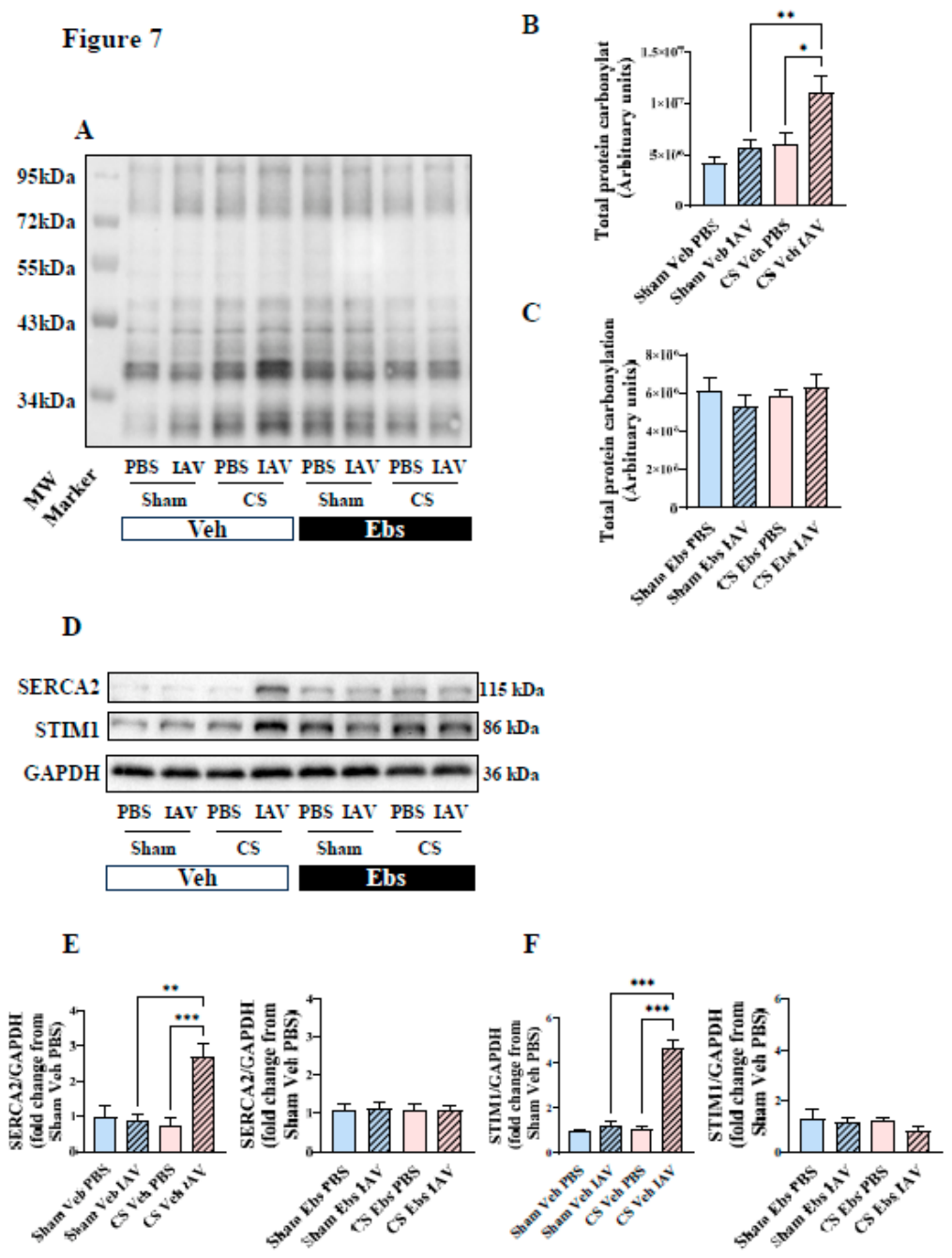
To further define the redox-sensitive mechanisms associated with skeletal muscle remodelling, we assessed oxidative protein modification and key calcium-handling proteins in the TA muscle. In vehicle-treated mice, oxyblot analysis showed an increase in oxidatively modified proteins, with the strongest signal observed in CS Veh IAV mice (Figure 7A). Densitometric quantification confirmed that CS Veh IAV group exhibited a marked elevation in oxyblot signal compared with Sham Veh IAV and CS Veh PBS groups, indicating that acute IAV challenge amplified oxidative protein modification only when combined with CS exposure (Figure 7B). In contrast, the increase in total lane carbonylation signal observed in vehicle-treated CS/IAV mice was not evident across the ebselen-treated groups (Figure 7C). We next examined whether this oxidative phenotype was accompanied by altered expression of calcium-handling proteins. Western blot analysis showed that SERCA2 and STIM1 protein expression were selectively increased in CS Veh IAV mice (Figure 7D). Quantification demonstrated ~2.5-fold increase in SERCA2/GAPDH and an ~4.8-fold increase in STIM1/GAPDH relative to the relevant vehicle controls (Figure 7E–F). These increases were not evident with IAV infection alone or CS exposure alone, indicating that calcium-handling protein remodelling was most pronounced when acute viral challenge occurred on the background of CS exposure. The increases in SERCA2 and STIM1 abundance observed in CS Veh IAV mice were not evident in the corresponding ebselen-treated groups (Figure 7E–F), supporting a redox-sensitive oxidative and calcium-handling protein remodelling response during viral exacerbation.
4. Discussion
This study demonstrates that CS exposure and acute IAV infection exert distinct but intersecting effects on the musculoskeletal system in a preclinical model of AECOPD. Three principal findings emerge from this work. First, acute IAV infection rapidly impaired trabecular bone microarchitecture, with reductions in BV/TV evident within 3 days of infection. Second, acute viral challenge worsened skeletal muscle force-generating capacity, particularly on a CS-exposed background, despite limited evidence of additional muscle wasting, supporting a dissociation between muscle quantity and muscle quality. Third, oxidative protein modification and calcium-handling protein remodelling were most pronounced in CS/IAV mice and were not evident in ebselen-treated animals, supporting a redox-sensitive component of exacerbation-associated skeletal muscle dysfunction.
A notable finding was that acute IAV infection alone reduced trabecular BV/TV, independent of CS exposure. COPD-associated bone loss is commonly attributed to chronic factors such as smoking, systemic inflammation, physical inactivity, altered body composition and disrupted muscle–bone crosstalk [3]. Our data extend this concept by suggesting that an acute respiratory viral insult can rapidly compromise trabecular bone structure. The short 3-day timeframe suggests that acute respiratory viral infection can rapidly alter measurable trabecular microarchitecture. However, because dynamic histomorphometry, osteoclast activity and bone turnover markers were not assessed at this acute endpoint, these data should be interpreted as evidence of altered bone microarchitecture rather than definitive proof of accelerated bone resorption or remodelling. This is consistent with osteoimmunological principles, whereby infection and immune activation can alter the balance between bone formation and resorption through inflammatory mediators and immune–bone interactions [18]. In parallel, CS exposure suppressed osteogenic matrix-related genes, including Ibsp, Spp1 and Bglap, and produced structural trabecular deterioration [30,31,32], supporting impaired bone-forming activity and disrupted bone homeostasis. The lack of additive deterioration in the CS + IAV group may indicate that a single viral challenge was insufficient to worsen an already CS exposure-injured bone compartment [19,20], or that different mechanisms converge on a similar trabecular endpoint.
The differential effects of CS exposure and IAV infection on hind limb bone and muscle suggest that these musculoskeletal compartments respond differently to chronic and acute respiratory insults. Both CS exposure and IAV infection impaired trabecular bone structure, whereas measurable muscle mass loss and fibre-type remodelling were primarily driven by CS exposure. This supports the concept that bone may be acutely sensitive to systemic inflammatory or osteoimmune perturbation during viral infection [18,33], while muscle wasting may require a more sustained catabolic exposure [34,35]. This interpretation is consistent with our previous work showing that IAV-driven airway inflammation can worsen TA force-generating capacity without necessarily causing additional limb muscle mass loss in smoke-exposed mice [9]. Thus, muscle mass and muscle function appear partially dissociated: CS exposure promotes structural wasting and myofibre remodelling, whereas IAV infection superimposed on CS exposure primarily worsens muscle quality, force generation and biochemical stress. The muscle data indicate that acute viral exacerbation primarily compromised muscle quality and function rather than producing additional measurable wasting, consistent with the observation that force deficits worsened in the absence of clear further reductions in muscle mass.
The most important mechanistic implication is that excessive oxidative stress may link the respiratory, skeletal muscle and bone compartments during COPD exacerbation [3]. CS and IAV generated a strong airway inflammatory phenotype, while skeletal muscle displayed redox-sensitive impairments in force generation, oxidative fibre maintenance, protein oxidation and calcium-handling remodelling. Oxidative stress is widely implicated in COPD skeletal muscle dysfunction, including altered fibre composition, mitochondrial dysfunction, impaired oxidative metabolism and proteostatic imbalance [12,15,16,17]. In this study, the key muscle abnormalities associated with CS and CS/IAV exposure were not evident or were less pronounced in ebselen-treated mice, supporting the involvement of redox-sensitive mechanisms in exacerbation-associated muscle dysfunction [9]. Although ebselen was not directly tested against the bone phenotype, the IAV-induced reduction in trabecular BV/TV raises the possibility that respiratory infection may also affect skeletal homeostasis through inflammatory and redox-sensitive pathways [21]. Together, the data support a lung–musculoskeletal axis in which excessive inflammatory and oxidative stress during COPD exacerbation compromises muscle and bone integrity.
The TA muscle may be particularly vulnerable to redox stress because of its fibre composition. The TA is a predominantly fast-twitch, glycolytic muscle, whereas more oxidative muscles generally possess greater mitochondrial content and antioxidant reserve [12,36]. Oxidative muscles have been reported to maintain more robust mitochondrial homeostasis than glycolytic muscles, while glycolytic muscles such as TA appear more susceptible to mitochondrial functional decline under stress conditions [36]. This is relevant here because the TA showed CS exposure-induced mass loss, impaired force generation, reduced myofibre CSA, loss of oxidative fibre proportion and increased oxidative/calcium-handling stress responses after CS/IAV exposure. One interpretation is that the fast-fibre-predominant TA has lower intrinsic oxidative reserve and may therefore be less redox robust during combined smoke and viral stress. The CS-induced loss of oxidative fibres may further reduce metabolic flexibility [37], making the muscle more susceptible to acute viral exacerbation [3,12]. However, this remains a hypothesis and should be tested directly by assessing fibre-type-specific oxidative damage, antioxidant capacity and mitochondrial respiration.
The induction of SERCA2 and STIM1 in CS/IAV-exposed mice is consistent with a link between oxidative stress, calcium-handling remodelling and impaired contractile function. SERCA proteins regulate sarcoplasmic reticulum calcium reuptake, while STIM1 contributes to store-operated calcium entry and calcium homeostasis [38]. In this study, SERCA2 and STIM1 were selectively increased in the CS Veh IAV mice, coinciding with the highest oxyblot signal and greatest force deficit. This suggests that acute IAV challenge on a CS exposure background unmasks or amplifies a skeletal muscle stress programme involving oxidative protein modification and calcium-handling remodelling. The absence of detectable SERCA2 and STIM1 increases in ebselen-treated mice supports the interpretation that this biochemical response is redox-sensitive. It is also possible that the increased SERCA2 and STIM1 abundance reflects altered fibre-type biology, a calcium-handling stress response, or a compensatory remodelling response rather than direct evidence of calcium dysfunction. These possibilities require direct experimental evaluation. These findings are consistent with the broader view that oxidative stress can impair muscle quality beyond changes in muscle mass alone, particularly by altering excitation–contraction coupling, fibre metabolism and protein homeostasis [37,39].
Clinically, these findings reinforce the need to consider COPD exacerbations as systemic events rather than isolated respiratory episodes [3,12]. The observation that acute IAV infection alone impaired trabecular bone structure suggests that viral exacerbations may contribute to skeletal fragility, particularly in patients with pre-existing osteopenia, osteoporosis or smoking-related bone loss [3,19,21]. Similarly, the dissociation between muscle mass and muscle function suggests that exacerbation-related weakness may occur even without overt additional wasting, which has been observed clinically [40]. This has implications for clinical assessment, as body weight or lean mass alone may underestimate muscle dysfunction during or after exacerbation [41]. The pattern observed in ebselen-treated mice also supports further investigation of antioxidant or redox-modulating strategies as adjuncts to pulmonary rehabilitation, nutritional support and exacerbation prevention, particularly for patients with exacerbation-prone COPD and musculoskeletal comorbidities [12].
Several limitations should be acknowledged. First, the study used a single acute IAV challenge after 8 weeks of CS exposure, whereas patients with COPD often experience repeated exacerbations over time [42]. Recurrent viral insults may produce more cumulative effects on bone, muscle mass, redox balance and recovery [3]. In addition, although ebselen was used to investigate redox-sensitive skeletal muscle responses, its effects on the bone phenotype were not directly assessed. Second, this study used male BALB/c mice, so sex-specific and strain-dependent responses were not addressed. Furthermore, bone histomorphometry, osteoclast activity, osteoblast function and circulating bone turnover markers were not assessed at the acute IAV endpoint, limiting mechanistic insight into the processes responsible for the observed trabecular changes. Third, although ebselen provided evidence for redox sensitivity, direct measurements of mitochondrial respiration, antioxidant enzyme activity, ROS-generating pathways and fibre-type-specific oxidative damage were not performed. Fourth, TA muscle was selected because of its CS-associated mass loss, anatomical association with the tibia and suitability for downstream contractile and molecular analyses, but other muscles may show different redox or calcium-handling responses. Finally, while SERCA2 and STIM1 changes suggest altered calcium-handling biology, calcium flux or excitation–contraction coupling should be measured in future studies.
In conclusion, this study shows that CS exposure and acute IAV infection differentially affect bone and skeletal muscle during experimental viral exacerbation of COPD. CS exposure drove systemic wasting, myofibre atrophy, oxidative fibre loss and muscle weakness, whereas acute IAV infection independently impaired trabecular bone structure and amplified muscle dysfunction when combined with CS exposure. Several CS/IAV-associated muscle abnormalities, including oxidative protein modification and calcium-handling protein remodelling, were not evident in ebselen-treated mice, supporting redox imbalance as a contributor to exacerbation-associated skeletal muscle pathology. These findings support a model in which viral exacerbation amplifies redox-sensitive muscle stress on a smoke-exposed background, while also revealing trabecular bone as an unexpectedly sensitive target of acute respiratory viral infection.
Author Contributions
Concept and design: RV, SMCH; acquisition of data: KM, SMCH, KB, AD, SND, HJS; data analysis and interpretation: KM, SMCH, RV, SND, HW, SS, SB; technical assistance: KB, AD, HJS; drafting, editing, and/or critical revision of the manuscript for intellectual content: all authors; RV also provided all resources for the work and is the senior investigator, ensuring accuracy and integrity.
Funding
The authors would like to thank the National Health and Medical Research Council of Australia (Project Grant Numbers APP1084627 and APP1138915) for funding this work.
References
- Vogelmeier, C.F.; Gaga, M.; Aalamian-Mattheis, M.; Greulich, T.; Marin, J.M.; Castellani, W.; et al. Efficacy and safety of direct switch to indacaterol/glycopyrronium in patients with moderate COPD: the CRYSTAL open-label randomised trial. Respir. Res. 2017, 18, 140. [Google Scholar] [CrossRef] [PubMed]
- Barnes, P.J. Inflammatory endotypes in COPD. Allergy 2019, 74, 1249–1256. [Google Scholar] [CrossRef] [PubMed]
- Mou, K.; Chan, S.M.H.; Vlahos, R. Musculoskeletal crosstalk in chronic obstructive pulmonary disease and comorbidities: Emerging roles and therapeutic potentials. Pharmacol. Ther. 2024, 257, 108635. [Google Scholar] [CrossRef] [PubMed]
- Zhang, L.; Sun, Y. Muscle-Bone Crosstalk in Chronic Obstructive Pulmonary Disease. Front Endocrinol. 2021, 12, 724911. [Google Scholar] [CrossRef] [PubMed]
- Cazzola, M.; Bajpai, J.; Calzetta, L.; Matera, M.G.; Rogliani, P. (2026) GOLD 2026: Transforming COPD Management with Early Intervention, Multi-dimensional Assessment, and Personalized Care. Drugs 86, 581–597. [CrossRef] [PubMed]
- Dimopoulos, G.; Lerikou, M.; Tsiodras, S.; Chranioti, A.; Perros, E.; Anagnostopoulou, U.; et al. Viral epidemiology of acute exacerbations of chronic obstructive pulmonary disease. Pulm. Pharmacol. Ther. 2012, 25, 12–18. [Google Scholar] [CrossRef] [PubMed]
- Kefala, A.M.; Fortescue, R.; Alimani, G.S.; Kanavidis, P.; McDonnell, M.J.; Magiorkinis, E.; et al. Prevalence and clinical implications of respiratory viruses in stable chronic obstructive pulmonary disease (COPD) and exacerbations: a systematic review and meta-analysis protocol. BMJ Open 2020, 10, e035640. [Google Scholar] [CrossRef] [PubMed]
- Chen, C.Y.J.; Yew, M.S.; Abisheganaden, J.A.; Xu, H. Predictors of Influenza PCR Positivity in Acute Exacerbations of Chronic Obstructive Pulmonary Disease. Int. J. Chron. Obstruct Pulmon Dis. 2022, 17, 25–32. [Google Scholar] [CrossRef] [PubMed]
- Mou, K.; Chan, S.M.H.; Brassington, K.; Dobric, A.; De Luca, S.N.; Seow, H.J.; et al. Influenza A Virus-Driven Airway Inflammation may be Dissociated From Limb Muscle Atrophy in Cigarette Smoke-Exposed Mice. Front Pharmacol. 2022, 13, 859146. [Google Scholar] [CrossRef] [PubMed]
- Barnes, P.J. Oxidative Stress in Chronic Obstructive Pulmonary Disease. Antioxidants 2022, 11. [Google Scholar] [CrossRef] [PubMed]
- Bernardo, I.; Bozinovski, S.; Vlahos, R. Targeting oxidant-dependent mechanisms for the treatment of COPD and its comorbidities. Pharmacol. Ther. 2015, 155, 60–79. [Google Scholar] [CrossRef] [PubMed]
- Chan, S.M.H.; Selemidis, S.; Vlahos, R. The Double-Edged Sword of ROS in Muscle Wasting and COPD: Insights from Aging-Related Sarcopenia. Antioxidants-Basel 2024, 13. [Google Scholar] [CrossRef] [PubMed]
- Oostwoud, L.C.; Gunasinghe, P.; Seow, H.J.; Ye, J.M.; Selemidis, S.; Bozinovski, S.; et al. Apocynin and ebselen reduce influenza A virus-induced lung inflammation in cigarette smoke-exposed mice. Sci. Rep. 2016, 6, 20983. [Google Scholar] [CrossRef] [PubMed]
- Bezerra, F.S.; Lanzetti, M.; Nesi, R.T.; Nagato, A.C.; Silva, C.P.E.; Kennedy-Feitosa, E.; et al. Oxidative Stress and Inflammation in Acute and Chronic Lung Injuries. Antioxidants 2023, 12. [Google Scholar] [CrossRef] [PubMed]
- Chan, S.M.H.; Bernardo, I.; Mastronardo, C.; Mou, K.; De Luca, S.N.; Seow, H.J.; et al. Apocynin prevents cigarette smoking-induced loss of skeletal muscle mass and function in mice by preserving proteostatic signalling. Br. J. Pharmacol. 2021, 178, 3049–3066. [Google Scholar] [CrossRef] [PubMed]
- Khan, A.; Fouda, S.; Mahzari, A.; Chan, S.M.H.; Zhou, X.; Ratnam, C.; et al. Cigarette smoking blocks the benefit from reduced weight gain for insulin action by shifting lipids deposition to muscle. Clin. Sci. (Lond) 2020, 134, 1659–1673. [Google Scholar] [CrossRef] [PubMed]
- Chan, S.M.H.; Cerni, C.; Passey, S.; Seow, H.J.; Bernardo, I.; van der Poel, C.; et al. Cigarette Smoking Exacerbates Skeletal Muscle Injury without Compromising Its Regenerative Capacity. Am. J. Respir. Cell Mol. Biol. 2020, 62, 217–230. [Google Scholar] [CrossRef] [PubMed]
- Galliera, E.; Massaccesi, L.; Logoluso, N.; Mangiavini, L.; Peretti, G.; Corsi Romanelli, M.M. Bone and Infections: An Osteoimmunological Interplay. Int. J. Mol. Sci. 2026, 27. [Google Scholar] [CrossRef] [PubMed]
- Caetano, C.C.S.; Azamor, T.; Meyer, N.M.; Onwubueke, C.; Calabrese, C.M.; Calabrese, L.H.; et al. Mechanistic insights into bone remodelling dysregulation by human viral pathogens. Nat. Microbiol. 2024, 9, 322–335. [Google Scholar] [CrossRef] [PubMed]
- Haudenschild, A.K.; Christiansen, B.A.; Orr, S.; Ball, E.E.; Weiss, C.M.; Liu, H.; et al. Acute bone loss following SARS-CoV-2 infection in mice. J. Orthop. Res. 2023, 41, 1945–1952. [Google Scholar] [CrossRef] [PubMed]
- Reis, J.; Ramos, A. In Sickness and in Health: The Oxygen Reactive Species and the Bone. Front Bioeng. Biotechnol. 2021, 9, 745911. [Google Scholar] [CrossRef] [PubMed]
- Brassington, K.; Chan, S.M.H.; De Luca, S.N.; Dobric, A.; Almerdasi, S.A.; Mou, K.; et al. Ebselen abolishes vascular dysfunction in influenza A virus-induced exacerbations of cigarette smoke-induced lung inflammation in mice. Clin. Sci. (Lond) 2022, 136, 537–555. [Google Scholar] [CrossRef] [PubMed]
- Brassington, K.; Chan, S.M.H.; Seow, H.J.; Dobric, A.; Bozinovski, S.; Selemidis, S.; et al. Ebselen reduces cigarette smoke-induced endothelial dysfunction in mice. Br. J. Pharmacol. 2021, 178, 1805–1818. [Google Scholar] [CrossRef] [PubMed]
- Percie du Sert, N.; Hurst, V.; Ahluwalia, A.; Alam, S.; Avey, M.T.; Baker, M.; et al. The ARRIVE guidelines 2.0: Updated guidelines for reporting animal research. Br. J. Pharmacol. 2020, 177, 3617–3624. [Google Scholar] [CrossRef] [PubMed]
- Vlahos, R.; Bozinovski, S.; Jones, J.E.; Powell, J.; Gras, J.; Lilja, A.; et al. Differential protease, innate immunity, and NF-kappaB induction profiles during lung inflammation induced by subchronic cigarette smoke exposure in mice. Am. J. Physiol. Lung Cell Mol. Physiol. 2006, 290, L931–945. [Google Scholar] [CrossRef] [PubMed]
- Wald, N.; Idle, M.; Bailey, A. Carboxyhaemoglobin levels and inhaling habits in cigarette smokers. Thorax 1978, 33, 201–206. [Google Scholar] [CrossRef] [PubMed]
- Wang, W.; Chan, S.M.H.; Almerdasi, S.A.; De Luca, S.N.; Nguyen, Q.A.; Adderley, J.; et al. Defining the cross-tissue communication between lungs and white adipose tissue in chronic obstructive pulmonary disease. Clin. Sci. (Lond) 2026, 140, 427–443. [Google Scholar] [CrossRef] [PubMed]
- Chan, S.M.H.; Brassington, K.; Almerdasi, S.A.; Dobric, A.; De Luca, S.N.; Coward-Smith, M.; et al. Inhibition of oxidative stress by apocynin attenuated chronic obstructive pulmonary disease progression and vascular injury by cigarette smoke exposure. Br. J. Pharmacol. 2023, 180, 2018–2034. [Google Scholar] [CrossRef] [PubMed]
- Bouxsein, M.L.; Boyd, S.K.; Christiansen, B.A.; Guldberg, R.E.; Jepsen, K.J.; Muller, R. Guidelines for assessment of bone microstructure in rodents using micro-computed tomography. J. Bone Min. Res. 2010, 25, 1468–1486. [Google Scholar] [CrossRef] [PubMed]
- Malaval, L.; Wade-Gueye, N.M.; Boudiffa, M.; Fei, J.; Zirngibl, R.; Chen, F.; et al. Bone sialoprotein plays a functional role in bone formation and osteoclastogenesis. J. Exp. Med. 2008, 205, 1145–1153. [Google Scholar] [CrossRef] [PubMed]
- Ma, R.; Su, Y.; Cao, R.; Wang, K.; Yang, P. Enhanced Osteogenic Activity and Bone Repair Ability of PLGA/MBG Scaffolds Doped with ZIF-8 Nanoparticles Loaded with BMP-2. Int. J. Nanomed. 2023, 18, 5055–5072. [Google Scholar] [CrossRef] [PubMed]
- Ding, W.; Lv, D.; Wang, M.; Pei, D. IBSP Promotes Breast Cancer Bone Metastasis and Proliferation via BMP-SMAD Signaling Pathway. Cancer Rep. 2024, 7, e2153. [Google Scholar] [CrossRef] [PubMed]
- Maltby, S.; Lochrin, A.J.; Bartlett, B.; Tay, H.L.; Weaver, J.; Poulton, I.J.; et al. Osteoblasts Are Rapidly Ablated by Virus-Induced Systemic Inflammation following Lymphocytic Choriomeningitis Virus or Pneumonia Virus of Mice Infection in Mice. J. Immunol. 2018, 200, 632–642. [Google Scholar] [CrossRef] [PubMed]
- Zhang, J.; Luo, W.; Miao, C.; Zhong, J. Hypercatabolism and Anti-catabolic Therapies in the Persistent Inflammation, Immunosuppression, and Catabolism Syndrome. Front Nutr. 2022, 9, 941097. [Google Scholar] [CrossRef] [PubMed]
- Ji, Y.; Li, M.; Chang, M.; Liu, R.; Qiu, J.; Wang, K.; et al. Inflammation: Roles in Skeletal Muscle Atrophy. Antioxidants 2022, 11. [Google Scholar] [CrossRef] [PubMed]
- Crupi, A.N.; Nunnelee, J.S.; Taylor, D.J.; Thomas, A.; Vit, J.P.; Riera, C.E.; et al. Oxidative muscles have better mitochondrial homeostasis than glycolytic muscles throughout life and maintain mitochondrial function during aging. Aging 2018, 10, 3327–3352. [Google Scholar] [CrossRef] [PubMed]
- Supruniuk, E.; Gorski, J.; Chabowski, A. Endogenous and Exogenous Antioxidants in Skeletal Muscle Fatigue Development during Exercise. In Antioxidants (Basel); 2023. [Google Scholar] [CrossRef] [PubMed]
- Zhuang, Z.; Meng, Y.; Xue, Y.; Wang, Y.; Cheng, X.; Jing, J. Adaptation of STIM1 structure-function relationships for optogenetic control of calcium signaling. J. Biol. Chem. 2024, 300, 107636. [Google Scholar] [CrossRef] [PubMed]
- Qaisar, R.; Bhaskaran, S.; Premkumar, P.; Ranjit, R.; Natarajan, K.S.; Ahn, B.; et al. Oxidative stress-induced dysregulation of excitation-contraction coupling contributes to muscle weakness. J. Cachexia Sarcopenia Muscle 2018, 9, 1003–1017. [Google Scholar] [CrossRef] [PubMed]
- Spruit, M.A.; Gosselink, R.; Troosters, T.; Kasran, A.; Gayan-Ramirez, G.; Bogaerts, P.; et al. Muscle force during an acute exacerbation in hospitalised patients with COPD and its relationship with CXCL8 and IGF-I. Thorax 2003, 58, 752–756. [Google Scholar] [CrossRef] [PubMed]
- Kim, S.H.; Shin, M.J.; Shin, Y.B.; Kim, K.U. Sarcopenia Associated with Chronic Obstructive Pulmonary Disease. J. Bone Metab. 2019, 26, 65–74. [Google Scholar] [CrossRef] [PubMed]
- Bhatt, S.P.; Agusti, A.; Bafadhel, M.; Christenson, S.A.; Bon, J.; Donaldson, G.C.; et al. Phenotypes, Etiotypes, and Endotypes of Exacerbations of Chronic Obstructive Pulmonary Disease. Am. J. Respir. Crit. Care Med. 2023, 208, 1026–1041. [Google Scholar] [CrossRef] [PubMed]
Figure 1.
Experimental design and treatment groups. (A) Schematic overview of the experimental protocol. Mice were exposed to room air (Sham) or cigarette smoke (CS) for 8 weeks, with daily oral gavage of either vehicle or ebselen (10 mg·kg⁻¹). At day 0, mice were intranasally inoculated with phosphate-buffered saline (PBS; diluent control) or influenza A virus (IAV) to model acute viral exacerbation on a CS-exposed background. Terminal experiments and tissue collection were performed at day 3 post-infection, including bronchoalveolar lavage fluid collection, tibial bone assessment, hindlimb muscle analyses, TA contractile testing and molecular assays. (B) Experimental groups included Sham Veh PBS, Sham Veh IAV, Sham Ebs PBS, Sham Ebs IAV, CS Veh PBS, CS Veh IAV, CS Ebs PBS and CS Ebs IAV. Veh, vehicle; Ebs, ebselen; PBS, phosphate-buffered saline.
Figure 1.
Experimental design and treatment groups. (A) Schematic overview of the experimental protocol. Mice were exposed to room air (Sham) or cigarette smoke (CS) for 8 weeks, with daily oral gavage of either vehicle or ebselen (10 mg·kg⁻¹). At day 0, mice were intranasally inoculated with phosphate-buffered saline (PBS; diluent control) or influenza A virus (IAV) to model acute viral exacerbation on a CS-exposed background. Terminal experiments and tissue collection were performed at day 3 post-infection, including bronchoalveolar lavage fluid collection, tibial bone assessment, hindlimb muscle analyses, TA contractile testing and molecular assays. (B) Experimental groups included Sham Veh PBS, Sham Veh IAV, Sham Ebs PBS, Sham Ebs IAV, CS Veh PBS, CS Veh IAV, CS Ebs PBS and CS Ebs IAV. Veh, vehicle; Ebs, ebselen; PBS, phosphate-buffered saline.
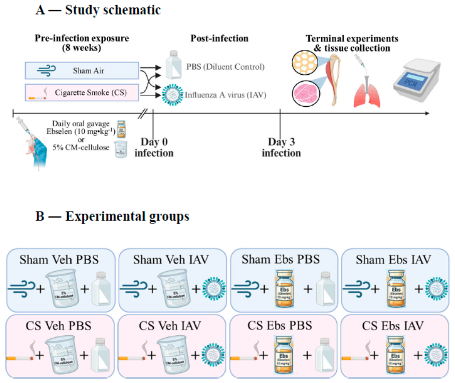
Figure 2.
Cigarette smoke exposure and acute influenza A virus infection increase airway inflammatory cell burden. Bronchoalveolar lavage fluid (BALF) was collected at day 3 post-infection from vehicle-treated mice exposed to room air (Sham) or CS, followed by PBS or influenza A virus (IAV) challenge. Quantification of (A) total BALF cells, (B) macrophages, (C) neutrophils and (D) lymphocytes showed increased airway inflammatory cellularity following CS exposure and/or IAV infection, with the combined CS Veh IAV group showing the greatest inflammatory burden. Data are presented as mean + SEM (n = 8 mice/group). *p < 0.05, **p < 0.01, ***p < 0.001.
Figure 2.
Cigarette smoke exposure and acute influenza A virus infection increase airway inflammatory cell burden. Bronchoalveolar lavage fluid (BALF) was collected at day 3 post-infection from vehicle-treated mice exposed to room air (Sham) or CS, followed by PBS or influenza A virus (IAV) challenge. Quantification of (A) total BALF cells, (B) macrophages, (C) neutrophils and (D) lymphocytes showed increased airway inflammatory cellularity following CS exposure and/or IAV infection, with the combined CS Veh IAV group showing the greatest inflammatory burden. Data are presented as mean + SEM (n = 8 mice/group). *p < 0.05, **p < 0.01, ***p < 0.001.
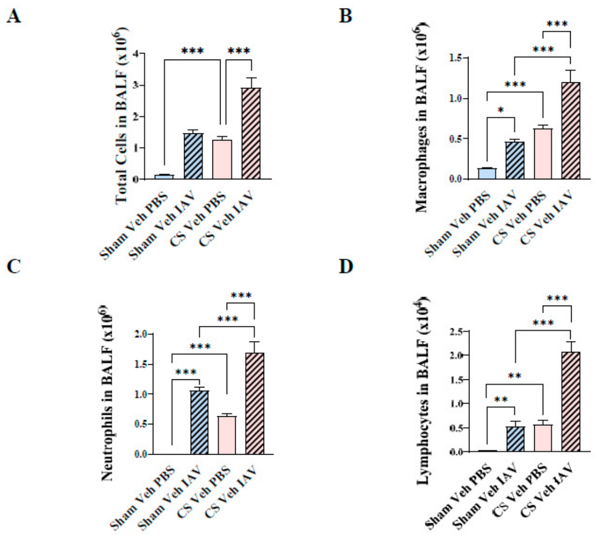
Figure 3.
Cigarette smoke exposure and acute influenza A virus infection independently impair tibial bone microarchitecture. Tibial micro-computed tomography was performed in vehicle-treated mice exposed to room air (Sham) or CS, followed by PBS or IAV challenge. (A) Representative micro-CT images of the proximal tibial trabecular region. Quantification of trabecular parameters included (B) bone volume fraction (BV/TV), (C) trabecular thickness (Tb.Th), (D) trabecular separation (Tb.Sp) and (E) trabecular number (Tb.N). (F) Representative micro-CT images of the cortical tibial region. Quantification of cortical parameters included (G) cortical area (Ct.Ar) and (H) cortical thickness (Ct.Th). CS exposure and IAV infection each impaired aspects of tibial bone microarchitecture, with no clear evidence of additive deterioration in the combined CS Veh IAV group. Data are presented as mean + SEM (n = 8 mice/group). *p < 0.05, **p < 0.01, ***p < 0.001.
Figure 3.
Cigarette smoke exposure and acute influenza A virus infection independently impair tibial bone microarchitecture. Tibial micro-computed tomography was performed in vehicle-treated mice exposed to room air (Sham) or CS, followed by PBS or IAV challenge. (A) Representative micro-CT images of the proximal tibial trabecular region. Quantification of trabecular parameters included (B) bone volume fraction (BV/TV), (C) trabecular thickness (Tb.Th), (D) trabecular separation (Tb.Sp) and (E) trabecular number (Tb.N). (F) Representative micro-CT images of the cortical tibial region. Quantification of cortical parameters included (G) cortical area (Ct.Ar) and (H) cortical thickness (Ct.Th). CS exposure and IAV infection each impaired aspects of tibial bone microarchitecture, with no clear evidence of additive deterioration in the combined CS Veh IAV group. Data are presented as mean + SEM (n = 8 mice/group). *p < 0.05, **p < 0.01, ***p < 0.001.
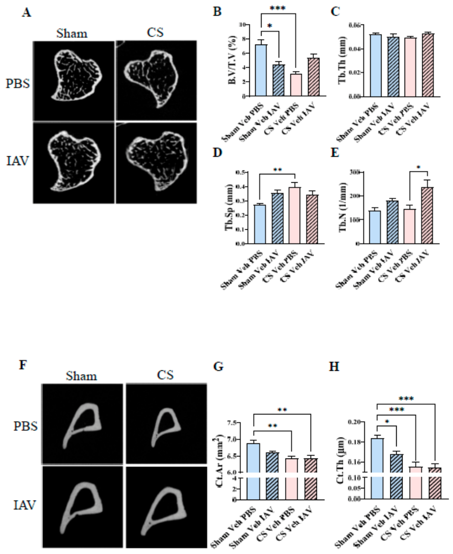
Figure 4.
Cigarette smoke exposure selectively reduces hindlimb muscle mass in vehicle-treated mice. Hindlimb muscles were dissected and weighed at day 3 post-infection from vehicle-treated mice exposed to room air (Sham) or CS, followed by PBS or IAV challenge. Muscle weights were quantified for (A) tibialis anterior, (B) soleus, (C) plantaris, (D) gastrocnemius and (E) quadriceps muscles. CS exposure reduced tibialis anterior, gastrocnemius and quadriceps muscle weights, whereas soleus and plantaris muscle weights were not detectably altered. Acute IAV infection alone did not clearly reduce hindlimb muscle mass or further exacerbate CS-associated muscle mass loss. Data are presented as mean+ SEM (n = 8 mice/group). ***p < 0.001.
Figure 4.
Cigarette smoke exposure selectively reduces hindlimb muscle mass in vehicle-treated mice. Hindlimb muscles were dissected and weighed at day 3 post-infection from vehicle-treated mice exposed to room air (Sham) or CS, followed by PBS or IAV challenge. Muscle weights were quantified for (A) tibialis anterior, (B) soleus, (C) plantaris, (D) gastrocnemius and (E) quadriceps muscles. CS exposure reduced tibialis anterior, gastrocnemius and quadriceps muscle weights, whereas soleus and plantaris muscle weights were not detectably altered. Acute IAV infection alone did not clearly reduce hindlimb muscle mass or further exacerbate CS-associated muscle mass loss. Data are presented as mean+ SEM (n = 8 mice/group). ***p < 0.001.
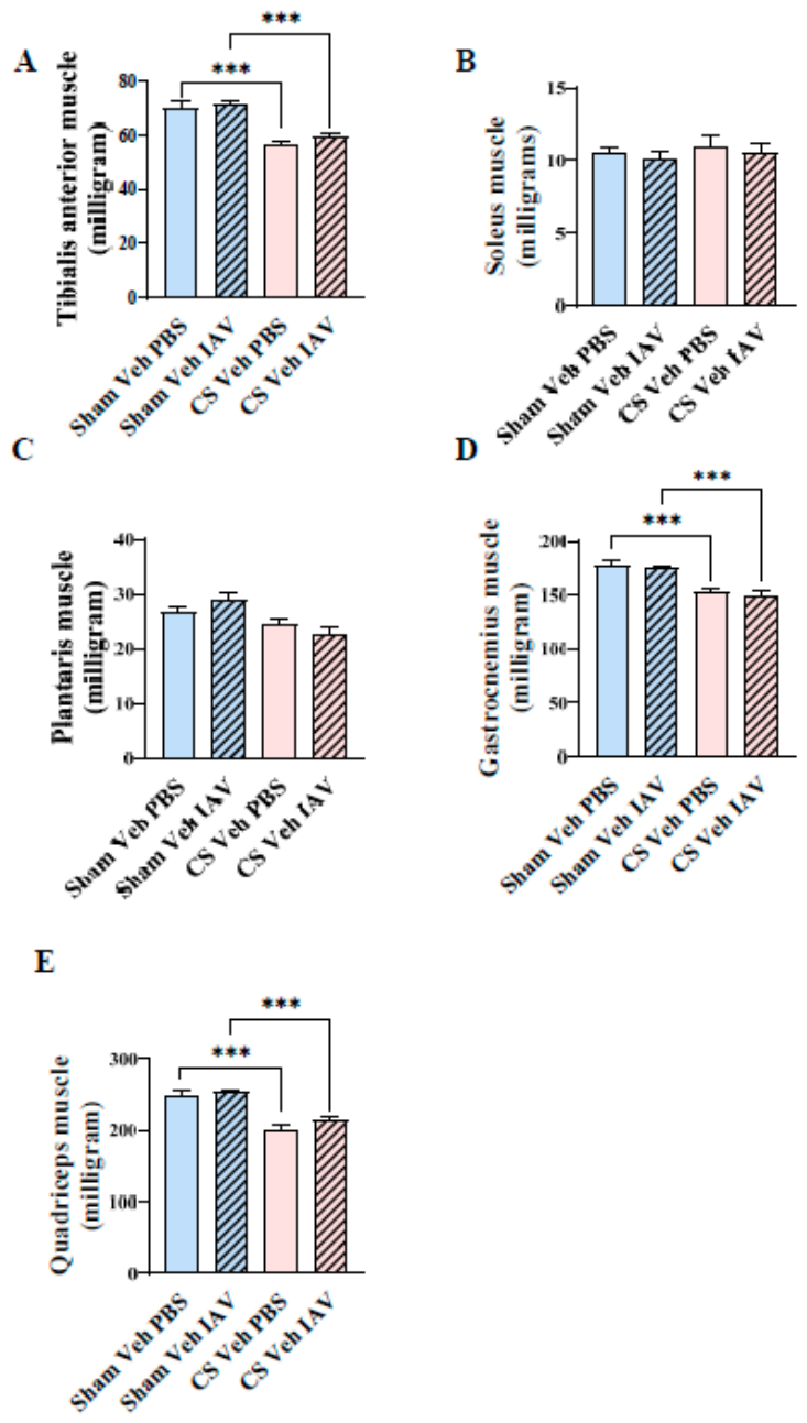
Figure 5.
Cigarette smoke- and influenza A virus-associated impairment of tibialis anterior contractile function is not evident in ebselen-treated mice. In situ tibialis anterior contractile function and spontaneous locomotor activity were assessed at day 3 post-infection. Force–frequency curves were generated in (A) vehicle-treated and (B) ebselen-treated mice exposed to room air (Sham) or CS, followed by PBS or IAV challenge. (C) Specific force was reduced in vehicle-treated mice following CS exposure or IAV infection, with the greatest deficit observed in the CS Veh IAV group. (D) Ebselen-treated mice showed no detectable reduction in specific force across experimental groups. Total distance travelled during open-field assessment was quantified in (E) vehicle-treated and (F) ebselen-treated mice, with no obvious group differences observed. Data are presented as mean + SEM (n = 6 mice/group). *p < 0.05, ***p < 0.001.
Figure 5.
Cigarette smoke- and influenza A virus-associated impairment of tibialis anterior contractile function is not evident in ebselen-treated mice. In situ tibialis anterior contractile function and spontaneous locomotor activity were assessed at day 3 post-infection. Force–frequency curves were generated in (A) vehicle-treated and (B) ebselen-treated mice exposed to room air (Sham) or CS, followed by PBS or IAV challenge. (C) Specific force was reduced in vehicle-treated mice following CS exposure or IAV infection, with the greatest deficit observed in the CS Veh IAV group. (D) Ebselen-treated mice showed no detectable reduction in specific force across experimental groups. Total distance travelled during open-field assessment was quantified in (E) vehicle-treated and (F) ebselen-treated mice, with no obvious group differences observed. Data are presented as mean + SEM (n = 6 mice/group). *p < 0.05, ***p < 0.001.
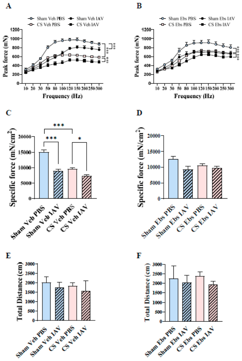
Figure 6.
Ebselen-treated mice show preservation of tibialis anterior myofibre morphology and oxidative fibre proportion following cigarette smoke exposure. TA muscle sections were analysed for myofibre morphology and oxidative fibre composition at day 3 post-infection. (A) Representative images of laminin-stained tibialis anterior cross-sections from vehicle-treated mice exposed to room air (Sham) or CS, followed by PBS or IAV challenge. (B) Quantification of mean myofibre cross-sectional area showed reduced myofibre size in CS-exposed vehicle-treated mice. (C) Myofibre size-distribution analysis showed a shift towards smaller myofibre cross-sectional area bins in CS-exposed vehicle-treated groups. (D) Representative laminin-stained tibialis anterior sections from ebselen-treated mice. (E) Mean myofibre cross-sectional area was preserved in ebselen-treated mice across experimental groups. (F) Myofibre size distribution was also maintained in ebselen-treated groups. (G) Representative oxidative fibre staining and quantification in vehicle-treated mice showed reduced oxidative fibre proportion following CS exposure, irrespective of IAV challenge. (H) Representative oxidative fibre staining and quantification in ebselen-treated mice showed preservation of oxidative fibre proportion across groups. Data are presented as mean + SEM (n = 6 mice/group). *p < 0.05, **p < 0.01, ***p < 0.001. CSA, cross-sectional area.
Figure 6.
Ebselen-treated mice show preservation of tibialis anterior myofibre morphology and oxidative fibre proportion following cigarette smoke exposure. TA muscle sections were analysed for myofibre morphology and oxidative fibre composition at day 3 post-infection. (A) Representative images of laminin-stained tibialis anterior cross-sections from vehicle-treated mice exposed to room air (Sham) or CS, followed by PBS or IAV challenge. (B) Quantification of mean myofibre cross-sectional area showed reduced myofibre size in CS-exposed vehicle-treated mice. (C) Myofibre size-distribution analysis showed a shift towards smaller myofibre cross-sectional area bins in CS-exposed vehicle-treated groups. (D) Representative laminin-stained tibialis anterior sections from ebselen-treated mice. (E) Mean myofibre cross-sectional area was preserved in ebselen-treated mice across experimental groups. (F) Myofibre size distribution was also maintained in ebselen-treated groups. (G) Representative oxidative fibre staining and quantification in vehicle-treated mice showed reduced oxidative fibre proportion following CS exposure, irrespective of IAV challenge. (H) Representative oxidative fibre staining and quantification in ebselen-treated mice showed preservation of oxidative fibre proportion across groups. Data are presented as mean + SEM (n = 6 mice/group). *p < 0.05, **p < 0.01, ***p < 0.001. CSA, cross-sectional area.
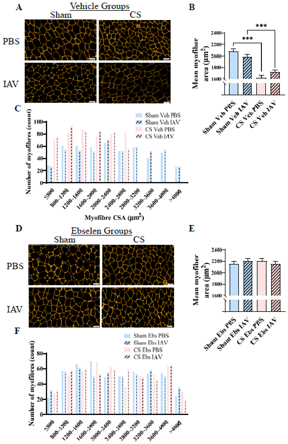
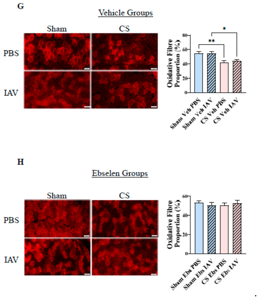
Figure 7.
Ebselen attenuates viral exacerbation-associated oxidative protein modification and calcium-handling protein remodelling. Oxidative protein modification and calcium-handling protein expression were assessed in tibialis anterior muscle homogenates from vehicle- and ebselen-treated mice exposed to room air (Sham) or CS, followed by PBS or IAV challenge. (A) Representative oxyblot image showing oxidatively modified proteins across vehicle- and ebselen-treated groups. (B) Total lane carbonylation signal was increased in CS Veh IAV mice. (C) No detectable increase in total lane carbonylation signal was observed across the ebselen-treated groups. (D) Representative Western blot images for SERCA2, STIM1 and GAPDH. (E) Quantification of SERCA2/GAPDH expression showed increased SERCA2 abundance in CS Veh IAV mice, a corresponding increase was not evident in ebselen-treated mice. (F) Quantification of STIM1/GAPDH expression showed increased STIM1 abundance in CS Veh IAV mice which was not evident in ebselen-treated mice. Data are presented as mean + SEM (n = 6 mice/group). *p < 0.05, **p < 0.01, ***p < 0.001. SERCA2, sarco/endoplasmic reticulum Ca²⁺-ATPase 2; STIM1, stromal interaction molecule 1.
Figure 7.
Ebselen attenuates viral exacerbation-associated oxidative protein modification and calcium-handling protein remodelling. Oxidative protein modification and calcium-handling protein expression were assessed in tibialis anterior muscle homogenates from vehicle- and ebselen-treated mice exposed to room air (Sham) or CS, followed by PBS or IAV challenge. (A) Representative oxyblot image showing oxidatively modified proteins across vehicle- and ebselen-treated groups. (B) Total lane carbonylation signal was increased in CS Veh IAV mice. (C) No detectable increase in total lane carbonylation signal was observed across the ebselen-treated groups. (D) Representative Western blot images for SERCA2, STIM1 and GAPDH. (E) Quantification of SERCA2/GAPDH expression showed increased SERCA2 abundance in CS Veh IAV mice, a corresponding increase was not evident in ebselen-treated mice. (F) Quantification of STIM1/GAPDH expression showed increased STIM1 abundance in CS Veh IAV mice which was not evident in ebselen-treated mice. Data are presented as mean + SEM (n = 6 mice/group). *p < 0.05, **p < 0.01, ***p < 0.001. SERCA2, sarco/endoplasmic reticulum Ca²⁺-ATPase 2; STIM1, stromal interaction molecule 1.
Disclaimer/Publisher’s Note: The statements, opinions and data contained in all publications are solely those of the individual author(s) and contributor(s) and not of MDPI and/or the editor(s). MDPI and/or the editor(s) disclaim responsibility for any injury to people or property resulting from any ideas, methods, instructions or products referred to in the content. |
© 2026 by the authors. Licensee MDPI, Basel, Switzerland. This article is an open access article distributed under the terms and conditions of the Creative Commons Attribution (CC BY) license (http://creativecommons.org/licenses/by/4.0/).
Copyright: This open access article is published under a Creative Commons CC BY 4.0 license, which permit the free download, distribution, and reuse, provided that the author and preprint are cited in any reuse.