

Submitted:
10 September 2026
Posted:
16 September 2026
You are already at the latest version
Abstract
Transcatheter tricuspid valve-in-valve replacement offers an alternative to high-risk redo surgery for failed tricuspid bioprostheses. We report a 36-year-old woman with severe right-heart failure, hepatic dysfunction, thrombocytopenia, and a mechanical mitral prosthesis after a previously abandoned transcatheter attempt. Redo implantation was performed under dexmedetomidine-ketamine sedation with spontaneous ventilation and a femoral nerve block. Transoesophageal echocardiography was avoided because of bleeding risk. Fluoroscopy and transthoracic echocardiography provided procedural guidance. Valve deployment was successful, the transvalvular gradient decreased and she was discharged on postoperative day 3, highlighting individualized management of competing haemorrhagic and thrombotic risks.
Keywords:
dexmedetomidine
; right-heart failure
; thrombocytopenia
; transcatheter tricuspid valve-in-valve replacement
; procedural sedation
; transthoracic echocardiography
Introduction
Redo tricuspid surgery carries substantial perioperative risk, for which transcatheter tricuspid valve-in-valve replacement has emerged as a less invasive alternative.[1,2] We present a case of a valve-in-valve TTVR patient with multiple co-morbidities, anaesthesia management, clinical outcome, and periprocedural management.
Patient Details and Clinical Course
A 36-year-old woman with rheumatic heart disease (RHD) presented in 2009 with features of severe mitral stenosis and tricuspid regurgitation. She underwent open mitral commissurotomy (OMC) + ring annuloplasty with concomitant tricuspid valve repair. At a second operation in 2020 she received a mechanical mitral valve replacement together with a bioprosthetic tricuspid valve(EPIC) replacement after her first pregnancy when her symptoms recurred. This procedure was complicated by a prolonged postoperative course requiring tracheostomy and eventual discharge. Specific details were not available with patient possible cause being pulmonary infection.
In 2024 she came with recurrence of right-sided symptoms attributable to failure of the bioprosthetic tricuspid valve. Transcatheter tricuspid valve in valve replacement (TTViVR) was planned via femoral route but after multiple failed attempts, trans jugular approach was taken. Owing to complex anatomy with grossly dilated right atrium (RA) and small right ventricle (RV)(Figure 1), the guide catheter had no stability and procedure was abandoned as there was already significant blood loss. Patient was discharged on high dose of diuretics.
In 2026 she presented with progressive dyspnoea on exertion New York Heart Association (NYHAIII) for redo TTViVR. She had progressed to right heart failure with gross ascites, hepatic dysfunction and transthoracic echocardiography (TTE) demonstrated grossly dilated RA with poor RV function, severe tricuspid stenosis (TS), moderate tricuspid regurgitation (TR) and a degenerated bioprosthesis(Figure 2). Laboratory evaluation reflected congestive hepatopathy, severe thrombocytopenia, raised liver enzymes, hypoalbuminemia and clinical jaundice. She was accepted for redo TTViVR using a newer approach.
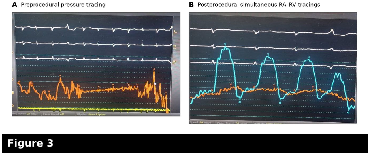
Using a steerable BZ sheath to maneuver the device it was successfully placed. The procedure was completed under sedation with a femoral block for sheath insertion. Post procedure gradient was reduced(Figure 3). She was monitored for 48 hours postoperatively and discharged on the third day. The patient was advised to continue anticoagulants, diuretics and regular bi weekly follow-up.
Discussion: Anaesthesia Challenges
This case sits at the intersection of a difficult airway, a failing right heart, decompensated congestive liver disease and a coagulopathic, thrombocytopenic patient carrying a mechanical prosthesis-a combination that constrained almost every anaesthetic decision.
Congestive Hepatopathy and Coagulopathy
A delicate balance between the hemorrhagic risk secondary to hepatic congestion and thrombotic risk associated with mechanical valve and procedural catheters was required. Anticoagulation was indicated for the patients preexisting mechanical heart valve. Given the concurrent hepatic dysfunction Child-Pugh class B, heparin bridging therapy was withheld.[3] Three units of fresh frozen plasma were transfused overnight with urine output monitoring avoiding fluid overload. INR on morning of procedure didn’t show much improvement. (Platelet count of 44 × 10⁹/L), hence 3 random donor platelets were transfused and a backup of 3 FFP 1 SDPs were kept and a strict minimum blood loss strategy was incorporated . Encephalopathy risk further argues against heavy or long-acting sedation, reinforcing the appeal of the short-acting, titratable agents chosen. Thrombocytopenia, together with liver-related coagulopathy, created a genuine bleeding diathesis that shaped both access and imaging choice. Following multidisciplinary heart team discussion, TEE was avoided because of the perceived gastrointestinal bleeding risk, and the procedure was guided by fluoroscopy supplemented by TTE.[4] Ultrasound-guided vascular access was used for femoral vein and radial artery access. Systemic heparin was withheld because of the substantial bleeding risk; the arterial catheter was maintained using the standard heparinised saline flush.
Airway
Past history of tracheostomy is a risk for tracheal or subglottic stenosis, tracheomalacia, and altered airway anatomy at the old stoma site. Tracheal USG for the same was done which showed no significant narrowing. Due to coagulopathy and past history of tracheostomy a decision for deep sedation for the procedure was made and a backup plan for general anaesthesia with endotracheal tube intubation seemed feasible. Emergency drugs like rocuronium and sugammadex were kept on standby along with video laryngoscope and fibre optic bronchoscope.
Right Heart Failure and Severe Tricuspid Pathology
TTE revealed there was minimal contractile reserve and a circulation dependent on adequate preload, sinus rhythm, and low pulmonary vascular resistance (PVR). Hemodynamic goals were adequate preload, avoid fluid overload, preserve systemic vascular resistance and coronary perfusion to the RV, avoidance of tachyarrhythmia, avoid hypercarbia, hypoxia, acidosis, and high airway pressures that raise PVR. General anaesthesia with positive-pressure ventilation would have threatened all of these: induction agents blunt preload and systemic tone, and mechanical ventilation raises intrathoracic pressure and PVR. Maintaining spontaneous ventilation under sedation preserved the negative-pressure venous return on which this RV depended.[5]
Choice of Anaesthetic Technique
Processed EEG entropy monitoring was used as an adjunct to clinical assessment to titrate the depth of sedation and avoid unintended over-sedation. This was achieved using a combination of dexmedetomidine 0.5ug/kg/hr with ketamine 1.2mg/kg/hr as infusion. Supplementary fentanyl at 1ug/kg and midazolam 0.03mg/kg was kept ready in case pacing was required and during inflation of valve to avoid patient discomfort. Dexmedetomidine provides sedation and anxiolysis with preserved respiratory drive, while ketamine contributes analgesia and sedation while maintaining systemic vascular tone and cardiac output to counterweight the sympatholytic and potential bradycardia of dexmedetomidine. The femoral nerve block for sheath access reduced the systemic sedative and opioid requirement, limiting respiratory depression and further protecting spontaneous ventilation. The potential complication of over-sedation is hypoventilation, hypercarbia (which itself raises PVR) and loss of a secured airway. The technique demanded vigilant titration, continuous capnography and a low threshold for airway support. Ketamine has potential to raise PVR and its sympathomimetic tachycardia are theoretical concerns in right heart failure. Though at sedative doses alongside dexmedetomidine the net haemodynamic profile proved favourable here.[6,7,8]
Imaging: Reliance on TTE and Omission of TEE
TEE is the conventional imaging technique for tricuspid structural interventions, guiding device positioning and detecting paravalvular leaks.[4,6] Due to aforementioned limitations described in this patient, procedural guidance relied on fluoroscopy supplemented by transthoracic echocardiography, accepting the poorer acoustic windows of TTE. The imaging strategy and the anaesthetic strategy were thus tailored for the same underlying bleeding constraint.
Vascular Access and Monitoring
Owing to severe RV dysfunction and bioprosthetic TV TAPSE and tricuspid annular S′ were considered unreliable.[9] This was confounded by the fact that spontaneous respiration makes PPV and SVV unreliable.[10] A pulmonary artery catheter was considered disproportionate to the expected benefit and carried additional arrhythmic, thrombotic and bleeding risks. Fluid administration was therefore guided by an integrated assessment of arterial pressure trends, heart rate, urine output and serial focused TTE with small, reassessed aliquots used to avoid both inadequate RV preload and worsening systemic venous congestion.
Conclusions
The central anaesthetic lesson is that in a patient with a hostile airway, a failing right heart, and a significant bleeding diathesis, a technique built around preserved spontaneous ventilation with deliberate omission of TEE in favour of TTE and fluoroscopy, compressible femoral access, and meticulous contingency planning can deliver a complex structural tricuspid intervention safely. Tricuspid valve in valve procedures are still novel and growing in India as the complexity of cases increases so should our plan of anaesthesia and perioperative management.
Declaration of patient consent
The authors certify that they have obtained written informed consent from the patient for publication of her clinical information and accompanying images. The patient understands that her name and initials will not be published and that reasonable efforts will be made to conceal her identity, although complete anonymity cannot be guaranteed.
Financial support and sponsorship
Nil.
Acknowledgments
The authors acknowledge the interventional cardiology team and nursing staff for their contribution to the patient’s care and thank the patient for her cooperation.
Conflicts of interest
There are no conflicts of interest.
Previous presentation
This manuscript has not been presented at any meeting or conference.
Clinical-trial registration
Not applicable (case report).
References
- Montenegro da Costa, M.J.; Quintella, E.F.; Kohn, L.; Lacoste, M.O.; Leite, G.L.; Hadid, L.; et al. Transcatheter tricuspid valve-in-valve procedure—an illustrative case report and review. J. Clin. Med. 2021, 10(17), 4004. [Google Scholar] [CrossRef] [PubMed]
- Du Preez, L.J.; Le Roux, P.; Coetzee, A.R. Anaesthesia for transvenous transcatheter tricuspid valve-in-valve implantation. S Afr. J. Anaesth. Analg. 2011, 17(4), 293–298. [Google Scholar] [CrossRef]
- Douketis, J.D.; Spyropoulos, A.C.; Murad, M.H.; Arcelus, J.I.; Dager, W.E.; Dunn, A.S.; et al. Perioperative management of antithrombotic therapy: an American College of Chest Physicians clinical practice guideline. Chest 2022, 162(5), e207–e243. [Google Scholar] [CrossRef] [PubMed]
- Hahn, R.T.; Abraham, T.; Adams, M.S.; Bruce, C.J.; Glas, K.E.; Lang, R.M.; et al. Guidelines for performing a comprehensive transesophageal echocardiographic examination: recommendations from the American Society of Echocardiography and the Society of Cardiovascular Anesthesiologists. J. Am. Soc. Echocardiogr. 2013, 26(9), 921–964. [Google Scholar] [CrossRef] [PubMed]
- Harjola, V.P.; Mebazaa, A.; Celutkiene, J.; Bettex, D.; Bueno, H.; Chioncel, O.; et al. Contemporary management of acute right ventricular failure: a statement from the Heart Failure Association and the Working Group on Pulmonary Circulation and Right Ventricular Function of the European Society of Cardiology. Eur. J. Heart Fail. 2016, 18(3), 226–241. [Google Scholar] [CrossRef] [PubMed]
- Zoller, J.K.; Gregory, S.H. Anesthetic considerations for transcatheter tricuspid valve repair. J. Cardiothorac. Vasc. Anesth. 2020, 34(7), 1942–1951. [Google Scholar] [CrossRef] [PubMed]
- Haurand, J.M.; Kavsur, R.; Ochs, L.; Tanaka, T.; Iliadis, C.; Sugiura, A.; et al. Deep sedation vs. general anesthesia for transcatheter tricuspid valve repair. Front Cardiovasc Med. 2022, 9, 976822. [Google Scholar] [CrossRef] [PubMed]
- Rajagopal, D.R.; Stewart, J.P.; Thourani, V.H.; Yadav, P.K. Transcatheter tricuspid valve replacement using conscious sedation and intracardiac echocardiography. JACC Case Rep. 2025, 30(41), 105987. [Google Scholar] [CrossRef] [PubMed]
- Rudski, L.G.; Lai, W.W.; Afilalo, J.; Hua, L.; Handschumacher, M.D.; Chandrasekaran, K.; et al. Guidelines for the echocardiographic assessment of the right heart in adults: a report from the American Society of Echocardiography. J. Am. Soc. Echocardiogr. 2010, 23(7), 685–713. [Google Scholar] [CrossRef] [PubMed]
- Monnet, X.; Marik, P.E.; Teboul, J.L. Prediction of fluid responsiveness: an update. Ann. Intensive Care 2016, 6(1), 111. [Google Scholar] [CrossRef] [PubMed]
Figure 1.
Preprocedural frontal chest radiograph showing marked cardiomegaly with a prominent right cardiac contour, corresponding to severe right atrial dilatation on echocardiography. Postsurgical changes of previous median sternotomy and prosthetic valve replacement are visible.
Figure 1.
Preprocedural frontal chest radiograph showing marked cardiomegaly with a prominent right cardiac contour, corresponding to severe right atrial dilatation on echocardiography. Postsurgical changes of previous median sternotomy and prosthetic valve replacement are visible.
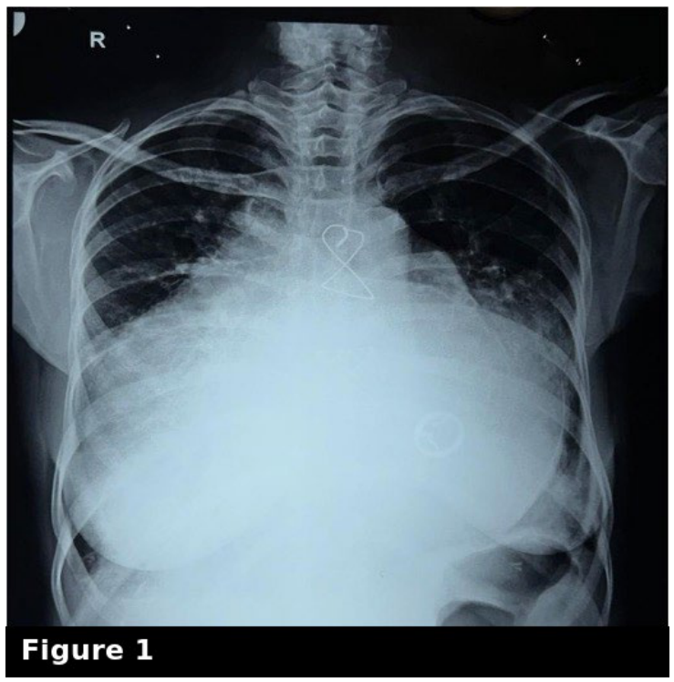
Figure 2.
Transthoracic echocardiographic findings before and after transcatheter tricuspid valve-in-valve replacement. (A) Preprocedural apical four-chamber view showing gross right atrial dilatation and a small right ventricle. (B) Preprocedural continuous-wave Doppler across the tricuspid bioprosthesis showing a maximum gradient of 18 mmHg and mean gradient of 8 mmHg. (C) Postprocedural continuous-wave Doppler showing reduction of the maximum and mean gradients to 8 and 4 mmHg, respectively.
Figure 2.
Transthoracic echocardiographic findings before and after transcatheter tricuspid valve-in-valve replacement. (A) Preprocedural apical four-chamber view showing gross right atrial dilatation and a small right ventricle. (B) Preprocedural continuous-wave Doppler across the tricuspid bioprosthesis showing a maximum gradient of 18 mmHg and mean gradient of 8 mmHg. (C) Postprocedural continuous-wave Doppler showing reduction of the maximum and mean gradients to 8 and 4 mmHg, respectively.
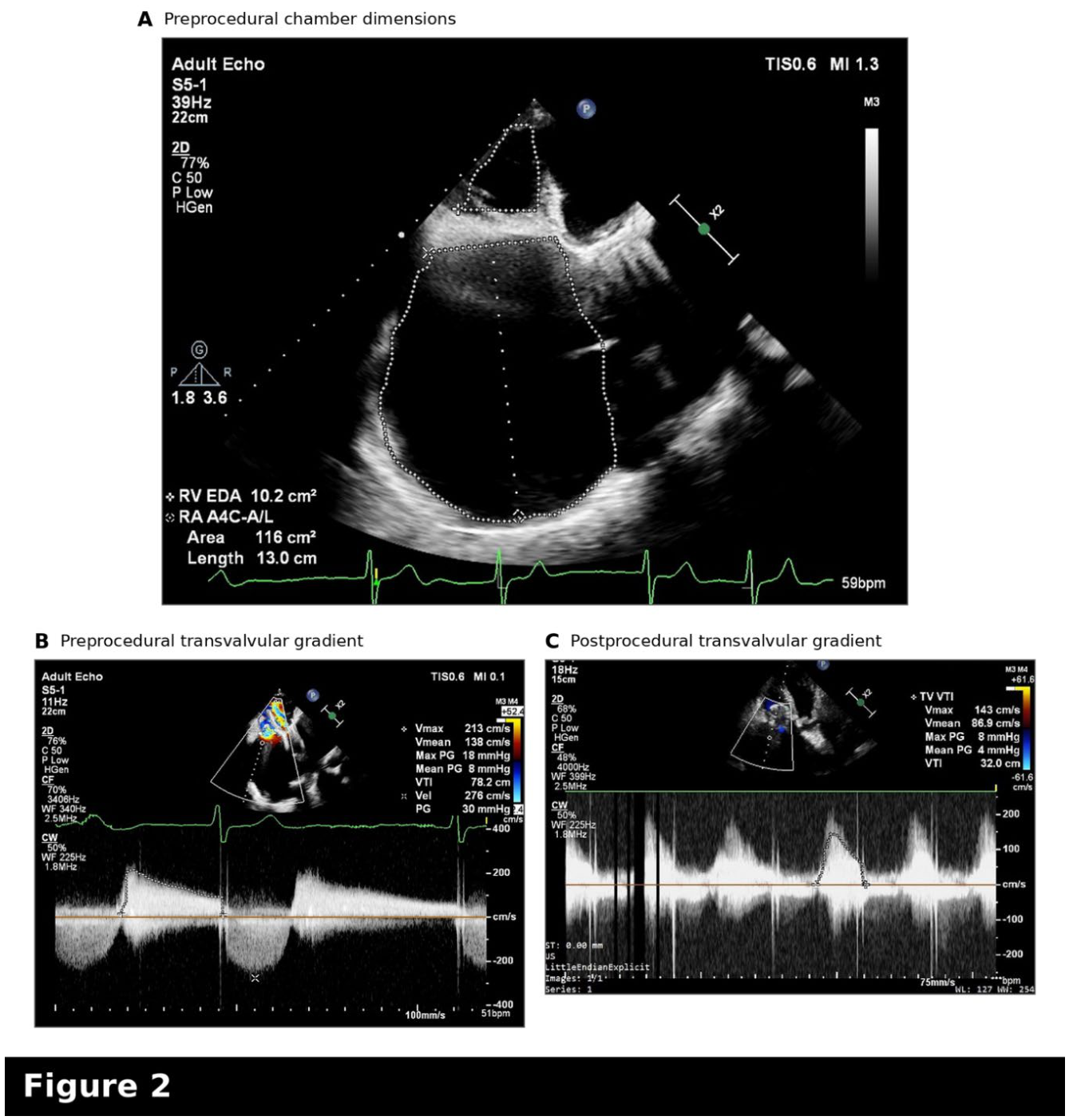
Figure 3.
Invasive haemodynamic pressure tracings. (A) Preprocedural right atrial pressure tracing. (B) Postprocedural simultaneous right atrial and right ventricular pressure tracings following transcatheter tricuspid valve-in-valve implantation.
Figure 3.
Invasive haemodynamic pressure tracings. (A) Preprocedural right atrial pressure tracing. (B) Postprocedural simultaneous right atrial and right ventricular pressure tracings following transcatheter tricuspid valve-in-valve implantation.
Disclaimer/Publisher’s Note: The statements, opinions and data contained in all publications are solely those of the individual author(s) and contributor(s) and not of MDPI and/or the editor(s). MDPI and/or the editor(s) disclaim responsibility for any injury to people or property resulting from any ideas, methods, instructions or products referred to in the content. |
© 2026 by the authors. Licensee MDPI, Basel, Switzerland. This article is an open access article distributed under the terms and conditions of the Creative Commons Attribution (CC BY) license (http://creativecommons.org/licenses/by/4.0/).
Copyright: This open access article is published under a Creative Commons CC BY 4.0 license, which permit the free download, distribution, and reuse, provided that the author and preprint are cited in any reuse.