
Submitted:
14 September 2026
Posted:
16 September 2026
You are already at the latest version
Abstract
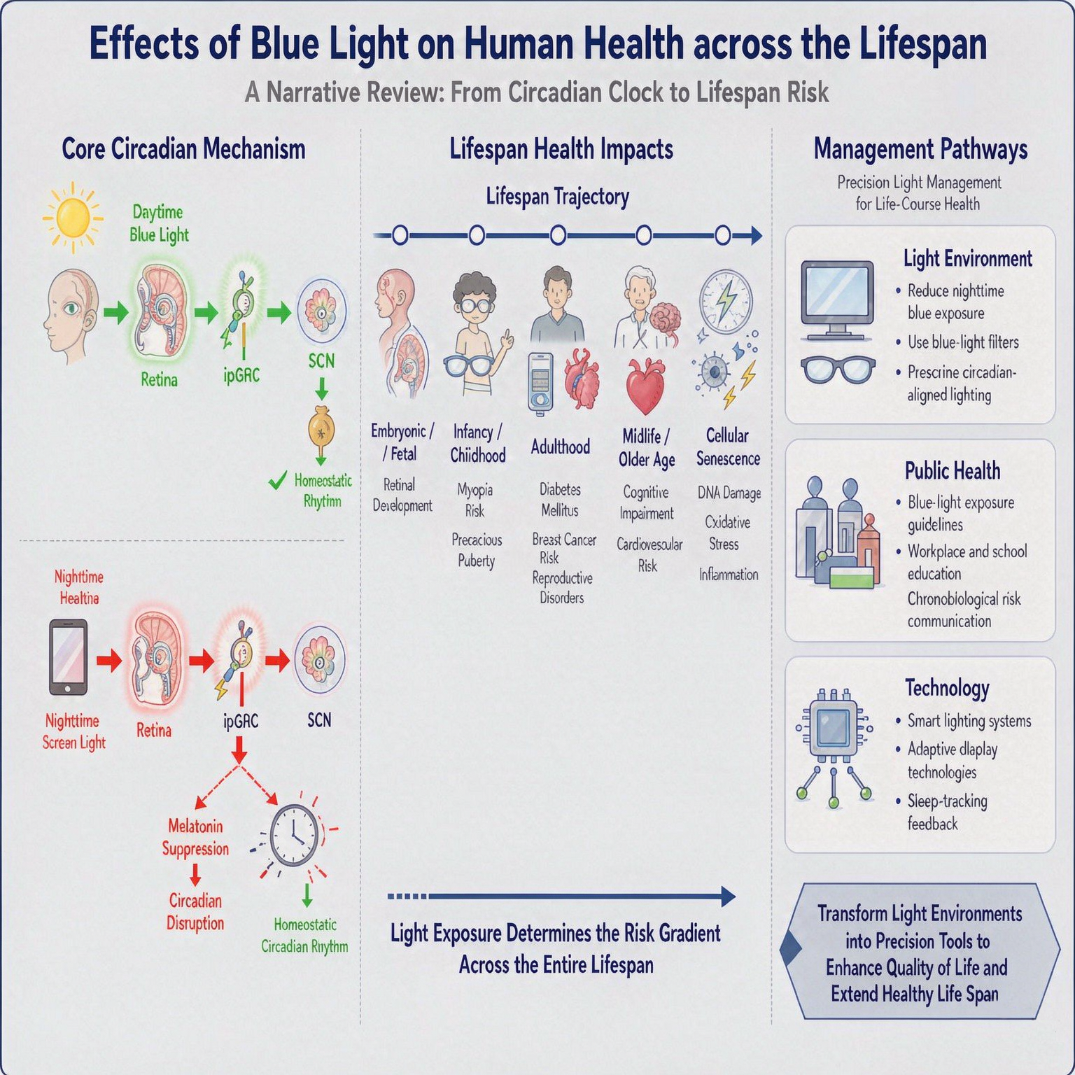
Natural light has shaped the evolution of human visual perception and circadian biology. However, the rapid proliferation of LEDs (Light Emitting Diode) and electronic screens has coincided with escalating global burdens of myopia, sleep disorders, and endocrine pathologies, underscoring a growing public health concern. This narrative review synthesizes current evidence on how blue light differentially modulates visual and non-visual (circadian) pathways, thereby exerting significant effects across the entire lifespan—from embryonic development to cellular senescence. Central to this review is the concept of diurnal biphasic effects: while daytime exposure is essential for maintaining homeostatic rhythmicity, nocturnal exposure precipitates a profound evolutionary mismatch. This disruption may destabilize the central clock and systemic physiology, thereby conferring multiple health risks. This review proposes that the path forward entails transforming artificial lights into precision tools for enhancing life quality and extending human health span.
Keywords:
blue light hazard
; myopia
; circadian rhythms
; photobiological effects
1. Introduction
Throughout human evolution, natural light has acted as an information and energy carrier, driving the formation of visual and non-visual photoreceptive mechanisms. These mechanisms not only govern visual perception, but also internalize the flux of natural light into fundamental biological timing, regulate sleep-wake cycles, and participate deeply throughout the lifespan. Meanwhile, human have also evolved a sophisticated physiological system precisely matched to the natural light, such as the emmetropic alignment of the ocular refractive apparatus to the dominant spectral band of natural light, selective filtration of different wavelengths lights by the ocular media, the dynamic modulation of irradiance by the pupil according to ambient brightness, and the biphasic response of the central nervous system to light adapted to diurnal variations. However, this ‘light ~ life’ synergistic system is facing structural challenges. Since the 1990s, the novel artificial light sources such as LEDs and electronic screens have become the predominant artificial light sources, particularly constituting the majority of Artificial Light at Night (ALAN). The proliferation of the novel light sources coincides spatiotemporally with rising global incidences of adolescent myopia, retinal degenerative diseases, sleep disorders, depressive moods, and certain hormone-dependent tumors. Given that the spectral composition of these light sources contains a higher proportion of blue light—which readily penetrates the ocular media, it is important to clarify the physiological mechanisms. This study therefore adopts an interdisciplinary optical–biological approach to clarify how blue light shapes visual and non-visual effects across immediate and sustained timescales. It is posited that amidst the rapid iteration of novel artificial lighting technology, priority should be given to the biological effects of light, thereby providing a scientific basis for improving public health.
2. Mechanisms of Action of Blue Light
Within the visible spectrum (380 nm–780 nm), blue light occupies the 400 nm–500 nm range and is subdivided into short-wavelength blue light (400 nm–450 nm) and long-wavelength (450 nm–500 nm) blue light.
2.1. Visual Mechanisms
2.1.1. Longitudinal Chromatic Aberration (LCA)
According to Cauchy’s dispersion formula,
the refractive index n of a medium is inversely proportional to the square of the wavelength λ. Consequently, light of different wavelengths exhibits varying refractive index within the ocular media, resulting in distinct focal positions on the retinal plane—a phenomenon known as Longitudinal Chromatic Aberration (LCA). Within the visible spectrum, blue light has a shorter wavelength and thus a higher refractive index than longer-wavelength light. This results in stronger refraction in the ocular media, shifting its focal point anterior to the retina and generating myopic defocus. Green and red light, having longer wavelengths and lower refractive indices, are refracted less; the focal plane of green light falls close to the retina, yielding emmetropic focus, whereas red light focuses posterior to the retina, inducing hyperopic defocus [1]. The human visual system has evolved under spectrally balanced natural lights: after refraction by the ocular media, the dominant spectral band of sunlight converges near the retinal plane, establishing emmetropic focus. In contrast, novel artificial light sources exhibit a higher proportion of blue light and a lower proportion of red light. This spectral imbalance amplifies the LCA effect, shifting the overall focal plane of the retina anteriorly and inducing myopic defocus [2].
2.1.2. Retinal Blue Light Hazards
Retinal Blue Light Hazards (BLH) is primarily concentrated in the short-wavelength blue light wave band [3]. Although the energy of a single blue-light photon (e.g., 2.85 eV for 435 nm wavelength photon) is insufficient to break DNA strands or protein peptide bonds (bond energy > 3.1 eV) directly, excessive exposure has been associated with retinopathy via “direct photo-oxidation” and “indirect photo-oxidation” [4,5].
- Direct damage mediated by non-photosensitizers mainly targets photoreceptor cells. Blue light is absorbed by endogenous chromophores within the visual cycle (e.g., all-trans-retinal) and components of the mitochondrial respiratory chain (e.g., cytochrome c), then excites electrons to higher energy states and precipitously generates Reactive Oxygen Species (ROS). When ROS levels exceed the scavenging threshold of macular xanthophylls (lutein and zeaxanthin), it leads to chronic inflammation and mitochondrial-dependent apoptosis, driving irreversible macular degeneration.
- Indirect damage mediated by photosensitizers predominantly disrupts Retinal Pigment Epithelium (RPE). Blue light excites lipofuscin (A2E) within the RPE. During the energy transfer to molecular oxygen, ROS are produced, triggering lipid peroxidation and protein damage; this process is considered a significant pathological basis for Age-related Macular Degeneration (AMD) [6,7,8].
It is noteworthy that long-term, low-intensity blue light may remain pathogenic through chronic photochemical stress: although ROS induced by low-intensity blue light can be compensated by the endogenous antioxidant system, repeated exposures lead to the sustained accumulation of oxidative damage markers (e.g., 8-OHdG) [9,10]. This results in the persistent over activation of core antioxidant response pathways (e.g., Nrf2/ARE pathway), leading to “antioxidant fatigue” and the exhaustion of defense mechanisms, potentially contributing to the degeneration of retinal structure and function at the subcellular level.
2.2. Non-Visual Mechanisms
Cortisol and melatonin are key hormones that regulate the human circadian rhythm. Cortisol acts as an “alertness” hormone, peaking in the early morning to promote an active state; melatonin, the “sleep” hormone, peaks during the late night. The intensity of blue light in natural light varies regularly with the diurnal cycle, modulating the mirror-image release of cortisol and melatonin and thus maintaining the steady-state pattern of daytime alertness and nighttime sleep—establishing itself as a key environmental factor driving circadian rhythms [11]. Blue light influences circadian phase through both indirect pathways mediated by melatonin and direct pathways mediated by intrinsically photosensitive retinal ganglion cells (ipRGCs):
- Melatonin-Mediated Indirect Pathway
Blue light exerts rhythmic regulation via the “retina-suprachiasmatic nucleus (SCN)-pineal gland” axis. Upon activation by blue light, ipRGCs transmit signals to the SCN via the retinohypothalamic tract (RHT) [12], thereby inhibiting melatonin synthesis in the pineal gland. The secretion profile of melatonin serves as a biochemical marker for the endogenous dark phase. Furthermore, the rhythm of melatonin regulates the sleep-wake cycle and interacts with the hypothalamic-pituitary-adrenal (HPA) axis, synchronizing with cortisol secretion to maintain circadian homeostasis [13]. Blue light exposure at night suppresses melatonin and disrupts cortisol rhythmicity, constituting a common basis for sleep and stress system dysregulation [14]. Conversely, melatonin exerts age-dependent inhibitory effects through receptors located at various levels of the hypothalamic-pituitary-gonadal (HPG) axis [15,16].
- ipRGC-Mediated Direct Pathway
Blue light also utilizes the ipRGC-mediated direct pathway to achieve neuroendocrine regulation that rapidly responds to light changes in the environment, providing a physiological basis for the instantaneous transmission of photic information to the reproductive center. Animal experiments have confirmed that even in models with pinealectomy or melatonin receptor knockout, blue light can activate the HPG axis [17,18,19]. This indicates the pathway does not rely on the pineal-melatonin axis; instead, it directly activates kisspeptin and Gonadotropin-Releasing Hormone (GnRH) neurons in deep hypothalamic nuclei via the retinohypothalamic tract, thereby inducing the rapid release of GnRH.
Novel artificial light sources introduce blue light that causes the diurnal rhythm synchronizer to appear ectopically at night. This induces a structural mismatch between the current human physiological state and the photic environment, potentially triggering neuroendocrine dysregulation. Consequently, this serves as a potential environmental pathological basis for abnormal pubertal onset, menstrual disorders, declining fertility, and even an increased risk of hormone-dependent tumors [12,20,21].
3. Health Effects of Blue Light
Beyond its visual role, blue light acts as a neuroendocrine signal via non-visual pathways, exerting systemic, life-course modulation across critical physiological domains—including visual health, development, reproduction, metabolism, and aging.
3.1. Induction and Exacerbation of Ocular Diseases
3.1.1. A Precipitant of Adolescent Myopia
Since the 1990s, the sharp rise in adolescent myopia prevalence in East Asia coincides spatiotemporally with the wide adoption of novel artificial light sources. This convergence suggests that a change in ambient light characteristics may serve as a significant environmental driver of the myopia epidemic. Evidence indicates that blue light disrupts ocular homeostasis through a dual mechanism: physical myopic defocus induced by LCA and physiological circadian disruption mediated by ipRGCs, thereby exerting a double impact on ocular health.
- Physical Defocus
Through evolutionary adaptation, the human visual system has acquired a refractive mechanism aligned with the spectral characteristics of natural light, enabling the focal plane of incident light to coincide with the retina and thereby establish emmetropic vision. However, the discrete spectra and high blue-light composition of novel artificial light sources disrupt this homeostasis: LCA generates myopic defocus stimuli that may drive compensatory axial elongation via biofeedback mechanisms, thereby disturbing refractive equilibrium. Although the human eye compensates for LCA to some extent, pupil dilation under low illuminance increases the proportion of marginal rays, thereby intensifying the LCA effect. Using Gullstrand simplified eye model(assuming no accommodation or aberrations under resting conditions) grounded in classical LCA data (450 nm vs. 680 nm focal difference ≈ 0.45–0.55 mm; ≈ 1.5–2.0 D) [22,23], it was calculated that the transition from photopic (> 300 lx, ≈ 3.0 mm pupil) to scotopic (< 50 lx, ≈ 8.0 mm pupil) conditions amplifies the effective focal separation between 450 nm and 680 nm light from ≈ 0.15 mm to ≈ 0.40 mm. This shift elevates the equivalent refractive error from ≈ 0.4 D to ≈ 1.0 D—a 150% increase. These findings suggest that pupil dilation under low-light conditions significantly exacerbates myopic defocus induced by LCA, representing one of the potential mechanisms underlying the onset and progression of myopia.
- Neuromodulation
The non-visual pathway mediated by ipRGCs also regulates refractive development and contributes to the onset of myopia. Animal studies confirm that selective ablation or chemogenetic activation of ipRGCs induces hyperopic or myopic shifts, respectively. Melanopsin signals from ipRGCs primarily modulate axial length, whereas conventional photoreceptor signals mainly modulate corneal curvature, both contributing to ocular development. In form-deprived eyes, ipRGCs show upregulated melanopsin expression and enhanced photoresponses, implicating these cells as a key hub that converts light cues into ocular growth signals [24]. Under circadian regulation, aberrant nighttime blue light hyperactivates ipRGCs, projecting via the RHT to the SCN, first destabilizing the central circadian clock. This may consequently desynchronizes local retinal clock genes (e.g., Cry1, Per3) and disrupts the mirror-image release rhythm of retinal dopamine and pineal melatonin. The consequent decline in dopaminergic inhibition of scleral expansion, coupled with downstream cAMP/PKA signaling, drives pathological scleral remodeling and excessive axial elongation [25].
3.1.2. An Aggravator of Age-Related Retinal Degeneration
While a single blue-light photon is insufficient to directly cleave biomolecular structures, chronic cumulative exposure from adolescence to adulthood may precipitate the accelerated degeneration of the senescent retina, thereby elevating the risk of AMD. Epidemiological evidence indicates a positive correlation between light exposure accumulated during childhood/youth and the early pathological hallmarks of AMD in later life, suggesting a substantial latent effect [26]. Compared to adults, children exhibit greater ocular transparency to blue light and larger pupil diameters under equivalent lighting conditions, resulting in higher retinal irradiance. Concurrently, the immature macular pigment optical density (MPOD) in children confers weaker antioxidant protection, rendering this population critically sensitive to BLH. While MPOD peaks in adulthood, sustained blue-light exposure continues to compound retinal damage. With advancing age, the physiological decline—characterized by a reduction in MPOD from adult levels (0.5–0.8) to senile levels (0.3–0.5), diminished antioxidant enzyme activity, and impaired autophagy—synergizes with A2E accumulation and inflammatory pathway activation. This multifaceted deterioration enhances phototoxicity, potentially accelerating AMD onset and facilitating neovascular conversion [27].
3.2. Effects of Blue Light on Neuroendocrine Regulation
3.2.1. An Initiator of Fetal and Infant Development and Rhythmicity
The chronobiological system originates during embryogenesis. Beyond genetic determinants, maternal endocrine rhythms—specifically melatonin and cortisol—are transmitted to the fetus via the materno-placental axis. These hormones entrain the fetal SCN, establishing a nascent circadian framework that underpins postnatal autonomous rhythmicity [28,29]. However, chronic maternal rhythm disturbances can relay aberrant endocrine signals to the fetus, disrupting the HPG/HPA axes programming . This malprogramming elevates the risk of neonatal circadian misalignment and predisposes the offspring to aberrant pubertal timing [30,31].
Infancy constitutes a critical window for the ontogeny of endogenous rhythmicity. Ambient blue light acts as a primary zeitgeber, entraining the secretory rhythms of melatonin and cortisol to systematically establish the foundation for sleep-wake cycles, cognitive maturation, and growth regulation. Clinical evidence confirms that light-dark cycle interventions mimicking natural photoperiods effectively promote neurodevelopmental outcomes in preterm infants [32,33]. Given that elevated nocturnal melatonin is indispensable for orchestrating Growth Hormone (GH) secretion [34], nocturnal blue light exposure may pose dual challenges: compromising the architectural integrity of the circadian system while simultaneously disrupting essential somatotropic signaling.
3.2.2. A Modulator of Pubertal Timing, Metabolism and Emotional Homeostasis
Adolescence constitutes a critical window for metabolic and endocrine maturation. During this stage, blue light modulates pubertal timing and progression, while concurrently orchestrating metabolic balance and emotional homeostasis.
- Modulation of Puberty timing
Blue light exerts profound regulatory effects on pubertal timing via melatonin secretion, functioning dually as both a “brake” and an “accelerator.” In childhood, high-amplitude nocturnal melatonin binds to MT1 receptors on hypothalamic GnRH neurons, providing a critical neuroendocrine brake on the HPG axis [35,36]. The physiological decline of this inhibition—termed HPG axis disinhibition— a prerequisite for pubertal onset [37]. Following puberty, the circadian rhythm of melatonin secretion stabilizes with reduced amplitude compared to childhood, and a further decline in adulthood reflects the age-specific characteristics of endocrine homeostasis [38]. Notably, human sexual maturation exhibits remarkable sensitivity to photic flux; epidemiological data confirm that the age of menarche correlates inversely with total annual sunlight exposure across latitudes [39,40]. By perpetuating a “long-day” signal that suppresses melatonin, nocturnal blue light can desynchronize the temporal patterning of GnRH pulse generation, triggering precocious puberty in susceptible individuals [41]. Consequently, alongside nutrition, ALAN is increasingly implicated as a key environmental driver of the global trend of earlier puberty [41,42,43].
- Modulation of Metabolic Homeostasis
Emerging evidence demonstrates that alterations in the light-dark cycle disrupt central rhythmicity and impair peripheral metabolism independently of caloric intake [44]. Large-scale epidemiological studies corroborate a dose-dependent association between ALAN exposure and obesity risk [45], a phenomenon mechanistically linked to the fact that genetic disruption of clock genes directly precipitates insulin resistance and adiposity [46]. In adolescents, nocturnal blue light constitutes a critical environmental driver of obesity by inducing central-peripheral circadian misalignment. Specifically, light exposure acts on the hypothalamic arcuate nucleus to disrupt the functional equilibrium between Neuropeptide Y/Agouti-related peptide and Pro-opiomelanocortin neurons, thereby enhancing orexigenic drive while blunting anorexigenic signaling. Concurrently, suppression of melatonin signaling via MT1/MT2 receptors disrupts the rhythmic expression of core clock genes (e.g., Bmal1, Per) in peripheral metabolic organs such as the liver, precipitating dysregulated lipogenesis and insulin resistance, which may collectively contribute to the pathogenesis of adolescent obesity.
- Bidirectional Modulation of Emotional States
The psychotropic effects of blue light show a distinct diurnal dichotomy: daytime exposure potentiates arousal via the ipRGCs–SCN–HPA axis, whereas nocturnal exposure increases vulnerability to depression by suppressing melatonin and disrupting HPG axis homeostasis [47,48]. In East Asian adolescents, this relationship carries significant clinical weight, as exposure to artificial light sources correlates with increased severity of depressive symptoms (β=0.208, p<0.01) and suicidal ideation (β=0.125, p<0.05) [49]. Mechanistically, blue light perturbs the HPG axis, exacerbating emotional dysregulation through a pathophysiological cascade rooted in steroid hormone dysrhythmia [16]. Beyond the neuroendocrine circuitry, ALAN-induced chronodisruption likely engages the gut–brain axis, precipitating depression by compromising intestinal barrier integrity, remodeling tryptophan-metabolizing microbiota, and consequently suppressing serotonergic biosynthesis [50].
3.2.3. A Risk Factor for Adult Reproductive Dysfunction, Diabetes, and Hormone-Dependent Malignancies
Extending beyond transient circadian disruption, nocturnal blue light acts as a systemic stressor that compromises adult physiological homeostasis. It may pose a significant threat not only to reproductive function and glucose metabolic balance but also potentially elevates the risk of hormone-dependent tumorigenesis.
- Impact on Reproductive Function
The pathophysiology of reproductive compromise induced by nocturnal blue light is mechanistically anchored in melatoninergic disruption. Activation of MT1/MT2 receptors alters the pulsatility of hypothalamic GnRH neurons, thereby desynchronizing the downstream secretory rhythm of gonadotropins [51,52]. In females, this chronodisruption precipitates menstrual dysregulation and anovulation, while simultaneously compromising folliculogenesis and oocyte competence via dysregulation of ovarian clock genes [53]. Concurrently, in males, rhythm disturbances blunt the diurnal testosterone profile and disrupt clock gene expression in Sertoli and Leydig cells, potentially culminating in impaired spermatozoa motility, morphometry, and genomic integrity [54]. These mechanistic insights are substantiated by epidemiological data confirming a markedly elevated risk of subfertility among individuals subjected to chronic shift work or excessive nocturnal screen exposure [55,56].
- Instigation of Type 2 Diabetes Mellitus (T2DM)
Large-scale cross-sectional analyses (n=98,658) reveal a robust dose-response relationship between ALAN intensity and diabetes prevalence; compared to the lowest exposure quintile, the highest quintile exhibits a 28% elevation in diabetes risk, independent of conventional confounders such as BMI, diet, and physical activity [57]. Beyond mere correlation, blue light-induced chronodisruption serves as a critical environmental instigator of T2DM. Circadian misalignment may promotes sympathetic overactivity and blunted cortisol rhythmicity, which directly suppresses insulin biosynthesis and secretion in pancreatic β-cells while inducing peripheral insulin resistance. This cascade is exacerbated by glucolipotoxicity, accelerating β-cell failure, and synergizes with sympathetic overdrive to drive central obesity and metabolic syndrome, thereby potentially establishing a self-perpetuating vicious cycle that precipitates T2DM and its microvascular complications [58,59].
- Association with Hormone-Dependent Malignancies
The rising global incidence of breast, prostate, and thyroid cancers may be etiologically linked to nocturnal blue light exposure. Beyond the proliferative drive conferred by metabolic dysfunction—specifically hyperinsulinemia and elevated Insulin-like Growth Factor-1 (IGF-1)—emerging evidence suggests that nocturnal light suppresses melatonin while potentially modulating the kisspeptin/GnRH neuronal activity. This desynchronizes the HPG axis, alters endogenous sex steroid profiles, and elevates the risk of hormone-dependent carcinogenesis [21,60,61], substantiated by epidemiological data: shift work is associated with a significantly increased risk of breast cancer [60], while global meta-analyses report pooled relative risks of 1.12 for breast cancer and 1.43 for prostate cancer under chronic ALAN exposure, exhibiting clear dose-response relationships [62,63,64]. High-resolution geospatial studies further confirmed a positive correlation between the ALAN intensity and the incidence of these malignancies, with the association being the most pronounced in regions dominated by high-blue-light spectra, aligning with Zhang et al.’s study proposed spectral-specific dose-response relationship [65,66]. However, the strength of evidence varies by site: while the links for breast and prostate cancers are robust, the association with thyroid carcinoma—which involves estrogen signaling and the hypothalamic-pituitary-thyroid (HPT) axis—is increasingly suggested by the recent study [66], whereas the role of blue light in ovarian carcinogenesis remains inconclusive due to dominant genetic and reproductive determinants [67].
3.2.4. An Accelerator of Multiorgan Degenerative Pathologies in Aging
Blue light may constitute a fundamental environmental risk factor that propels the trajectory of senescence, serving as a catalyst for the onset and progression of multiorgan degenerative pathologies.
3.2.4.1. An Accelerator of Neurodegenerative Pathologies
In model organisms such as Drosophila melanogaster, chronic exposure to high-intensity blue light has been shown to impair mitochondrial bioenergetics and neurotransmitter homeostasis, potentially contributing to reduced lifespan [68,69,70]. Importantly, these peripheral mitochondrial deficits are compounded by a parallel decline in central circadian circuitry, the phenomenon mirrored in human aging. This is compounded by senescent decompensation of the “light-rhythm axis,” where diminished ipRGC sensitivity, attenuated SCN amplitude, and blunted melatonin output flatten circadian waveforms [71,72,73], rendering the aging brain vulnerable to photic disruption. Mechanistically, light-induced sleep fragmentation and slow-wave sleep deficit impair glymphatic clearance, reducing cerebrospinal fluid exchange. This may precipitate pathological accumulation of Amyloid-β and Tau, triggering neuroinflammation and synaptic toxicity [73,74]. Critically, degeneration of hypothalamic and brainstem hubs establishes a self-amplifying loop: circadian disruption→sleep loss→glymphatic failure→proteotoxic aggregation→neuronal injury, elevating the risk of Alzheimer’s disease [73,74,75].
3.2.4.2. A Catalyst for Cardiovascular and Cerebrovascular Degeneration
Nocturnal blue light may disrupt cardiovascular homeostasis via a “melatonin suppression–sympathetic overdrive–metabolic dysregulation” cascade, undermining vasoprotection while perpetuating sympathetic-adrenal activation to induce non-dipping blood pressure and attenuated heart rate variability (HRV), thereby fueling atherogenesis. This mechanistic insight is substantiated by population-level data revealing a steep dose-response relationship: relative to the lowest exposure quintile, the highest quintile faces a 32% higher risk of coronary heart disease, a 47% surge in myocardial infarction, and a 28% increase in stroke risk [76,77] . Experimentally, chronic light overexposure induces hypertensive remodeling—encompassing left ventricular hypertrophy and aortic fibrosis—while translational human data corroborate that reduced urinary 6-sulfatoxymelatonin is independently associated with incident hypertension, suggesting melatonin deficiency as a potential contributor of this pathological trajectory [78,79].
4. Public Health Implications and Future Directions
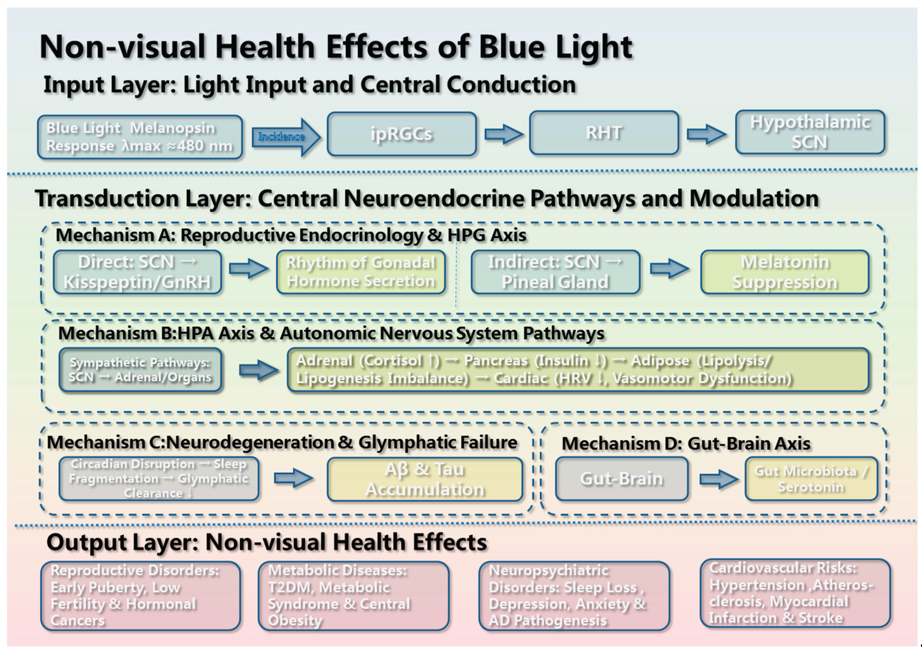
The visual and non-visual action mechanisms & health effects of blue light are illustrated in Figure 1 and Figure 2.
4.1. Public Health Implications
To mitigate the health risks imposed by blue light, an integrated framework centered on “Light as a Health Determinant” is proposed, bridging public awareness, clinical practice, and policy governance:
- Public Awareness and Behavioral Intervention
The principle of “Circadian-Appropriate Lighting” should be widely adopted, emphasizing spectral specificity and diurnal differentiation. This necessitates promoting adequate daytime light exposure while strictly curtailing nocturnal screen time and ambient illumination. Priority should be given to optimizing light environments across educational, occupational, and domestic settings.
- Clinical Integration and Interdisciplinary Management
- Clinical protocols shall evolve to incorporate “light hygiene” into routine healthcare delivery. It is essential to develop personalized lighting prescriptions tailored to specific populations. Furthermore, fostering interdisciplinary collaboration among ophthalmology, endocrinology, and psychiatry is critical to establishing early screening and comprehensive intervention strategies for light-induced pathologies.
- Policy Formulation and Governance
Institutionalizing light health effects within public health policy is imperative. Enacting health-oriented lighting standards ensures that scientifically optimized lighting becomes a universal public good. Strategically, it aims to reduce the long-term societal burden of myopia, sleep disorders, and endocrine-related diseases, thereby lowering overall public healthcare expenditure.
4.2. Future Directions
To harness light as a non-pharmacological determinant of health, the field must pivot toward three strategic imperatives:
- Deciphering Molecular Circuitry.
A primary challenge is to identify the precise molecular substrates linking light spectra to neuroendocrine and circadian regulation. Quantifying the coupling between light exposure and long-term health trajectories is required to establish the theoretical basis for precision phototherapy.
- Constructing Predictive Frameworks.
Overcoming the data bottleneck requires integrating lifespan physiological responses into quantitative models. Using large-scale longitudinal cohorts, research will map the dynamic “Spectral Property–Physiological Effect–Critical Window” nexus to enable predictive interventions.
- Engineering Adaptive Ecosystems.
The current paradigm of static and uniform lighting should shift to AI-driven, chronotype-specific ecosystems. This technological leap aims to deliver dynamic precision lighting that proactively aligns with individual biology, representing a translational strategy to extend human healthspan.
5. Conclusions
Blue light exerts diurnal biphasic effects on human health. During the day, it serves as an essential zeitgeber that maintains homeostatic rhythmicity. However, the proliferation of novel artificial light sources has created a profound evolutionary mismatch, amplifying the LCA effect to induce myopia while disrupting SCN-mediated circadian homeostasis and downstream neuroendocrine axes. This may present a systemic challenge spanning from visual impairment to multiorgan degeneration across the lifespan. Consequently, public health paradigms must evolve beyond the conventional framework of “vision-centered prevention” toward comprehensive “life-course light health management.” This calls for interdisciplinary standards for dynamic lighting environments grounded in human physiology—a fundamental shift from passively mitigating photic interference to actively optimizing light as a non-pharmacological determinant of health.
Author Contributions
Conceptualization, writing—original draft preparation, L.L.; writing—review and editing, HJ.W.; supervision, HJ.W. All authors have read and agreed to the published version of the manuscript.
Funding
This research received no external funding.
Institutional Review Board Statement
Not applicable.
Informed Consent Statement
Not applicable.
Data Availability Statement
No new data were created or analyzed in this study. Data sharing is not applicable to this review.
Acknowledgments
During the preparation of this manuscript, the authors used AI tools for language polishing, reference organization, and screening assistance. No AI tool was used for study design, data analysis, or figure generation. The authors have reviewed and edited all AI-assisted output and take full responsibility for the content.
Conflicts of Interest
The authors declare no conflict of interest.
References
- Atchison, D.A.; Smith, G. Chromatic dispersions of the ocular media of human eyes. J. Opt. Soc. Am. A 2005, 22, 29–37. [Google Scholar] [CrossRef]
- Swiatczak, B.; Schaeffel, F. Myopia: Why the retina stops inhibiting eye growth. Sci. Rep. 2022, 12, 21704. [Google Scholar] [CrossRef]
- International Electrotechnical Commission. Photobiological Safety of Lamps and Lamp Systems—Part 7: Light Sources and Luminaires Primarily Emitting Visible Radiation; IEC: Geneva, Switzerland, 2023. [Google Scholar]
- Behar-Cohen, F.; Martinsons, C.; Vienot, F.; Zissis, G.; Barlier-Salsi, A.; Cesarini, J.P.; Enouf, O.; Garcia, M.; Picaud, S.; Attia, D. Light-emitting diodes (LED) for domestic lighting: Any risks for the eye? Retin. Eye Res. 2011, 30, 239–257. [Google Scholar] [CrossRef]
- Françon, A.; Delaunay, K.; Jaworski, T.; Girard, P.; Cohen-Tannoudji, D.; Simonutti, M.; Picaud, S. Phototoxicity of low doses of light and influence of the spectral composition on human RPE cells. Sci. Rep. 2024, 14, 6839. [Google Scholar] [CrossRef]
- Sparrow, J.R.; Miller, A.S.; Zhou, J. Blue light-absorbing intraocular lens and retinal pigment epithelium protection in vitro. J. Cataract Refract. Surg. 2004, 30, 873–878. [Google Scholar] [CrossRef]
- Organisciak, D.T.; Vaughan, D.K. Retinal light damage: mechanisms and protection. Prog. Retin. Eye Res. 2010, 29, 113–134. [Google Scholar] [CrossRef]
- Sparrow, J.R.; Nakanishi, k.; Parish, C.A. The lipofuscin fluorophore A2E mediates blue light-induced damage to retinal pigmented epithelial cells [J]. Invest Ophthalmol. Vis. Sci. 2000, 41(7), 1981–1989. [Google Scholar]
- Song, W.; Zhu, R.; Gao, W.; Xing, C.; Yang, L. Blue light induces RPE cell necroptosis, which can be inhibited by minocycline. Front. Med. 2022, 9, 831463. [Google Scholar] [CrossRef]
- Xie, T.; Cai, J.; Yao, Y.; Sun, C.; Yang, Q.; Wu, M.; Xu, Z.; Sun, X.; Wang, X. LXA4 protects against blue-light induced retinal degeneration in human A2E-laden RPE cells and Balb-c mice. Ann. Transl. Med. 2021, 9(15), 1249. [Google Scholar] [CrossRef]
- Reppert, S.M. Melatonin metabolism: A circadian rhythm. In Melatonin in Humans; Wurtman, R.J., Waldhauser, F., Eds.; Center for Brain Sciences and Metabolism Charitable Trust: Cambridge, MA, USA, 1986; pp. 3–11. [Google Scholar]
- Berson, D.M.; Dunn, F.A.; Takao, M. Phototransduction by retinal ganglion cells that set the circadian clock. Science 2002, 295, 1070–1073. [Google Scholar] [CrossRef]
- Robertson-Dixon, I.; Murphy, M.J.; Crewther, S.G.; Riddell, N. The influence of light wavelength on human HPA axis rhythms: A systematic review. Life 2023, 13(10), 1968. [Google Scholar] [CrossRef]
- Ouyang, J.Q.; Davies, S.; Dominoni, D. Hormonally mediated effects of artificial light at night on behavior and fitness: Linking endocrine mechanisms with function. J. Exp. Biol. 2018, 221, jeb156893. [Google Scholar] [CrossRef]
- Luboshitzky, R.; Shen-Orr, Z.; Nave, R.; Lavi, S.; Lavie, P. Melatonin administration alters semen quality in healthy men. J. Androl. 2002, 25(4), 572–578. [Google Scholar] [CrossRef]
- Li, L.; Xie, H.; Dai, J.F. Advances in research on the correlation between the hypothalamic-pituitary-gonadal axis and depression. Med. Recapitul. (in Chinese). 2014, 20, 3872–3874. [Google Scholar]
- Fernandez, D.C.; Fogerson, P.M.; Lazzerini Ospri, L.; Thomsen, M.B.; Layne, R.M.; Severin, D.; Zhan, J.; Singer, J.H.; Stryker, M.P.; Leinwand, S.G.; et al. Light affects mood and learning through distinct retina-brain pathways. Cell 2018, 175, 71–84.e18. [Google Scholar] [CrossRef]
- An, K.; Zhao, H.; Miao, Y.; Xu, Q.; Li, Y.F.; Ma, Y.Q.; Shi, S.; Zhang, Z.; Wang, Y.; Zhang, X.; et al. A circadian rhythm-gated subcortical pathway for nighttime-light-induced depressive-like behaviors in mice. Nat. Neurosci. 2020, 23, 869–880. [Google Scholar] [CrossRef]
- Li, S.; Givalois, L.; Pelletier, G. Effects of aging and melatonin administration on gonadotropin-releasing hormone (GnRH) gene expression in the male and female rat. Peptides 1997, 18, 1023–1028. [Google Scholar] [CrossRef]
- Hall, J.C.; Rosbash, M.; Young, M.W. Molecular genetic analysis of circadian rhythms in Drosophila. Annu. Rev. Neurosci. 1990, 13, 373–393. [Google Scholar] [CrossRef]
- International Agency for Research on Cancer. Carcinogenicity of night shift work. Lancet Oncol. 2019, 20, 1058–1059. [Google Scholar] [CrossRef]
- Atchison, D.A.; Smith, G. Optics of the Human Eye; Butterworth-Heinemann: Oxford, UK, 2000. [Google Scholar]
- Howarth, P.A.; Bradley, A. The longitudinal chromatic aberration of the human eye and its correction. Vis. Res. 1986, 26, 361–366. [Google Scholar] [CrossRef]
- Liu, A.; Liu, Y.; Wang, G.; Shao, Y.; Yu, C.; Yang, Z.; Zhou, J.; Zhang, Q.; Li, X.; Chen, W.; et al. The role of ipRGCs in ocular growth and myopia development. Sci. Adv. 2022, 8, eabm9027. [Google Scholar] [CrossRef]
- Peng, T.L.; Li, S.M.; Wang, N.L. Circadian rhythm disruption-induced myopia: Mechanisms via the eye-brain axis and implications for intervention. Chin. J. Optom. Ophthalmol. Vis. Sci. (in Chinese). 2026, 28, 161–167. [Google Scholar] [CrossRef]
- Cruickshanks, K.J.; Klein, R.; Klein, B.E.; Nondahl, D.M.; Chappell, R.J.; Tweed, T.S.; Wiley, L.A.; Fry, R.C.; Klein, B.E.K.; Lee, K.E.; et al. Sunlight and the 10-year incidence of age-related maculopathy: The Beaver Dam Eye Study. Arch. Ophthalmol. 2001, 119, 246–254. [Google Scholar] [CrossRef]
- Yan, Y.; Wu, Y.; Zhao, Y.; Wang, J.; Liu, S.; Chen, L.; Zhang, H.; Li, Q.; Sun, X.; Huang, R.; et al. A review on eye diseases induced by blue light: Pathology, model, active ingredients and mechanisms. Front. Pharmacol. 2025, 16, 1513406. [Google Scholar] [CrossRef]
- Seron-Ferre, M.; Valenzuela, G.J.; Torres-Farfan, C. Circadian clocks during embryonic and fetal development. Birth Defects Res. C Embryo Today 2010, 81, 204–214. [Google Scholar] [CrossRef]
- Ivanov, D.O.; Evsyukova, I.I.; Mironova, E.S.; Petrova, A.S.; Fedorova, N.V.; Kozlov, V.A.; Sokolov, A.V.; Zaitseva, O.V.; Karpova, T.S.; Smirnova, E.A.; et al. Maternal melatonin deficiency leads to endocrine pathologies in children in early ontogenesis. Int. J. Mol. Sci. 2021, 22, 2058. [Google Scholar] [CrossRef]
- Yao, N.; Kinouchi, K.; Katoh, M.; Ono, M.; Sato, T.; Takahashi, S.; Yamamoto, T.; Tanaka, H.; Suzuki, K.; Watanabe, M.; et al. Maternal circadian rhythms during pregnancy dictate metabolic plasticity in offspring. Cell Metab. 2025, 37, 395–412.e6. [Google Scholar] [CrossRef]
- Lin, Y.; Cheng, W.; Weng, H.; Zhang, L.; Chen, X.; Liu, Y.; Wang, J.; Li, Z.; Sun, Q.; Huang, W.; et al. The impact of maternal circadian rhythm disruption on offspring. Innov. Med. 2026, 4, 100192. [Google Scholar] [CrossRef]
- Rivkees, S.A. Developing circadian rhythmicity in infants. Pediatrics 2003, 112, 373. [Google Scholar] [CrossRef]
- Mann, N.P.; Haddow, R.; Stokes, L.; Goodley, S.; Rutter, N. Effect of night and day on preterm infants in a newborn nursery: Randomised trial. BMJ 1986, 293, 1265–1267. [Google Scholar] [CrossRef]
- Van Cauter, E.; Plat, L. Physiology of growth hormone secretion during sleep. J. Pediatr. 1996, 128, S32–S37. [Google Scholar] [CrossRef]
- Waldhauser, F.; Weizenbacher, G.; Frisch, H.; Zeitlhuber, U.; Waldhauser, M.; Wurtman, R.J. Fall in nocturnal serum melatonin during prepuberty and pubescence. Lancet 1984, 1, 362–365. [Google Scholar] [CrossRef]
- Brzezinski, A. Melatonin in humans. N. Engl. J. Med. 1997, 336, 186–195. [Google Scholar] [CrossRef]
- Chen, Z.X.; Si, L.N.; Shu, W.H.; Wang, Y.; Liu, J.; Zhang, H. Melatonin delays female pubertal onset in mice by modulating the hypothalamic PI3K/Akt/mTOR signaling pathway. Acta Anat. Sin. (in Chinese). 2023, 54, 644–651. [Google Scholar]
- Wang, J.; Chen, Q.; Li, J.F. Age-related differences in the interaction between melatonin and the hypothalamic-pituitary-gonadal axis. J. Med. Res. (in Chinese). 2005, 34, 68–70. [Google Scholar] [CrossRef]
- Roenneberg, T.; Aschoff, J. Annual rhythm of human reproduction: I. Biology, sociology, or both? J. Biol. Rhythm. 1990, 5, 195–216. [Google Scholar] [CrossRef]
- Bellastella, A.; Criscuolo, T.; Mango, A.; Perrone, L.; Sinisi, A.A.; Faggiano, M. Circannual rhythms of plasma luteinizing hormone, follicle-stimulating hormone, testosterone, prolactin and cortisol in prepuberty. Clin. Endocrinol. 1983, 24, 453–459. [Google Scholar] [CrossRef]
- Yi, Z.; Wen-Qin, D.; Ying, L.; Zhang, X.; Liu, Y.; Wang, J.; Chen, L.; Sun, Q.; Li, H.; Zhao, R.; et al. Brighter bedroom light at night predicts risk for earlier pubertal onset: A 2-year longitudinal study. J. Clin. Endocrinol. Metab. 2025, 110, dgaf526. [Google Scholar] [CrossRef]
- Harbard, E.; Allen, N.B.; Trinder, J.; Bei, B. What’s keeping teenagers up? Prebedtime behaviors and actigraphy-assessed sleep over school and vacation. J. Adolesc. Health 2016, 58, 426–432. [Google Scholar] [CrossRef]
- Parent, A.S.; Teilmann, G.; Juul, A.; Skakkebaek, N.E.; Toppari, J.; Bourguignon, J.P. The timing of normal puberty and the age limits of sexual precocity: Variations around the world, secular trends, and changes after migration. Endocr. Rev. 2003, 24, 668–693. [Google Scholar] [CrossRef]
- Fonken, L.K.; Workman, J.L.; Walton, J.C.; Weil, Z.M.; Morris, J.S.; Haim, A.; Nelson, R.J. Light at night increases body mass by shifting the time of food intake. Proc. Natl. Acad. Sci. USA 2010, 107, 18664–18669. [Google Scholar] [CrossRef]
- Park, Y.M.; White, A.J.; Jackson, C.L.; Weinberg, C.R.; Sandler, D.P. Association of exposure to artificial light at night while sleeping with risk of obesity in women. JAMA Intern. Med. 2019, 179, 1061–1071. [Google Scholar] [CrossRef]
- Shi, S.Q.; Ansari, T.S.; McGuinness, O.P.; Wasserman, D.H.; Johnson, C.H. Circadian disruption leads to insulin resistance and obesity. Curr. Biol. 2013, 23, 372–381. [Google Scholar] [CrossRef]
- LeGates, T.A.; Fernandez, D.C.; Hattar, S. Light as a central modulator of circadian rhythms, sleep and affect. Nat. Rev. Neurosci. 2014, 15, 443–454. [Google Scholar] [CrossRef]
- Terman, M.; Terman, J.S. Light therapy for seasonal and nonseasonal depression: Efficacy, protocol, safety, and side effects. CNS Spectr. 2005, 10, 647–663. [Google Scholar] [CrossRef]
- Kim, S.; Lee, J. Smartphone use, gender, and adolescent mental health: Longitudinal evidence from South Korea. J. Adolesc. Health 2024, 75, 1120–1128. [Google Scholar] [CrossRef]
- Liu, R.; Wang, X.; Jin, Y.; Zhang, Y.; Li, H.; Chen, W.; Zhao, L.; Sun, J.; Xu, Q.; Huang, M.; et al. A peripheral mechanism of depression: Disturbed intestinal epithelial Per2 gene expression causes depressive behaviors in mice with circadian rhythm disruption via gut barrier damage and microbiota dysbiosis. Adv. Sci. 2025, 12, 2509034. [Google Scholar] [CrossRef]
- Zhang, R.; Lahens, N.F.; Ballance, H.I.; Hughes, M.E.; Hogenesch, J.B. A circadian gene expression atlas in mammals: Implications for biology and medicine. Proc. Natl. Acad. Sci. USA 2014, 111, 16219–16224. [Google Scholar] [CrossRef]
- Touitou, Y.; Reinberg, A.; Touitou, D. Association between light at night, melatonin secretion, sleep deprivation, and the internal clock: Health impacts and mechanisms of circadian disruption. Life Sci. 2017, 173, 94–106. [Google Scholar] [CrossRef]
- Shao, S.; Zhao, H.; Lu, Z.; Zhang, Y.; Wang, J.; Li, X.; Chen, L.; Liu, Y.; Sun, Q.; Zhou, W.; et al. Circadian rhythms within the female HPG axis: From physiology to etiology. Endocrinology 2021, 162, bqab117. [Google Scholar] [CrossRef]
- Green, A.; Barak, S.; Shine, L.; Kahane, A.; Dagan, Y. Light emitted from media devices at night is associated with decline in sperm quality. Sleep 2020, 43, A12. [Google Scholar] [CrossRef]
- Cho, Y.; Ryu, S.H.; Lee, B.R.; Kim, K.H.; Lee, E.; Choi, J. Effects of artificial light at night on human health: A literature review of observational and experimental studies applied to exposure assessment. Chronobiol. Int. 2015, 32, 1294–1310. [Google Scholar] [CrossRef]
- Jensen, T.K.; Andersen, A.M.; Skakkebaek, N.E.; Jorgensen, N.; Jensen, E.; Kolstad, H.A.; Bonde, J.P.; Olsen, J. Association of sleep disturbances with reduced semen quality: A cross-sectional study among 953 healthy young Danish men. Am. J. Epidemiol. 2013, 177, 1027–1037. [Google Scholar] [CrossRef]
- Zheng, R.; Xin, Z.; Li, M.; Wang, Y.; Chen, L.; Zhang, H.; Liu, J.; Sun, Q.; Zhao, W.; Li, H.; et al. Outdoor light at night in relation to glucose homoeostasis and diabetes in Chinese adults: A national and cross-sectional study of 98,658 participants from 162 study sites. Diabetologia 2023, 66, 336–345. [Google Scholar] [CrossRef]
- Kalsbeek, A.; La Fleur, S.; Fliers, E. Circadian control of glucose metabolism. Mol. Metab. 2014, 25, 3–7. [Google Scholar] [CrossRef]
- Sadacca, L.A.; Lamia, V.; De Lemus-Chaparro, C.; Kaestner, K.H.; Zaret, K.S. An intrinsic circadian clock of the pancreas is required for normal insulin release and glucose homeostasis in mice. Diabetologia 2011, 54, 120–124. [Google Scholar] [CrossRef]
- Papantoniou, K.; Castaño-Vinyals, G.; Espinosa, A.; Aragonés, N.; Pérez-Gómez, B.; Burgos, J.; Gómez, M.; Alguacil, J.; Peiro, R.; Jiménez-Moleón, J.J.; et al. Breast cancer risk and night shift work in a case-control study in a Spanish population. Eur. J. Epidemiol. 2016, 31, 867–878. [Google Scholar] [CrossRef]
- Li, T.; Fan, Y.; Liu, F. Regulatory effect of melatonin on the male hypothalamic-pituitary-gonadal axis. Prog. Mod. Biomed. (in Chinese). 2006, 6, 119–121. [Google Scholar]
- Garcia-Saenz, A.; De Miguel, A.S.; Espinosa, A.; Valentin, A.; Aragonés, N.; Llorca, J.; Amiano, P.; Martín Sánchez, V.; Guevara, M.; Capelo, R.; et al. Association between outdoor light-at-night exposure and colorectal cancer in Spain. Epidemiology 2020, 31, 718–727. [Google Scholar] [CrossRef]
- Garcia-Saenz, A.; De Miguel, A.S.; Espinosa, A.; Valentin, A.; Aragonés, N.; Llorca, J.; Amiano, P.; Martín Sánchez, V.; Guevara, M.; Capelo, R.; et al. Evaluating the association between artificial light-at-night exposure and breast and prostate cancer risk in Spain (MCC-Spain study). Environ. Health Perspect. 2018, 126, 047011. [Google Scholar] [CrossRef]
- Palomar-Cros, A.; Kogevinas, M.; Espinosa, A.; Aragonés, N.; Pérez-Gómez, B.; Burgos, J.; Gómez, M.; Alguacil, J.; Peiro, R.; Jiménez-Moleón, J.J.; et al. Indoor and outdoor artificial light-at-night (ALAN) and cancer risk: A systematic review and meta-analysis of multiple cancer sites with critical appraisal of exposure assessment. Sci. Total Environ. 2024, 955, 177059. [Google Scholar] [CrossRef]
- James, P.; Bertone-Johnson, E.R.; Hart, J.E.; Laden, F.; Schernhammer, E.S. Outdoor light at night and breast cancer incidence in the Nurses’ Health Study II. Environ. Health Perspect. 2017, 125, 087004. [Google Scholar] [CrossRef]
- Zhang, D.; Jones, R.R.; James, P.; Kitahara, C.M.; Xiao, Q. Associations between artificial light at night and risk for thyroid cancer: A large US cohort study. Cancer 2021, 127, 1448–1458. [Google Scholar] [CrossRef]
- Arafa, A.; Sobh, M.; Elsayed, S.; Ewis, A.; Hashim, A.; El-Shafei, M.; El-Setouhy, M.; El-Ghitany, E.; El-Bahnasawy, M.; El-Sayed, A.; et al. Is night shift work associated with ovarian cancer? A systematic review and meta-analysis. Genes 2025, 16, 628. [Google Scholar] [CrossRef]
- Giebultowicz, J.M.; Chen, Y.; Nash, T.R.; Hanafusa, K.; Sun, X.; Kapahi, P. Chronic exposure to blue light leads to accelerated aging in Drosophila melanogaster. Front. Aging 2022, 3, 867100. [Google Scholar] [CrossRef]
- Song, Y.; Ryoo, H.D.; Delaney, J.R.; Kim, S.; Lee, J.; Park, S. Age-dependent effects of blue light exposure on lifespan and neurodegeneration in Drosophila melanogaster. npj Aging 2022, 8, 16. [Google Scholar] [CrossRef]
- Turner, P.L.; Mainster, M.A. Circadian photoreception: Ageing and the eye’s important role in systemic health. Br. J. Ophthalmol. 2008, 92, 1439–1444. [Google Scholar] [CrossRef]
- Karasek, M. Melatonin, human aging, and age-related diseases. Exp. Gerontol. 2004, 39, 1723–1729. [Google Scholar] [CrossRef]
- Daneault, V.; Dumais, A.; Filippini, M.; Vandewalle, G.; Dubé, J.; Roy, M.; Carrier, J.; Doyon, J.; Gosselin, N.; Mongrain, V.; et al. Aging reduces the stimulating effect of blue light on cognitive brain functions. Sleep 2011, 34, 765–774. [Google Scholar] [CrossRef]
- Ding, J.; Zhao, C.; Hao, X.; Li, Y.; Wang, Z.; Zhang, L.; Chen, W.; Liu, H.; Sun, J.; Xu, Q.; et al. Glymphatic and meningeal lymphatic dysfunction in Alzheimer’s disease: Mechanisms and therapeutic perspectives. Alzheimers Dement. 2025, 21, e70709. [Google Scholar] [CrossRef]
- Singh, J.; Kumar, D.; Kaur, J.; Sharma, R.; Gupta, A.; Verma, S.; Singh, P.; Kaur, M.; Singh, A.; Kumar, V.; et al. The rhythm of decline: Circadian disruption in neurodegeneration. J. Food Drug Anal. 2025, 33, 388–403. [Google Scholar] [CrossRef]
- Zhu, X.Y.; Zhou, T.Y.; Huang, Q.; Zhang, Y.; Liu, J.; Wang, S.; Chen, L.; Li, H.; Sun, Q.; Zhao, W.; et al. Research progress in evaluating glymphatic system function in patients with central nervous system diseases based on DTI-ALPS. Chin. J. Magn. Reson. Imaging (in Chinese). 2024, 15, 166–171. [Google Scholar] [CrossRef]
- Min, J.K.; Blaha, M.J.; Arain, S.A.; Nasir, K.; Blumenthal, R.S.; Jones, S.R.; Martin, S.S.; Michos, E.D.; Virani, S.S.; Toth, P.P.; et al. Light exposure at night and cardiovascular disease incidence. JAMA Netw. Open 2025, 8, e2534782. [Google Scholar] [CrossRef]
- Wu, Y.; Shen, P.; Yang, Z.; Chen, L.; Zhang, H.; Liu, J.; Sun, Q.; Zhao, W.; Li, H.; Wang, Y.; et al. Outdoor light at night, air pollution, and risk of cerebrovascular disease: A cohort study in China. Stroke 2024, 55, 1012–1021. [Google Scholar] [CrossRef]
- Simko, F.; Pechanova, O.; Repova-Bednarova, K.; Paulis, L.; Krajcirovicova, K.; Celec, P.; Zeman, M.; Herichova, I.; Stebelova, K.; Baka, T.; et al. Hypertension and cardiovascular remodelling in rats exposed to continuous light: Protection by ACE-inhibition and melatonin. Mediat. Inflamm. 2014, 2014, 703175. [Google Scholar] [CrossRef]
- Obayashi, K.; Saeki, K.; Iwamoto, J.; Okamoto, N.; Tomioka, K.; Nezu, S.; Ikada, Y.; Kurumatani, N. Exposure to light at night, nocturnal urinary melatonin excretion, and obesity/dyslipidemia in the elderly: A cross-sectional analysis of the HEIJO-KYO study. J. Clin. Endocrinol. Metab. 2013, 98, 337–344. [Google Scholar] [CrossRef]
Figure 1.
Visual health effects of blue light.
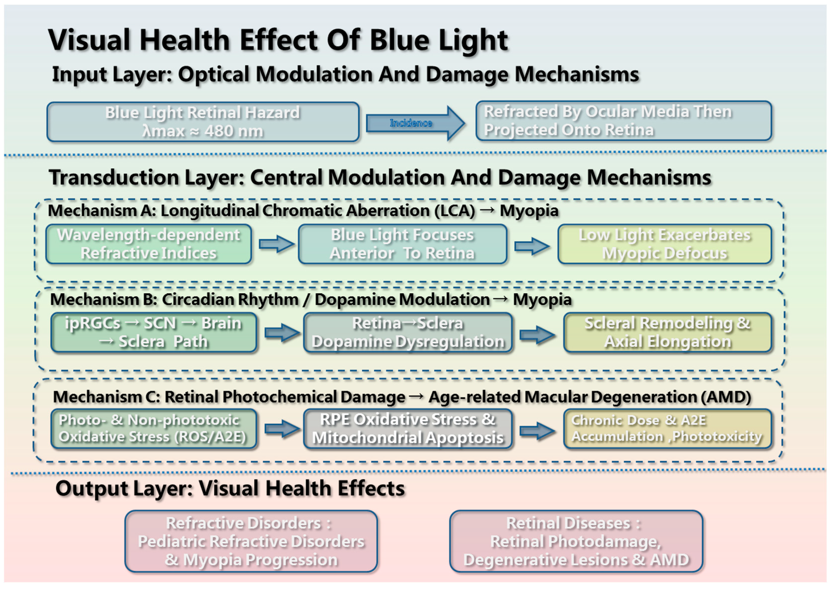
Figure 2.
Non-visual health effects of blue light.
Disclaimer/Publisher’s Note: The statements, opinions and data contained in all publications are solely those of the individual author(s) and contributor(s) and not of MDPI and/or the editor(s). MDPI and/or the editor(s) disclaim responsibility for any injury to people or property resulting from any ideas, methods, instructions or products referred to in the content. |
© 2026 by the authors. Licensee MDPI, Basel, Switzerland. This article is an open access article distributed under the terms and conditions of the Creative Commons Attribution (CC BY) license (http://creativecommons.org/licenses/by/4.0/).
Copyright: This open access article is published under a Creative Commons CC BY 4.0 license, which permit the free download, distribution, and reuse, provided that the author and preprint are cited in any reuse.