

Submitted:
13 September 2026
Posted:
15 September 2026
You are already at the latest version
Abstract
Background and Clinical Significance: Herpes zoster (HZ) results from reactivation of latent varicella-zoster virus and disproportionately affects older adults. Antiviral therapy and systemic analgesia are well established, but local wound care of the eroded skin remains largely empirical: no dressing has been validated in controlled trials for this indication, and denuded HZ lesions are frequently mismanaged with dry gauze that adheres to the wound bed and aggravates pain. Case Presentation: An 85-year-old man with no known chronic disease presented with a one-week history of thoracic HZ unresponsive to outpatient antiviral therapy, requiring admission for uncontrolled pain (9/10 on the Numeric Rating Scale, NRS), vomiting, asthenia, and hyporexia. Examination revealed confluent, superficial-partial-thickness erosions across the T6–T8 dermatomes (12.5 × 24 cm). After a negative wound culture, a single polylactic-acid-based membrane (Suprathel®250) was applied over the entire denuded area. Pain fell from 9/10 to 2/10 on the NRS within seven days, the membrane fully resorbed with complete re-epithelialization by day 12, and the patient resumed independent daily activities by the third week without scarring. Conclusions: A polylactic acid membrane produced rapid pain relief and complete wound closure after a single application in an extensive HZ lesion, comparing favorably with published outcomes for hydrocolloid dressings and standard gauze-based care. Synthetic skin substitutes deserve controlled evaluation as a dedicated wound-care option for HZ, particularly for patients with extensive dermatomal involvement or poor tolerance of conventional dressings.
Keywords:
herpes zoster
; shingles
; polylactic-acid-based biomaterials
; skin substitute
; wound healing
; pain management
1. Introduction and Clinical Significance
Herpes zoster (HZ), commonly known as shingles results from reactivation of latent varicella-zoster virus (VZV, human herpesvirus 3), which persists in a non-replicating state within dorsal root and cranial nerve ganglia after primary chickenpox infection. Reactivation is thought to reflect a decline in VZV-specific cell-mediated immunity, whether due to physiological immunosenescence, psychological stress, or an immunosuppressive condition or treatment. Clinically, HZ evolves through a pre-eruptive (prodromal) phase of localized dysesthesia, an acute eruptive phase marked by a dermatomal maculopapular-vesicular rash, and a chronic phase in which pain may persist as post-herpetic neuralgia (PHN) [1,2]
The global burden of HZ is substantial and rising as populations age. A 2025 analysis of the Global Burden of Disease 2021 dataset covering 204 countries estimated that varicella and HZ cases increased by 19.0% between 1990 and 2021 (from 72.8 to 86.7 million), with age-standardized incidence remaining essentially stable while high-income regions saw a resurgence attributable to population aging [3]. Regional hospital-based series report similarly elevated figures: an Italian cohort of 47,933 HZ-related hospitalizations between 2011 and 2023 found an overall incidence of 6.46 per 1000 person-years, rising to 9.12 per 1000 person-years among adults over 65 [4]. Lifetime risk approaches one in three individuals, and may reach 50% among those who live into their ninth decade [2].
Several factors increase both the likelihood and severity of HZ. Advanced age is the single strongest predictor, reflecting progressive decline in VZV-specific immunity [2]. Diabetes mellitus is independently associated with a higher risk of HZ and of HZ-related hospitalization: a 10-year observational study from Piedmont, Italy, found that among diabetic patients, age over 65, obesity (BMI > 30), and poor glycemic control (HbA1c > 8.0%) increased the risk of HZ hospitalization, with diabetic patients facing a 40% higher rehospitalization risk and a 25% higher risk of severe complications compared with non-diabetic patients [5]. Other chronic conditions linked to reduced cell-mediated immunity malignancy, chronic kidney disease, and autoimmune disease — have also been associated with more severe HZ courses and higher in-hospital mortality, particularly in patients over 80 years old [4]. Our patient, notably, had none of these comorbidities, which makes the severity of his presentation extensive multidermatomal involvement requiring hospital admission somewhat atypical and underscores that advanced age alone can be sufficient to produce a severe course.
Systemic antiviral therapy (acyclovir, valacyclovir, or famciclovir) remains the cornerstone of acute management and is most effective when started within 72 hours of rash onset, although treatment may still offer benefit later if new vesicles continue to appear or if complications such as ophthalmic or neurological involvement are present [2]. Antivirals reduce the duration of viral shedding, accelerate lesion resolution, and lower the risk of PHN, but they do not directly address the cutaneous erosions that result once vesicles rupture.
By contrast, local wound care of the denuded skin in HZ has received comparatively little systematic attention. The eroded areas that follow vesicle rupture behave, in practice, as partial-thickness wounds: they are painful, prone to fluid loss, and vulnerable to secondary bacterial infection, which can itself precipitate rare but severe complications such as necrotizing fasciitis [6]. Historically, these lesions were treated with drying solutions and powders on the assumption that the rash is self-limited within two to three weeks; however, the recognition that HZ vesicles extend into the dermis behaving as true partial-thickness wounds rather than superficial epidermal defects has prompted growing interest in occlusive, moisture-retentive dressings as an alternative to traditional dry gauze [7]. A comprehensive literature review of patient-centered HZ wound outcomes found that healing time ranged from 2 to 7 weeks when lesions were dressed with a moisture-retentive dressing, compared with 9 to 10 weeks when lesions were left air-exposed [8]. Despite this, no dressing has been validated in a randomized controlled trial specifically for HZ, and an ongoing clinical trial comparing hydrocolloid occlusion, silicone gel, and standard drying-solution care for acute HZ remains, to our knowledge, the only registered controlled study addressing this question directly [9].
Suprathel®250 (ST250; PolyMedics Innovations GmbH, Denkendorf, Germany) is a thin, autoadhesive, acellular, biodegradable skin substitute composed of a synthetic polylactide-based copolymer (lactide 73–80%, trimethylene carbonate 12–16%, and caprolactone 8–11%) [10]. It was developed for deep dermal burns and is now used across a range of complex skin wounds [11]. In burn studies, ST250 causes significantly less pain during dressing changes than traditional paraffin-impregnated gauze, while producing comparable healing times and re-epithelialization rates [12,13]. Because it adheres directly to the wound bed and is gradually resorbed as the skin re-epithelializes underneath it, ST250 does not require the frequent dressing changes that make conventional gauze so poorly tolerated on denuded, hyperalgesic skin a property with direct relevance to the pain that dominates the clinical picture of acute HZ [12]). More recently, PLA-based matrices have also been applied outside the burn setting: a 2025 single-center European case series reported their use in chronic, therapy-resistant wounds of diverse etiology, describing favorable integration into the wound bed and a contribution to wound closure even in previously stalled lesions [14]. To our knowledge, however, no prior report has specifically examined the use of a polylactic acid membrane for the cutaneous lesions of HZ. We describe a case in which a single application of ST250 was associated with rapid pain relief and complete wound closure in a patient with extensive dermatomal HZ, and we place this outcome in the context of previously published wound-care strategies for HZ.
2. Case Presentation
2.1. Patient Information
An 85-year-old male, originally from San Luis Potosí and independent in his activities of daily living prior to this illness, presented to the emergency department with no history of diabetes mellitus, cardiovascular disease, malignancy, immunosuppressive therapy, or prior reported episodes of herpes zoster (HZ). He had no history of varicella-zoster virus vaccination and denied high-risk behaviors. He presented with a one week history of a severe painful thoracic rash with edema and painful, crusted vesicular lesions affecting the left side across the mid-back along the T6–T8 dermatomes. Despite having been managed as an outpatient with oral antiviral therapy and topical care, his condition progressively deteriorated, manifesting as severe neuropathic pain, persistent vomiting, severe asthenia, hyporexia, and inability to tolerate oral intake. He denied fever, chills, or shortness of breath. He lived with family members who assisted with his care during the acute presentation.
2.2. Clinical Findings
On admission, vital signs were within normal limits: body temperature was 36.7 °C, blood pressure was 126/88 mmHg, pulse rate was 76 beats per minute, oxygen saturation was 97%, and respiratory rate was 16 breaths per minute. He was alert, oriented, and conscious, but restless due to severe pain and burning at the site of the lesions. Systemic examination, including the respiratory and cardiovascular systems, showed no abnormalities. Dermatological examination revealed a large confluent area of erosion with a well demarcated dermatomal distribution across T6-T8 on the left hemithorax, extending from the paravertebral region anteriorly toward the midline (Figure 1.). The lesion measured approximately 12.5cm x 24cm and presented with the clinical appearance of a superficial partial-thickness (second-degree) burn, erythematous wound bed with loss of epidermis and scattered residual vesicle roofs, but without evidence of full-thickness skin loss, active bleeding, or purulent discharge. The surrounding skin was edematous and tender to palpation. No satellite lesions, mucosal involvement, or signs suggestive of disseminated disease were observed. Pain was rated as 9/10 on the Numeric Rating Scale (NRS), described as a constant burning sensation with superimposed sharp exacerbations on contact with clothing or bedding, which significantly disrupted his sleep and rendered him unable to dress or bathe independently.
Components of the laboratory exam include complete blood count with differential, basic metabolic panel, and inflammatory markers showed no systemic abnormalities suggestive of bacterial infection or sepsis. White blood cell count was normal at 7,620/mm³ with 60.8% neutrophils, 26.6% lymphocytes, and 12.5% monocytes, while C-reactive protein (CRP) was elevated at 40.3 mg/L (reference < 5 mg/L). Other routine biochemical parameters were within normal limits. The diagnosis of herpes zoster was established clinically based on history, characteristic dermatomal distribution, and lesion morphology. In accordance with standard practice for classic presentations, no additional virological confirmation (e.g., PCR or direct fluorescent antibody testing) was performed.
2.3. Therapeutic Intervention and Follow-Up
The patient was admitted for inpatient management. A wound culture was obtained to rule out secondary bacterial infection before any dressing was applied; results were negative for pathogenic growth. Oral valacyclovir (1,000 mg every 8 hours) was continued to arrest ongoing viral replication. Analgesia was escalated to include systemic opioids for the nociceptive component of his pain and oral pregabalin for acute neuropathic pain and hyperalgesia. For wound care, the wound bed was gently cleansed with sterile saline to remove debris, exudate, and residual topical ointment from prior outpatient treatment, avoiding mechanical trauma to the friable surrounding skin. (Figure 2). The membrane adhered spontaneously to the moist wound bed without requiring additional adhesive tape or bandaging. A secondary layer of Vaseline-impregnated gauze was placed over the membrane to minimize mechanical friction against clothing and bedding, and the patient tolerated the application without any acute increase in pain.
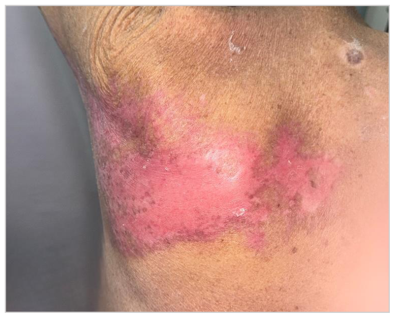
He was discharged after 24 hours of inpatient observation, continuing oral valacyclovir and his analgesic regimen. At outpatient follow-up seven days after membrane placement, his pain had decreased to 2/10 on the NRS and the membrane remained adherent with visible early re-epithelialization at its margins. At the second follow-up visit, 12 days after application, the membrane had fully resorbed and the wound demonstrated complete re-epithelialization (Figure 3) without dehiscence, exudate, or clinical signs of infection; analgesic tapering was initiated at this visit. By the third week, his caregivers reported that he had resumed dressing and bathing independently and was sleeping without positional discomfort related to the healed area. At a final follow-up approximately six weeks after presentation, the healed skin showed no hypertrophic scarring, keloid formation, or persistent dyspigmentation and the patient reported no residual pain suggestive of post-herpetic neuralgia at the treated site. No adverse reactions to the membrane were observed at any point during follow-up.
3. Discussion
3.1. Polylactic Acid Membrane Efficacy in Herpes Zoster Lesions
In this patient, a single application of a polylactic acid membrane was followed by a reduction in pain from 9/10 to 2/10 on the NRS within seven days and complete wound closure by day 12, without the need for repeated dressing changes. This trajectory compares favorably with previously reported outcomes for both standard gauze-based care and alternative occlusive dressings in HZ.
A comparable case describing hydrocolloid dressing use in a patient with severe HZ-related pain and extensive epidermal damage reported a reduction from 9/10 to 4/10 on the Visual Analogue Scale (VAS) a numerically similar but proportionally smaller drop than the reduction achieved in our patient, and one that additionally required repeated dressing changes over the course of treatment, which the hydrocolloid format is designed to minimize but does not eliminate [15]. In the broader literature review by Bolton and colleagues summarizing patient-centered HZ wound outcomes across published case series, healing time with moisture-retentive dressings ranged from 2 to 7 weeks, compared with 9 to 10 weeks when lesions were managed with simple air exposure or dry gauze [8,16]. Our patient’s 12-day time to complete closure falls at the favorable end of that range despite the considerable size of the lesion (12.5 × 24 cm) and his advanced age, both factors generally associated with slower wound healing.
These comparisons should be interpreted cautiously, as they involve different dressing technologies, wound sizes, and patient populations, and none derive from a head-to-head controlled trial. However, the consistency of the direction of effect occlusive, moisture-retentive dressings outperforming dry or air-exposed management, and a single-application synthetic membrane potentially outperforming hydrocolloid dressings that require more frequent changes is in keeping with the underlying rationale that HZ erosions behave as partial-thickness wounds that benefit from the same principles of moist wound healing established for burns and other cutaneous defects [15,16]
3.2. Pain Control
The magnitude and speed of pain relief observed in this case is consistent with mechanisms described for ST250 in burn wounds, where it has been shown to significantly reduce pain scores compared with paraffin gauze and other comparator dressings in randomized clinical trials [12,13]. ST250 is a CE-certified, semi-permeable, absorbable alloplastic skin substitute that is highly permeable to oxygen, limits evaporative water loss, and controls the accumulation of inflammatory exudate. Because it adheres to and effectively becomes part of the wound bed rather than requiring removal and reapplication, it avoids the repeated mechanical trauma to exposed nerve endings that occurs each time a conventional gauze dressing is peeled away from denuded dermis — a mechanism that plausibly explains why our patient required no further analgesic escalation and why his opioid and pregabalin requirements could be tapered starting at the second follow-up visit [12]. As the polylactic acid matrix degrades, it also releases lactate into the wound microenvironment, which has been proposed to promote angiogenesis and modulate local inflammation, potentially contributing to the accelerated healing observed here and in burn populations [14].
Direct comparators for skin-substitute use in HZ specifically are scarce, which itself reflects the evidence gap this case seeks to address. The most relevant prior report, discussed above, is the hydrocolloid dressing case published by Kiedrowski, describing an elderly woman with recurrent HZ and extensive epidermal damage whose VAS pain score fell from 9/10 to 4/10 with hydrocolloid occlusion, alongside improved quality of life and accelerated healing relative to her own prior HZ episodes managed conventionally [17]. Our patient’s proportionally larger pain reduction (9/10 to 2/10) and single-application course suggest that a fully occlusive, adherent synthetic membrane may offer an incremental advantage over hydrocolloid dressings, which typically still require periodic replacement as they saturate with exudate.
Beyond HZ, ST250 and related polylactic-acid-based matrices have an established comparative track record against other wound-care modalities. Compared with collagen dressings and acellular xenografts in chronic wounds such as diabetic foot ulcers and venous leg ulcers, polylactic acid-based composite dressings have been reported to shorten healing time and improve healing rates [18]. In burns specifically, a randomized bicentric trial found that ST250 produced significantly lower pain scores than the comparator dressing with no significant difference in healing time or re-epithelialization [13]. A 10-year pediatric cohort further reported favorable adherence to the wound bed and a reduced need for frequent dressing changes across nearly 2,100 patients treated with ST250 for second-degree burns [19]. Taken together with the 2025 European case series describing successful use of a polylactic acid matrix in chronic, non-burn wounds [14], this body of evidence supports the biological plausibility of extending polylactic-acid-based membranes to an acute, painful dermatomal wound such as HZ, even though the present report constitutes, to our knowledge, the first such application described in the literature.
3.3. Limitations and Clinical Practice Implications
This report describes a single patient, and as with any case report, the observed outcome cannot establish efficacy or be generalized without controlled comparison. Pain scores were recorded by clinical staff at scheduled visits rather than through a structured, blinded pain diary. Nonetheless, the magnitude and consistency of the clinical response rapid pain reduction, single-application wound closure, and an uneventful healing course in a patient of advanced age with an unusually extensive lesion — support further investigation.
Vaccination remains the most effective strategy for preventing HZ and its complications, and its promotion should remain a public health priority, particularly given the rising incidence associated with global population aging [3,4]. However, patients who are unvaccinated, who decline vaccination, or who develop HZ despite vaccination will continue to require an effective acute wound-care strategy. Given the existing evidence base for ST250 in burns and chronic wounds, and the encouraging signal from this case alongside the single prior hydrocolloid report, we suggest that polylactic-acid-based membranes warrant evaluation in a prospective, ideally controlled, series of HZ patients particularly those with extensive dermatomal involvement, in whom conventional gauze-based care is often poorly tolerated and in whom the risk of prolonged pain and delayed healing is highest.
4. Conclusions
In this patient with extensive thoracic HZ, a single application of a polylactic acid membrane (Suprathel®250) was associated with a rapid and substantial reduction in pain (from 9/10 to 2/10 on the NRS within seven days) and complete wound closure by day 12, without adverse events or scarring. This outcome compares favorably with the limited existing literature on occlusive wound care for HZ, including a previously reported case using hydrocolloid dressings. Polylactic acid membranes may represent a reasonable wound care option for HZ, particularly in patients with extensive dermatomal involvement or poor tolerance of conventional dressings, but larger prospective studies are needed to confirm this benefit and define the place of these membranes within HZ treatment protocols.
Author Contributions
Conceptualization, V.M.L.-G. and M.A.M.-J.; supervision, writing—review and editing, P.A.C.-B. and A.P.S.-Q.; visualization, M.A.M.-J. and P.A.C.-B.; writing—original draft preparation, V.M.L.-G., M.A.M.-J. and P.A.C.-B.; data curation, M.A.M.-J. and V.M.L.-G.; investigation, P.A.C.-B. and J.L.R.-G.; resources, P.A.C.-B., A.P.S.-Q. and J.L.R.-G. All authors have read and agreed to the published version of the manuscript.
Funding
This research received no external funding.
Institutional Review Board Statement
Not applicable.
Informed Consent Statement
Informed consent was obtained from the patient involved in this study. Written informed consent, including consent to publish accompanying clinical images, was obtained from the patient for publication of this paper.
Data Availability Statement
No new data were created or analyzed in this study. The original contributions presented in this study are included in the article. Further inquiries can be directed to the corresponding author.
Conflicts of Interest
J.L.R.-G. has received or receives honoraria as a consultant for PolyMedics Innovations GmbH (PMI). All other authors declare no conflicts of interest. The funders had no role in the design of the study; in the collection, analysis, or interpretation of data; in the writing of the manuscript; or in the decision to publish the results.
Abbreviations
The following abbreviations are used in this manuscript:
| HZ | Herpes zoster |
| NRS | Numeric Rating Scale |
| PHN | Post-herpetic neuralgia |
| ST250 | Suprathel®250 |
| VAS | Visual Analogue Scale |
| VZV | Varicella-zoster virus |
References
- Behera, D.; Belemkar, S. A Short Review and Update on Epidemiology, Treatment, and Management of Herpes Zoster (Shingles) Infection. Curr. Pharm. Des. 2026. [Google Scholar] [CrossRef] [PubMed]
- Waggan, A.I.; Imtiaz, K.; Ayo-Farai, O.; Okwilagwe, H.; Ehi-Iyoha, I.; Akhtar, R.; et al. Current advances in the epidemiology, risk factors, prevention, management, and long-term outcomes of herpes zoster and post-herpetic neuralgia. Front Microbiol. 2026, 17. [Google Scholar] [CrossRef] [PubMed]
- Wu, Z.; Zhang, J.; Wei, J.; Huang, C.; Xu, W.; Wu, J.; et al. Global burden of varicella and herpes zoster across 204 countries, 1990–2021: a temporal trend analysis in the era of the COVID-19 pandemic and projections to 2036. Front Public Health 2025, 13, 1654535. [Google Scholar] [CrossRef] [PubMed] [PubMed Central]
- Mattei, A.; Cialfi, D.; D’Annunzio, A.; Fabiani, L.; Fiasca, F.; Gabutti, G. Comorbidities in hospitalized patients with herpes zoster: an Italian retrospective observational study in the years 2011-2023. BMC Infect Dis. 2025, 26, 185. [Google Scholar] [CrossRef] [PubMed] [PubMed Central]
- Hospitalisation for herpes zoster in people with and without diabetes: A 10-year-observational study | Request PDF. ResearchGate. [CrossRef] [PubMed]
- Alamri, A.M.; AlWadai, H.H.A.; Isaway, N.A.I. Critical Chest Wall Necrotizing Fasciitis Triggered by Herpes Zoster: A Case Report. Am J Case Rep. 2024, 25, e944186. [Google Scholar] [CrossRef] [PubMed] [PubMed Central]
- Kaler, J.; Hussain, A.; Flores, G.; Kheiri, S.; Desrosiers, D.; Kaler, J.; et al. Monkeypox: A Comprehensive Review of Transmission, Pathogenesis, and Manifestation. Cureus 2022, 14. [Google Scholar] [CrossRef] [PubMed]
- Bolton, L.L.; Faller, N.; Kirsner, R.S. Herpes Zoster (Shingles) Patient-Centered Wound Outcomes: A Literature Review. Adv Skin Wound Care;PubMed 2021, 34, 239–48. [Google Scholar] [CrossRef] [PubMed] [PubMed Central]
- Cecil.md Clinical Trial Occlusion vs Standard Treatment for the Treatment of Herpes Zoster [Internet]. [cited 2026 Aug 31]. Available online: https://veterans.networkofcare.org/cecil-md/CommunityResources/ClinicalTrials/Detail/NCT04258930?keyword=%22Zoster%22.
- SUPRATHEL. PMI – The Wound Healing Company [Internet]. 2024 Dec 6 [cited 2026 Aug 31]. Available online: https://polymedics.com/en/product/suprathel/.
- Kalantzi, S.; Alexakis, C.; Anagnostaki, M.E.; Anagnostaki, I.; Zacharis, K. Genital Herpes Zoster: A Report of a Rare Case. Cureus 2024, 16(10), e72290. [Google Scholar] [CrossRef] [PubMed] [PubMed Central]
- Heitzmann, W.; Mossing, M.; Fuchs, P.C.; Akkan, J.; Seyhan, H.; Grieb, G.; et al. Comparative Clinical Study of Suprathel® and Jelonet® Wound Dressings in Burn Wound Healing after Enzymatic Debridement. Biomedicines 2023, 11, 2593. [Google Scholar] [CrossRef] [PubMed] [PubMed Central]
- Uhlig, C.; Rapp, M.; Hartmann, B.; Hierlemann, H.; Planck, H.; Dittel, K.K. Suprathel-an innovative, resorbable skin substitute for the treatment of burn victims. Burn. J. Int. Soc. Burn Inj. 2007, 33, 221–9. [Google Scholar] [CrossRef] [PubMed]
- Bioresorbable Polylactic Acid Matrix for Chronic Non-Healing Wounds: First Clinical Experience in Europe [Internet]. [cited 2026 Aug 31]. Available online: https://www.mdpi.com/2075-4426/16/1/10.
- Severe pain from shingles: a hydrocolloid dressing helps | Request PDF. ResearchGate. 2026 May 5. [CrossRef] [PubMed]
- Martínez-Jiménez, M.A.; Novoa-Moreno, A.L.; Loza-González, V.M.; Pérez-Medina-Carballo, R.; Cervantes-Báez, P.A. Use of Polylactic Acid Dermal Matrix for the Management of Wounds with Exposed Avascular Structures. J Clin Med. 2025, 15, 3. [Google Scholar] [CrossRef] [PubMed] [PubMed Central]
- Kiedrowski, M. Severe pain from shingles: a hydrocolloid dressing helps. BMJ Support Palliat. Care 2023, 13(e1), e91–2. [Google Scholar] [CrossRef] [PubMed]
- Liden, B.A.; Liu, T.; Regulski, M.; Foster, M.; DeLeon, R.; Palazzi, G.; et al. A multicenter retrospective study comparing a polylactic acid CAMP with intact fish skin graft or a collagen dressing in the management of diabetic foot ulcers and venous leg ulcers. Wounds Compend Clin. Res. Pract. 2024, 36, 297–302. [Google Scholar] [CrossRef] [PubMed]
- Schriek, K.; Ott, H.; Sinnig, M. Paradigm Shift in Treatment Strategies for Second-Degree Burns Using a Caprolactone Dressing (Suprathel®)? A 15-Year Pediatric Burn Center Experience in 2084 Patients. Eur. Burn J.;PubMed 2021, 3, 1–9. [Google Scholar] [CrossRef] [PubMed] [PubMed Central]
Figure 1.
Extensive lesion with confluent areas of erosion across the T6-T8 dermatomes.
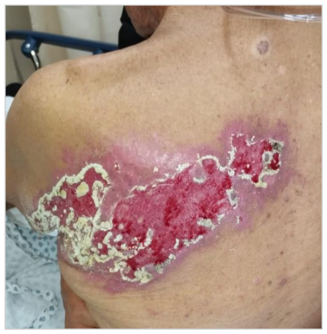
Figure 2.
Polylactic acid membrane placed directly over the wound covering the whole lesion.
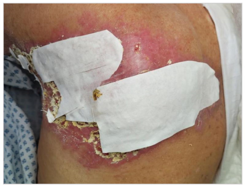
Figure 3.
Complete re-epithelialization at the 12-day follow-up visit.
Disclaimer/Publisher’s Note: The statements, opinions and data contained in all publications are solely those of the individual author(s) and contributor(s) and not of MDPI and/or the editor(s). MDPI and/or the editor(s) disclaim responsibility for any injury to people or property resulting from any ideas, methods, instructions or products referred to in the content. |
© 2026 by the authors. Licensee MDPI, Basel, Switzerland. This article is an open access article distributed under the terms and conditions of the Creative Commons Attribution (CC BY) license (http://creativecommons.org/licenses/by/4.0/).
Copyright: This open access article is published under a Creative Commons CC BY 4.0 license, which permit the free download, distribution, and reuse, provided that the author and preprint are cited in any reuse.