

Submitted:
11 September 2026
Posted:
14 September 2026
You are already at the latest version
Abstract
Background and Aim: Exportin 1 (XPO1) is the principal nuclear export receptor for numerous tumor-suppressor proteins, regulatory factors, and selected oncogenic mRNAs and has emerged as a therapeutically relevant vulnerability in multiple myeloma (MM). Selinexor, a first-in-class oral selective inhibitor of nuclear export (SINE), targets XPO1 and has demonstrated antimyeloma activity both as a doublet and in combination with established therapeutic agents. This review summarizes the biological rationale and molecular mechanisms of XPO1 inhibition, clinical evidence supporting selinexor-based therapy, mechanisms of resistance and potential predictive biomarkers, and practical considerations regarding toxicity management.
Materials and Methods: A narrative review of the published literature on XPO1 biology and selinexor in MM was performed, with emphasis on preclinical mechanistic studies, prospective clinical trials, and studies addressing resistance, biomarkers, pharmacology, and treatment-related toxicity. Particular attention was given to pivotal and clinically relevant studies evaluating selinexor-based regimens, including Selinexor Treatment of Refractory Myeloma (STORM), BOSTON, and combination cohorts from the STOMP program, together with recent translational studies expanding the molecular framework of XPO1 inhibition.
Results: XPO1 inhibition exerts pleiotropic antimyeloma effects through nuclear retention and reactivation of tumor-suppressor proteins, suppression of NF-κB-dependent signaling and oncogenic mRNA translation, enhancement of glucocorticoid signaling, and disruption of cellular protein homeostasis. More recent findings implicate treatment-induced XPO1 degradation through ubiquitin-regulatory mechanisms and suppression of lipid biosynthesis through the Lipin1–SREBP axis. Clinically, selinexor demonstrated activity in heavily pretreated triple-class refractory MM and subsequently showed improved efficacy when incorporated into once-weekly combination regimens, particularly with bortezomib and dexamethasone. Additional combinations with carfilzomib, pomalidomide, lenalidomide, and daratumumab have demonstrated clinically meaningful activity in selected relapsed/refractory multiple myeloma (RRMM) populations. Candidate determinants of response and resistance include XPO1 Cys528 alterations, hnRNPU, USP7-dependent regulation of XPO1 stability, and Lipin1–SREBP signaling, although none is currently prospectively validated for patient selection. Treatment-related cytopenias, gastrointestinal toxicity, fatigue, and hyponatremia remain important but can frequently be addressed through proactive supportive care and dose optimization.
Conclusion: XPO1 inhibition represents a mechanistically distinct therapeutic strategy in MM, and selinexor has established clinical activity across multiple relapsed/refractory settings. Increasing use of lower-dose, once-weekly combination strategies has improved its therapeutic feasibility, while emerging insights into XPO1 degradation, metabolic regulation, and molecular determinants of response are broadening the biological framework of this drug class. Prospective biomarker validation and optimization of treatment sequencing will be important for defining the future role of XPO1-directed therapy in the evolving MM treatment landscape.
Keywords:
multiple myeloma
; selinexor
; XPO1
; exportin 1
; nuclear export
; SINE
; biomarkers
; drug resistance
1. Biology of Xpo1 and the Nuclear Export Mechanism
Macromolecular transport between the nucleus and cytoplasm in eukaryotic cells plays a fundamental role in the regulation of gene expression, the cell cycle, the DNA damage response, apoptosis, and cellular homeostasis. This molecular trafficking occurs through nuclear pore complexes, whereas the regulated transport of proteins and large macromolecular complexes is largely mediated by nuclear transport receptors belonging to the karyopherin family. XPO1, also known as chromosome region maintenance 1, is the major nuclear export receptor in the cell and controls the transport of numerous proteins as well as various RNA species from the nucleus to the cytoplasm [1].
1.1. Xpo1 Structure and Cargo Recognition
XPO1 is an evolutionarily conserved protein with a molecular weight of approximately 120 kDa. Structurally, it consists of 21 consecutive Huntingtin, elongation factor 3, A subunit of protein phosphatase 2A, and TOR1 repeats. The superhelical architecture formed by these Huntingtin, elongation factor 3, A subunit of protein phosphatase 2A, and TOR1 repeats enables XPO1 to interact with cargo proteins, Ran-GTP, and other components of the nuclear transport machinery [1]. A substantial proportion of proteins transported by XPO1 contain short amino acid sequences termed nuclear export signals (NES). Classical NES motifs are enriched in hydrophobic amino acids, particularly leucine, as well as isoleucine, valine, phenylalanine, and methionine. NES-containing cargos bind to the hydrophobic NES-binding groove located on the outer convex surface of XPO1 [2].
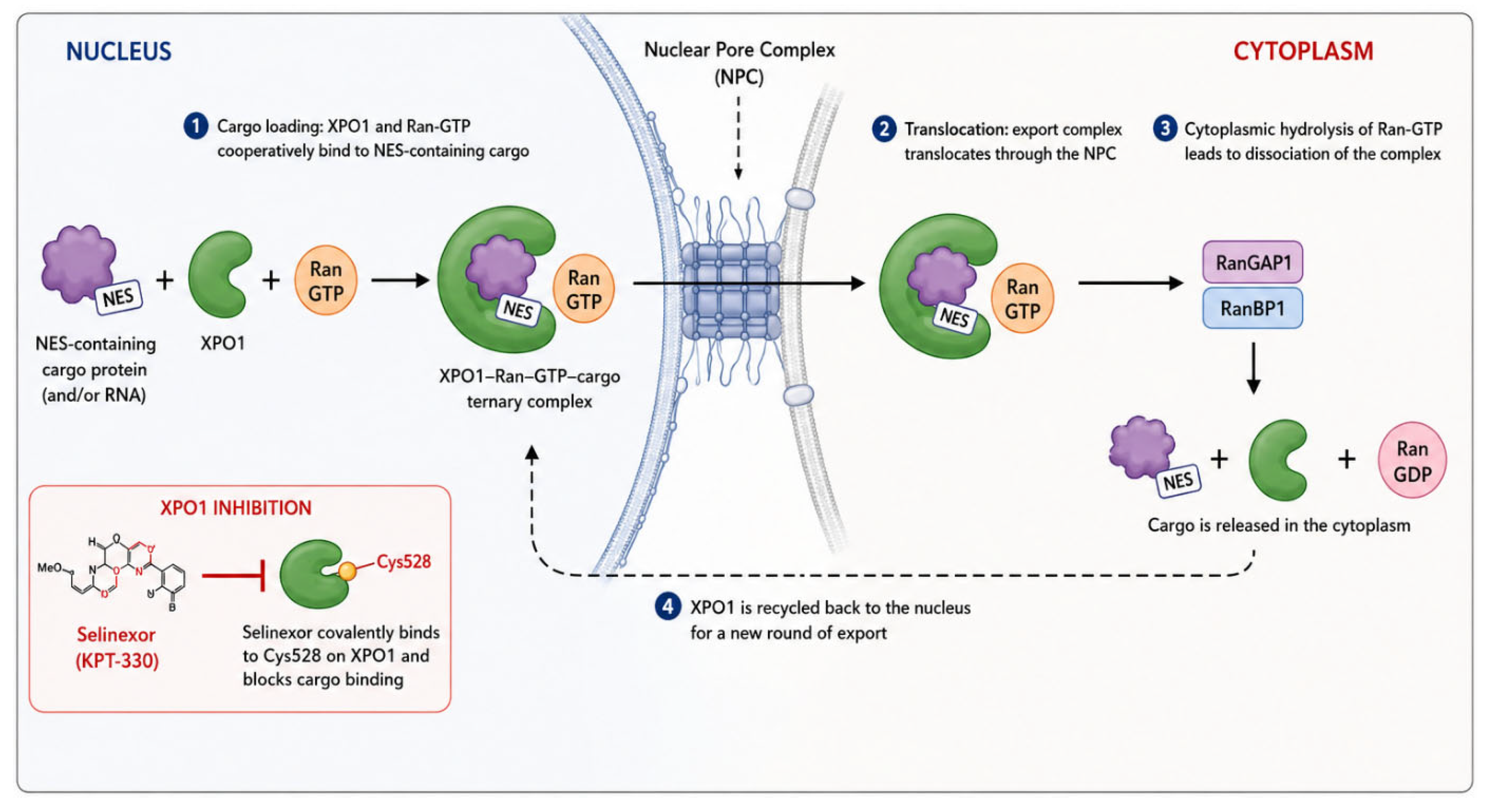
Crystallographic studies have demonstrated that NES motifs with different amino acid sequences and secondary structures can engage the same hydrophobic binding region on XPO1. Five hydrophobic pockets within this region accommodate the hydrophobic side chains of the NES. The ability of different NES motifs to adopt distinct backbone conformations while engaging the same hydrophobic pockets explains how XPO1 can recognize highly diverse NES sequences [1,3,4]. Cys528, located within the NES-binding groove, is a critical residue for the pharmacological targeting of XPO1. SINE target Cys528 within the cargo-binding pocket of XPO1, thereby preventing the binding and nuclear export of NES-containing cargos. The demonstration that the XPO1 C528S mutation confers resistance to selinexor further confirms the critical role of Cys528 in the selinexor–XPO1 interaction [5].
1.2. Ran-Gtp-Dependent Nuclear Export Cycle
XPO1-mediated nuclear export depends on the Ran-GTP/Ran-GDP gradient generated by the small GTPase Ran (Ras-related nuclear protein). In the nucleus, the chromatin-associated guanine nucleotide exchange factor regulator of chromosome condensation 1 maintains a high concentration of GTP-bound Ran. Within the nucleus, XPO1 binds cooperatively to Ran-GTP and an NES-containing cargo to form the XPO1–Ran-GTP–cargo ternary complex [1]. This export complex traverses the nuclear pore complex and reaches the cytoplasm. In the cytoplasm, Ran GTPase-activating protein 1 and Ran-binding protein 1 facilitate Ran-GTP hydrolysis and disassembly of the export complex. Consequently, the cargo is released into the cytoplasm, whereas XPO1 returns to the nucleus to participate in another transport cycle. Thus, the high Ran-GTP concentration in the nucleus and low concentration in the cytoplasm provide directionality to XPO1-mediated nuclear transport [1].
Figure 1.
XPO1-mediated Ran-GTP-dependent nuclear export cycle and the mechanism of XPO1 inhibition by selinexor.
Figure 1.
XPO1-mediated Ran-GTP-dependent nuclear export cycle and the mechanism of XPO1 inhibition by selinexor.
1.3. Biological Significance of Xpo1-Mediated Nuclear Export
The biological significance of XPO1 extends beyond the physical translocation of macromolecules from the nucleus to the cytoplasm. By controlling the subcellular localization of numerous regulatory proteins and RNA species, XPO1 influences multiple cellular processes, including cell-cycle progression, DNA-damage responses, transcriptional regulation, apoptosis, and cellular survival [1]. Dysregulation of XPO1-mediated nuclear export has been increasingly implicated in malignant transformation and tumor progression. XPO1 is overexpressed in a variety of hematologic and solid malignancies, where excessive nuclear export can result in the aberrant cytoplasmic localization and functional inactivation of tumor-suppressor and growth-regulatory proteins. Because the biological activity of many of these proteins depends on their nuclear localization, their inappropriate export may simultaneously impair cell-cycle checkpoints, apoptotic signaling, and cellular responses to genomic stress [6,7]. Importantly, XPO1 functions as a central regulatory node rather than as a component of a single oncogenic signaling pathway. Consequently, pharmacologic disruption of XPO1-mediated export has the potential to restore the nuclear localization and activity of multiple tumor-suppressive cargos while concurrently interfering with survival-promoting mechanisms. This pleiotropic biological effect provides a strong rationale for targeting XPO1 in malignancies characterized by dependence on dysregulated nuclear export [7].
1.4. Pharmacologic Targetability of Xpo1
The structural organization of XPO1 provides a well-defined pharmacologic vulnerability within its NES-binding groove. Cys528, located within this hydrophobic cargo-binding pocket, represents a critical residue for the interaction of SINE compounds with XPO1. Occupation of this site interferes with the binding of NES-containing cargos and thereby disrupts XPO1-dependent nuclear export [1,5]. The functional importance of Cys528 has been demonstrated experimentally. Substitution of this residue with serine (C528S) markedly reduces sensitivity to SINE compounds, providing genetic evidence that the antitumor activity of these agents is mediated through direct engagement of XPO1. These structural and functional observations established XPO1 as a druggable component of the nuclear transport machinery and provided the molecular foundation for the development of SINE compounds [8]. Among these agents, selinexor (KPT-330) became the first XPO1 inhibitor to achieve clinical implementation. Its pharmacologic characteristics and the downstream molecular consequences of XPO1 inhibition are discussed in detail in the subsequent sections [9].
2. Biological and Therapeutic Significance of Xpo1 in Multiple Myeloma
In MM, the proliferation and survival of malignant plasma cells are supported not only by alterations in intracellular signaling pathways but also by complex interactions with the bone marrow microenvironment. Because inhibition of XPO1-mediated nuclear export can simultaneously alter the intracellular localization of multiple tumor suppressor and survival-regulatory proteins, it has emerged as a potential therapeutic strategy in MM [7].
2.1. Xpo1 Expression and Clinical/Biological Relevance in Mm
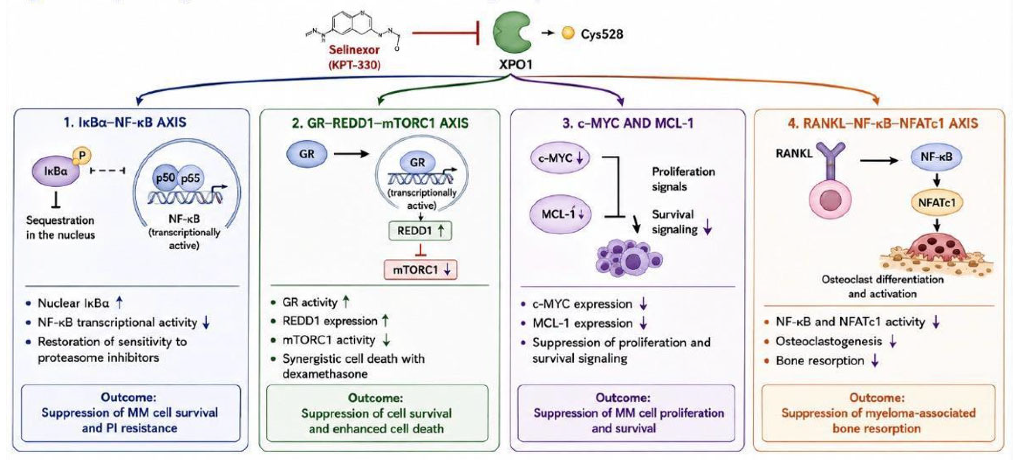
Evidence from patient-derived material supports a biologically relevant role for XPO1 in plasma-cell malignancies. Tai et al. found elevated XPO1/CRM1 expression in MM and plasma cell leukemia samples, with particularly high levels in bortezomib-resistant cells [7]. Higher expression was also linked to a greater burden of lytic bone disease and inferior survival. Consistent with these clinical associations, experimental depletion of XPO1 reduced MM-cell viability, indicating that XPO1 contributes functionally to malignant plasma-cell survival [7]. XPO1 blockade exerts antimyeloma activity through several convergent mechanisms. In preclinical models, SINE retain XPO1 cargo tumor-suppressor proteins within the nucleus, a change associated with cell-cycle arrest and apoptosis. XPO1 inhibition also reduces c-MYC and MCL-1 expression and attenuates nuclear factor kappa B (NF-κB) activity. Consequently, XPO1 represents a distinct therapeutic node through which several pathways supporting MM-cell growth and survival can be disrupted simultaneously [7,10].
2.2. Tumor-Suppressor Retention and Mm Cell Survival
A central consequence of XPO1 inhibition in MM is the restoration of nuclear localization of tumor-suppressor and growth-regulatory proteins. Preclinical studies have demonstrated that selective inhibitors of nuclear export retain XPO1 cargo proteins within the nucleus, thereby restoring tumor-suppressive functions that are compromised by excessive nuclear export. This redistribution is associated with cell-cycle arrest and induction of apoptosis in malignant plasma cells [7]. The antimyeloma effects of XPO1 inhibition are therefore intrinsically pleiotropic. Rather than targeting a single oncogenic driver, inhibition of nuclear export simultaneously alters the intracellular localization and functional activity of multiple proteins involved in proliferation, survival, and apoptosis. In parallel, XPO1 inhibition reduces the expression of key survival-associated proteins, including c-MYC and MCL-1, further contributing to suppression of myeloma-cell viability. These observations provide a mechanistic link between increased XPO1 activity in MM and the survival advantage of malignant plasma cells, while supporting nuclear export as a therapeutically actionable vulnerability [7].
2.3. Xpo1–iκbα–nf-Κb Axis and Pi Resistance
The NF-κB signaling pathway is an important regulator of MM-cell proliferation, survival, and treatment resistance. In the context of combined XPO1 and proteasome inhibition, the inhibitor of nuclear factor kappa B alpha (IκBα)–NF-κB axis appears to be particularly relevant. Preclinical models of acquired proteasome-inhibitor resistance provide further mechanistic support for this approach. Addition of selinexor restored responsiveness to bortezomib or carfilzomib in resistant MM cells. This effect was accompanied by increased total and nuclear IκBα, greater formation of IκBα–NF-κB complexes, and lower NF-κB transcriptional activity. Importantly, reducing IκBα expression weakened the cytotoxicity of the selinexor–bortezomib combination, supporting a causal contribution of this pathway. A similar sensitizing effect was observed in cells obtained from patients with proteasome inhibitor-refractory MM. These findings suggest that the synergy between XPO1 inhibition and proteasome inhibition is mediated, at least in part, by nuclear retention of IκBα and consequent suppression of NF-κB signaling [10].
2.4. Bone Marrow Microenvironment and Myeloma Bone Disease
The effects of XPO1 inhibition are not restricted to malignant plasma cells. SINE compounds retain cytotoxic activity when MM cells are cocultured with bone marrow stromal cells or osteoclasts. This suggests that the antimyeloma activity of XPO1 inhibition can persist despite protective signals provided by the bone marrow microenvironment. XPO1 inhibition also has biological effects relevant to myeloma bone disease. SINE compounds suppress receptor activator of nuclear factor κB ligand (RANKL)-induced NF-κB and nuclear factor of activated T cells 1 (NFATc1) activation, thereby inhibiting osteoclastogenesis and bone resorption, while exerting more limited effects on osteoblasts and bone marrow stromal cells. In in vivo models of MM-associated bone lesions, XPO1 inhibition reduced tumor burden and bone lysis and prolonged survival. These findings suggest that targeting XPO1 may simultaneously affect both malignant plasma cells and microenvironmental mechanisms contributing to MM bone disease [7].
2.5. Gr–redd1–mtorc1 Axis and Synergy with Dexamethasone
Glucocorticoids, particularly dexamethasone, are key components of many MM treatment combinations. The glucocorticoid receptor (GR) and mechanistic target of rapamycin (mTOR) signaling pathway have been shown to play important roles in the synergy between selinexor and dexamethasone. Mechanistic studies by Argueta et al. showed that selinexor increases both GR transcript and protein levels in MM cells [11]. When dexamethasone was added, GR-dependent transcription was further enhanced and the two agents produced synergistic cell death. Notably, GR expression was required for the interaction with dexamethasone, whereas the antimyeloma effect of selinexor alone did not depend on GR. A major downstream consequence of the selinexor–dexamethasone interaction is inhibition of mechanistic target of rapamycin complex 1 (mTORC1) signaling. Enhanced GR activity promotes expression of regulated in development and DNA damage responses 1 (REDD1), which contributes to reduced mTORC1 activity and loss of cell viability. These observations provide a mechanistic basis for combining selinexor with dexamethasone in MM [11].
Figure 2.
Major biological effects of XPO1 inhibition in multiple myeloma. Abbreviations: XPO1, exportin 1; GR, glucocorticoid receptor; REDD1, regulated in development and DNA damage responses 1; mTORC1, mechanistic target of rapamycin complex 1; IκBα, inhibitor of nuclear factor kappa B alpha; NF-κB, nuclear factor kappa B; PI, proteasome inhibitor; RANKL, receptor activator of nuclear factor κB ligand; NFATc1, nuclear factor of activated T cells 1; Multiple Myeloma, MM.
Figure 2.
Major biological effects of XPO1 inhibition in multiple myeloma. Abbreviations: XPO1, exportin 1; GR, glucocorticoid receptor; REDD1, regulated in development and DNA damage responses 1; mTORC1, mechanistic target of rapamycin complex 1; IκBα, inhibitor of nuclear factor kappa B alpha; NF-κB, nuclear factor kappa B; PI, proteasome inhibitor; RANKL, receptor activator of nuclear factor κB ligand; NFATc1, nuclear factor of activated T cells 1; Multiple Myeloma, MM.
2.6. Positioning Xpo1 as A Therapeutic Target
Taken together, the available biological and preclinical evidence positions XPO1 as a therapeutically relevant vulnerability in MM. Increased XPO1 expression and its association with adverse disease characteristics, together with the reduction in myeloma-cell viability following XPO1 suppression, support a functional dependence of malignant plasma cells on dysregulated nuclear export [7]. The therapeutic rationale for XPO1 inhibition is further strengthened by its ability to simultaneously restore the nuclear localization of tumor-suppressor proteins, suppress survival-associated mechanisms including c-MYC, MCL-1, and NF-κB signaling, and interfere with protective interactions within the bone marrow microenvironment. In addition, inhibition of osteoclastogenic signaling provides a potential biological link between XPO1 targeting and myeloma-associated bone disease. The ability of XPO1 inhibition to restore sensitivity to proteasome inhibitors (PIs) through the IκBα–NF-κB axis and to enhance dexamethasone-induced cell death through GR–REDD1–mTORC1 signaling further provides a mechanistic rationale for combination-based therapeutic strategies [7,10,11]. Collectively, these observations established the biological foundation for the clinical development of selective inhibitors of nuclear export in MM. Selinexor, the first-in-class clinically implemented XPO1 inhibitor, represents the principal translation of this therapeutic concept and is discussed in detail in the following section [9].

Table 1.
Major XPO1-Related Biological Mechanisms in MM and Effects of XPO1 Inhibition.
| XPO1-related pathway / cargo | Role in MM | Effect of XPO1 inhibition | Biological consequence |
|---|---|---|---|
| Tumor suppressor proteins (p53, p21, p27, FOXO) | XPO1-mediated export reduces nuclear tumor-suppressor activity | Nuclear retention and restoration of transcriptional activity | Cell-cycle arrest and apoptosis |
| IκBα–NF-κB signaling | Export of IκBα facilitates NF-κB-dependent survival signaling | Nuclear accumulation of IκBα and inhibition of NF-κB transcriptional activity | Suppression of prosurvival and proliferative signaling |
| Oncogenic mRNA–eIF4E axis | XPO1-dependent export supports translation of oncogenic transcripts, including MYC, CCND1, MCL1, and cIAP | Nuclear retention of oncogenic mRNAs and reduced cytoplasmic translation | Reduced expression of prosurvival proteins |
| Glucocorticoid receptor signaling | Nuclear export limits sustained GR-mediated transcriptional activity | Nuclear retention/activation of GR and enhancement of dexamethasone-induced REDD1 signaling | mTORC1 suppression and enhanced glucocorticoid-mediated apoptosis |
| Proteostasis and proteasome-related signaling | XPO1-dependent survival pathways contribute to adaptation to proteotoxic stress | Complementary disruption of protein homeostasis when combined with proteasome inhibition | Enhanced proteotoxic stress and apoptosis |
| XPO1 protein stability (USP7–CRL5/ASB8 axis) | USP7-mediated deubiquitination contributes to XPO1 stabilization | Selinexor disrupts XPO1 stabilization and promotes ubiquitin-dependent XPO1 degradation | Depletion of the cellular XPO1 protein pool and amplification of target inhibition |
| Lipin1–SREBP signaling | XPO1-mediated Lipin1 export supports SREBP-dependent lipid metabolic programs | Nuclear Lipin1 accumulation suppresses SREBP transcriptional activity | Reduced fatty-acid and cholesterol biosynthesis |
| Tumor–immune microenvironment | XPO1-dependent processes may influence NK/T-cell function and immune-checkpoint expression | XPO1 inhibition may modulate immune-cell activity and checkpoint signaling | Potential enhancement of antitumor immunity; clinical relevance remains investigational |
3. Selinexor: Pharmacology and Molecular Mechanisms of Action
3.1. Pharmacological and Pharmacokinetic Profile
Selinexor (KPT-330) is an orally bioavailable, small-molecule SINE and the first agent of this class to achieve clinical implementation. Its principal pharmacologic target is XPO1, the major nuclear export receptor responsible for the transport of numerous proteins and RNA species from the nucleus to the cytoplasm [9,12]. Following oral administration, selinexor reaches maximum plasma concentrations within approximately 2–4 h and has a terminal elimination half-life of approximately 6–8 h. Pharmacokinetic analyses indicate a generally predictable exposure profile across clinically relevant dosing schedules. Selinexor is widely distributed and highly bound to plasma proteins. These pharmacokinetic properties, together with the persistence of its downstream pharmacodynamic effects on nuclear export, have supported the development of intermittent dosing schedules, including once- and twice-weekly administration [12].
3.2. Direct Target Engagement: Cys528 and Xpo1 Inhibition
The primary molecular event underlying selinexor activity is direct engagement of XPO1 within its hydrophobic NES-binding groove. Selinexor interacts with the critical Cys528 residue within the cargo-binding pocket, thereby preventing productive binding of NES-containing cargos and disrupting XPO1-mediated nuclear export [1,13]. Unlike irreversible XPO1 inhibitors, selinexor forms a slowly reversible covalent interaction with Cys528. Functional blockade of the NES-binding groove consequently alters the intracellular distribution of a broad spectrum of XPO1 cargos, resulting in their nuclear retention and initiating multiple downstream antitumor effects. The central importance of Cys528 for SINE activity is further supported by experimental models in which substitution of this residue confers marked resistance to XPO1 inhibition [1,8].
3.3. Nuclear Retention of Tumor-Suppressor Proteins
A major downstream consequence of XPO1 inhibition is the nuclear retention and functional reactivation of tumor-suppressor and cell-cycle regulatory proteins. Selinexor prevents the XPO1-mediated export of cargos including p53, p21, p27, FOXO-family proteins, and retinoblastoma protein (Rb), thereby increasing their nuclear availability. Restoration of these nuclear tumor-suppressive functions can activate complementary antineoplastic programs. Increased p53 activity promotes pro-apoptotic signaling, including induction of PUMA and BAX, whereas restoration of p21- and p27-dependent cell-cycle control contributes to inhibition of cyclin-dependent kinase activity and G1/S cell-cycle arrest. Through simultaneous modulation of multiple XPO1 cargos, selinexor therefore produces a broader tumor-suppressive response than would be expected from inhibition of a single signaling pathway [7,9,10]. Selinexor also promotes nuclear retention of IκBα, resulting in suppression of NF-κB transcriptional activity. The biological relevance of this pathway in MM, particularly its contribution to proteasome-inhibitor resistance and combination therapy, is discussed in Section 2.
3.4. Suppression of Oncogenic Mrna Export and Translation
Suppression of NF-κB signaling: IκBα, the endogenous inhibitor of NF-κB, is an XPO1 cargo. Selinexor-mediated inhibition of XPO1 promotes the nuclear retention of IκBα. Nuclear IκBα subsequently interferes with NF-κB DNA binding and transcriptional activity, thereby suppressing survival and proliferative signaling in MM cells [7,10]. Inhibition of oncogenic mRNA translation: The mRNAs encoding several pro-survival proteins, including c-Myc, Cyclin D1, MCL-1, and cIAP, depend on the XPO1–eIF4E complex for their export from the nucleus to the cytoplasm. By inhibiting XPO1-mediated nuclear export, selinexor promotes the nuclear retention of these transcripts and consequently reduces their cytoplasmic translation and downstream oncoprotein expression [7,9,10].
3.5. Xpo1 Degradation Through the Asb8–usp7 Axis
Exposure to selinexor also promotes the proteasomal degradation of its molecular target, XPO1. Using cryo-electron microscopy, Wing et al. (2025) demonstrated that SINE compounds, including selinexor, expose a cryptic site on XPO1 that enables recruitment of ASB8, a substrate receptor of a Cullin-RING E3 ubiquitin ligase complex [14]. This allosteric, “molecular glue-like” mechanism promotes XPO1 ubiquitination and subsequent proteasomal degradation. This mechanism is complemented by an additional regulatory layer described by Wang et al. (2025): under physiological conditions, the deubiquitinase USP7 stabilizes XPO1 through deubiquitination, whereas selinexor disrupts the USP7–XPO1 interaction, thereby impairing XPO1 stabilization and facilitating its degradation [15]. Thus, the activity of selinexor extends beyond functional inhibition of XPO1-mediated nuclear export to include active depletion of the cellular XPO1 protein pool [14,15].
3.6. Lipin1–srebp Axis and Metabolic Reprogramming
Recent evidence suggests that the antimyeloma activity of selinexor extends beyond the regulation of nuclear transport and classical survival pathways to include alterations in cellular lipid metabolism. Lipin1 has been identified as an XPO1-dependent nuclear export cargo, and inhibition of XPO1 by selinexor promotes the nuclear accumulation of Lipin1. Nuclear Lipin1 suppresses the transcriptional activity of sterol regulatory element-binding proteins (SREBPs), leading to reduced expression of key genes involved in lipid and cholesterol biosynthesis, including FASN, SCD, DHCR24, and FDPS. Consistent with these transcriptional changes, selinexor treatment reduces fatty-acid and cholesterol synthesis in MM cell lines and primary CD138+ myeloma cells. Genetic suppression experiments further support a functional role for Lipin1 in the inhibition of SREBP signaling and in determining sensitivity to selinexor, both in vitro and in murine xenograft models. Collectively, these findings identify the Lipin1–SREBP axis as an additional metabolic component of selinexor activity, expanding its mechanism of action beyond nuclear retention of tumor-suppressor proteins and suppression of classical oncogenic survival pathways [16].
3.7. Immunomodulatory Effects on the Tumor Microenvironment:
Emerging evidence suggests that the effects of selinexor may extend beyond tumor cell-intrinsic mechanisms and may modulate NK- and T-cell function as well as immune checkpoint expression. Although these findings indicate a potential non-cell-autonomous dimension to the mechanism of action of selinexor, the available evidence remains preliminary, and its clinical implications have yet to be fully established [17].
3.8. Integrated Mechanistic Model
The antimyeloma activity of selinexor reflects the coordinated disruption of multiple cellular processes rather than inhibition of a single oncogenic pathway. Direct engagement of XPO1 at Cys528 inhibits XPO1-mediated nuclear export, resulting in the nuclear retention and functional reactivation of tumor-suppressor and cell-cycle regulatory proteins. In parallel, nuclear retention of IκBα suppresses NF-κB transcriptional activity, while impaired export of oncogenic mRNAs reduces the translation of key survival-associated proteins, including c-Myc, Cyclin D1, MCL-1, and cIAP [18]. More recent findings have further expanded this mechanistic framework. Selinexor not only functionally inhibits XPO1 but also promotes depletion of the XPO1 protein pool through ubiquitin–proteasome-dependent mechanisms involving the USP7–XPO1 and CRL5–ASB8 regulatory axes. In addition, nuclear accumulation of Lipin1 suppresses SREBP-dependent transcription and lipid and cholesterol biosynthesis, revealing a metabolic component of XPO1 inhibition. Emerging evidence also suggests that selinexor may influence the tumor microenvironment through modulation of NK- and T-cell function and immune checkpoint expression, although the clinical relevance of these immunomodulatory effects remains to be established [14,15,16,17]. Collectively, these complementary mechanisms integrate alterations in nuclear transport, transcriptional regulation, apoptotic and cell-cycle control, oncogenic protein synthesis, protein homeostasis, and cellular metabolism. This pleiotropic mode of action provides a biological rationale for combining selinexor with agents targeting complementary vulnerabilities in MM and forms the mechanistic basis for the combination strategies discussed in the following section.
Table 2.
Pharmacologic and Pharmacokinetic Characteristics of Selinexor.
| Parameter | Selinexor characteristic | Clinical / pharmacologic relevance |
|---|---|---|
| Drug class | SINE | First-in-class oral XPO1 inhibitor |
| Molecular target | XPO1/CRM1 | Binds to the NES-binding groove of XPO1 |
| Binding site | Cys528 | Covalent but slowly reversible interaction with XPO1 |
| Route of administration | Oral | Enables incorporation into outpatient combination regimens |
| Oral bioavailability | Approximately 80% | Supports effective systemic exposure following oral administration |
| Time to peak concentration (Tmax) | Approximately 2–4 h | Relatively rapid systemic absorption |
| Plasma protein binding | Approximately 95% | Predominantly protein-bound in circulation |
| Metabolism | Primarily hepatic; mainly CYP3A4, with contributions from UGTs and GSTs | Potential relevance of concomitant medications affecting metabolic pathways |
| Elimination half-life | Approximately 6–8 h | Plasma half-life is shorter than the duration of downstream biological effects of XPO1 inhibition |
| Elimination | Predominantly hepatobiliary/fecal; limited renal excretion | Renal clearance represents a minor component of overall elimination |
| Pharmacodynamic effect | Inhibition of XPO1-mediated nuclear export and nuclear retention of XPO1 cargo proteins | Provides the mechanistic basis for downstream antimyeloma effects |
4. Preclinical Rationale for Combination Therapy
4.1. Synergy with Proteasome Inhibitors
The combination of selinexor with PIs, particularly bortezomib and carfilzomib, is supported by a strong mechanistic rationale centered on complementary inhibition of the IκBα–NF-κB signaling axis. NF-κB is a major regulator of myeloma-cell survival, proliferation, and treatment resistance, and its activity is tightly controlled by the endogenous inhibitor IκBα. Proteasome inhibition prevents the proteasomal degradation of IκBα, whereas selinexor inhibits XPO1-mediated nuclear export and promotes its nuclear retention. The combination therefore increases both the abundance and nuclear localization of IκBα, resulting in enhanced formation of IκBα–NF-κB complexes and more profound suppression of NF-κB transcriptional activity [10].
Preclinical studies have demonstrated that this interaction is particularly relevant in the setting of acquired PI resistance. Selinexor restored sensitivity to both bortezomib and carfilzomib in PI-resistant MM cell lines and enhanced apoptosis when combined with either PI. Importantly, similar sensitization was observed in myeloma cells obtained from patients with PI-refractory disease, supporting the translational relevance of this mechanism. The functional importance of the IκBα–NF-κB axis was further demonstrated by IκBα knockdown experiments, in which depletion of IκBα markedly attenuated the cytotoxicity of the selinexor–bortezomib combination [10]. In addition to suppression of NF-κB signaling, combined XPO1 and proteasome inhibition enhances the nuclear accumulation of tumor-suppressor proteins and disrupts multiple survival pathways, resulting in synergistic antimyeloma activity in preclinical models. Collectively, these findings indicate that selinexor and PIs exert complementary effects on protein homeostasis and nuclear export and provide a biological rationale for combining selinexor with bortezomib or carfilzomib, including in PI-resistant disease. This preclinical framework subsequently supported the clinical development of PI-containing selinexor regimens [19].
4.2. Synergy with Dexamethasone
The combination of selinexor with dexamethasone is supported by a distinct mechanistic interaction involving glucocorticoid receptor signaling and suppression of the mTORC1 pathway. Selinexor increases GR expression in MM cells, thereby enhancing their responsiveness to dexamethasone. Upon dexamethasone-mediated GR activation, the combination markedly increases GR-dependent transcription and promotes the expression of downstream regulators of mTOR signaling, particularly regulated in development and DNA damage response 1 (REDD1) [11]. Enhanced REDD1 expression contributes to suppression of mTORC1 activity, a central regulator of cellular growth, metabolism, and survival in MM. Consistent with this mechanism, silencing of REDD1 attenuated the inhibitory effect of the selinexor–dexamethasone combination on mTOR signaling, supporting a functional role for the GR–REDD1–mTORC1 axis in the observed synergy. Importantly, although the direct antimyeloma activity of selinexor is largely independent of GR expression, its synergistic interaction with dexamethasone appears to be GR-dependent. These findings provide a biological rationale for incorporating dexamethasone into selinexor-based regimens and illustrate how XPO1 inhibition can enhance glucocorticoid-mediated antimyeloma activity through coordinated suppression of mTORC1 signaling [11].
4.3. Rationale for Combination with IMiDs
Preclinical studies have also demonstrated synergistic antimyeloma activity when XPO1 inhibition is combined with immunomodulatory drugs (IMiDs), providing a rationale for the clinical evaluation of selinexor with pomalidomide or lenalidomide. In experimental MM models, the addition of an IMiD to XPO1 inhibition enhanced cytotoxicity compared with either therapeutic approach alone, including activity observed with selinexor–pomalidomide combinations [9]. The biological basis for this interaction is likely multifactorial. Selinexor simultaneously promotes nuclear retention and reactivation of tumor-suppressor proteins, suppresses NF-κB signaling, and reduces the expression of survival-associated oncoproteins such as c-MYC and MCL-1. These effects may complement the direct antimyeloma and immunomodulatory activities of IMiDs, thereby increasing cellular susceptibility to treatment. Importantly, however, the molecular determinants of selinexor–IMiD synergy have not been defined as clearly as those underlying its interactions with PIs or dexamethasone [9]. The demonstration of synergistic activity in preclinical MM models provided the rationale for subsequent clinical investigation of selinexor–IMiD combinations, particularly selinexor plus pomalidomide and dexamethasone (SPd) and selinexor plus lenalidomide and dexamethasone (SRd). Their clinical efficacy and safety are discussed in the subsequent sections [9].
4.4. Rationale for Combination with Anti-Cd38 Monoclonal Antibodies
The combination of selinexor with anti-CD38 monoclonal antibodies, particularly daratumumab, represents a mechanistically complementary therapeutic strategy in MM. Whereas selinexor exerts predominantly tumor cell-intrinsic effects through inhibition of XPO1-mediated nuclear export, reactivation of tumor-suppressor pathways, suppression of oncogenic signaling, and induction of apoptosis, daratumumab targets CD38-expressing plasma cells through immune-mediated mechanisms. The distinct mechanisms of action of these agents therefore provide a biological rationale for their combined use [20]. In addition, emerging evidence that selinexor may modulate NK- and T-cell function and immune checkpoint expression raises the possibility that XPO1 inhibition could influence the immune microenvironment in ways that complement antibody-based therapy. However, these immunomodulatory effects remain incompletely characterized, and a direct mechanistic basis for synergy between selinexor and anti-CD38 antibodies has not yet been established as clearly as that observed with PIs or dexamethasone [17]. Despite this mechanistic uncertainty, the non-overlapping modes of action and clinical activity of both therapeutic classes supported the evaluation of selinexor–daratumumab combinations in relapsed/refractory MM. Subsequent clinical studies have demonstrated activity of selinexor, daratumumab, and dexamethasone-containing regimens, including in heavily pretreated populations, and these clinical data are discussed in the following sections [20].
4.5. Translation of Preclinical Synergy into Clinical Combination Strategies
The pleiotropic effects of XPO1 inhibition provide a strong biological rationale for incorporating selinexor into combination-based treatment strategies in MM. Rather than relying on a single pathway, selinexor simultaneously restores tumor-suppressor activity, suppresses oncogenic and NF-κB-dependent survival signaling, alters protein homeostasis, and promotes apoptosis. These effects create multiple opportunities for complementary interactions with established antimyeloma agents [7,10]. Among these combinations, the mechanistic rationale is particularly well established for PIs and dexamethasone, through modulation of the IκBα–NF-κB and GR–REDD1–mTORC1 axes, respectively. Preclinical studies have also demonstrated enhanced antimyeloma activity when selinexor is combined with IMiDs, while combinations with anti-CD38 monoclonal antibodies are supported by complementary mechanisms of action and emerging preclinical and clinical evidence [9,10,11,17,20]. Collectively, these observations provided the foundation for the systematic clinical evaluation of selinexor-based doublet, triplet, and, more recently, quadruplet regimens. Importantly, the development of once-weekly selinexor-containing combinations has also sought to optimize the balance between antimyeloma activity and tolerability. The clinical translation of these mechanistically informed strategies, including combinations with dexamethasone, bortezomib, carfilzomib, pomalidomide, lenalidomide, and daratumumab, is discussed in the following section [9,21].
5. Clinical Development of Selinexor in Multiple Myeloma
5.1. Early Clinical Development and Selinexor–dexamethasone
Early clinical development of selinexor in MM established proof of concept for therapeutic XPO1 inhibition while also demonstrating the importance of combination therapy. In a multicenter phase I study, Chen et al. evaluated selinexor with or without dexamethasone in patients with heavily pretreated RRMM. Single-agent selinexor demonstrated modest activity, with an objective response rate (ORR) of 4% and a clinical benefit rate of 21%. In contrast, the addition of dexamethasone substantially enhanced antimyeloma activity; in the cohort receiving selinexor 45 mg/m² (approximately 80 mg) plus dexamethasone 20 mg twice weekly, the ORR reached 50%. These findings provided early clinical support for the mechanistic synergy between XPO1 inhibition and glucocorticoid signaling and established selinexor plus dexamethasone as the backbone for subsequent clinical development [22].
The activity of this combination was subsequently evaluated by Vogl et al. in a phase II study involving 79 patients with highly refractory MM who had received a median of seven prior treatment regimens. Patients received selinexor 80 mg plus dexamethasone 20 mg twice weekly. Forty-eight patients had disease refractory to bortezomib, carfilzomib, lenalidomide, and pomalidomide, whereas 31 patients were additionally refractory to an anti-CD38 monoclonal antibody. The overall response rate was 21%, with comparable response rates in the quad-refractory (21%) and penta-refractory (20%) subgroups. Notably, an ORR of 35% was observed among patients with high-risk cytogenetic abnormalities, including t(4;14), t(14;16), and del(17p). The median duration of response was approximately 5 months [23]. Collectively, these early studies demonstrated that selinexor retained clinically meaningful activity in heavily pretreated and multidrug-refractory MM and that dexamethasone substantially enhanced its antimyeloma effect. These observations established the selinexor–dexamethasone backbone and provided the foundation for its subsequent evaluation in the larger STORM study.
5.2. Storm Trial
The phase IIb STORM study represented a pivotal step in the clinical development of selinexor by evaluating its activity in a population with highly refractory MM and limited remaining therapeutic options. In Part 2 of the study, 122 patients with penta-exposed, triple-class refractory MM received oral selinexor 80 mg plus dexamethasone 20 mg twice weekly. Patients had previously been exposed to bortezomib, carfilzomib, lenalidomide, pomalidomide, and daratumumab and were required to have disease refractory to at least one proteasome inhibitor, one immunomodulatory drug, and daratumumab. The population was heavily pretreated, with a median of seven previous treatment regimens, and 53% of patients had high-risk cytogenetic abnormalities [24]. Selinexor plus dexamethasone achieved an ORR of 26% (95% CI, 19–35%), including two stringent complete responses, six very good partial responses, and 24 partial responses. An additional 13% of patients achieved a minimal response, resulting in a clinical benefit rate of 39%. Responses occurred rapidly, with a median time to response of approximately 4 weeks. The median duration of response was 4.4 months, while median progression-free survival (PFS) and overall survival (OS) were 3.7 and 8.6 months, respectively. Notably, among patients achieving at least a minimal response, median OS extended to 15.6 months [24]. The clinical relevance of these findings should be interpreted in the context of the highly treatment-refractory population enrolled in STORM. All patients had progressive disease at study entry, and most had exhausted the major therapeutic classes available at that time. Despite this adverse clinical profile, selinexor–dexamethasone produced clinically meaningful responses across several patient subgroups, providing proof that XPO1 remained a therapeutically actionable target even in advanced multidrug-refractory MM. The STORM results provided the pivotal efficacy evidence supporting the initial accelerated regulatory approval of selinexor in heavily pretreated RRMM and established XPO1 inhibition as a clinically validated therapeutic strategy. However, the toxicity profile and relatively limited durability of disease control observed with twice-weekly selinexor also highlighted the need for more tolerable dosing schedules and rational combination approaches. These considerations subsequently shaped the development of once-weekly selinexor-containing regimens, most notably the selinexor–bortezomib–dexamethasone combination evaluated in the phase III BOSTON trial.
5.3. Boston Trial
The phase III BOSTON trial provided randomized evidence supporting the clinical efficacy of selinexor-based combination therapy in previously treated MM. In this multicenter, open-label study, 402 patients who had received one to three prior lines of therapy were randomly assigned to receive once-weekly selinexor, bortezomib, and dexamethasone (SVd; n = 195) or standard bortezomib and dexamethasone (Vd; n = 207). In the SVd arm, selinexor was administered at 100 mg once weekly together with once-weekly bortezomib, representing a substantially different treatment schedule from the twice-weekly selinexor–dexamethasone regimen used in STORM [21]. The study met its primary endpoint, demonstrating a significant improvement in PFS with SVd. Median PFS was 13.93 months with SVd compared with 9.46 months with Vd (HR, 0.70; 95% CI, 0.53–0.93; p = 0.0075), corresponding to a 30% reduction in the risk of disease progression or death. The overall response rate was also significantly higher with SVd than with Vd (76.4% vs. 62.3%; p = 0.0012), and responses were deeper, with very good partial response or better achieved in 44.6% and 32.4% of patients, respectively. An important feature of the BOSTON regimen was the use of once-weekly bortezomib in the SVd arm, compared with twice-weekly bortezomib in the control arm. Despite the addition of selinexor, clinically significant peripheral neuropathy was less frequent with SVd: grade ≥2 peripheral neuropathy occurred in 21% of patients receiving SVd compared with 34% receiving Vd (p = 0.0013). Conversely, selinexor-associated toxicities remained clinically relevant, particularly thrombocytopenia, fatigue, and anemia, emphasizing the importance of supportive care, dose modification, and toxicity monitoring during treatment [25]. The clinical benefit of SVd was also observed across biologically relevant subgroups. In a prespecified analysis of patients with high-risk cytogenetic abnormalities, the ORR was 78.6% with SVd compared with 57.7% with Vd, while median PFS was 12.91 versus 8.61 months, respectively. Although the PFS difference within the high-risk subgroup did not reach statistical significance, these findings suggested that the activity of selinexor-based therapy was maintained in patients with adverse cytogenetic features. BOSTON therefore marked an important transition in the clinical development of selinexor, from the twice-weekly selinexor–dexamethasone approach used in heavily pretreated triple-class refractory disease to a once-weekly, mechanism-based triplet regimen applicable earlier in the relapsed/refractory treatment course. The trial also provided clinical validation of the preclinical synergy between XPO1 and proteasome inhibition described above. On the basis of the BOSTON results, selinexor in combination with bortezomib and dexamethasone received regulatory approval for patients with MM who had received at least one prior therapy.
5.4. Stomp and Other Selinexor-Based Triplets
The phase Ib/II STOMP (Selinexor and Backbone Treatments of Multiple Myeloma Patients) study provided a platform for the systematic evaluation of once-weekly selinexor in combination with several established antimyeloma agents. Multiple parallel cohorts investigated combinations with carfilzomib, pomalidomide, daratumumab, lenalidomide, and other therapeutic backbones. Collectively, these studies demonstrated that selinexor could be incorporated into diverse triplet regimens and supported the transition from the twice-weekly selinexor–dexamethasone schedule used in STORM toward lower-dose, once-weekly combination strategies. Selinexor, carfilzomib, and dexamethasone (SKd). The combination of selinexor with carfilzomib and dexamethasone was evaluated in patients with RRMM, based on the preclinical synergy between XPO1 and proteasome inhibition. In the STOMP SKd cohort, 32 patients with a median of four prior lines of therapy received once-weekly selinexor in combination with carfilzomib and dexamethasone. Among patients who were not refractory to carfilzomib, the overall response rate was 78%, with 44% achieving a very good partial response or better. Median PFS was approximately 15 months and median duration of response was 22.7 months. Importantly, activity was maintained in patients with high-risk cytogenetic features, supporting the potential utility of dual XPO1 and proteasome inhibition in biologically adverse disease [26].
Selinexor, pomalidomide, and dexamethasone (SPd). The all-oral SPd regimen has also demonstrated clinically meaningful activity in RRMM. Early STOMP experience showed responses in both pomalidomide-naïve and IMiD-refractory populations, supporting the feasibility of combining XPO1 inhibition with an IMiD backbone. Subsequent dose-optimization analyses evaluated once-weekly selinexor at 40 or 60 mg in combination with pomalidomide and dexamethasone. Although the 60-mg regimen produced a higher ORR (65% versus 50%), the 40-mg regimen was associated with a longer median PFS (18.4 versus 9.5 months) and a more favorable overall tolerability profile. These observations illustrate the importance of dose optimization when selinexor is incorporated into combination therapy and support the use of lower once-weekly doses to improve the therapeutic index [27].
Selinexor, daratumumab, and dexamethasone (SDd). In the STOMP SDd cohort, 34 patients with RRMM received selinexor in combination with daratumumab and dexamethasone. Patients had received a median of three prior treatment regimens and were frequently refractory to PIs and IMiDs. Among daratumumab-naïve patients, the ORR was 73% and median PFS was 12.5 months. Once-weekly selinexor at 100 mg was selected as the recommended phase II dose in combination with standard daratumumab and dexamethasone. In contrast, responses were not observed among the small number of patients with pre-existing daratumumab-refractory disease, although the limited sample size precludes definitive conclusions regarding activity in this setting [20].
Selinexor, lenalidomide, and dexamethasone (SRd). An all-oral combination of selinexor, lenalidomide, and dexamethasone was also explored within STOMP. At the recommended phase II dose of once-weekly selinexor 60 mg, lenalidomide 25 mg on days 1–21, and weekly dexamethasone, particularly high response rates were observed in lenalidomide-naïve RRMM, with an ORR of approximately 92%. In contrast, substantially lower activity was observed among patients previously exposed to lenalidomide, emphasizing the influence of prior IMiD exposure and resistance on the efficacy of this combination. These findings suggest that SRd may be most relevant in selected lenalidomide-sensitive populations rather than in patients with established lenalidomide-refractory disease [27].
Taken together, the STOMP experience demonstrated that selinexor can be combined with multiple therapeutic classes and that once-weekly dosing can preserve substantial antimyeloma activity while facilitating combination treatment. The heterogeneity in efficacy across individual cohorts also highlights the importance of prior drug exposure, refractory status, partner-agent selection, and selinexor dose optimization when selecting a selinexor-based regimen.
5.5. Emerging Quadruplet Strategies
The clinical development of selinexor has increasingly expanded beyond doublet and triplet regimens toward quadruplet combinations designed to simultaneously target multiple therapeutic vulnerabilities in MM. These approaches remain less mature than the established selinexor-based triplets and should therefore be regarded as emerging strategies rather than standard treatment options. One of the most extensively evaluated quadruplet approaches combines selinexor with daratumumab, bortezomib, and dexamethasone (Dara-SVd). This strategy builds upon the established activity of SVd while incorporating CD38-directed immune therapy. In a phase II study, the combination was evaluated in patients with RRMM, demonstrating the feasibility of simultaneously targeting XPO1-mediated nuclear export, proteasome-dependent protein homeostasis, and CD38-expressing plasma cells. These findings provide clinical support for further investigation of selinexor-containing quadruplets, although longer follow-up and comparative studies are required to define their relative efficacy and tolerability [28].
Another emerging strategy combines selinexor with carfilzomib, pomalidomide, and dexamethasone (SKPd). This approach is supported by the independently demonstrated activity of both SKd and SPd and aims to exploit complementary effects of XPO1 inhibition, proteasome inhibition, and immunomodulation. A phase I/II clinical study is evaluating selinexor, pomalidomide, and dexamethasone with or without carfilzomib in RRMM, particularly in patients with disease resistant to PIs and IMiDs [29]. More broadly, the development of selinexor-containing quadruplets reflects a shift toward lower-dose, once-weekly selinexor schedules integrated into multidrug regimens. Such strategies may improve depth of response by targeting complementary biological pathways; however, this potential benefit must be balanced against cumulative hematologic, gastrointestinal, constitutional, and infectious toxicities. Careful dose optimization and patient selection are therefore likely to be particularly important when selinexor is incorporated into four-drug combinations. At present, selinexor-based quadruplets remain investigational, and available data are substantially less mature than those supporting SVd or other established selinexor-containing triplets. Ongoing and randomized studies will be required to determine whether the addition of selinexor to contemporary three-drug backbones translates into clinically meaningful improvements in depth and durability of response without unacceptable toxicity.
5.6. Clinical Interpretation and Patient Selection
The clinical role of selinexor in MM should be considered within an increasingly complex treatment landscape that now includes PIs, IMiDs, anti-CD38 monoclonal antibodies, CAR T-cell therapies, bispecific antibodies, and other targeted approaches. Rather than representing a universally preferred option at relapse, selinexor may be particularly valuable in selected clinical scenarios in which its distinct mechanism of action, oral administration, and lack of cross-resistance with several established drug classes provide therapeutic advantages. One potentially relevant population comprises patients with high-risk cytogenetic disease. Subgroup analyses of the BOSTON trial demonstrated that the benefit of selinexor–bortezomib–dexamethasone was preserved in patients with high-risk abnormalities, including del(17p), t(4;14), t(14;16), and amplification of 1q21. These findings suggest that adverse cytogenetic features do not preclude responsiveness to XPO1 inhibition and support consideration of selinexor-containing combinations in biologically high-risk disease [25].
Renal impairment represents another clinically relevant setting. Because renal dysfunction is common in advanced MM and may restrict the use or dosing of several antimyeloma agents, treatments that can be administered without major renal dose adjustment are potentially advantageous. Subgroup analyses from BOSTON have demonstrated preserved efficacy of once-weekly selinexor–bortezomib–dexamethasone across patients with renal impairment, supporting the feasibility of this regimen in appropriately selected patients with compromised renal function [30].
The positioning of selinexor has also evolved with the introduction of T-cell–redirecting therapies. CAR T-cell products and bispecific antibodies can achieve deep and durable responses in eligible patients; however, access, manufacturing time, infectious complications, immune fitness, comorbidities, and rapidly progressive disease may limit their applicability in individual patients. In this context, selinexor-based regimens may provide an alternative for patients who are unsuitable for or unable to access T-cell–redirecting therapy. More recently, selinexor has also been explored as a potential holding or bridging strategy before CAR T-cell therapy, although this application remains investigational and requires prospective validation.
Prior treatment exposure should substantially influence regimen selection. Patients with previous anti-CD38 monoclonal antibody exposure may retain sensitivity to selinexor-based combinations because XPO1 inhibition operates through a distinct therapeutic mechanism. Real-world analyses have reported clinically meaningful outcomes with selinexor-containing triplets in heavily pretreated populations, including patients previously exposed to anti-CD38 therapy. Conversely, the efficacy of specific selinexor combinations remains dependent on sensitivity to the partner agent; for example, activity of SRd is substantially reduced in lenalidomide-refractory disease, whereas SDd appears considerably more active in daratumumab-naïve than in daratumumab-refractory patients [31].
Patient selection must also account for the characteristic toxicity profile of selinexor. Baseline thrombocytopenia, poor nutritional status, significant fatigue, recurrent gastrointestinal symptoms, and susceptibility to electrolyte disturbances may adversely affect tolerability. In contrast, contemporary once-weekly schedules and proactive supportive care can substantially improve treatment feasibility compared with the higher-intensity twice-weekly dosing initially used in STORM. Therefore, the choice of selinexor dose, combination partner, and supportive-care strategy should be individualized according to prior therapy, disease biology, comorbidities, performance status, and anticipated treatment tolerance [32].
Overall, selinexor is best viewed as a mechanism-driven therapeutic option whose clinical value depends on appropriate patient and regimen selection. Its distinct XPO1-directed activity may be particularly useful in multidrug-exposed disease, selected high-risk subgroups, patients with renal impairment, and individuals for whom cellular or bispecific immunotherapies are unsuitable or temporarily inaccessible. Further prospective studies are needed to define its optimal sequencing relative to contemporary immunotherapies and to determine whether biomarker-guided patient selection can further improve its therapeutic index.
Table 3.
Key Clinical Studies of Selinexor-Based Therapy in Multiple Myeloma.
| Study / Regimen | Phase | Population | Selinexor-Based Regimen | Key Efficacy Outcomes | Main Clinical Message |
|---|---|---|---|---|---|
| Chen et al. | I | Heavily pretreated RRMM | Selinexor ± dexamethasone | Single-agent ORR 4%; ORR with selected selinexor–dexamethasone cohort 50% | Established proof of concept for XPO1 inhibition and demonstrated enhanced activity with dexamethasone |
| Vogl et al. | II | Highly refractory RRMM; median 7 prior regimens | Selinexor 80 mg + dexamethasone 20 mg twice weekly | ORR 21%; quad-refractory 21%; penta-refractory 20%; high-risk cytogenetics ORR 35%; median DOR ≈ 5 months | Demonstrated clinically meaningful activity in multidrug-refractory disease |
| STORM | IIb | Penta-exposed, triple-class refractory MM; n=122 | Selinexor 80 mg + dexamethasone 20 mg twice weekly | ORR 26%; CBR 39%; median DOR 4.4 mo; median PFS 3.7 mo; median OS 8.6 mo | Pivotal evidence supporting clinical validation of selinexor in heavily pretreated RRMM |
| BOSTON | III | RRMM after 1–3 prior lines; n=402 | SVd vs Vd | ORR 76.4% vs 62.3%; median PFS 13.93 vs 9.46 mo; HR 0.70; ≥VGPR 44.6% vs 32.4% | Established once-weekly SVd as an effective earlier-line selinexor-based triplet |
| STOMP – SKd | Ib/II | RRMM; n=32 | Selinexor + carfilzomib + dexamethasone | ORR 78% in carfilzomib non-refractory patients; ≥VGPR 44%; median PFS ≈ 15 mo; median DOR 22.7 mo | Supports dual XPO1 and proteasome inhibition, including activity in high-risk disease |
| STOMP – SPd | Ib/II | RRMM | Selinexor + pomalidomide + dexamethasone | ORR 65% vs 50% with selinexor 60 vs 40 mg weekly; median PFS 9.5 vs 18.4 mo, respectively | Demonstrated activity with IMiD-based therapy and highlighted importance of dose optimization |
| STOMP – SDd | Ib/II | RRMM; n=34 | Selinexor + daratumumab + dexamethasone | In daratumumab-naïve patients: ORR 73%; median PFS 12.5 mo | Demonstrated feasibility and activity of XPO1 inhibition combined with anti-CD38 therapy |
| STOMP – SRd | Ib/II | RRMM, particularly lenalidomide-naïve disease | Selinexor + lenalidomide + dexamethasone | ORR ≈ 92% in lenalidomide-naïve patients; markedly lower activity after prior lenalidomide exposure | Suggests greatest activity in lenalidomide-sensitive populations |
| Dara-SVd | II | RRMM | Daratumumab + selinexor + bortezomib + dexamethasone | Early-phase feasibility/activity reported; mature comparative efficacy not established in the current draft | Emerging quadruplet strategy; remains investigational |
| SKPd | I/II | RRMM | Selinexor + carfilzomib + pomalidomide + dexamethasone | Ongoing/early clinical evaluation | Investigational quadruplet aiming to combine XPO1 inhibition, PI activity, and IMiD activity |
6. Mechanisms of Resistance and Biomarkers of Response to Selinexor
6.1. Target-Based Resistance: Xpo1 Cys528 Alterations
A direct mechanism of resistance to selective inhibitors of nuclear export involves alteration of the drug-binding site within XPO1. Selinexor interacts with Cys528 located in the NES-binding groove of XPO1, and substitution of this residue can markedly reduce sensitivity to SINE compounds. CRISPR/Cas9-based studies demonstrated that replacement of cysteine with serine (C528S) is sufficient to confer profound resistance to selinexor, even in the heterozygous state, confirming the critical importance of Cys528 for target engagement. Despite the strength of this experimental evidence, XPO1 C528S has not been established as a common mechanism of acquired selinexor resistance in patients with MM. Thus, although alteration of the XPO1 drug-binding site provides a proof-of-principle mechanism of target-based resistance, clinical resistance to selinexor appears to be substantially more complex and is likely driven predominantly by adaptive and non-mutational mechanisms [5,8].
6.2. Adaptive and Non-Mutational Resistance Mechanisms
Resistance to selinexor does not appear to depend exclusively on mutations affecting XPO1. Experimental models generated through prolonged drug exposure demonstrate that resistant MM cells can develop broad adaptive changes that attenuate the cellular consequences of XPO1 inhibition. These changes include reduced nuclear accumulation of tumor-suppressor proteins, impaired induction of apoptosis, and transcriptional alterations involving apoptotic signaling, inflammatory pathways, cellular adhesion, and stress-response mechanisms. Adaptive remodeling of nucleocytoplasmic transport may represent an additional mechanism of resistance. Molecular profiling of selinexor-refractory disease has identified enrichment of pathways involved in mRNA processing, splicing, and nucleocytoplasmic transport, together with alterations in alternative nuclear transport components. These observations raise the possibility that malignant plasma cells may partially compensate for XPO1 blockade by increasing their dependence on alternative transport pathways, thereby reducing the biological consequences of XPO1 inhibition. Collectively, these findings support a model in which selinexor resistance frequently reflects cellular adaptation to sustained disruption of nuclear export rather than the emergence of a single dominant escape mutation. Such plasticity may also explain the heterogeneity of clinical responses to selinexor and highlights the need for biomarkers capable of identifying tumors that remain dependent on XPO1-mediated transport [33].
6.3. Hnrnpu and Nuclear Transport/rna Metabolism
The RNA-binding protein heterogeneous nuclear ribonucleoprotein U (hnRNPU) has emerged as a potential determinant of selinexor sensitivity in MM. hnRNPU participates in several aspects of RNA metabolism and is closely linked to the nuclear transport machinery. Experimental studies demonstrated that MM cells with reduced hnRNPU expression were more susceptible to selinexor-induced apoptosis, whereas high hnRNPU expression was associated with a more resistant phenotype. Consistent with these findings, patients who responded to selinexor exhibited relatively lower hnRNPU expression, suggesting its potential relevance as a predictive biomarker. Mechanistically, hnRNPU influences XPO1-dependent nuclear transport through several complementary processes. It regulates the intracellular localization of LTV1 and NMD3, proteins involved in the nuclear export of ribosomal subunits, and also binds to RAN and MDM2 mRNAs, thereby modulating their translation. Reduction of hnRNPU consequently alters both ribosomal transport and the broader RNA–protein network supporting XPO1-mediated nuclear export. These observations suggest that the response to selinexor is determined not only by XPO1 itself but also by the broader regulatory machinery controlling nuclear transport and RNA metabolism. Although hnRNPU remains an investigational biomarker and requires prospective clinical validation, its association with selinexor sensitivity provides a potential framework for biomarker-guided selection of patients most likely to benefit from XPO1 inhibition [33].
6.4. Xpo1 Degradation and the Usp7 Axis
The extent of XPO1 degradation following selinexor exposure may represent an additional determinant of treatment sensitivity. Beyond functional inhibition of XPO1-mediated nuclear export, selinexor promotes ubiquitin–proteasome-dependent depletion of the XPO1 protein pool. Experimental studies have demonstrated a positive association between the extent of XPO1 degradation and cellular sensitivity to selinexor, suggesting that efficient target depletion may contribute to the depth of the antimyeloma response. The deubiquitinase USP7 has emerged as an important regulator of this process. Under basal conditions, USP7 interacts with XPO1 and promotes its stabilization through deubiquitination. Selinexor disrupts the USP7–XPO1 interaction, thereby increasing XPO1 ubiquitination and facilitating its subsequent proteasomal degradation. Conversely, preservation of XPO1 stability may attenuate the functional consequences of selinexor exposure. Consistent with this model, USP7 expression has been reported to correlate negatively with selinexor sensitivity in MM cells, whereas genetic or pharmacologic suppression of USP7 enhances the antimyeloma activity of selinexor in both in vitro and in vivo models. These findings suggest that the balance between XPO1 ubiquitination and deubiquitination may influence therapeutic response and raise the possibility that USP7 expression or the extent of treatment-induced XPO1 degradation could serve as biomarkers of selinexor sensitivity. However, their predictive value has not yet been prospectively validated in patients [15].
6.5. Lipin1–srebp as A Metabolic Determinant
Emerging evidence suggests that metabolic dependencies may also influence sensitivity to XPO1 inhibition. Lipin1, recently identified as an XPO1-dependent nuclear export cargo, provides a mechanistic link between selinexor activity and lipid metabolism in malignant plasma cells. Following XPO1 inhibition, nuclear accumulation of Lipin1 suppresses SREBP-dependent transcription, leading to reduced expression of genes involved in fatty-acid and cholesterol biosynthesis, including FASN, SCD, DHCR24, and FDPS. Importantly, experimental manipulation of Lipin1 expression has demonstrated that this pathway is not merely a downstream pharmacodynamic consequence of XPO1 inhibition. Suppression of Lipin1 attenuates the inhibitory effect of selinexor on SREBP signaling and reduces sensitivity to treatment, whereas preserved Lipin1-dependent signaling contributes to the metabolic and antimyeloma effects of selinexor in vitro and in murine xenograft models. These findings identify the Lipin1–SREBP axis as a potential metabolic determinant of selinexor response and suggest that variability in lipid-metabolic dependencies may contribute to heterogeneous treatment sensitivity among MM cells. Nevertheless, the clinical utility of Lipin1 or SREBP-related signatures as predictive biomarkers remains investigational and requires validation in prospectively characterized patient cohorts [16].
6.6. Emerging Predictive Biomarkers and Future Perspectives
At present, no prospectively validated biomarker is available for selecting patients with MM for selinexor-based therapy. Although XPO1 expression provides a biological rationale for therapeutic targeting, XPO1 abundance alone is unlikely to fully account for the substantial heterogeneity in clinical response. Emerging evidence instead supports a multidimensional model in which target engagement, nuclear transport dependency, protein homeostasis, RNA metabolism, and metabolic state collectively influence sensitivity to XPO1 inhibition. Several candidate biomarkers have emerged from preclinical and translational studies. Alterations at the XPO1 Cys528 binding site provide a mechanistically definitive but apparently uncommon model of target-based resistance. In contrast, hnRNPU expression may reflect dependence on nuclear transport and RNA-regulatory networks, whereas USP7 activity and the efficiency of treatment-induced XPO1 degradation may influence the extent of target depletion. The Lipin1–SREBP axis further suggests that metabolic characteristics of MM cells may contribute to selinexor responsiveness [8,15,16,33]. Rather than relying on a single molecular marker, future biomarker strategies may therefore require integrated signatures incorporating nuclear transport, XPO1 turnover, RNA-processing pathways, and cellular metabolism. Prospective correlative studies embedded within clinical trials will be essential to determine whether these candidate biomarkers can predict response independently of established clinical and disease-related factors. Such approaches may ultimately enable biomarker-guided selection of patients most likely to benefit from XPO1 inhibition while avoiding unnecessary toxicity in those with intrinsically resistant disease.
7. Selinexor Toxicity and Practical Management
7.1. Overall Safety Profile
Selinexor is associated with a characteristic and generally predictable toxicity profile comprising hematologic, gastrointestinal, metabolic, and constitutional adverse events. The most commonly reported toxicities include thrombocytopenia, anemia, neutropenia, fatigue, nausea, decreased appetite, weight loss, diarrhea, vomiting, and hyponatremia. The incidence and severity of these adverse events may vary according to selinexor dose and schedule, combination partners, baseline patient characteristics, and prior treatment exposure. Early clinical experience with twice-weekly selinexor highlighted the clinical impact of cytopenias, gastrointestinal symptoms, fatigue, and nutritional deterioration. More recent treatment strategies have increasingly adopted lower-dose, once-weekly selinexor schedules, together with proactive supportive care and early dose modification, to improve tolerability while maintaining antimyeloma activity. Accordingly, careful monitoring and early management of treatment-related adverse events are essential for maintaining treatment adherence and optimizing the therapeutic benefit of selinexor-based regimens [21,24,32].
7.2. Hematologic Toxicities and Their Management
Hematologic toxicity represents an important component of the safety profile of selinexor, particularly in heavily pretreated patients with limited bone marrow reserve. Thrombocytopenia is among the most clinically relevant adverse events, while anemia and neutropenia are also frequently observed during treatment. Regular complete blood count monitoring is therefore essential, particularly during the early treatment period and in patients with baseline cytopenias. Management is based on the severity of cytopenia and associated clinical complications and may include temporary treatment interruption, dose reduction, transfusion support, growth-factor administration, and antimicrobial therapy when indicated. Severe thrombocytopenia or clinically significant bleeding generally requires treatment interruption and supportive care, whereas severe anemia may require red blood cell transfusion. For clinically significant neutropenia or febrile neutropenia, temporary treatment interruption and granulocyte colony-stimulating factor support may be considered. Once adequate hematologic recovery is achieved, selinexor can generally be resumed at a reduced dose according to the applicable dose-modification recommendations [24,32].
7.3. Gastrointestinal Toxicity, Anorexia, and Weight Loss
Gastrointestinal adverse events are common during selinexor-based therapy and include nausea, vomiting, diarrhea, decreased appetite, and weight loss. These toxicities may negatively affect oral intake, hydration, nutritional status, and overall treatment tolerance, particularly during the early phases of therapy. Proactive supportive care is therefore important and should include preventive antiemetic therapy, together with close monitoring of body weight, nutritional intake, hydration status, and serum electrolytes. Patients who develop persistent gastrointestinal symptoms or clinically significant anorexia and weight loss may require additional antiemetic therapy, nutritional support, hydration, appetite-stimulating interventions, and temporary selinexor dose interruption or reduction according to toxicity severity. Early recognition and management are particularly important to prevent progressive nutritional deterioration and to maintain treatment adherence.
A step-up dosing strategy may represent an additional approach to improve early gastrointestinal tolerability. In a small single-center retrospective series of eight patients, selinexor was initiated at 60 mg weekly and increased by 20 mg each week to a target dose of 100 mg, together with prophylactic aprepitant, a 5-HT3 antagonist, and dexamethasone. No grade ≥2 nausea/vomiting or diarrhea was observed, while five of eight patients achieved at least a partial response. Although limited by the small sample size and retrospective design, these findings suggest that gradual dose escalation may warrant further evaluation as a strategy to mitigate early gastrointestinal toxicity [24,32,34].
7.4. Hyponatremia and Metabolic Abnormalities
Hyponatremia is a characteristic metabolic adverse event associated with selinexor therapy and may occur in the context of reduced oral intake, gastrointestinal toxicity, dehydration, or other treatment-related factors. Serum sodium levels should therefore be assessed at baseline and monitored regularly during treatment, with particular attention to patients experiencing persistent nausea, vomiting, anorexia, or weight loss. Management should include evaluation and correction of potentially contributing factors, optimization of hydration and nutritional status, and appropriate electrolyte replacement. Clinically significant or severe hyponatremia may require temporary interruption of selinexor, with treatment resumed at a reduced dose following adequate correction [32]
7.5. Fatigue and Constitutional Toxicity
Fatigue is a common and clinically relevant adverse event during selinexor-based therapy and may adversely affect functional status, quality of life, and treatment adherence. Because fatigue is frequently multifactorial, potentially contributing factors such as anemia, dehydration, nutritional impairment, electrolyte abnormalities, infection, sleep disturbance, and concomitant medications should be assessed and corrected whenever possible. Management is primarily supportive and includes optimization of hydration and nutritional status, activity and energy-conservation strategies, and treatment of reversible contributing factors. Persistent or severe fatigue may require temporary treatment interruption or selinexor dose reduction to improve tolerability and maintain long-term treatment adherence [32].
7.6. Neurologic and Ocular Adverse Events
Neurologic and ocular adverse events have been reported less frequently during selinexor-based therapy. Neurologic manifestations may include dizziness, confusion, cognitive disturbances, or other changes in mental status, whereas ocular events may include blurred vision and, less commonly, cataract-related changes. Patients who develop new or worsening neurologic or visual symptoms should undergo appropriate clinical evaluation to exclude alternative causes, including metabolic abnormalities, concomitant medications, infection, or underlying comorbidities. Persistent or clinically significant toxicity may require treatment interruption or dose modification, with specialist evaluation when appropriate [24,32].
7.7. Supportive Care and Dose Optimization
Optimal use of selinexor requires a proactive rather than reactive approach to supportive care. Before treatment initiation, baseline blood counts, serum electrolytes, hydration and nutritional status, body weight, and relevant comorbidities should be assessed. During therapy, regular monitoring allows early recognition of cytopenias, gastrointestinal toxicity, electrolyte disturbances, nutritional deterioration, and other treatment-related adverse events. Preventive antiemetic therapy, adequate hydration, and nutritional support are particularly important during the early treatment period. Dose and schedule optimization are also central to improving tolerability. Contemporary selinexor-based regimens increasingly favor once-weekly dosing, while temporary treatment interruption and stepwise dose reduction can be used to manage clinically significant or persistent toxicities. Supportive measures, including transfusion support, growth factors, antiemetics, electrolyte replacement, and nutritional interventions, should be individualized according to the toxicity profile and clinical condition of the patient. Importantly, early toxicity management may help prevent treatment discontinuation and preserve exposure to an otherwise active therapy. Therefore, appropriate patient selection, anticipatory supportive care, close monitoring, and timely dose modification should be considered integral components of selinexor-based treatment. Nutritional and metabolic support. Given the high frequency of anorexia, nausea, reduced oral intake, and weight loss during selinexor therapy, nutritional status should be assessed at baseline and monitored throughout treatment. In patients at nutritional risk, an energy intake of approximately 25–30 kcal/kg/day and a protein intake of 1.0–1.5 g/kg/day may be considered, with requirements individualized according to nutritional status, age, comorbidities, renal function, and degree of catabolic stress. Small, frequent, energy- and protein-dense meals and early use of oral nutritional supplements may be helpful when adequate intake cannot be achieved with the usual diet. Adequate hydration should also be maintained, with approximately 2 L/day of fluid encouraged when clinically appropriate and in the absence of contraindications such as significant cardiac or renal dysfunction. More recently, tolerability-driven dose-optimization strategies have incorporated planned treatment-free intervals into once-weekly selinexor schedules. In clinical practice, omission of the late-cycle dose (e.g., Day 29 in a 35-day schedule) may provide an additional recovery period before the subsequent cycle, potentially facilitating hematologic, nutritional, and constitutional recovery. This approach is consistent with the broader trend toward lower-dose and intermittent selinexor schedules, although specific Day-29 omission has not been prospectively validated as an alternative to the original BOSTON regimen and should therefore be individualized according to treatment tolerance [24,32,35,36].
Table 4.
Practical Management of Common Selinexor-Associated Adverse Events.
| Adverse event | Monitoring / trigger | Supportive management | Dose modification |
|---|---|---|---|
| Thrombocytopenia | Regular platelet monitoring; increased frequency with declining counts | Platelet transfusion and supportive care as clinically indicated | 25–75 ×10⁹/L: dose reduction; <25 ×10⁹/L or clinically significant bleeding: interrupt treatment; resume at reduced dose after recovery to ≥50 ×10⁹/L |
| Anemia | Regular hemoglobin monitoring; assess symptoms and contributing causes | Red blood cell transfusion as clinically indicated | Hb <8 g/dL: consider dose reduction/interruption; severe or life-threatening anemia requires interruption; resume at reduced dose after adequate recovery |
| Neutropenia | Regular ANC monitoring; assess for fever/infection | G-CSF and antimicrobial therapy when clinically indicated | ANC 0.5–1.0 ×10⁹/L: consider dose reduction; ANC <0.5 ×10⁹/L or febrile neutropenia: interrupt; resume at reduced dose after recovery |
| Nausea / vomiting | Monitor symptoms, oral intake, hydration, and electrolytes | Prophylactic antiemetic therapy; additional antiemetics and hydration as required | Persistent or severe toxicity may require interruption and subsequent dose reduction |
| Decreased appetite / weight loss | Monitor body weight, nutritional intake, and hydration | Nutritional counseling/support, hydration, and appetite-stimulating interventions when appropriate | Consider interruption or dose reduction for clinically significant or persistent toxicity |
| Hyponatremia | Baseline and regular serum sodium; evaluate hydration and nutritional status | Correct contributing factors, optimize hydration/nutrition, and replace electrolytes as appropriate | Na ≤130 mmol/L: treatment interruption may be required; resume at reduced dose following adequate correction |
| Fatigue | Assess severity and reversible contributors, including anemia, dehydration, electrolyte abnormalities, infection, and nutritional impairment | Treat reversible causes; hydration, nutritional support, and activity/energy-conservation strategies | Persistent or severe fatigue may require treatment interruption or dose reduction |
Abbreviations: ANC, absolute neutrophil count; G-CSF, granulocyte colony-stimulating factor; Hb, hemoglobin. Management should be individualized according to toxicity severity, baseline clinical status, concomitant therapy, and the applicable selinexor prescribing information. Supportive care and dose modification recommendations may vary according to the selinexor-containing regimen.
8. Next-Generation XPO1 Inhibitors: Eltanexor
Eltanexor (KPT-8602) is a second-generation SINE developed to preserve XPO1-directed antitumor activity while improving the tolerability limitations associated with selinexor. Like selinexor, eltanexor targets the NES-binding groove of XPO1; however, it exhibits substantially reduced penetration across the blood–brain barrier. Preclinical studies have associated this pharmacologic property with reduced anorexia, weight loss, and constitutional toxicity, potentially allowing more frequent dosing while maintaining XPO1 inhibition. Preclinical studies in MM demonstrated antimyeloma activity of eltanexor and synergistic effects when combined with established agents, including PIs and other antimyeloma therapies. Early phase I/II evaluation in heavily pretreated RRMM subsequently demonstrated preliminary clinical activity. Among 31 evaluable patients receiving eltanexor with or without dexamethasone, the reported ORR was 13% and the clinical benefit rate was 45%; addition of dexamethasone appeared to enhance activity in a small evaluable subgroup. Although these findings support the feasibility of second-generation XPO1 inhibition, eltanexor has not undergone clinical development in MM comparable to that of selinexor, and its therapeutic role remains undefined. Nevertheless, its reduced CNS penetration and potentially improved tolerability provide proof of concept that pharmacologic refinement of XPO1 inhibition may broaden the therapeutic window of this drug class. Further development of next-generation XPO1 inhibitors, together with biomarker-guided patient selection, therefore represents an important direction for future research [37,38].
9. Future Directions
Despite the established activity of selinexor in MM, several questions regarding the optimal use of XPO1 inhibition remain unresolved. A major priority is the development of predictive biomarkers capable of identifying patients most likely to benefit from selinexor-based therapy. Emerging candidates, including hnRNPU, the USP7–XPO1 regulatory axis, and Lipin1–SREBP signaling, provide promising biological leads but require prospective clinical validation before they can be incorporated into treatment selection. The optimal dose, combination partners, and treatment sequencing of selinexor also remain areas of active investigation. In an increasingly complex therapeutic landscape dominated by anti-CD38 antibodies, CAR T-cell therapies, and bispecific antibodies, future studies should define where XPO1 inhibition provides the greatest clinical benefit and whether it can be effectively integrated before, between, or after T-cell–redirecting therapies. Contemporary development increasingly emphasizes lower-dose, once-weekly combination strategies to optimize the balance between efficacy and tolerability. Finally, continued development of next-generation XPO1-directed approaches may further improve the therapeutic index of this drug class. In parallel, novel strategies for assessing target engagement—including emerging pharmacodynamic approaches such as XPO1-directed molecular imaging—may ultimately enable more individualized application of XPO1 inhibition [9,15,16,21,33].
10. Conclusions
XPO1 has emerged as a biologically and therapeutically relevant target in MM, where dysregulated nuclear export contributes to tumor-cell survival, treatment resistance, and disruption of tumor-suppressor function. Selinexor, the first-in-class SINE, exerts pleiotropic antimyeloma effects through inhibition of XPO1-mediated nuclear export, restoration of nuclear tumor-suppressor activity, suppression of oncogenic signaling and protein synthesis, and modulation of XPO1 protein stability and cellular metabolism. Recent mechanistic findings involving the USP7–XPO1 axis, CRL5–ASB8-mediated XPO1 degradation, and Lipin1–SREBP signaling have further expanded the biological framework of XPO1 inhibition beyond conventional nuclear cargo retention. Clinical development has established selinexor as an active component of combination therapy in relapsed/refractory MM, while dose optimization and proactive supportive care have improved its therapeutic feasibility. Nevertheless, treatment-related toxicity, heterogeneous sensitivity, and uncertainty regarding optimal sequencing remain important challenges. Future progress will depend on prospective validation of predictive biomarkers, refinement of combination and dosing strategies, and clarification of the role of XPO1 inhibition within the rapidly evolving myeloma treatment landscape. Collectively, available evidence supports XPO1 inhibition as a mechanistically distinct therapeutic strategy in MM, with the potential for more individualized application as our understanding of response and resistance continues to evolve.
Author Contributions
Conceptualization: S.K., H.E., B.A., T.Z., B.A.C., T.U., M.S.D.; Methodology: S.K., H.E., B.A., T.Z., B.A.C., T.U., M.S.D.; Writing—original draft preparation, S.K., H.E., B.A., T.Z., B.A.C., T.U., M.S.D.; writing—review and editing, S.K., H.E., B.A., T.Z., B.A.C., T.U., M.S.D.; All authors have read and agreed to the published version of the manuscript.
Funding
This research received no external funding.
Institutional Review Board Statement
Not applicable.
Informed Consent Statement
Not applicable.
Data Availability Statement
No new data were created or analyzed in this study. Data sharing is not applicable to this article.
Conflicts of Interest
The authors declare no conflict of interest.
References
- Fung, H.Y.J.; Chook, Y.M. Atomic basis of CRM1-cargo recognition, release and inhibition. Semin. Cancer Biol. 2014, 27, 52–61. [Google Scholar] [CrossRef]
- Dong, X.; Biswas, A.; Süel, K.E.; Jackson, L.K.; Martinez, R.; Gu, H.; Chook, Y.M. Structural basis for leucine-rich nuclear export signal recognition by CRM1. Nature 2009, 458, 1136–1141. [Google Scholar] [CrossRef]
- Güttler, T.; Madl, T.; Neumann, P.; Deichsel, D.; Corsini, L.; Monecke, T.; et al. NES consensus redefined by structures of PKI-type and Rev-type nuclear export signals bound to CRM1. Nat. Struct. Mol. Biol. 2010, 17, 1367–1376. [Google Scholar] [CrossRef]
- Fung, H.Y.J.; Fu, S.C.; Chook, Y.M. Nuclear export receptor CRM1 recognizes diverse conformations in nuclear export signals. eLife 2017, 6, e23961. [Google Scholar] [CrossRef]
- Neggers, J.E.; Vercruysse, T.; Jacquemyn, M.; Vanstreels, E.; Baloglu, E.; Shacham, S.; et al. Identifying drug-target selectivity of small-molecule CRM1/XPO1 inhibitors by CRISPR/Cas9 genome editing. Chem. Biol. 2015, 22, 107–116. [Google Scholar] [CrossRef]
- Turner, J.G.; Dawson, J.; Cubitt, C.L.; Baz, R.; Sullivan, D.M. Inhibition of CRM1-dependent nuclear export sensitizes malignant cells to cytotoxic and targeted agents. Semin. Cancer Biol. 2014, 27, 62–73. [Google Scholar] [CrossRef]
- Tai, Y.T.; Landesman, Y.; Acharya, C.; Calle, Y.; Zhong, M.Y.; Cea, M.; et al. CRM1 inhibition induces tumor cell cytotoxicity and impairs osteoclastogenesis in multiple myeloma: Molecular mechanisms and therapeutic implications. Leukemia 2014, 28, 155–165. [Google Scholar] [CrossRef]
- Neggers, J.E.; Vanstreels, E.; Baloglu, E.; Shacham, S.; Landesman, Y.; Daelemans, D. Heterozygous mutation of cysteine 528 in XPO1 is sufficient for resistance to selective inhibitors of nuclear export. Oncotarget 2016, 7, 68842–68850. [Google Scholar] [CrossRef]
- Mo, C.C.; Yee, A.J.; Midha, S.; Hartley-Brown, M.A.; Nadeem, O.; O’Donnell, E.K.; et al. Selinexor: Targeting a novel pathway in multiple myeloma. EJHaem 2023, 4, 792–810. [Google Scholar] [CrossRef]
- Turner, J.G.; Kashyap, T.; Dawson, J.L.; Gomez, J.; Bauer, A.A.; Grant, S.; et al. XPO1 inhibitor combination therapy with bortezomib or carfilzomib induces nuclear localization of IκBα and overcomes acquired proteasome inhibitor resistance in human multiple myeloma. Oncotarget 2016, 7, 78896–78909. [Google Scholar] [CrossRef]
- Argueta, C.; Kashyap, T.; Klebanov, B.; Unger, T.J.; Guo, C.; Harrington, S.; et al. Selinexor synergizes with dexamethasone to repress mTORC1 signaling and induce multiple myeloma cell death. Oncotarget 2018, 9, 25529–25544. [Google Scholar] [CrossRef]
- Bader, J.C.; Abdul Razak, A.R.; Shacham, S.; Xu, H. Pharmacokinetics of selinexor: The first-in-class selective inhibitor of nuclear export. Clin. Pharmacokinet. 2021, 60, 957–969. [Google Scholar] [CrossRef]
- Crochiere, M.L.; Baloglu, E.; Klebanov, B.; Donovan, S.; Del Alamo, D.; Lee, M.; Kauffman, M.; Shacham, S.; Landesman, Y. A method for quantification of exportin-1 (XPO1) occupancy by selective inhibitor of nuclear export (SINE) compounds. Oncotarget 2016, 7, 1863–1877. [Google Scholar] [CrossRef]
- Wing, C.E.; Fung, H.Y.J.; Kwanten, B.; Cagatay, T.; Niesman, A.B.; Jacquemyn, M.; et al. SINE compounds activate exportin 1 degradation through an allosteric mechanism. Nat. Chem. Biol. 2025, 21, 2002–2013. [Google Scholar] [CrossRef]
- Wang, J.; Chen, M.; Jiang, J.; Wan, Y.; Li, X.; Zhang, M.; et al. Targeting deubiquitinase USP7-mediated stabilization of XPO1 contributes to the anti-myeloma effects of selinexor. J. Transl. Med. 2025, 23, 62. [Google Scholar] [CrossRef]
- Wang, J.Y.; Chen, M.P.; Jiang, J.X.; Wan, Y.K.; Li, X.; Zhang, Y.W.; et al. Lipin1-dependent transcriptional inactivation of SREBPs contributes to selinexor sensitivity in multiple myeloma. Acta Pharmacol. Sin. 2025, 46, 2496–2508. [Google Scholar] [CrossRef]
- Tasbihi, K.; Bruns, H. Selinexor’s immunomodulatory impact in advancing multiple myeloma treatment. Cells 2025, 14, 430. [Google Scholar] [CrossRef]
- Li, S.; Fu, J.; Walker, C.J.; Yang, J.; Bhutani, D.; Chakraborty, R.; et al. Dual targeting of protein translation and nuclear protein export results in enhanced antimyeloma effects. Blood Adv. 2023, 7, 2926–2937. [Google Scholar] [CrossRef]
- Kashyap, T.; Argueta, C.; Aboukameel, A.; Unger, T.J.; Klebanov, B.; Mohammad, R.M.; et al. Selinexor, a selective inhibitor of nuclear export (SINE) compound, acts through NF-κB deactivation and combines with proteasome inhibitors to synergistically induce tumor cell death. Oncotarget 2016, 7, 78883–78895. [Google Scholar] [CrossRef]
- Gasparetto, C.; Lentzsch, S.; Schiller, G.; Callander, N.; Tuchman, S.; Chen, C.; et al. Selinexor, daratumumab, and dexamethasone in patients with relapsed or refractory multiple myeloma. EJHaem 2021, 2, 56–65. [Google Scholar] [CrossRef]
- Grosicki, S.; Simonova, M.; Spicka, I.; Pour, L.; Kriachok, I.; Gavriatopoulou, M.; et al. Once-per-week selinexor, bortezomib, and dexamethasone versus twice-per-week bortezomib and dexamethasone in patients with multiple myeloma (BOSTON): A randomised, open-label, phase 3 trial. Lancet 2020, 396, 1563–1573. [Google Scholar] [CrossRef]
- Chen, C.; Siegel, D.; Gutierrez, M.; Jacoby, M.; Hofmeister, C.C.; Gabrail, N.; et al. Safety and efficacy of selinexor in relapsed or refractory multiple myeloma and Waldenström macroglobulinemia. Blood 2018, 131, 855–863. [Google Scholar] [CrossRef]
- Vogl, D.T.; Dingli, D.; Cornell, R.F.; Huff, C.A.; Jagannath, S.; Bhutani, D.; et al. Selective inhibition of nuclear export with oral selinexor for treatment of relapsed or refractory multiple myeloma. J. Clin. Oncol. 2018, 36, 859–866. [Google Scholar] [CrossRef]
- Chari, A.; Vogl, D.T.; Gavriatopoulou, M.; Nooka, A.K.; Yee, A.J.; Huff, C.A.; et al. Oral selinexor–dexamethasone for triple-class refractory multiple myeloma. N. Engl. J. Med. 2019, 381, 727–738. [Google Scholar] [CrossRef]
- Richard, S.; Chari, A.; Delimpasi, S.; Simonova, M.; Spicka, I.; Pour, L.; et al. Selinexor, bortezomib, and dexamethasone versus bortezomib and dexamethasone in previously treated multiple myeloma: Outcomes by cytogenetic risk. Am. J. Hematol. 2021, 96, 1120–1130. [Google Scholar] [CrossRef]
- Gasparetto, C.; Schiller, G.J.; Tuchman, S.A.; Callander, N.S.; Baljevic, M.; Lentzsch, S.; et al. Once-weekly selinexor, carfilzomib, and dexamethasone in carfilzomib non-refractory multiple myeloma patients. Br. J. Cancer 2022, 126, 718–725. [Google Scholar] [CrossRef]
- White, D.; Schiller, G.J.; Madan, S.; Lentzsch, S.; Chubar, E.; Lavi, N.; et al. Efficacy and safety of once-weekly selinexor 40 mg versus 60 mg with pomalidomide and dexamethasone in relapsed and/or refractory multiple myeloma. Front. Oncol. 2024, 14, 1352281. [Google Scholar] [CrossRef]
- González-Calle, V.; Rodríguez-Otero, P.; Sureda, A.; De Arriba, F.; Reinoso, M.; Ribas, P.; et al. Selinexor, daratumumab, bortezomib, and dexamethasone for the treatment of patients with relapsed or refractory multiple myeloma: Results of the phase II, non-randomized, multicenter GEM-SELIBORDARA study. Haematologica 2024, 109, 2219–2229. [Google Scholar] [CrossRef]
- Martino, E.A.; Vigna, E.; Bruzzese, A.; Labanca, C.; Mendicino, F.; Lucia, E.; et al. Selinexor in multiple myeloma. Expert Opin. Pharmacother. 2024, 25, 421–434. [Google Scholar] [CrossRef]
- Delimpasi, S.; Mateos, M.V.; Auner, H.W.; Gavriatopoulou, M.; Dimopoulos, M.A.; Quach, H.; et al. Efficacy and tolerability of once-weekly selinexor, bortezomib, and dexamethasone in comparison with standard twice-weekly bortezomib and dexamethasone in previously treated multiple myeloma with renal impairment: Subgroup analysis from the BOSTON study. Am. J. Hematol. 2022, 97, E83–E86. [Google Scholar] [CrossRef]
- Afrough, A.; Hashmi, H.; Hansen, D.K.; Sidana, S.; Ahn, C.; Peres, L.C.; et al. Real-world impact of bridging therapy on outcomes of ide-cel for myeloma in the US Myeloma Immunotherapy Consortium. Blood Cancer J. 2024, 14, 63. [Google Scholar] [CrossRef]
- Mikhael, J.; Noonan, K.R.; Faiman, B.; Gleason, C.; Nooka, A.K.; Costa, L.J.; et al. Consensus recommendations for the clinical management of patients with multiple myeloma treated with selinexor. Clin. Lymphoma Myeloma Leuk. 2020, 20, 351–357. [Google Scholar] [CrossRef]
- Wang, X.; Xu, J.; Li, Q.; Zhang, Y.; Lin, Z.; Zhai, X.; et al. RNA-binding protein hnRNPU regulates multiple myeloma resistance to selinexor. Cancer Lett. 2024, 580, 216486. [Google Scholar] [CrossRef]
- Zorlu, T.; Aslan Candir, B.; Uncu Ulu, B.; Iskender, D.; Yigenoglu, T.N.; Ulas, T.; Dal, M.S.; Altuntas, F. Prevention of selinexor-induced nausea/vomiting and diarrhea in the first month of therapy: A novel step-up dosing approach. In Proceedings of the 12th World Congress on Controversies in Multiple Myeloma (COMy), Poster presentation. Paris, France, 14–17 May 2026. [Google Scholar]
- Baljevic, M.; Schiller, G.; Mark, T.M.; Van Domelen, D.R.; Gasparetto, C. Lower dose and weekly schedules of selinexor in multiple myeloma—Updated evidence on safety and efficacy. Front. Oncol. 2025, 15, 1649493. [Google Scholar] [CrossRef]
- Muscaritoli, M.; Arends, J.; Bachmann, P.; Baracos, V.; Barthelemy, N.; Bertz, H.; et al. ESPEN practical guideline: Clinical nutrition in cancer. Clin. Nutr. 2021, 40, 2898–2913. [Google Scholar] [CrossRef]
- Hing, Z.A.; Fung, H.Y.J.; Ranganathan, P.; Mitchell, S.; El-Gamal, D.; Woyach, J.A.; et al. Next-generation XPO1 inhibitor shows improved efficacy and in vivo tolerability in hematological malignancies. Leukemia 2016, 30, 2364–2372. [Google Scholar] [CrossRef]
- Cornell, R.F.; Baz, R.; Richter, J.R.; Rossi, A.; Vogl, D.T.; Chen, C.; et al. A phase 1 clinical trial of oral eltanexor in patients with relapsed or refractory multiple myeloma. Am. J. Hematol. 2022, 97, E54–E58. [Google Scholar] [CrossRef]
Disclaimer/Publisher’s Note: The statements, opinions and data contained in all publications are solely those of the individual author(s) and contributor(s) and not of MDPI and/or the editor(s). MDPI and/or the editor(s) disclaim responsibility for any injury to people or property resulting from any ideas, methods, instructions or products referred to in the content. |
© 2026 by the authors. Licensee MDPI, Basel, Switzerland. This article is an open access article distributed under the terms and conditions of the Creative Commons Attribution (CC BY) license (http://creativecommons.org/licenses/by/4.0/).
Copyright: This open access article is published under a Creative Commons CC BY 4.0 license, which permit the free download, distribution, and reuse, provided that the author and preprint are cited in any reuse.