

Submitted:
08 September 2026
Posted:
10 September 2026
You are already at the latest version
Abstract
Microglia are the main effector cells in neuroinflammation, and their chronic activation is a factor in the progressive development of neurological diseases. Evidence has shown that palmatine attenuates the proinflammatory response and reduces oxidative stress in murine microglia by suppressing NF-κB. However, its effect on NF-κB translocation and the production of reactive oxygen species (ROS) in human microglia has not been explored. Consequently, we evaluated the effect of palmatine on NF-κB nuclear translocation and superoxide anion (O2∙-) production in human microglia (HMC3) stimulated by lipopolysaccharide (LPS). Methods: The PyMOL visualization system, molecular docking, and the protein-ligand interaction profiler (PLIP) were used to analyze the affinity of palmatine for NF-κB (p50) and the possible putative binding site. Microglia were stimulated with LPS for 1 h in the absence or presence of palmatine at different concentrations. We quantified NF-κB translocation by immunofluorescence and O2∙- production using the nitroblue tetrazolium assay. Results: In silico analysis suggests that palmatine interacts stably with NF-κB through electrostatic and hydrophobic bonds. Consistently, in vitro studies revealed that palmatine significantly reduced NF-κB translocation after LPS stimulation. We observed that O2∙- production was significantly decreased under the same treatments. Conclusion: Palmatine has important capabilities to reduce NF-κB translocation and O2∙- levels, suggesting a potential regulatory effect on the proinflammatory response. Further research is required to elucidate the expression levels of pro-inflammatory cytokines and to explore the anti-inflammatory effect of palmatine in human microglia.
Keywords:
neuroinflammation
; microglia
; palmatine
; NF-κB
1. Introduction
Neuroinflammation is a cellular response to infection or tissue injury, and when chronic, it causes significant damage to the central nervous system (CNS), becoming a common denominator in the development and progression of neurodegenerative diseases and neurological disorders. [Wang, 2026]. Microglia, the principal effector cell of the innate and adaptive immune response in the CNS, acts as a resident macrophage in the brain and maintains communication with almost all its cellular components, which makes it involved in several biological processes: immune response, neural development, response to injury, and neuroprotective functions [Stöberl, 2023]. Under normal physiological conditions, microglia constantly monitor the brain environment, maintaining a resting phenotypic state; however, when tissue homeostasis is altered, they can adopt phenotypes described within the classical terminology: proinflammatory and neuroprotective phenotypes. The neuroprotective microglial phenotype is characterized by the secretion of neurotrophic factors and anti-inflammatory cytokines that promote neuroprotection and tissue repair. On the other hand, the proinflammatory phenotype is an activation mediated by pattern recognition receptors (PRR), among which are those that recognize damage-associated molecular patterns (DAMPs) and pathogen-associated molecular patterns (PAMPs). Toll like receptors (TLR) are a subfamily of receptors that recognize PAMPs; among them, the TLR4-MD2 complex is the main receptor for lipopolysaccharides (LPS), a component of the outer membrane of Gram-negative bacteria [Kwon, 2020; Heiss, 2026; Zusso, 2019].
Stimuli through the TLR4-MD2 complex initiate an intracellular signaling cascade that results in the translocation of the transcription factor NF-κB to promoter regions of the DNA, activating a cellular response and increasing the expression of proinflammatory cytokines, among which are tumor necrosis factor alpha (TNF-α), interleukin-1β (IL-1β), and interleukin-6 (IL-6), along with increased expression of enzymes and inflammatory markers such as inducible nitric oxide synthase (iNOS), NADPH oxidase (NOX), and cluster differentiation (CD) 16/32 and CD68, which have negative effects on adjacent cells by increasing oxidative stress, inflammation, and tissue damage [Zusso, 2019; Kann, 2022; Rodríguez, 2020]. For this reason, it is important to identify therapeutic agents capable of modulating the proinflammatory activity of microglia to counteract the cytotoxic effects induced by chronic neuroinflammation [Gao, 2023; Mahmood A, 2022].
Palmatine is a protoberberine alkaloid present in various plant species such as Coptis chinensis, Phellodendron amurense, Tinospora sagittata, and Stephania yunnanensis. This alkaloid has drawn growing interest in therapeutic compound research due to its diverse properties [Long, 2019; Zhang, 2025]. In this context, it has been shown that palmatine attenuated LPS-induced neuroinflammation by suppressing NF-κB translocation in murine histological samples [Zeng, 2024]. It has also been reported that palmatine countered neuronal damage due to microglial overactivation in cerebral ischemia induced by middle cerebral artery occlusion in mice; furthermore, in the same study, palmatine was shown in vitro to decrease inflammatory activity, shifting from a proinflammatory phenotype to a neuroprotective phenotype in the BV2 murine microglial cell line [Wang, 2025]. However, although animal models are an integral part of research in various fields, interspecies differences in neuroimmunology may limit translation of results to humans [Gros, 2022; Flanagan, 2026; the effects of palmatine on human microglia have not been explored, so it is important to assess the effects of this alkaloid in human models and whether its ability to attenuate NF-κB translocation and ROS production resembles that demonstrated in murine lineages. The aim of this study was to assess the effect of palmatine on NF-κB translocation and O2∙- production in human microglia (HMC3) stimulated with LPS. Our findings indicate that palmatine significantly decreases O2∙-production and NF-κB nuclear translocation, suggesting an attenuation of oxidative stress and proinflammatory activity in human microglia.
2. Materials and Methods
2.1. Reagents
The immortalized human microglia cell line clone 3 (HMC3) was kindly provided by Dr. Alejandro Luarte Navarro (Universidad de los Andes, Santiago, Chile). Dulbecco's modified Eagle's medium with high glucose (DMEM), fetal bovine serum, penicillin/streptomycin (10,000 IU/mL), 0.25x trypsin-EDTA, and 1x phosphate-buffered saline (PBS) were obtained from Cytiva Hyclone (Massachusetts, United States). The lipopolysaccharide from Escherichia coli O111:B4 (10 mg), palmatine chloride (100 mg), Nitroblue tetrazolium (NBT) (50 mg), Dimethyl sulfoxide (DMSO), Potassium hydroxide (KOH), trypan blue solution (0.4 w/v), 4,6-diamidino-2-phenylindole (DAPI), Triton X-100 and paraformaldehyde were acquired from Sigma-Aldrich (Milan, Italy). The human anti-NF-κB p50 polyclonal antibody, Alexa Fluor 647, Fluoromount-G, normocin (1mg/mL) were acquired from Thermofisher Scientific (Massachusetts, United States).
2.2. Cell Culture
The immortalized human microglial cell line clone 3 (HMC3) was used. Unless otherwise indicated, cells were maintained at 37°C with a humidified atmosphere and 5% CO2 for all conditions. Cultures were maintained in Dulbecco’s Modified Eagle Medium with 4.5 g/L glucose (DMEM), 4 mM L-glutamine, and sodiumpyruvate, supplemented with 10% heat-inactivated fetal bovine serum (56°C, 30 min), penicillin/streptomycin (100 UI/mL), and normocin (100 μg/mL). Cells were cultured in T25 cm2 and T75 cm2 culture flasks with a medium volume range of 0.2–0.5 mL/cm2 depending on cell density and were subcultured when reaching approximately 70-80% confluence.
2.3. Molecular Docking
The structure of the human NF-κB (p50) protein (PDB ID: 1svc) was obtained from the Protein Data Bank (PDB). Palmatine (PubChem CID: 19009; CAS: 3486-67-7) was obtained from the PubChem database. The preparation of the three-dimensional structures of NF-κB and palmatine was performed using the Swissdock software, which runs a molecular docking engine based on Autodock Vina [Bugnon, 2024; Eberhardt, 2021]. Docking was performed using a global approach for the interaction between palmatine and NF-κB. The molecular docking parameters were set as follows: 1) The center of the box was aligned with the protein’s geometric center to ensure coverage of NF-κB. 2) Box dimensions were 45 x 20 x 41 points in the X, Y, Z directions. 3) An exhaustiveness level of 16 was set for adequate exploration of binding sites; and 4) this configuration allows for sampling of different possible ligand binding sites on NF-κB. Files generated by Autodock Vina 107 were analyzed with PyMOL software to evaluate the ligand’s position and its distance from the potential binding site on NF-κB [Tian Tian, 2025]. Finally, target-ligand interactions were characterized using PLIP, allowing identification of bond types formed.
2.4. Cell Viability Assay
To confirm that palmatine does not have long-term cytotoxic effects, cell viability was assessed by the trypan blue exclusion method (0.4% w/v) [Zusso, 2019]. 7.5 x10⁴ cells/mL were seeded in 12-well plates at increasing concentrations of palmatine (40, 50, 80, and 100 μg/mL) for 24h. After incubation, supernatants were collected and cells were trypsinized for 10 minutes. Then, cells were centrifuged at 1200 revolutions per minute (RPM) for 10 minutes and resuspended in unsupplemented DMEM. Cell viability was calculated from the supernatants and suspended cells using the following equation: Cell viability (%) = No. of live cells / (No. of live cells + No. of dead cells) *100
2.5. Nitroblue Tetrazolium (NBT) Assay
The production of O2∙– in microglia was quantified through the Nitroblue tetrazolium (NBT) assay. The principle of this assay is based on the reduction of NBT in the presence of O2∙–, transforming into formazan, an insoluble blue precipitate. [Stojanović, 2025] Cells were seeded in 96-well plates at a density of 1.0 x 10⁵ cells/mL with DMEM supplemented with 10% Fetal Bovine Serum, penicillin/streptomycin (100 IU/mL), and incubated overnight. Subsequently, the medium was removed and replaced with unsupplemented DMEM. Cells were pretreated with palmatine (40 and 50 μg/mL) and incubated for 2h, then stimulated with LPS (250 ng/mL) for an additional 1h. After the treatment times, the medium was removed from the wells and NBT (1mg/mL) was added for 1h. Then, NBT was removed and methanol was added for 10 minutes, and finally formazan was solubilized with DMSO and 2M KOH under agitation for 20 min. The absorbance of the solubilized product was analyzed with a microplate reader at a wavelength of 560 nm [Matyszczuk, 2022].
2.6. Immunofluorescence
Microglia were cultured at a density of 7.5x10⁴ cells/mL in 6-well plates coated with a coverslip and incubated overnight. After incubation, microglia were pretreated with palmatine (40 μg/mL and 50 μg/mL) for 2h and then stimulated with LPS (250 ng/mL) for 1h. Cells were fixed with 4% paraformaldehyde for 15 minutes and then permeabilized with 0.25% Triton for 30 minutes at room temperature (RT), followed by blocking of non-specific staining by incubation with 5% goat serum/0.25% Triton in 1% PBS for 1h. After blocking, cells were incubated with the primary anti-NF-κB (p50, 1:500) antibody overnight at 4°C, then the secondary antibody conjugated to Alexa Fluor 647 (1:500) was added for 1h in the previous blocking solution at 37°C. It should be noted that cells were carefully washed between steps with sterile 1% PBS. Nuclei were stained with DAPI (0.5 μg/mL) for 10 minutes and coverslips were mounted inversely with Fluoromount-G mounting medium on slides. Images were acquired with the multichannel Inverted Axio Observer 7 microscope (Carl Zeiss GmbH) SN 3869001810, with EC Plan-Neofluar 2.5x/0.085 M27 [2.5x, Air] preview objectives and EC Plan-Neofluar 20x/0.50 M27 [20x, Air] acquisition objective. ImageJ software (National Institutes of Health, Bethesda, MD, USA) was used. For each image, channels were separated and converted to 8-bit grayscale. Nuclei were identified through the signal from DAPI and the regions of interest (ROI) corresponding to the nuclear area for each cell were defined. Fluorescence intensity was calculated under the gray parameter (Mean gray value) using it as an indicator to compare fluorescence expression levels between treatments [Zhu, 2026]. Values were normalized with respect to the LPS group and were expressed as a percentage (%).
2.7. Statistical Analysis
All data are presented as results from 3 independent experiments for each condition. Statistical analysis was performed using JASP software version 0.95.4.0 (University of Amsterdam, Netherlands). Differences between groups were evaluated using one-way ANOVA. For multiple comparisons, post hoc tests were used applying the Tukey correction. The Dunnett test was performed, considering statistically significant * p < 0.05, ** p < 0.01, *** p < 0.001. Data are presented as mean ± SEM.
3. Results
3.1. Binding Mode of Palmatine to NF-κB 156
We performed an in-silico analysis to estimate the binding mode of palmatine to NF-κB. The structures of NF-κB, palmatine, and the result of molecular docking between protein and ligand are presented, with the binding site of palmatine on NF-κB shown in white (Figure 3, a-b). The analysis revealed that palmatine binds stably within a cavity of NF-κB, forming bonds with the amino acid residues PHE56, PRO71, LYS80, and GLY69. The interactions with PHE56, PRO71, and LYS80 correspond to hydrophobic interactions, while with GLY69, hydrogen bonds are formed. The distances of the hydrophobic interactions and hydrogen bonds formed between NF-κB and palmatine are expressed in ångström (Å), with PHE56 at 3.56 Å, PRO71 at 3.82 Å, LYS80 at 3.91 Å, and GLY69 at 3.54 Å (supplementary table, S1). The bond formation energies suggest a stable association between NF-κB and palmatine (-6.58 kcal/mol) (Figure 1, c).
3.2. Determination of Non-Cytotoxic Concentrations of Palmatine in Microglia
To determine whether palmatine has a cytotoxic effect on microglia, we quantified cell viability under increasing concentrations of palmatine (40, 50, 80, and 100 μg/mL) over 24h to establish a safe working concentration range. We observed no significant difference between treatments compared to the control group without palmatine; however, the lowest concentrations (40 and 50 μg/mL) were chosen for subsequent experiments (Figure 2).
3.3. Palmatine Inhibits LPS-Induced Translocation of NF-κB in Microglia
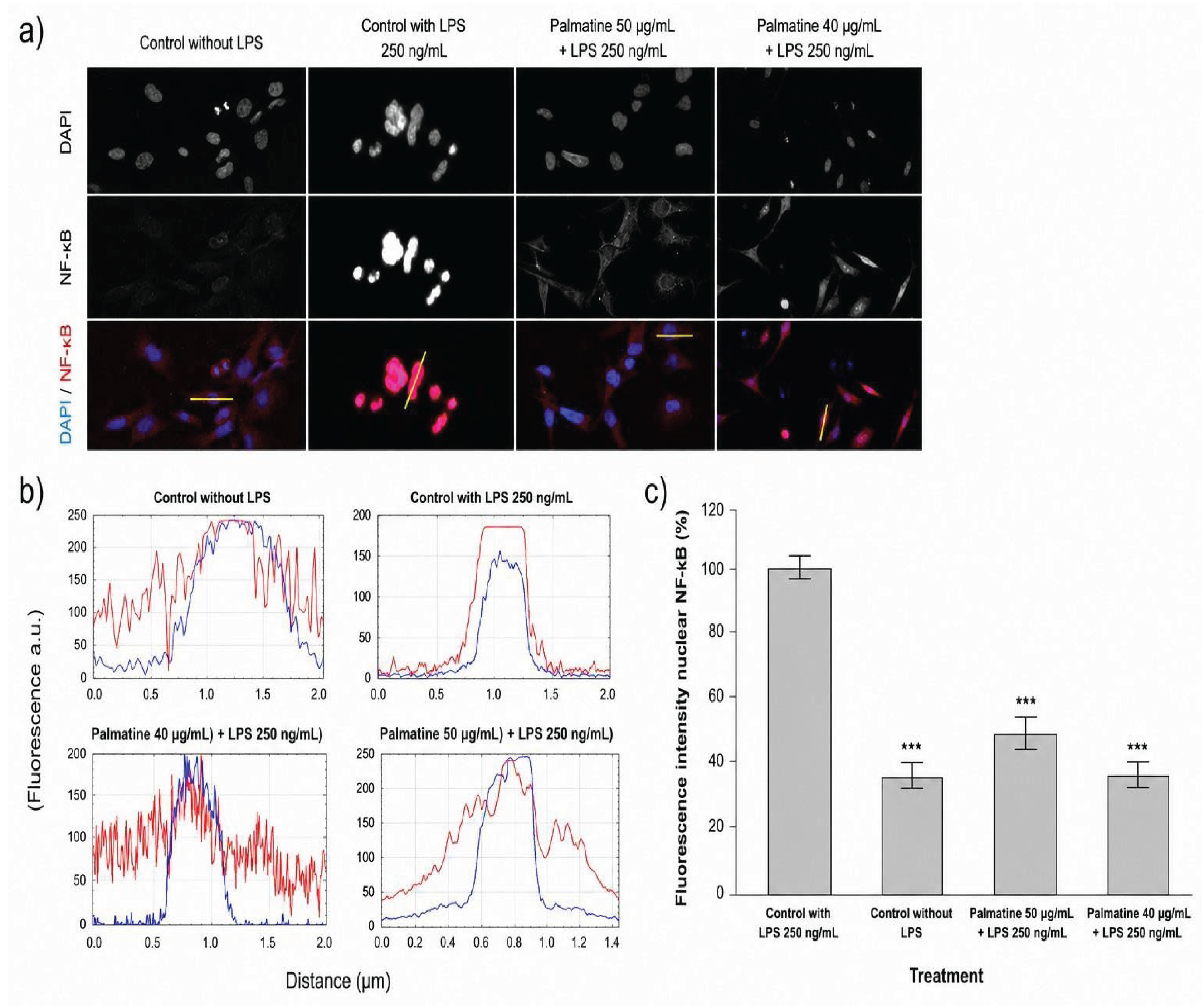
We analyzed the translocation of the transcription factor NF-κB, which mainly occurs in response to proinflammatory stimuli, increasing the expression of chemokines and proinflammatory cytokines in microglia [Zusso, 2019; Yan, 2024]. To this end, we quantified the nuclear translocation of the p50 subunit as an indicator of NF-κB activation. Stimulation with LPS prompted significant translocation of the cytoplasmic p50 subunit to the nucleus compared to the unstimulated group, indicating clear NF-κB activation. Conversely, pretreatment with palmatine significantly reduced LPS-induced nuclear translocation of the p50 subunit, suggesting that palmatine exerts anti-inflammatory effects by decreasing NF-κB translocation (Figure 3).
Figure 3.
Effect of palmatine on NF-κB p50 translocation in unstimulated and LPS-stimulated microglia (HMC3) (250 ng/mL). (a) Representative confocal images show the subcellular distribution of p50. (b) Fluorescence intensity profiles across a line crossing the cytoplasm-nucleus-cytoplasm of a representative cell in the third row of panel (a). (c) Profiles were obtained with ImageJ. Nuclear p50 fluorescence intensity was calculated using ImageJ software. Data are presented as mean ± SEM. Results are expressed as percentages. Analysis was performed by one-way ANOVA, comparing the control group stimulated with LPS to the 199 group without LPS (p < 0.001) and the group with LPS to the groups with LPS and palmatine treatments (p < 0.001).
Figure 3.
Effect of palmatine on NF-κB p50 translocation in unstimulated and LPS-stimulated microglia (HMC3) (250 ng/mL). (a) Representative confocal images show the subcellular distribution of p50. (b) Fluorescence intensity profiles across a line crossing the cytoplasm-nucleus-cytoplasm of a representative cell in the third row of panel (a). (c) Profiles were obtained with ImageJ. Nuclear p50 fluorescence intensity was calculated using ImageJ software. Data are presented as mean ± SEM. Results are expressed as percentages. Analysis was performed by one-way ANOVA, comparing the control group stimulated with LPS to the 199 group without LPS (p < 0.001) and the group with LPS to the groups with LPS and palmatine treatments (p < 0.001).
3.4. Palmatine Treatments Reduced O2∙- Production in LPS-Activated Microglia
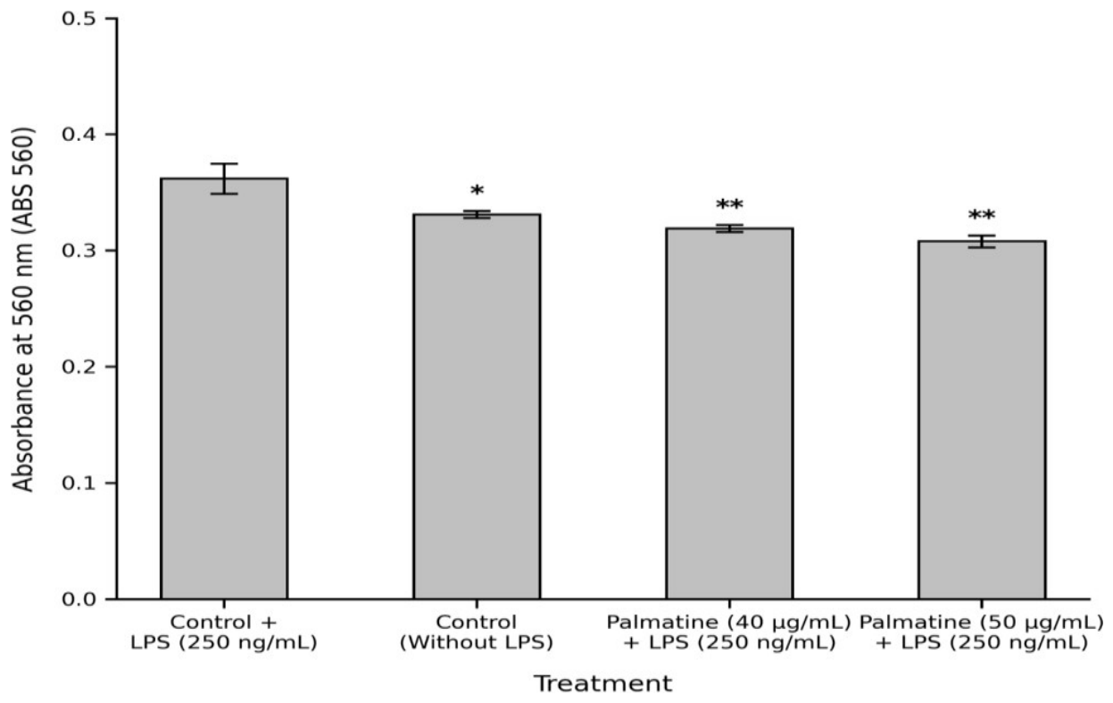
We analyzed whether palmatine decreases ROS production in microglia activated by LPS, particularly measuring O2∙- production, considered the first precursor radical generated by the cells [Kaushal, 2019]. To do this, we quantified the reduction of Nitroblue tetrazolium (NBT) as an indicator of O2∙- production [Stojanović, 2025]. Stimulation with LPS significantly increased absorbance levels compared to the unstimulated group. In contrast, pretreatment with palmatine significantly reduced O2∙- production compared to the LPS-stimulated group (Figure 4).
4. Discussion
As the principal orchestrator of neuroinflammation [Wang 2025], microglia has become a target in research into the etiology of neurological diseases as well as in the search for therapeutic agents that modulate its inflammatory response. Being highly dynamic in the central nervous system, it has been described in different phenotypic or functional states depending on its environment. These various cellular states in response to specific stimuli can be identified via morphological parameters, molecular markers, or inflammatory mediators [Zong, 2026]. The NF-κB complex is considered pleiotropic as it regulates various cellular processes related to cell proliferation, apoptosis, and the proinflammatory response [Shih, 2015]. Therefore, its classical activation with LPS via the TLR4-MD2 complex is a key indicator of a proinflammatory phenotype in micro- glia and induces the secretion of proinflammatory cytokines. On the other hand, there is upregulation of enzymes related to oxidative stress that contribute to progressive neuronal damage [Zusso, 2019; Yan, 2024]. To address our hypothesis that palmatine modulates the translocation of NF-κB in human microglia, we performed an in-silico analysis of the possible putative binding site of palmatine on NF-κB (Figure 1). The molecular docking and PLIP analysis predict a favorable interaction between palmatine and NF-κB, forming hydrophobic interactions and hydrogen bonds with specific amino acid residues, while the estimated binding free energy (ΔG = −6.58 kcal/mol) suggests that palmatine has affinity for NF-κB by binding in a cavity. The in-silico interaction and possible attenuation of palmatine on NF-κB have not been explored, although molecular modeling of other alkaloids with anti-inflammatory potential that may act on NF-κB has been conducted. In this context, molecular docking has shown that certain compounds of different nature can interact directly with NF-κB, with binding energies ranging between −5.85 kcal/mol and −6.65 kcal/mol [Jain, 2022]. These results support the possibility of a strong interaction between NF-κB and palmatine that may prevent its nuclear translocation and negatively regulate the expression of proinflammatory mediators. However, it is interesting to assess in future in silico studies palmatine affinity for other proteins involved in the upstream TLR4-MD2/NF-κB signaling pathway (Figure 1). Previous studies have used different concentrations and exposure times with palmatine [Zeng, 2024; Wang, 2025]. Initially, we assessed a concentration range (40–100 μg/mL) over 24 h (Figure 2) to determine a dose allowing us to evaluate the anti-inflammatory effect of palmatine without compromising cell viability. None of the concentrations tested produced significant cytotoxic effects; this was confirmed by the trypan blue exclusion assay (0.4% w/v), based on the principle that dead cell membranes are permeable to the dye, staining them blue [Zusso, 2019]. Although no concentration compromised viability, we established 40 and 50 μg/mL as the concentrations for immunofluorescence and nitroblue tetrazolium experiments because cell viability remained above 95% at these doses (Figure 2). In vitro and in vivo studies in murine models have reported that palmatine can attenuate proinflammatory activity in microglia stimulated with LPS via the NF-κB pathway and/or inflammatory mediators [Zeng, 2023; Wang, 2025]. As mentioned above, the differences described between the murine and human immune response may limit the reproducibility of results in humans [Flanagan, 2026; Bjornson-Hooper, 2019; Zhou, 2025]. One of the key factors regulating the expression of a diversity of genes involved in the microglial proinflammatory response is the transcription factor NF-κB, which promotes the expression of cytokines and proteins associated with oxidative stress and inflammation [Kowalewska, 2026]. Under homeostatic conditions, NF-κB is inhibited by the IkB protein, remaining in cytoplasm. Stimulation of cellular PRRs with different signals (e.g., LPS) triggers phosphorylation of IkB by the IKK kinase and its subsequent ubiquitination, leading to proteasomal degradation, which results in the release of NF-κB to DNA promoter sequences, increasing the transcription of proinflammatory cytokines [Kowalewska, 2026]. We monitored by immunofluorescence the p50 subunit, which forms a homodimer or heterodimer with RelA (p65) and represents an active form of NF-κB in TLR4-MD2 signaling [Zusso, 2019; Shih, 2015]. Pretreatment with palmatine in microglia significantly reduced nuclear translocation after LPS stimulation (Figure 3). Recent studies in human 259 microglia stimulated with LPS have shown that lycopene inhibits NF-κB signaling and decreases proinflammatory cytokines, facilitating a neuroprotective phenotypic transition [Jifu, 2026]. These findings support that palmatine interferes with a crucial regulator in the production of proinflammatory mediators in response to LPS stimulation. The production of reactive oxygen species (ROS) is a cellular defense mechanism against pathogenic or damaging stimuli; however, chronic ROS production leads to cell damage that results in apoptosis or necrosis of surrounding cells [Ye, 2022]. We investigated whether palmatine decreases ROS production by quantifying O2∙−, a radical produced by the enzyme NADPH oxidase (NOX), the principal enzyme involved in ROS production against proinflammatory stimuli, including LPS and cytokines [Ye, 2022; Yau, 2026]. We observed by the NBT assay that treatments with palmatine significantly reduced O2∙− production compared to the LPS-stimulated control group (250 ng/mL). On the other hand, we also observed that O2∙− production in palmatine-treated groups did not differ significantly from a second control group without LPS stimulation or palmatine treatment. These results are consistent with those observed in the NF-κB p50 immunofluorescence assays, as reduced DNA translocation could contribute to lower ROS production.
Previous studies have reported that activation of NF-κB induces the expression of ROS-producing enzymes, particularly NOX2 [Yan su, 2026]. Therefore, the reduction in ROS following palmatine treatment could be associated with lower NF-κB activation. These findings suggest that palmatine exerts antioxidant effects capable of attenuating oxidative stress in microglia stimulated with LPS (Figure 4).
5. Conclusions
Although our study was conducted in silico and in an in vitro model of human microglia, our findings provide information on an attenuating effect on NF-κB translocation, a crucial factor in proinflammatory activa-tion in immune cells, which has also been observed in murine cell lines both in vivo and in vitro. This study has partially addressed some knowledge gaps about the effect of palmatine on NF-κB that had not been studied in human microglia, opening the possibility for future investigations into the anti-inflammatory effect of palmatine through molecular analyses to strengthen.
Supplementary Materials
The following supporting information can be downloaded at the website of this paper posted on Preprints.org, Table S1: Molecular interactions between palmatine and NF-κB p50 predicted by PLIP.
Author Contributions
Experiment design and support in manuscript writing, MPH; Cell cultures, design and experimental execution, data analysis and manuscript support, ABF; Project management, funding acquisition and data analysis, ETS; NCV. All authors read and approved the final version of this manuscript for publication.
Funding
This research received funding through the UFRO research directorate (DIUFRO) project code: DI24-0109.
Institutional Review Board Statement
This research was approved by the Bioethics Committee of the Universidad de La Frontera, Acta N°: 092-24, Folio: 046-24.
Informed Consent Statement
Not applicable.
Data Availability Statement
The original data are contained within the article/supplementary material. For any additional inquiries, please contact the corresponding authors.
Acknowledgments
We thank Dr. Alejandro Navarro Luarte from the Center for Biomedical Innovation and Research of the Universidad de los Andes, Santiago, Chile, for kindly providing a vial of the HMC3 microglia cell line for this study. We also thank Dr. Enrique Montiel Eulefi from the Department of Medical Morphology at the Universidad de La Frontera, Temuco, Chile, for providing access to his cell culture room. We acknowledge DIUFRO for funding this project under code: DI24-0109. Finally, we thank ANID FONDEQUIP EQM200228 for granting use of the multichannel microscope.
Conflicts of Interest
The authors declare no conflict of interest.
Abbreviations
The following abbreviations are used in this manuscript:
DAPI: 4,6-diamino-2-phenylindole
DMEM: Modified Dulbecco's Eagle Medium DMSO: Dimethyl sulfoxide
KOH: Potassium hydroxide NBT: Nitroblue tetrazolium NOX: NADPH oxidase LPS: Lipopolysaccharide O2- : Superoxide anion
PAMPs: Pathogen-associated molecular patterns PBS: Phosphate-buffered saline
PRR: Pattern recognition receptors ROS: Reactive oxygen species SNC: Central nervous system
References
- Wang, W., Tan, S., Zuo, X. et al. Brain Organoids in Neurodegenerative Disease Modeling: Advances, Applications and Future Perspectives. Mol Neurobiol 63, 142 (2026). [CrossRef]
- Stöberl, N., Maguire, E., Salis, E. et al. Modelos de glía derivados de iPSC humanas para el estudio de la neuroinflamación. J Neuroinflammation 20, 231 (2023). [CrossRef]
- Kwon, H. S., & Koh, S. H. (2020). Neuroinflammation in neurodegenerative disorders: The roles of microglia and astrocytes. Translational Neurodegeneration, 9(42), 1-12. [CrossRef]
- Heiss, C. N., Naylor, A. S., Pesämaa, I., Meshi, A., Cespedes-Cortes, B., Lounès, C., Taki, A., Stenström, K. T., Vizlin-Hodzic, D., Zetterberg, H., Palma, V., Brinkmalm, G., Brinkmalm, A. & Fruhwürth, S. (2026). The temporal and stimuli-specific effects of LPS and IFNγ on microglial activation. Frontiers in Aging Neuroscience, 18. [CrossRef]
- Zusso, M., Lunardi, V., Franceschini, D., Lodola, R., Pagetta, A., Stifani, S., Frasson, A. C., Giusti, P., & Moro, S. (2019). Ciprofloxacin and levofloxacin attenuate microglia-induced neuroinflammation by inhibiting the TLR4/NF-κB signaling pathway. Journal of Neuroinflammation, 16(148), 1-16.
- Kann, O., Almouhanna, F. & Chausse, B. (2022). Interferon γ: a master cytokine in microglia-mediated neural network dysfunction and neurodegeneration. Trends in Neurosciences. [CrossRef]
- Rodríguez-Gómez, J. A., Kavanagh, E., Engskog-Vlachos, P., Engskog, M. K. R., Herrera, A. J., Espinosa-Oliva, A. M., Joseph, B., Hajji, N., Venero, J. L., & Burguillos, M. A. (2020). Microglia: Agents of the CNS pro-inflammatory response. Cells, 9(7), 1717. [CrossRef]
- Gao, C., Jiang, J., Tan, Y. & Chen, S. (2023). Microglia in neurodegenerative diseases: mechanism and potential therapeutic targets. Signal Transduction and Targeted Therapy, 8. [CrossRef]
- Mahmood, A. and Miron, V. E. (2022). Microglia as therapeutic targets for central nervous system remyelination. Current opinion in pharmacology, 63. [CrossRef]
- Long, J., Song, J., Zhong, L., Liao, Y., Liu, L., & Li, X. (2019). Palmatine: A review of its pharmacology, toxicity and pharmacokinetics. Biochimie, 162, 176–184. [CrossRef]
- Zhang, Y., Tong, Y., Li, P., Zhu, L., Zhang, H., Xie, H., Nasir, S., Li, W., Fang, M., Wang, J., Qing, S., Wang, X. & Zhang, W. (2025). Palmatine potentiates cefquinome efficacy against multidrug-resistant Escherichia coli via sulfur/taurine metabolism and oxidative stress modulation. Frontiers in Microbiology, 16. [CrossRef]
- Zeng, J., Pei, H., Wu, H., Chen, W., Du, R., & He, Z. (2024). Palmatine attenuates LPS-induced neuroinflammation through the PI3K/Akt/NF-κB pathway. Journal of Biochemical and Molecular Toxicology, 38(1), e23544. [CrossRef]
- Wang, N., Wei, X., Zhou, P., Wu, W., Liu, J., Zhu, H., Tang, Z., Chai, X., Yang, N., Zhang, Q., Dong, Y., Guo, R., & Zhang, Q. (2025). Palmatine mitigates ischemic brain injury by regulating microglial polarization and sphingolipid metabolism. International Immunopharmacology, 165, 115444. [CrossRef]
- Gros, P. and Casanova, J. (2022). Reconciling Mouse and Human Immunology at the Altar of Genetics. Annual Review of Immunology. [CrossRef]
- Flanagan, B. A., Fuess, L. E., Vrtílek, M., Roth-Monzón, A. J. & Bolnick, D. I. (2024). Macroevolutionary changes in gene expression response to an immune stimulus across the diversity of fishes. bioRxiv. [CrossRef]
- Bjornson-Hooper, Z. B., Fragiadakis, G. K., Spitzer, M. H., Madhireddy, D., McIlwain, D. & Nolan, G. (2019). A comprehensive atlas of immunological differences between humans, mice and non-human primates. bioRxiv. [CrossRef]
- Zhou, W., Xia, S., Wang, C., Yang, Q., Verkhratsky, A. & Niu, J. (2025). Critical analysis of translational potential of rodent models of white matter pathology across a wide spectrum of human diseases. Cell Death and Disease, 16(1). [CrossRef]
- Yan, Y., Bai, S., Han, H., Dai, J., Niu, L., Wang, H., Dong, Q., Yin, H., Yuan, G. & Pan, Y. (2024). Knockdown of trem2 promotes proinflammatory microglia and inhibits glioma progression via the JAK2/STAT3 and NF-κB pathways. Cell communication and signaling, 22. [CrossRef]
- Kaushal, G. P., Chandrashekar, K. & Juncos, L. A. (2019). Molecular Interactions Between Reactive Oxygen Species and Autophagy in Kidney Disease. International Journal of Molecular Sciences, 20. [CrossRef]
- Stojanović, T., Rakić, M., Ćosić, M. V., Pavlović, M. O., Sabovljević, A., Sabovljević, M., Božić, B., Nedeljković, B. B., Vujičić, M. M. & Lunić, T. (2025). Moss Extracts as Natural Neuroprotective Agents: Mitigating LPS-Induced Neuroinflammation and Microglial Activation. Cells, 14. [CrossRef]
- Zong, S., Cui, X., Wu, S. & Lu, Z. (2026). Microglia and neuroinflammation: function, heterogeneity, and crosstalk. Cellular and Molecular Immunology. [CrossRef]
- Shih, R., Wang, C. & Yang, C. M. (2015). NF-kappaB Signaling Pathways in Neurological Inflammation: A Mini Review. Frontiers in Molecular Neuroscience, 8. [CrossRef]
- Jain, P. and Sudandiradoss, C. (2022). Andrographolide-based potential anti-inflammatory transcription inhibitors against nuclear factor NF-kappa-B p50 subunit (NF-κB p50): an integrated molecular and quantum mechanical approach. 3 Biotech, 13. [CrossRef]
- Kowalewska, E., Kamińska, J., Żebrowska-Nawrocka, M., Balcerczak, E., Rybaczek, M., Łysoń, T., Tylicka, M., Wawrusiewicz-Kurylonek, N., Matowicka-Karna, J. & Koper-Lenkiewicz, O. M. (2026). Expression of NF-κB Isoforms and IKK Complex Subunits Differs in Peripheral Blood Mononuclear Cells (PBMCs) of Patients with Meningiomas: A Pilot Study. Life, 16(6). [CrossRef]
- Jifu, C., Lü, L., Li, Y., Gao, X., Pei, B., Liu, L., Xu, Z., Xia, J. & Wang, J. (2026). Lycopene regulates microglial M1/M2 polarization by inhibiting MAPK/NF-κB signaling and alleviates neuroinflammation. International Immunopharmacology, 184. [CrossRef]
- Ye, Y., Jian, Z., Jin, T., Li, Y., Zeng, Z., Xu, Z., Xiong, X. & Gu, L. (2022). NOX2-mediated reactive oxygen species are double-edged swords in focal cerebral ischemia in mice. Journal of Neuroinflammation, 19. [CrossRef]
- Su, Y., Kwon, S. K., Choi, H. S., Han, Y., Park, J., Jang, Y., Choi, J. & Seo, J. (2026). EPA-Derived diHEPAs Attenuate Lipopolysaccharide-Induced Acute Lung Injury by Regulating Inflammation and Redox Homeostasis. International Journal of Molecular Sciences, 27(8). [CrossRef]
- Bugnon, M., Röhrig, U. F., Goullieux, M., Perez, M. A. S., Daina, A., Michielin, O. & Zoete, V. (2024). SwissDock 2024: major enhancements for small-molecule docking with Attracting Cavities and AutoDock Vina. Nucleic Acids Research. [CrossRef]
- Eberhardt, J., Santos-Martins, D., Tillack, A. F. & Forli, S. (2021). AutoDock Vina 1.2.0: New Docking Methods, Expanded Force Field, and Python Bindings. Journal of Chemical Information and Modeling, 61(8). [CrossRef]
- Tian, T., Wu, F., Li, Z., Yang, X., Cao, H., Chen, L., Yin, Y., Hu, L. & Liang, Y. (2026). Oxygen-dependent conformation change of hemoglobin modulates methylmercury binding. Journal of Environmental Science, 163. [CrossRef]
- Matyszczuk, K., Krzepiłko, A., Matyszczuk, K. & Krzepiłko, A. (2022). Model Study for Interaction of Sublethal Doses of Zinc Oxide Nanoparticles with Environmentally Beneficial Bacteria Bacillus thuringiensis and Bacillus megaterium. International Journal of Molecular Sciences, 23. [CrossRef]
- Zhu, A., Liu, J., Liu, Y., Xu, R., Yuan, Y., Sun, Q., Wang, F. & Qu, L. (2026). Metapanax delavayi extract as a neurocutaneous modulator via CRHR1/POMC/MC1R signaling. Scientific Reports. [CrossRef]
Figure 1.
Molecular docking analysis between NF-κB and palmatine. Structure of NF-κB with the predicted palmatine binding site shown in white (a). Three-dimensional structure of palmatine (b). NF-κB–palmatine complex obtained through molecular docking. Interaction analysis carried out with PLIP identified hydrophobic interactions between palmatine and residues Phe56, Pro71, and Lys80, as well as a hydrogen bond with Gly69 (c). The structures were visualized using PyMOL.
Figure 1.
Molecular docking analysis between NF-κB and palmatine. Structure of NF-κB with the predicted palmatine binding site shown in white (a). Three-dimensional structure of palmatine (b). NF-κB–palmatine complex obtained through molecular docking. Interaction analysis carried out with PLIP identified hydrophobic interactions between palmatine and residues Phe56, Pro71, and Lys80, as well as a hydrogen bond with Gly69 (c). The structures were visualized using PyMOL.
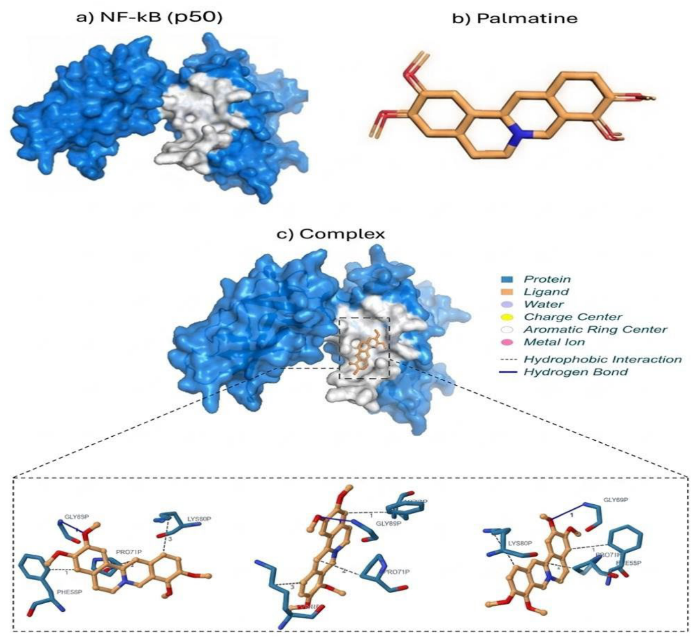
Figure 2.
Viability analysis of human microglia (HMC3) exposed to increasing concentrations of palmatine. Cultures with and without palmatine were incubated for 24h in non-supplemented DMEM medium. Data are presented as mean ± SEM. No statistically significant differences were observed compared to the control group (p > 0.05).
Figure 2.
Viability analysis of human microglia (HMC3) exposed to increasing concentrations of palmatine. Cultures with and without palmatine were incubated for 24h in non-supplemented DMEM medium. Data are presented as mean ± SEM. No statistically significant differences were observed compared to the control group (p > 0.05).
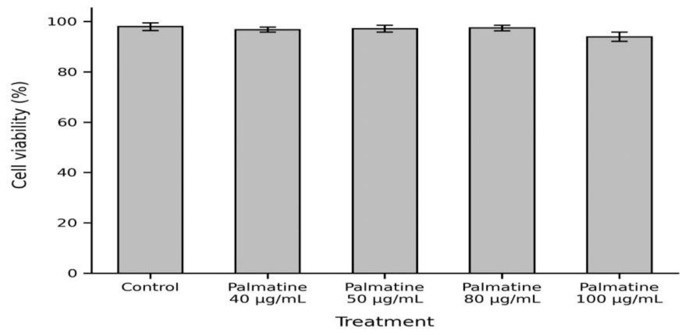
Figure 4.
Effect of palmatine on O2∙- production in unstimulated and LPS-stimulated microglia (HMC3). Measurements were carried out from three independent experiments. Data are presented as mean ± SEM. Analysis was performed by one-way ANOVA, comparing the control group stimulated with LPS to the group without LPS (p < 0.05) and the group with LPS to the groups with LPS and palmatine treatments (p < 0.01).
Figure 4.
Effect of palmatine on O2∙- production in unstimulated and LPS-stimulated microglia (HMC3). Measurements were carried out from three independent experiments. Data are presented as mean ± SEM. Analysis was performed by one-way ANOVA, comparing the control group stimulated with LPS to the group without LPS (p < 0.05) and the group with LPS to the groups with LPS and palmatine treatments (p < 0.01).
Disclaimer/Publisher’s Note: The statements, opinions and data contained in all publications are solely those of the individual author(s) and contributor(s) and not of MDPI and/or the editor(s). MDPI and/or the editor(s) disclaim responsibility for any injury to people or property resulting from any ideas, methods, instructions or products referred to in the content. |
© 2026 by the authors. Licensee MDPI, Basel, Switzerland. This article is an open access article distributed under the terms and conditions of the Creative Commons Attribution (CC BY) license (http://creativecommons.org/licenses/by/4.0/).
Copyright: This open access article is published under a Creative Commons CC BY 4.0 license, which permit the free download, distribution, and reuse, provided that the author and preprint are cited in any reuse.