

Submitted:
09 September 2026
Posted:
09 September 2026
You are already at the latest version
Abstract
Yellow fever (YF) remains a significant public health threat globally. Despite the absence of confirmed outbreaks since the 1992-1993 outbreak, Kenya is classified as a medium-to-high risk country. In January 2022, clusters of acute febrile illness and unexplained deaths were reported in Isiolo County. An integrated epidemiological and entomological investigation was conducted to confirm transmission, characterize risk factors, determine the geographic extent, and assess the risk of spread to the neighboring northeastern counties. Samples from suspected YF cases were tested using molecular and serological methods following WHO guidelines. Concurrent entomological surveys assessed vector species, densities, and breeding habitats to estimate transmission risk and guide vector control. N=194 serum samples (Isiolo and Garissa, n=166 [85.6%]; Wajir, Marsabit and Meru, n=20 [14.4%]) from suspected cases were tested. Serological testing identified 11 presumptive YFV IgM-positive cases (Isiolo, n=7; Garissa, n=1; Meru=2, Nairobi=1). Three of the positive cases from Isiolo (n=2), and Garissa (n=1) were confirmed by PRNT. Entomological surveys identified Aedes aegypti, Ae. furcifer, and Ae. simpsoni, all recognized YFV vectors. This investigation demonstrated active YFV transmission and elevated risk in the affected areas; highlighting the significance of integrated surveillance, vector control, and vaccination efforts to prevent future outbreaks.
Keywords:
yellow fever virus
; outbreak investigation
; Kenya
; arbovirus
; entomological surveillance
; Aedes aegypti
; PRNT
; vector-borne diseases
1. Introduction
Yellow fever (YF) is an acute viral hemorrhagic disease caused by yellow fever virus (YFV), a mosquito-borne flavivirus naturally maintained through sylvatic, intermediate, and urban transmission cycles involving mosquitoes, non-human primates (NHPs), and humans [1]. In East Africa, YFV transmission occurs predominantly through sylvatic cycles involving NHPs and forest-dwelling Aedes africanus, Ae. simpsoni s.l., Ae. luteocephalus, and Ae. vittatus vectors whereas urban transmission in West and Central Africa is largely driven by Ae. aegypti [2]. In Kenya, several confirmed or potential YFV vectors have been documented, including Ae. africanus, Ae. bromeliae, Ae. keniensis, Ae. aegypti, the Ae. simpsoni s.l., Ae. furcifer, Ae. taylori, and Ae. adersi [2,3,4].
Yellow fever virus (YFV), a member of the genus Flavivirus within the family Flaviviridae, is believed to have originated in Africa after its first isolation in Ghana in 1927 [5]. Despite decades of control efforts, it remains endemic across many tropical regions of Africa and South America [6,7], where it frequently causes outbreaks, particularly in regions with low vaccination coverage and suitable ecological conditions for vector proliferation. Africa bears the greatest burden of YF, accounting for approximately 90% of the global estimated 200,000 annual cases and 30,000 deaths [7]. Clinical manifestations range from asymptomatic, mild infection to severe hemorrhagic disease, with reported case-fatality rates (CFR) of 20-50% among severe cases [8]. Climate change, population growth, urbanization, and increased human travel have significantly contributed to the frequent emergence and re-emergence of arboviral diseases, including YF. These factors continue to drive YFV transmission and periodic outbreaks across West, Central, and East Africa, underscoring the virus's persistent public health threat [9].
The Greater East African region has experienced multiple YF outbreaks over the past two decades, including South Sudan (2003, 2018, and 2020), Sudan (2005, 2012, and 2013), Ethiopia (2013, 2018, and 2020), and Uganda (2011, 2016, 2019, 2020, and 2022) [10]. These outbreaks have caused substantial public health, social, and economic disruptions across the region [10].
Historically, Kenya experienced the first major YF outbreak between 1992 and 1993 in the Kerio Valley of the then Rift Valley Province with a CFR of 19% [11]. Entomologic investigations demonstrated a predominantly sylvatic transmission cycle, with Ae. africanus and Ae. keniensis implicated as the principal vectors [12]. The outbreak established Kenya as a country at risk for recurrent YF transmission, and remains a significant historical reference for current and future YFV circulation [11,12]. Prior to this outbreak, serological surveys in northern region of Kenya including Marsabit and Garissa in the early 1960s had reported YFV-neutralizing antibodies in humans [13]. These findings pointed to either undetected endemic transmission or spillover from neighboring countries such as Ethiopia, which had experienced epidemics from 1960-1962 [11,13,14].
Kenya is considered a medium-to-high risk country for YF based on WHO risk assessments under the Eliminate Yellow Fever Epidemics (EYE) Strategy, owing to its ecological suitability for transmission, history of outbreaks, presence of competent vectors, and proximity to endemic countries in the East African region, such as Ethiopia, South Sudan and Uganda. Consequently, Kenya continues to maintain heightened surveillance and preparedness for suspected YF cases. In January 2022, cases of acute febrile illness accompanied by jaundice and unexplained deaths were reported from Merti and Garbatulla sub-counties of Isiolo County in the upper eastern region, raising suspicion of a viral hemorrhagic fever outbreak. The clinical presentation and epidemiological pattern prompted intensified investigations for YF and other hemorrhagic fever pathogens. Given Isiolo's strategic location as a major transport corridor linking northern and central Kenya, concerns emerged regarding the potential for outbreak amplification and spread to other regions. On 4 March 2022, the Ministry of Health (MOH) officially declared a YF outbreak following preliminary results of laboratory testing of samples from suspected cases. By mid-March 2022, 53 suspected cases, including six deaths, had been reported from Isiolo County. The outbreak generated significant public health concern due to low population immunity due to the absence of previous vaccination, and the presence of competent vectors capable of sustaining transmission. Consequently, the WHO assessed the public health risk as high at both national and regional levels [16].
This study describes the integrated human and entomological investigations conducted to confirm the circulation of YFV, characterize epidemiological patterns, assess vector ecology, and evaluate the risk of spread to the wider upper eastern as well as the neighboring northeastern regions.
2. Materials and Methods
Study Sites
Isiolo County (0°21′N, 37°35′E; 25,336 km²; population 268,002 in the 2019 census) lies approximately 285 km north of Nairobi in Kenya's upper eastern region. The county comprises predominantly arid and semi-arid lowlands that support a largely pastoralist economy, with acacia woodlands occurring along seasonal drainage lines and an average population density of approximately 11 people/km². Sampling was conducted in four villages reporting suspected cases: Kambi Juu, Manyatta Ganna, and Biliqo Marara in Merti Sub-county, and Gafarsa in Garbatulla Sub-county. Merti and Garbatulla were the sub-counties at the epicentre of the outbreak.
Garissa County (0°27′S, 39°40′E; 44,753 km²; population 841,353) lies approximately 366 km east-northeast of Nairobi and is situated within the arid landscapes of eastern Kenya. The county is bisected by the Tana River. Of its six sub-counties, Garissa Township Sub-county was selected for sampling because it reported a suspected case on the national line list. As the most densely populated part of the county, it also experiences substantial in-migration from neighboring counties and sub-counties. Six clusters were surveyed: Bula Punda, Bula Reek, Bula Vumbi, Bula Khalif, Bula Mathengeni, and Bula College. At the time of sampling, Garissa had experienced approximately one year without significant rainfall.
Meru County (0°03′N, 37°38′E; population 1,545,714) lies approximately 220 km northeast of Nairobi on the northern and northeastern slopes of Mount Kenya and extends into the volcanic Nyambene Hills. The county exhibits considerable ecological heterogeneity, with annual rainfall ranging from approximately 300 mm in the lower midlands to 2,500 mm in the southeastern highlands. Rainfall is bimodal, with long rains occurring from mid-March to May and short rains from October to December. Consequently, Meru is ecologically distinct from the other study counties. Three clusters reporting suspected cases were sampled: Kinna and Mpeketoni in Igembe North Sub-county, and Kathithini in Igembe Central Sub-county.
Wajir County (1°45′N, 40°04′E; 55,841 km²; population 781,263) lies approximately 625 km northeast of Nairobi and is the most remote and arid of the four study counties. The county receives a mean annual rainfall of approximately 240 mm and experiences recurrent droughts. Suspected yellow fever cases were confined to Wajir South (Habaswein) and Wajir West (Hadado) sub-counties. Sampling was conducted at eight sites/ villages across these sub-counties and Wajir Town: Township, Alimoaw, ADC, Barwahgo, God-Ade, Bula Wagberi, Hadado, and Habaswein (Figure 1).
Human Sample Collection and Transportation
The laboratory was not directly involved in human sample collection. Whole blood samples were obtained at the reporting health facilities using standard phlebotomy procedures by trained healthcare personnel and collected into appropriate Vacutainer tubes (red-top). Following collection, samples were appropriately labeled with unique identifiers and accompanied by the relevant clinical and epidemiological information. The specimens were maintained under cold-chain conditions (2-8°C) and transported to the Kenya Medical Research Institute (KEMRI) Viral Hemorrhagic Fevers (VHF) laboratory in Nairobi within the recommended time frame to preserve sample integrity. Upon receipt, samples were logged into the database and inspected for proper labeling, leakage, hemolysis, clotting, and transport conditions as part of quality assurance (QA) procedures. Whole blood samples were then centrifuged to obtain se-rum. The resulting serum aliquots were transferred into sterile cryovials and either processed immediately for downstream serological and molecular analyses or stored at appropriate temperatures until testing.
Mosquito Collection
Mosquitoes were sampled in the four counties during the dry season. Across the study area, unreliable or the absence of piped water supplies necessitate the long-term storage of water in household containers such as jerrycans, drums, buckets, and tanks. These conditions create favorable habitats for Ae. aegypti, facilitating its proliferation and increasing the risk of arboviral disease transmission.
Adult mosquito collection: Entomological surveillance was conducted in selected indoor, outdoor, peridomestic, and forested habitats to characterize the abundance, distribution, and breeding habitats of Aedes species across all life stages (adults, larvae, pupae, and eggs). Adult mosquitoes were collected using CO₂-baited BG-Sentinel traps deployed during daylight hours to target host-seeking, diurnally active Aedes mosquitoes, CDC light traps deployed overnight to sample nocturnally active mosquitoes, and prokopack aspirators targeting resting adult mosquitoes from indoor and peridomestic environments.
Immature mosquito collection: Larvae and pupae were collected from artificial and natural water-holding containers located indoors and outdoors. Following household stratification, a door-to-door larval survey was conducted according to WHO guidelines [17] to assess Aedes breeding preferences and habitat productivity. Potential breeding sites were systematically inspected using flashlights, and immatures were collected using pipettes for small containers and 300 mL white dippers for larger water bodies. Information on the type, location, and characteristics of each breeding habitat was recorded for mosquito-habitat-association analysis.
Egg collection: To investigate sylvatic breeding sites, dry soil samples were collected from tree holes and transported to the insectary for controlled hatching. This approach enabled the detection of Stegomyia species utilizing natural breeding habitats and is consistent with established methods for sampling cryptic sylvatic Aedes populations [18].
Laboratory rearing immature mosquitoes: The soil samples containing mosquito eggs were processed in a Biosafety Level 2 insectary at KEMRI, where flooding was used to stimulate egg hatching. Emerging larvae were reared to adult mosquitoes.
Mosquito identification: All mosquitoes collected as adults and those emerging from immature stages were identified morphologically to species using standard taxonomic keys [19] under a stereomicroscope. Identification was based on key morphological features, including coloration, scaling patterns, wing characteristics, and leg markings.
Laboratory Methods
Laboratory testing of human suspected YF cases was conducted in accordance with the WHO YF outbreak diagnostic algorithm. The serum samples were subjected to molecular analysis using reverse transcription polymerase chain reaction (RT-PCR) to detect viral RNA, and serological testing for the detection of anti-YFV immunoglobulin M (IgM) antibodies using WHO-recommended assays. Samples yielding positive or equivocal results were referred to the WHO-designated regional YF Reference Laboratory at the Uganda Virus Research Institute (UVRI), Entebbe, Uganda, for differential PRNT to distinguish YFV-specific neutralizing antibodies from those elicited by other antigenically related flaviviruses that also circulate in the region.
Nucleic acid extraction
Viral RNA was extracted from serum samples using the QIAamp Viral RNA Mini Kit (QIAGEN, Hilden, Germany) according to the manufacturer's instructions. Briefly, RNA was purified from serum specimens and eluted in 60 µL of elution buffer. The extracted RNA was immediately used as template for downstream YFV assays by RT-PCR.
Yellow Fever Virus RT-PCR
Detection of YFV RNA was performed using the RealStar® YFV RT-PCR Kit 1.0 (Altona Diagnostics GmbH, Hamburg, Germany) according to the manufacturer's instructions. Amplification and detection were carried out on an ABI 7500 Real-Time PCR System (Thermo Fisher Scientific, Waltham, MA, USA). Briefly, the RT-PCR assay comprised a reverse transcription step at 55°C for 20 minutes, followed by an initial denaturation at 95°C for 2 minutes. Amplification was then performed for 45 cycles at 95°C for 15 seconds, 55°C for 45 seconds, and 72°C for 15 seconds. Fluorescence data were collected during each amplification cycle and analyzed using the instrument's software. Each PCR run included positive, negative, and internal controls provided with the kit to monitor assay performance and ensure result validity. Run acceptance criteria were based on the expected performance of all controls and target amplification profiles (Basile et al., 2022).
CDC 72-Hour MAC-ELISA
A 72-hour Centers for Disease Control and Prevention (CDC) IgM antibody capture enzyme-linked immunosorbent assay (MAC-ELISA) was used to detect YFV-specific IgM antibodies in the serum samples. Briefly, the inner 60 wells of a 96-well Immulon® microtiter plate were coated with 75 µL per well of diluted goat anti-human IgM capture antibody and incubated at 2-8°C for a minimum of 12 hours. Following incubation, the coating solution was discarded, and the plate was blotted dry before the addition of 200 µL of blocking buffer to each well. Plates were incubated at room temperature for 30 minutes and subsequently washed five times with wash buffer. Diluted test sera and controls, including an in-house YF IgM-positive control, a kit-provided YF IgM-positive control, and a negative control, were added at a volume of 50 µL per well in a six-well format for each specimen. The plates were incubated at 37°C for 1 hour and then washed five times. Fifty microliters each of YF viral antigen was added to each designated test well, while a corresponding normal control antigen was added to paired wells. Plates were covered and incubated overnight at 2-8°C in a humidified chamber to facilitate antigen-antibody binding. The following day, antigen solutions were discarded and the plates washed five times. Subsequently, 50µL per well of diluted horseradish peroxidase (HRP)-conjugated monoclonal antibody was added, and the plates were incubated at 37°C for 1 hour, then washed ten times to remove unbound conjugate. Thereafter, 75µL of tetramethylbenzidine (TMB) substrate solution was added to the inner 60 wells and incubated at room temperature in the dark. Development of a blue color indicated the presence of antigen-antibody complexes. The enzymatic reaction was terminated by adding 75µL of TMB stop solution to all wells, resulting in a yellow color change. Optical density (OD) values were measured at 450 nm using a microplate reader. Results were interpreted according to CDC MAC-ELISA criteria using the positive-to-negative (P/N) ratio and normal background reaction (NBR) values. Samples were considered positive when the P/N ratio was ≥ 3.0 and the NBR was ≥ 2.0, provided all assay validity criteria were met. Specimens meeting these thresholds were- classified as presumptive YF IgM-positive and were subsequently considered for confirmatory testing in accordance with the WHO YF outbreak diagnostic algorithm.
Differential Plaque Reduction Neutralization Test (PRNT)
Differential Plaque Reduction Neutralization Test (PRNT) was performed on serum samples that tested presumptively positive for YFV-specific IgM by MAC-ELISA, together with a QA subset comprising 10% of IgM-negative specimens, at the UVRI, to distinguish YFV-specific neutralizing antibodies from those elicited by other antigenically related flaviviruses that also circulate in the region. The assay evaluated virus-specific neutralizing antibody responses against a panel of flaviviruses, including YFV, West Nile virus (WNV), dengue virus serotypes 1-4 (DENV-1 to DENV-4), and Zika virus (ZIKV). Briefly, serial dilutions of heat-inactivated serum samples were incubated with a standardized dose of each virus before inoculation onto confluent monolayers of Vero cells. Following incubation, the reduction in the number of virus-induced plaques relative to virus-only controls was determined, and neutralizing antibody titers calculated. Test results were interpreted based on established WHO criteria. A sample was considered positive for YFV when it demonstrated a significantly higher neutralizing antibody titer against YFV compared with the other flaviviruses tested, thereby providing serological evidence of YFV-specific infection and reducing the likelihood of cross-reactive flavivirus antibody responses.
Data analysis
Larval indices, proportions, and breeding-preference ratios were calculated from household and container inspection records. Ae. aegypti Stegomyia indices were computed according to the standard WHO guidelines as follows:
- House Index (HI): Number of houses positive for Ae. aegypti immatures ÷ number of houses inspected × 100
- Container Index (CI): Number of water-holding containers positive for Ae. aegypti immatures ÷ number of water-holding containers inspected × 100
- Breteau Index (BI): Number of containers positive for Ae. aegypti immatures ÷ number of houses inspected × 100
Indices were calculated at both village and county levels. Entomological risk was evaluated using two established classification frameworks. For YF transmission risk, we applied the WHO Stegomyia threshold criteria used by Agha et al. (2017), whereby areas were classified as low risk (HI < 4%), medium risk (HI = 4-35%), or high risk (HI > 35%).
Container productivity and breeding preferences were assessed by comparing the proportion of positive containers among different container types and by estimating breeding-preference ratios (BPR) using the formula:
A BPR > 1 indicates positive selection or preference for a breeding habitat, BPR = 1 indicates proportional use relative to habitat availability, while BPR < 1 suggests avoidance or under-utilization of the habitat.
3. Results
3.1. Human Surveillance
Between February 2022 and May 2023, a total of 194 suspected YF cases were sampled and tested from 12 counties across Kenya. Of these, 122 (62.9%) specimens were referred for laboratory testing between February and May 2022, coinciding with the peak of the outbreak. Five counties in the upper eastern region, together with Garissa County in the neighboring northeastern region, accounted for the majority of the samples tested (166/194; 85.6%). The remaining 28 (14.4%) samples were received from seven other counties, including Nairobi and Mombasa, Kenya's capital and port city, respectively. Most of these cases reported recent travel to, or epidemiological links with, the affected outbreak areas. None of the 194 samples tested positive for YFV by RT-PCR. In contrast, serological testing identified 11 presumptive YFV IgM-positive samples from four counties, comprising Isiolo (n = 7), Garissa (n = 1), Nairobi (n=1) and Meru (n = 2). Subsequent confirmatory testing using PRNT at the WHO YFV Reference Laboratory at UVRI confirmed three cases of recent YFV infection, including two from Isiolo and one from Garissa. These findings provided laboratory evidence of active YFV circulation in the affected areas despite the absence of RT-PCR-confirmed cases (Figure 2).
3.2. Entomological Findings
3.2.1. Immature Mosquito Collection
Entomological risk assessment was conducted in Isiolo, Garissa, Meru and Wajir counties to assess vector abundance, species composition, and the presence of potential YFV vectors in areas with reported human cases. Overall, 488 households and 2,004 water-holding containers were inspected. Ae. aegypti immatures were detected in 55 (11.3%) households, and 69 (3.4%) containers inspected. All immature mosquitoes recovered from domestic and peri-domestic containers were identified as Ae. aegypti. In Isiolo County, the most affected by the suspected YF outbreak, mosquito collections were undertaken in four villages with suspected YF cases. These included Kambi Juu, Manyatta Ganna, and Biligo Marara villages in Merti Sub-County, as well as Gafarsa village in Garbatulla Sub-County. A total of 118 households were inspected for Aedes breeding habitats, with evidence of Aedes breeding detected in 12 (10.2%) households. A total of 416 water-holding containers were inspected, and 16 (3.8%) were positive for Ae. aegypti larvae and pupae. The Container Index (CI), House Index (HI), Breteau Index (BI), and Pupal Index (PI) were 3.8%, 10.2%, 13.6%, and 0.5%, respectively. Based on these indices, Kambi Juu and Manyatta Ganna were classified as high-risk sites for YF transmission, exceeding the WHO risk threshold (HI > 5%, CI > 3%, and BI > 5), whereas Biliqo Marara and Gafarsa were classified as low-risk sites, with no Ae. aegypti immatures sampled. In Garissa County, surveys targeted sites proximal to Isiolo County which had confirmed human YF cases, as well as Garissa Township which had recorded one confirmed case. A total of 122 houses and 718 indoor and outdoor water-holding containers were inspected for Aedes breeding habitats, and Aedes immatures respectively. Ae. aegypti breeding was detected in 12 houses (HI = 9.8%) and 18 containers (CI = 2.5%), resulting in a BI of 14.8 and 56 immatures. These indices exceeded the WHO risk thresholds, indicating an elevated risk for YF transmission in Garissa Township. Isiolo, Garissa, and Wajir were classified as medium risk for YF based on WHO house index thresholds, while Meru was low risk. Vector infestation was highly clustered, with 14 of 21 clusters classified as medium risk and 7 as low risk (Table 1).
3.2.2. Container Type and Breeding Preference
Jerrycans were the most common container type, accounting for 1,378 (68.8%) of the 2,004 containers inspected. However, only 12 were positive for Aedes immatures, giving a positivity rate of 0.9% and a BPI ratio of 0.25. In contrast, plastic drums represented only 19.0% of inspected containers (380/2,004) but accounted for 42 (60.9%) of the 69 positive containers, with a BPI of 3.2. Collectively, plastic drums, metal drums, and plastic tanks comprised 20.0% of containers inspected but contributed 48 (69.6%) of the 69 positive containers. All three underground tanks and half of the plastic tanks inspected were positive for Ae. aegypti immatures However, these container types were represented by small sample sizes, limiting interpretation (Table 2).
3.2.4. Adult Mosquito Collection
Overall, 7,835 adult mosquitoes were collected from Isiolo, Garissa, and Meru. Culex pipiens s.l. dominated collections across the three counties, comprising 64.2% of specimens overall. Adult Ae. aegypti were few, with only 113 specimens collected. A total of 1,538 adult mosquitoes were collected in Isiolo County using BG-Sentinel and CDC light traps. Culex pipiens s.l. was the most abundant species, accounting for 1,115 (72.4%) of the specimens, followed by Ae. furcifer (218, 14.5%), Cx. univittatus (58, 3.8%), Cx. zombaensis (14, 1.1%), Ae. aegypti (64, 4.2%), and Anopheles gambiae 27 (1.8%). In Garissa County, 1,253 adult mosquitoes were collected and grouped into 79 pools for laboratory analysis. Culex pipiens s.l. dominated collections (91.0%), followed by Cx. univittatus (7.7%), while only a single Ae. aegypti specimen was collected, despite the presence of immature stages at the same sites. Meru recorded the largest and most diverse mosquito collection (5,044 specimens, 16 taxa). No adult sampling was conducted in Wajir County (Table 3).
3.2.4. Tree hole Samples
Flooding of tree-hole soil collected in Isiolo yielded 223 mosquitoes, predominantly Ae. furcifer (n=218, 97.8%), with Ae. simpsoni s.l (n=3, 1.3%) and Ae. aegypti (n=2, 0.9%) also emerging, all of which are important vectors involved in the sylvatic transmission cycle of YFV. Notably, Ae. furcifer was detected only from dormant eggs in tree-hole habitats and was not captured by adult trapping methods, underscoring the importance of egg-bank sampling for detecting sylvatic vectors.
4. Discussion
The 2022 YF outbreak in Kenya occurred in a region characterized by frequent YF activity, as demonstrated by the historical and recent outbreaks in Ethiopia, Uganda, and South Sudan [21,22]. The largest documented outbreak in the region occurred in Ethiopia from 1960 to 1962, with a CFR of 30% [21]. Evidence of YF immunity among unvaccinated populations in northern Kenya during a 1966-1967 survey was attributed to spillover from the Ethiopian outbreak, providing the earliest evidence of human exposure [13]. Together, these observations highlight the persistent risk of YF emergence in Kenya and the East African region [21,22].
The 2022 outbreak represents the first major YF outbreak in Kenya since the 1992-1993 outbreak, which had a reported 19% CFR [11]. Although no confirmed YF outbreaks were reported between 1993 and 2022, serological evidence has indicated sustained low-level circulation in the Rift Valley, western, and northern regions [23,24,25,26]. The detection of two travel-related YF cases in Nairobi during the 2016 Angola outbreak further underscored Kenya's vulnerability to virus importation that could trigger local transmission [27,28].
Our findings provide epidemiological, serological, and entomological evidence of active YFV transmission in Isiolo and Garissa counties in 2022. Although only three cases were confirmed by PRNT, the detection of YFV-specific antibodies, vectors, and high larval indices support local virus circulation. Among the 194 suspected cases, none was positive by RT-PCR, 11 were presumptive positives by IgM, while three were confirmed by PRNT. This reflects the transient nature of YFV viremia and the persistence of IgM antibodies following infection. The confirmation of only 3 of the 11 presumptive positive cases by PRNT underscores the significance of confirmatory testing in settings where multiple flaviviruses co-circulate, resulting in serological cross-reactivity [10,29], false-positive findings, and potentially leading to misdiagnoses.
The outbreak was confirmed in March 2022, after suspected cases were first identified in January, with a CFR of 11.3% [16]. The CFR was comparable to that reported in the Democratic Republic of the Congo but lower than those observed in Uganda and Ethiopia [30,31,32]. Differences in CFRs between countries may reflect variations in surveillance, prompt outbreak detection, healthcare access, and response effectiveness. These findings highlight the importance of early recognition and reporting of suspected arboviral diseases by healthcare workers and communities [33].
Entomological investigations identified active Ae. aegypti breeding in the two counties with confirmed cases, with larval indices exceeding the WHO thresholds associated with increased arboviral transmission risk. Isiolo recorded larval indices, HI = 10.2%, BI = 13.6%, and CI=3.8%, while Garissa had an HI of 9.8%, BI = 14.6, and CI = 2.5%. These high indices, together with PRNT-confirmed human infections, indicate favorable conditions for vector breeding and suggest a strong association between vector abundance and transmission risk [16,34,35].
Although Culex spp. predominated adult mosquito collections compared to Ae. aegypti, these findings should be interpreted with caution because sampling occurred several months after the onset and confirmation of the outbreak. Thus, the data may not provide an accurate representation of the mosquito species composition and densities during the peak transmission period. The collection of Ae. aegypti as both immatures and adults, even though relatively few specimens, confirms the presence of known vectors with the potential to sustain transmission [34,36].
The detection of Ae. simpsoni s.l. and Ae. furcifer in Isiolo County, both recognized sylvatic YFV vectors and previously implicated in outbreaks in Africa [2,3,4], was significant. Their presence in areas inhabited by NHPs, suggests that the outbreak likely involved a sylvatic transmission cycle with spillover to humans. This pattern is consistent with the epidemiology of YF in East Africa, where sylvatic and intermediate transmission cycles are commonly reported. The same vector species, including Ae. aegypti, Ae. africanus, and Ae. simpsoni s.l., were reported during the 2016 Uganda outbreak [30].
Household water-storage practices likely contributed to sustaining vector breeding and densities. Jerrycans and plastic drums were the most productive breeding habitats inspected, accounting for 87% of positive containers in Isiolo and 97% in Garissa, with immature Ae. aegypti being the only species collected. These findings are in agreement with previous studies identifying domestic water-storage containers as major Ae. aegypti breeding sites [20,37]. In the arid and semi-arid settings of both counties, limited access to piped water necessitates long-term water storage, with containers mostly remaining uncovered and rarely completely emptied, allowing for uninterrupted mosquito breeding. These results underscore the need for targeted vector control strategies that address local water-storage practices and environmental management [37].
Vaccination remains the most effective strategy for preventing YF outbreaks. At the time of the outbreak, routine YF vaccination in Kenya was limited to five high-risk counties and excluded Isiolo and Garissa [38]. Following outbreak confirmation, a vaccination campaign under the EYE strategy, alongside active case finding, risk communication, improved case management, and strengthened sample referral systems, helped control the outbreak in high-risk sub-counties of Isiolo (Merti and Garbatula) and Garissa (Lagdera and Balambala).
The Global YF Laboratory Network (GYFLaN) equally played an important role in outbreak confirmation and response by facilitating referral of presumptive YF IgM-positive samples to the UVRI for PRNT testing. Through GYFLaN, the KEMRI VHF Laboratory supported surveillance, diagnostic confirmation, quality assurance, and capacity strengthening, demonstrating the importance of regional laboratory networks in outbreak preparedness and response.
Limitation of the response: The response had several limitations. Entomological investigations were conducted months after outbreak onset and confirmation, potentially underestimating vector densities during peak transmission period. The absence of RT-PCR-positive samples limited direct confirmation of acute infections, likely due to delayed specimen collection beyond the viremic period. In addition, the small number of PRNT-confirmed cases restricted detailed assessment of transmission dynamics, while entomological sampling in selected outbreak areas may not fully represent vector distribution across the affected counties. Despite these limitations, the combined epidemiological, serological, and entomological findings provide strong evidence of local YFV transmission.
5. Conclusions
This outbreak provides the first evidence of active YFV circulation in Isiolo and Garissa counties, suggesting an expansion of YF risk in Kenya. PRNT-confirmed infec-tions, high Ae. aegypti vector densities, and the presence of sylvatic vectors (Ae. simpsoni s,l, and Ae. furcifer) indicate the involvement of both domestic and sylvatic transmission cycles. Given previous reports of dengue and Rift Valley fever virus circulation in these counties [39,40,41], sustained integrated surveillance, strengthened outbreak preparedness, and routine YF vaccination in Isiolo and Garissa are needed to mitigate against future outbreak risks.
Author Contributions
Conceptualization, S.L.K., J.L., S.K., D.L., and E.O.; methodology, S.L.K., J.L., S.L., A.N., S.O., F.M., B.C., J.M., E.C., H.K., V.O.; software, S.O.; validation, S.L.K., and J.L.; investigation, D.L., and E.O.; resources, S.L.K., S.K.; data curation, S.O., S.O., and S.L.K.; writing—original draft preparation, S.L.K., F.M., J.M.; writing—review and editing, J.L., E.C., F.M., J.M., S.O., S.L., H.K., A.N., V.O., and S.K.; supervision, S.K.; project administration, S.L.K. and S.K. All authors have read and agreed to the published version of the manuscript. All.
Funding
This research received no external funding.
Institutional Review Board Statement
Clinical specimens were collected at the respective health facilities, as part of standard patient care and public health surveillance activities, and subsequently referred to the KEMRI VHF Laboratory for YF diagnostic testing and outbreak confirmation. All data were anonymized before analysis to support outbreak investigation and response activities. Scientific and ethical approval to report findings from de-identified outbreak samples was obtained from the KEMRI Scientific and Ethics Review Unit (SERU) through a running Outbreak Investigation Protocol, approval No. SSC#3035.
Informed Consent Statement
The data presented in this report were generated as part of a routine public health response to a suspected YF outbreak and did not constitute human subjects research, hence, informed consenting was not performed.
Data Availability Statement
All data generated is provided in the manuscript.
Acknowledgments
We acknowledge the dedication and contributions of healthcare workers in the affected regions for their role in case detection, sample collection, and referral of suspected cases for confirmatory testing. We also thank the Global Yellow Fever Laboratory Network (GYFLaN) and the Global Alliance for Vaccines and Immunization (GAVI) for their invaluable support through the provision of reagents, technical assistance, and laboratory capacity strengthening, which were critical to the outbreak investigation and response.
Conflicts of Interest
The authors declare no conflicts of interest.
Abbreviations
The following abbreviations are used in this manuscript:
| Ae. | Aedes |
| BI | Breteau Index |
| BPR | Breeding preference ratio |
| CDC | Centre for Disease Control |
| CI | Container Index |
| CFR | Case-fatality rates |
| Cx. | Culex |
| DENV | Dengue virus |
| EYE | Eliminate Yellow Fever Epidemics |
| GAVI | Global Alliance for Vaccines and Immunization |
| GYFLaN | Global Yellow Fever Laboratory Network |
| HI | House Index |
| IgM | Immunoglobulin |
| KEMRI | Kenya Medical Research Institute |
| MAC-ELISA | Monoclonal antibody capture - enzyme-linked immunosorbent assay |
| NHPs | Non-human primates |
| NBR | Normal Background Reaction |
| PI | Pupal Index |
| PRNT | Plague Reduction Neutralization Test |
| QA | quality assurance |
| RNA | Ribonucleic acid |
| RT-PCR | Reverse Transcriptase polymerase chain reaction |
| SERU | Scientific and Ethics Review Unit |
| UVRI | Uganda Virus Research Institute |
| VHF | Viral hemorrhagic fever laboratory |
| WHO | World Health Organization |
| WNV | West Nile virus |
| YF | Yellow fever |
| YFV | Yellow fever virus |
| ZIKV | Zika virus |
References
- Oyono, M.G.; Kenmoe, S.; Abanda, N.N.; Takuissu, G.R.; Ebogo-Belobo, J.T.; Kenfack-Momo, R.; Kengne-Nde, C.; Mbaga, D.S.; Tchatchouang, S.; Kenfack-Zanguim, J.; Lontuo Fogang, R.; Zeuko'o Menkem, E.; Ndzie Ondigui, J.L.; Kame-Ngasse, G.I.; Magoudjou-Pekam, J.N.; Bowo-Ngandji, A.; Nkie Esemu, S.; Ndip, L. Epidemiology of yellow fever virus in humans, arthropods and non-human primates in sub-Saharan Africa: A systematic review and meta-analysis. PLoS Negl. Trop. Dis. 2022, 16(7), e0010610. [Google Scholar] [CrossRef] [PubMed]
- Ellis, B.R.; Sang, R.C.; Horne, K.M.; Higgs, S.; Wesson, D.M. Yellow fever virus susceptibility of two mosquito vectors from Kenya, East Africa. Trans. R Soc. Trop. Med. Hyg. 2012, 106(6), 387–9. [Google Scholar] [CrossRef] [PubMed]
- Nwangwu, U.C.; Soumare, H.; Oguzie, J.U.; Nwachukwu, W.E.; Onwude, C.O.; Dogunro, F.A.; Diallo, M.; Ezihe, C.K.; Agashi, N.O.; Eloy, E.I.; Anokwu, S.O.; Anioke, C.C.; Ikechukwu, L.C.; Nwosu, C.M.; Nwaogo, O.N.; Ngwu, I.M.; Onyeanusi, R.N.; Okoronkwo, A.I.; Orizu, F.U.; Etiki, M.O.; Onuora, E.N.; Okeke, P.C.; Chukwuekezie, O.C.; Ochu, J.C.; Happi, A.N.; Ibrahim, S.S.; Ifedayo, A.; Ihekweazu, C.; Happi, C.T. Nationwide surveillance detects yellow fever and chikungunya viruses in multiple Aedes mosquito species in Nigeria. Parasit. Vectors 2025, 18(1), 443. [Google Scholar] [CrossRef] [PubMed]
- Agha, S.B.; Tchouassi, D.P.; Turell, M.J.; Bastos, A.D.S.; Sang, R. Risk assessment of urban yellow fever virus transmission in Kenya: is Aedes aegypti an efficient vector? Emerg. Microbes Infect. 2022, 11(1), 1272–1280. [Google Scholar] [CrossRef] [PubMed]
- Staples, J.E.; Monath, T.P. Yellow fever: 100 years of discovery. JAMA 2008, 300(8), 960–2. [Google Scholar] [CrossRef] [PubMed]
- Barrett, A.D.; Higgs, S. Yellow fever: a disease that has yet to be conquered. Annu Rev. Entomol. 2007, 52, 209–29. [Google Scholar] [CrossRef] [PubMed]
- WHO. Yellow fever. 2025 20 October 2025. Available online: https://www.who.int/news-room/fact-sheets/detail/yellow-fever.
- Stock, N.K.; Laraway, H.; Faye, O.; Diallo, M.; Niedrig, M.; Sall, A.A. Biological and phylogenetic characteristics of yellow fever virus lineages from West Africa. J. Virol. 2013, 87(5), 2895–907. [Google Scholar] [CrossRef] [PubMed]
- Petersen, E.; Valdoleiros, S.R.; McCloskey, B.; Traore, T.; Sharma, A.; Azhar, E.I.; Zumla, A. Yellow fever across Africa, the Middle East, and Asia: New vaccines and closing the preparedness gap. Int. J. Infect. Dis. 2026. 169, 108768. [Google Scholar] [CrossRef] [PubMed]
- WHO. Yellow fever- Africa Region-East, West and Central African Region Disease Outbreak News. 2023 3 January 2023. Available online: https://www.who.int/emergencies/disease-outbreak-news/item/2022-DON431.
- Sanders, E.J.; Marfin, A.A.; Tukei, P.M.; Kuria, G.; Ademba, G.; Agata, N.N.; Ouma, J.O.; Cropp, C.B.; Karabatsos, N.; Reiter, P.; Moore, P.; Gubler, D.J. First recorded outbreak of yellow fever in Kenya, 1992-1993. I. Epidemiologic investigations. Am. J. Trop. Med. Hyg. 1998, 59(4), 644–9. [Google Scholar] [CrossRef] [PubMed]
- Sanders, E.J.; Marfin, A.A.; Tukei, P.M.; Kuria, G.; Ademba, G.; Agata, N.N.; Ouma, J.O.; Cropp, C.B.; Karabatsos, N.; Reiter, P.; Moore, P.S.; Gubler, D.J. First recorded outbreak of yellow fever in Kenya, 1992-1993. I. Epidemiologic investigations. Am. J. Trop. Med. Hyg. 1998, 59(4), 644–9. [Google Scholar] [CrossRef] [PubMed]
- Henderson, B.E.; Metselaar, D.; Cahill, K.; Timms, G.L.; Tukei, P.M.; Williams, M.C. Yellow fever immunity surveys in northern Uganda and Kenya and eastern Somalia, 1966-67. Bull. World Health Organ. 1968, 38(2), 229–37. [Google Scholar] [PubMed]
- Sutherland, L.J.; Cash, A.A.; Huang, Y.J.; Sang, R.C.; Malhotra, I.; Moormann, A.M.; King, C.L.; Weaver, S.C.; King, C.H.; LaBeaud, A.D. Serologic evidence of arboviral infections among humans in Kenya. Am. J. Trop. Med. Hyg. 2011, 85(1), 158–61. [Google Scholar] [CrossRef] [PubMed]
- WHO. Eliminate yellow fever epidemics (EYE) strategy 2017-2026. 2017. Available online: https://www.who.int/initiatives/eye-strategy.
- WHO. Disease Outbreak News; Yellow Fever - Kenya. 2022. Available online: https://www.who.int/emergencies/disease-outbreak-news/item/yellow-fever-kenya.
- WHO. Vector-borne diseases. 2020 2 Mar 2020. Available online: https://www.who.int/news-room/fact-sheets/detail/vector-borne-diseases.
- Diallo, D.; Diagne, C.T.; Hanley, K.A.; Sall, A.A.; Buenemann, M.; Ba, Y.; Dia, I.; Weaver, S.C.; Diallo, M. Larval ecology of mosquitoes in sylvatic arbovirus foci in southeastern Senegal. Parasit. Vectors 2012, 5, 286. [Google Scholar] [CrossRef] [PubMed]
- Edwards, F.W. Mosquitoes of the Ethiopian Region III. – Culicine adults and pupae; Printed by order of the Trustees, British Museum (Natural History): London, 1941. [Google Scholar]
- Agha, S.B.; Tchouassi, D.P.; Bastos, A.D.S.; Sang, R. Assessment of risk of dengue and yellow fever virus transmission in three major Kenyan cities based on Stegomyia indices. PLoS Negl. Trop. Dis. 2017, 11(8), e0005858. [Google Scholar] [CrossRef] [PubMed]
- Lilay, A.; Asamene, N.; Bekele, A.; Mengesha, M.; Wendabeku, M.; Tareke, I.; Girmay, A.; Wuletaw, Y.; Adossa, A.; Ba, Y.; Sall, A.; Jima, D.; Mengesha, D. Reemergence of yellow fever in Ethiopia after 50 years, 2013: epidemiological and entomological investigations. BMC Infect. Dis. 2017, 17(1), 343. [Google Scholar] [CrossRef] [PubMed]
- Adedolapo, S.; Idumah, G.; Nwachukwu, W.; Luis, M.H.; Nick, J.; Simeon, C. A systematic narrative review of risk drivers of yellow fever reemergence in Africa from 1974 to 2025. Discov. Public Health 2026, 23(1), 664. [Google Scholar] [CrossRef]
- Kibathi, M.H.; Chepkorir, E.; Mabeya, S.N.; Tchouassi, D.P.; Sang, R. Seroprevalence of dengue, yellow fever, and related flaviviruses among the rural human population in Nguruman and Kerio Valley, Kenya. Front Virol. 2024, 4. [Google Scholar] [CrossRef] [PubMed]
- Inziani, M.; Adungo, F.; Awando, J.; Kihoro, R.; Inoue, S.; Morita, K.; Obimbo, E.; Onyango, F.; Mwau, M. Seroprevalence of yellow fever, dengue, West Nile and chikungunya viruses in children in Teso South Sub-County, Western Kenya. Int. J. Infect. Dis. 2020, 91, 104–110. [Google Scholar] [CrossRef] [PubMed]
- Chepkorir, E.; Tchouassi, D.P.; Konongoi, S.L.; Lutomiah, J.; Tigoi, C.; Irura, Z.; Eyase, F.; Venter, M.; Sang, R. Serological evidence of Flavivirus circulation in human populations in Northern Kenya: an assessment of disease risk 2016-2017. Virol. J. 2019, 16(1), 65. [Google Scholar] [CrossRef] [PubMed]
- Kwallah, A.o.; Inoue, S.; Thairu-Muigai, A.W.; Kuttoh, N.; Morita, K.; Mwau, M. Seroprevalence of yellow fever virus in selected health facilities in Western Kenya from 2010 to 2012. Jpn. J. Infect. Dis. 2015, 68(3), 230–4. [Google Scholar] [CrossRef] [PubMed]
- WHO. Yellow fever outbreak in Kenya;Two imported cases, 2016. 2016 06/08/2026. Available online: https://www.who.int/emergencies/disease-outbreak-news/item/6-april-2016-yellow-fever-kenya-en.
- Kraemer, M.U.G.; Faria, N.R.; Reiner, R.C., Jr.; Golding, N.; Nikolay, B.; Stasse, S.; Johansson, M.A.; Salje, H.; Faye, O.; Wint, G.R.W.; Niedrig, M.; Shearer, F.M.; Hill, S.C.; Thompson, R.N.; Bisanzio, D.; Taveira, N.; Nax, H.H.; Pradelski, B.S.R.; Nsoesie, E.O.; Murphy, N.R.; Bogoch, I.I.; Khan, K.; Brownstein, J.S.; Tatem, A.J.; de Oliveira, T.; Smith, D.L.; Sall, A.A.; Pybus, O.G.; Hay, S.I.; Cauchemez, S. Spread of yellow fever virus outbreak in Angola and the Democratic Republic of the Congo 2015-16: a modelling study. Lancet Infect. Dis. Erratum in: Lancet Infect Dis. 2019 Apr;19(4):e109. doi: 10.1016/S1473-3099(19)30079-9. 2017, 17(3), 330–338. [Google Scholar] [CrossRef] [PubMed]
- WHO. Laboratory Manual for Yellow Fever, 2nd Edition; Geneva, 2024. [Google Scholar]
- Kwagonza, L.; Masiira, B.; Kyobe-Bosa, H.; Kadobera, D.; Atuheire, E.B.; Lubwama, B.; Kagirita, A.; Katushabe, E.; Kayiwa, J.T.; Lutwama, J.J.; Ojwang, J.C.; Makumbi, I.; Ario, A.R.; Borchert, J.; Zhu, B.P. Outbreak of yellow fever in central and southwestern Uganda, February-may 2016. BMC Infect. Dis. 2018, 18(1), 548. [Google Scholar] [CrossRef] [PubMed]
- Mengesha, T.M.; Beyene, B.; Ayele, W.; Abebe, A.; Tareke, I.; Sall, A.; Yactayo, S.; Shibeshi, M.E.; Staples, E.; Belay, D.; Lilay, A.; Alemu, A.; Alemu, E.; Kume, A.; H/Mariam, A.; Ronveaux, O.; Tefera, M.; Kassa, W.; Bekele; Weyessa, A.; Jima, D.; Kebede, A.; Tayachew, A. Sero-prevalence of yellow fever and related Flavi viruses in Ethiopia: a public health perspective. BMC Public Health 2018, 18(1), 1011. [Google Scholar] [CrossRef] [PubMed]
- Tague, C.; Budair, M.M.; Rusho, M.A.; Basaria, A.A.A.; Tariq, R.; Yokolo, H.; Ekouo, J.; Naaz, F.; Makeda, D.; Karegeya, A.; Mugisha, M.J.M.; Wei, C.R.; Hangi, S.; Kihanduka, E.; Onesime, J.; Rugendabanga, E.; Akilimali, A. Exploring the national burden and challenges in the fight against yellow fever in the Democratic Republic of Congo: a review. Ann. Med. Surg. (Lond) 2025, 87(10), 6574–6579. [Google Scholar] [CrossRef] [PubMed]
- Yahouédo, A.G.; Merle, C.S.; Chanda, E.; Rabe, I.B.; Fouque, F.; Rojas, D.P.; Aseffa, A.; Halleux, C.M.; Velayudhan, R. Engaging communities in the control of arboviral diseases: insights from the African region. PLoS Negl. Trop. Dis. 2025, 19(7), e0013300. [Google Scholar] [CrossRef] [PubMed]
- Barrett, A.D.; Monath, T.P. Epidemiology and ecology of yellow fever virus. Adv. Virus Res. 2003, 61, 291–315. [Google Scholar] [CrossRef] [PubMed]
- WHO. Comprehensive Guidelines for Prevention and Control of Dengue and Dengue Haemorrhagic Fever. Geneva, 2011. Available online: https://iris.who.int/handle/10665/204894.
- Gubler, D.J. Dengue, Urbanization and Globalization: The Unholy Trinity of the 21(st) Century. Trop. Med. Health 2011, 39((4) Suppl, 3–11. [Google Scholar] [CrossRef] [PubMed]
- Khan, J.; Adil, M.; Tsheten, T.; Manrique-Saide, P.; Zhang, D.; Aziz, A.; Lv, Z.; Chen, T. Assessing Aedes mosquito larval indicators, dengue virus infection rates, and risk factors in Khyber Pakhtunkhwa: Insights for improved vector control strategies. PLoS Negl. Trop. Dis. 2025, 19(7), e0013252. [Google Scholar] [CrossRef] [PubMed]
- Ministry of Health; K. Kenya National Immunization Policy Guidelines 2023 2023 06/08/2026. Available online: http://guidelines.health.go.ke:8000/media/Kenya_National_Immunization_Policy_Guidelines_Version_signed.pdf.
- Affognon, H.; Mburu, P.; Hassan, O.A.; Kingori, S.; Ahlm, C.; Sang, R.; Evander, M. Ethnic groups' knowledge, attitude and practices and Rift Valley fever exposure in Isiolo County of Kenya. PLoS Negl. Trop. Dis. 2017, 11(3), e0005405. [Google Scholar] [CrossRef] [PubMed]
- Konongoi, L.; Ofula, V.; Nyunja, A.; Owaka, S.; Koka, H.; Makio, A.; Koskei, E.; Eyase, F.; Langat, D.; Schoepp, R.J.; Rossi, C.A.; Njeru, I.; Coldren, R.; Sang, R. Detection of dengue virus serotypes 1, 2 and 3 in selected regions of Kenya: 2011-2014. Virol. J. 2016, 13(1), 182. [Google Scholar] [CrossRef] [PubMed]
- Sang, R.; Kioko, E.; Lutomiah, J.; Warigia, M.; Ochieng, C.; O'Guinn, M.; Lee, J.S.; Koka, H.; Godsey, M.; Hoel, D.; Hanafi, H.; Miller, B.; Schnabel, D.; Breiman, R.F.; Richardson, J. Rift Valley fever virus epidemic in Kenya, 2006/2007: the entomologic investigations. Am. J. Trop. Med. Hyg. 2010, 83((2) Suppl, 28–37. [Google Scholar] [CrossRef] [PubMed]
Figure 1.
Map of Kenya showing the counties where yellow fever (YF) outbreak investigations were conducted.
Figure 1.
Map of Kenya showing the counties where yellow fever (YF) outbreak investigations were conducted.
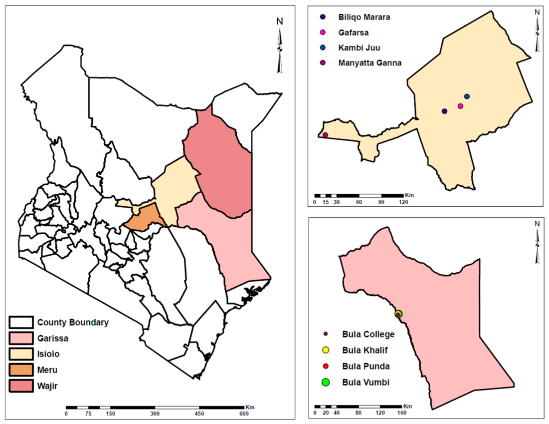
Figure 2.
Yellow fever diagnostic outcomes among suspected cases received from different counties in Kenya between February 2022 to May 2023. (A) Diagnostic cascade flowchart depicting outcomes for 194 suspected cases tested by qRT-PCR, IgM ELISA, and plaque reduction neutralization testing (PRNT). (B) County-level breakdown of samples tested, presumptive IgM positivity, and PRNT-confirmed cases.
Figure 2.
Yellow fever diagnostic outcomes among suspected cases received from different counties in Kenya between February 2022 to May 2023. (A) Diagnostic cascade flowchart depicting outcomes for 194 suspected cases tested by qRT-PCR, IgM ELISA, and plaque reduction neutralization testing (PRNT). (B) County-level breakdown of samples tested, presumptive IgM positivity, and PRNT-confirmed cases.
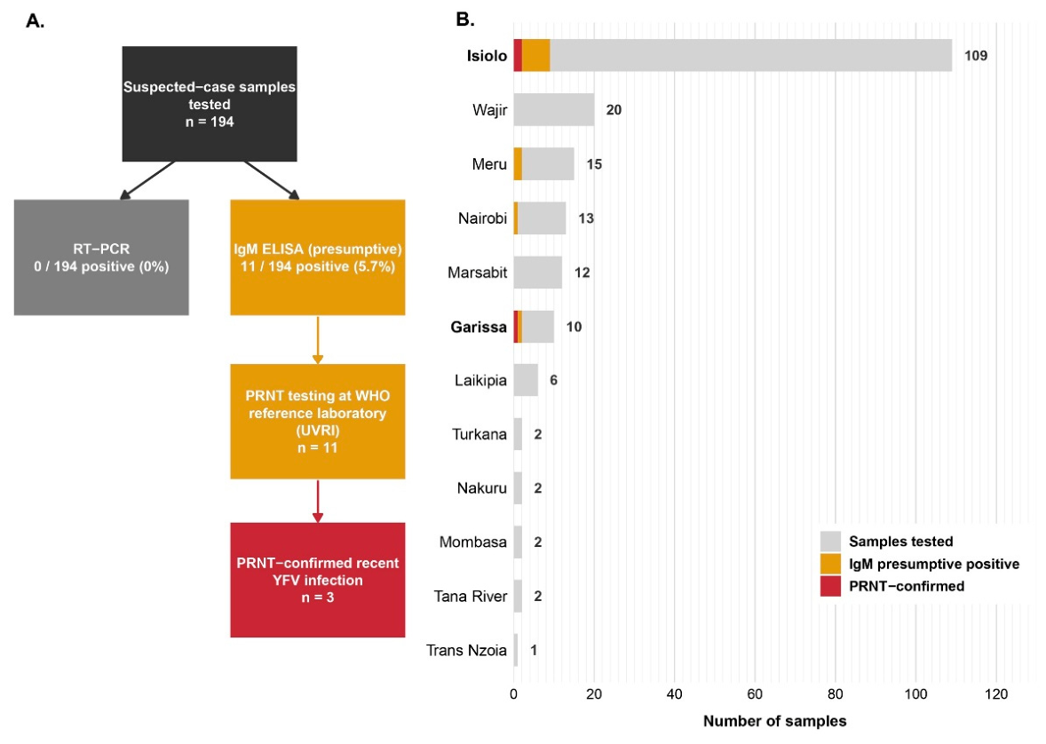

Table 1.
Households inspected, water-holding container types surveyed, Aedes aegypti infestation levels, and Stegomyia indices with corresponding risk classifications by cluster and county.
Table 1.
Households inspected, water-holding container types surveyed, Aedes aegypti infestation levels, and Stegomyia indices with corresponding risk classifications by cluster and county.
| County | Cluster | Houses inspected | Houses infested | Containers inspected | Containers infested |
HI |
CI |
BI |
YF risk |
| Isiolo | Kambi Juu Manyatta Ganna Biliqo Marara Gafarsa Overall |
30 45 25 18 118 |
7 5065 416 |
102 176 73012 |
10 60016 |
23.3 11.10010.2 |
9.8 3.4003.8 |
33.3 13.30013.6 |
Medium Medium Low Low Medium |
| Garissa | Bula Punda Bula Reek Bula Vumbi Bula Khalif Bula Mathengeni Bula College Overall |
25 36 11 13 24 13 122 |
3 60201 12 |
162 319 45 112 38 42 718 |
– – – – – – 18 |
12.0 16.7015.407.7 9.8 |
– – – – – – 2.5 |
– – – – – – 14.8 |
Medium Medium Low Medium Low Medium Medium |
| Meru | Kinna | 30 | 0 | 90 | 0 | 0 | 0 | 0 | data |
| peketoni | 50 | 0 | 148 | 0 | 0 | 0 | 0 | data | |
| Kathithini | 30 | 4 | 122 | 6 | 13.3 | 4.9 | 20 | data | |
| Overall | 110 | 4 | 360 | 6 | 3.6 | 1.7 | 5.5 | Low | |
| Township | 23 | 7 | 131 | – | 30.4 | – | – | Medium | |
| Alimoaw | 11 | 1 | 53 | – | 9.1 | – | – | Medium | |
| ADC | 20 | 7 | 63 | – | 35.0 | – | – | Medium | |
| Wajir | Barwahgo | 26 | 3 | 85 | – | 11.5 | – | – | Medium |
| God-Ade | 20 | 5 | 57 | – | 25.0 | – | – | Medium | |
| Bula Wagberi | 15 | 2 | 40 | – | 13.3 | – | – | Medium | |
| Hadado | 11 | 0 | 24 | – | 0 | – | – | Low | |
| Habaswein | 12 | 2 | 67 | – | 16.7 | – | – | Medium | |
| Overall | 138 | 27 | 520 | 29 | 19.6 | 5.6 | 21.0 | Medium | |
| All counties | Pooled | 488 | 2,004 | 55 | 69 | 11.3 | 3.4 | 14.1 | Medium |
Table 2.
Distribution of inspected container types and associated Ae. aegypti infestation and breeding preference ratios across all surveyed counties.
Table 2.
Distribution of inspected container types and associated Ae. aegypti infestation and breeding preference ratios across all surveyed counties.
| Isiolo | Garissa | Meru | Wajir | Overall | ||||||||
| Container type | No. of containers inspected | No. of containers infested | No. of containers inspected | No. of containers infested | No. of containers inspected | No. of containers infested | No. of containers inspected | No. of containers infested | No. of containers inspected | No. of containers infested | % infested | BPR |
| Ground pool | 1 | 1 | 0 | 0 | 0 | 0 | 0 | 0 | 1 | 1 | 100 | 29.04 |
| Underground tanks | 0 | 0 | 0 | 0 | 0 | 0 | 3 | 3 | 3 | 3 | 100 | 29.04 |
| Plastic tanks | 0 | 0 | 0 | 0 | 0 | 0 | 8 | 4 | 8 | 4 | 50 | 14.5 |
| Metal drums | 1 | 1 | 1 | 1 | 0 | 0 | 10 | 0 | 12 | 2 | 16.7 | 4.84 |
| Plastic drums | 114 | 14 | 103 | 6 | 37 | 5 | 126 | 17 | 380 | 42 | 11.1 | 3.21 |
| Buckets | 4 | 0 | 72 | 0 | 16 | 1 | 60 | 4 | 152 | 5 | 3.3 | 0.96 |
| Jerrycans | 289 | 0 | 525 | 11 | 277 | 0 | 287 | 1 | 1,378 | 12 | 0.9 | 0.25 |
| Plastic basins | 6 | 0 | 11 | 0 | 24 | 0 | 9 | 0 | 50 | 0 | 0 | 0 |
| Tires | 0 | 0 | 0 | 0 | 2 | 0 | 0 | 0 | 6 | 0 | 0 | 0 |
| Cooling pans | 0 | 0 | 1 | 0 | 0 | 0 | 2 | 0 | 3 | 0 | 0 | 0 |
| Plastic bottles | 0 | 0 | 0 | 0 | 0 | 0 | 3 | 0 | 3 | 0 | 0 | 0 |
| Metal basins | 0 | 0 | 0 | 0 | 0 | 0 | 2 | 0 | 2 | 0 | 0 | 0 |
| Clay pots | 0 | 0 | 1 | 0 | 0 | 0 | 0 | 0 | 1 | 0 | 0 | 0 |
| Banana leaf axils | 0 | 0 | 4 | 0 | 4 | 0 | 0 | 0 | 4 | 0 | 0 | 0 |
| Tree holes | 1 | 0 | 0 | 0 | 0 | 0 | 0 | 0 | 1 | 0 | 0 | 0 |
| Totals | 416 | 16 | 718 | 18 | 360 | 6 | 510 | 29 | 2,004 | 69 | 3.4 | |
Table 3.
Adult mosquito species composition and relative abundance by county.
| Species | Isiolo | Garissa | Meru | Total |
| Culex pipiens | 1,115 (72.5%) | 1,134 (90.5%) | 2,779 (55.1%) | 5,028 (64.2%) |
| Cx. zombaensis | 14 (0.9%) | 12 (1%) | 1,549 (30.7%) | 1,575 (20.1%) |
| Cx. univittatus | 58 (3.8%) | 97 (7.7%) | 147 (2.9%) | 302 (3.9%) |
| Cx. vansomereni | 28 (1.8%) | 1 (0.07%) | 215 (4.6%) | 244 (3.1%) |
| Anopheles funestus | – | – | 248 (4.9%) | 248 (3.2%) |
| Aedes furcifer | 218 (14.2%) | – | – | 218 (2.8%) |
| Aedes aegypti | 64 (4.1%) | 1 (0.07%) | 48 (0.5%) | 113 (1.4%) |
| An. gambiae s.l. | 27 (1.8%) | 8 (0.6%) | – | 35 (0.4%) |
| Ae. hirsutus | – | – | 26 (0.1%) | 26 (0.3%) |
| An. maculipalpis | – | – | 12 (0.2%) | 12 (0.2%) |
| Ae. chauseri | 4 (0.3%) | – | – | 4 (0.05%) |
| Ae. simpsoni s.l. | 3 (0.2%) | – | 1 (0.01%) | 4 (0.05%) |
| An. coustani | – | – | 4 (0.07%) | 4 (0.05%) |
| Ae. tricholabis | – | – | 3 (0.05%) | 3 (0.04%) |
| Cx. annulioris | – | – | 2 (0.03%) | 2 (0.03%) |
| Cx. cinereus | – | – | 2 (0.03%) | 2 (0.03%) |
| Aedes spp. | – | – | 4 (0.07%) | 4 (0.05%) |
| Culex spp. | 7 (0.5%) | – | 3 (0.05%) | 10 (0.1%) |
| Anopheles spp. | – | – | 1 (0.01%) | 1(0.01%) |
| Total | 1,538 (19.6%) | 1,253 (32.3%) | 5,044 (64.4%) | 7,835 (100%) |
Disclaimer/Publisher’s Note: The statements, opinions and data contained in all publications are solely those of the individual author(s) and contributor(s) and not of MDPI and/or the editor(s). MDPI and/or the editor(s) disclaim responsibility for any injury to people or property resulting from any ideas, methods, instructions or products referred to in the content. |
© 2026 by the authors. Licensee MDPI, Basel, Switzerland. This article is an open access article distributed under the terms and conditions of the Creative Commons Attribution (CC BY) license (http://creativecommons.org/licenses/by/4.0/).
Copyright: This open access article is published under a Creative Commons CC BY 4.0 license, which permit the free download, distribution, and reuse, provided that the author and preprint are cited in any reuse.