

Submitted:
09 September 2026
Posted:
09 September 2026
You are already at the latest version
Abstract
Fat embolism syndrome (FES) is a rare, life-threatening complication of sickle cell disease (SCD), often underrecognized due to overlap with acute chest syndrome, sepsis, and thrombotic microangiopathies. We report a 37-year-old man with sickle cell disease who presented with an acute vaso-occlusive crisis and rapidly deteriorated with respiratory failure, cardiovascular collapse, and neurological dysfunction. Common etiologies including pulmonary embolism, infection, and stroke were excluded. Laboratory evidence of hemolysis, cytopenias, elevated ferritin, and acute cor pulmonale suggested systemic FES secondary to bone marrow necrosis. The patient underwent urgent red cell exchange transfusion, followed by therapeutic plasma exchange and corticosteroids, resulting in significant clinical and hemodynamic recovery. This case highlights the importance of early recognition and aggressive multimodal therapy in SCD-FES.
Keywords:
sickle cell disease
; fat embolism
; RBC exchange
; plasma exchange
1. Introduction
Sickle cell disease (SCD) is a complex inherited hemoglobinopathy marked by recurrent vaso-occlusion, chronic hemolysis, and progressive end-organ damage. While complications such as acute chest syndrome and stroke are well recognized, fat embolism syndrome (FES) remains an uncommon but devastating entity that is frequently underdiagnosed. In patients with SCD, FES arises in the absence of trauma and is believed to result from extensive bone marrow necrosis, leading to systemic embolization of fat particles and a profound inflammatory response.
The clinical course of FES in SCD is often fulminant, beginning with an apparently typical painful crisis and rapidly evolving into hypoxemic respiratory failure, neurological deterioration, and multiorgan dysfunction. Diagnostic uncertainty is common due to significant overlap with other acute SCD complications, and delays in recognition are associated with high mortality. Emerging evidence, however, suggests that early intervention particularly red cell exchange transfusion, with or without therapeutic plasma exchange can be lifesaving.
We present a striking case of systemic fat embolism syndrome in a patient with sickle cell/β-thalassemia who experienced sudden cardiopulmonary collapse and neurological impairment. This report aims to heighten clinical awareness of FES in SCD, emphasize key diagnostic features, and illustrate the potential for favorable outcomes with timely, aggressive management.
Case presentation
A 37-year-old man with Sickle Cell–β⁰ Thalassemia and G6PD deficiency was admitted with acute vaso-occlusive and hemolytic crises. His past medical history included multiple ICU admissions for acute chest syndrome, avascular necrosis of the left shoulder and bilateral femoral heads (requiring right total hip replacement), vertebral compression fractures, and recurrent multifocal osteomyelitis requiring multiple incision and drainage procedures.
On admission, he was afebrile, BP 125/60 mmHg, HR 86 bpm, RR 18/min, and O₂ saturation 100% on room air. Within hours, he developed severe chest pain and respiratory distress, tachycardia (HR 136 bpm), and required 5 L/min supplemental oxygen via nasal cannula. Chest X-ray and CT pulmonary angiography excluded pneumonia and pulmonary embolism (Figure 1). Labs revealed worsening hemolysis, with a drop of hemoglobin to 7.7 gm/dL (Table 1).
He became vitally unstable, and required intubation for suspected acute chest syndrome. He experienced pulseless electrical activity (PEA) cardiac arrest and revived after six minutes. He subsequently developed supraventricular tachycardia and atrial fibrillation, managed with electrical cardioversion. Transthoracic echocardiography revealed mildly reduced EF (40%), septal motion abnormalities consistent with RV volume and pressure overload, moderately dilated RV, moderate-to-severe systolic dysfunction, elevated RVSP (60 mmHg), and moderate-to-severe tricuspid regurgitation.
He was admitted to the ICU, required vasopressors, and underwent urgent RBC exchange, reducing HbS from 73% to 11.7%. Electroencephalography (EEG) showed continuous bilateral theta/delta activity, consistent with diffuse cerebral dysfunction, without epileptiform discharges.
FES was suspected after exclusion of other causes. Subsequently, therapeutic plasma exchange (TPE) was initiated for seven days, alongside corticosteroids. Peripheral smear revealed neutrophilic leukocytosis with toxic granulation; ferritin was elevated (4481 ng/mL), and initial septic workup was negative. Supportive care included PRBC transfusions and antibiotics for hospital-acquired pneumonia. Brain MRI revealed subacute lacunar infarction in the right centrum semiovale, diffuse microhemorrhages, small vessel ischemic changes, and focal cortical enhancement in the right posterior parietal region (Figure 2).
Follow-up TTE demonstrated normalized EF (55%) and RVSP (31 mmHg). The patient showed remarkable clinical improvement, was started on an extensive rehabilitation program, and discharged on hydroxyurea 1500 mg daily.
4. Discussion
Sickle cell disease (SCD) is a hereditary hemoglobinopathy caused by a single nucleotide substitution in the β-globin gene, resulting in the production of abnormal hemoglobin S (HbS). The disease is characterized by recurrent vaso-occlusive crises (VOC) and multisystem complications (1).
In this paper we present fat embolism syndrome (FES) in a patient with sickle cell diseas which is an uncommon and frequently underrecognized complication, yet it is associated with substantial morbidity and mortality. FES most commonly occurs in the setting of trauma and orthopedic interventions, particularly following long-bone fractures with an incidence ranges from 1% to 29%, reflecting substantial variability in diagnostic criteria and reporting practices. In contrast, individuals with sickle cell hemoglobinopathies are at risk for non-trauma–related FES, which arises independently of mechanical injury. However, FES in sickle cell disease remains an infrequent but serious complication accounting for 10.3% in one metanalysis (2).
The pathological effects of fat embolism in both traumatic and non-traumatic contexts, including sickle cell disease, arise from a combination of mechanical vascular obstruction and biochemical injury mediated through inflammatory and cytotoxic pathways (11).
The mechanical theory posits that fat embolism results from the physical release of fat globules, either following traumatic injury or in the setting of extensive bone marrow necrosis in sickle cell disease (SCD). Once liberated, these fat particles enter the venous circulation and become trapped within the pulmonary microvasculature, producing ventilation–perfusion mismatch and potentially precipitating acute respiratory distress syndrome (ARDS) (11).
The biochemical theory proposes that circulating free fatty acids (FFAs) and other lipolytic degradation products released from fat globules induce direct cytotoxic injury to endothelial cells, pneumocytes, and other tissues. These toxic metabolites trigger a robust inflammatory cascade characterized by complement activation, cytokine release, and neutrophil recruitment, thereby amplifying vascular permeability, promoting diffuse alveolar damage, and contributing to multiorgan dysfunction. In sickle cell disease, this inflammatory response may be further exacerbated by underlying hemolysis, oxidative stress, and endothelial activation, heightening the vulnerability to severe manifestations of FES.This theory explains the 24-48 hour delay often seen in FES onset like the case in our patient (11).
Evidence suggests that subclinical marrow necrosis and chronic low-grade fat embolization may occur even during the steady state in SCD, supported by observations of persistently elevated secretory phospholipase A₂ (sPLA₂) levels in affected individuals (3).
A comprehensive literature review published through 2018 identified 87 cases and demonstrated that FES disproportionately affects milder sickle cell genotypes. Hemoglobin SC disease (HbSC) was the most frequently represented variant, accounting for approximately 50% of reported cases, whereas sickle/β⁺-thalassemia (HbSβ⁺) represented an additional 18% (8), which is the case in our patient. By contrast, homozygous sickle cell disease (HbSS) constituted only about 15% of cases (3).
One prevailing hypothesis posits that individuals with non-SS genotypes exhibit higher baseline hemoglobin and hematocrit levels, which increase blood viscosity and may heighten susceptibility to bone marrow ischemia and subsequent necrosis. However, emerging data complicate this paradigm. A recent single-center cohort reported that most FES cases (70%) occurred in patients with the HbSS genotype, suggesting that additional or alternative mechanisms may contribute to FES risk in this population (5). Another important contributing factor is that the prevalence of HbSS genotype in higher in some countries such as the case in our patient.
Infection is a well-recognized precipitating factor for fat embolism syndrome (FES) in sickle cell disease. Human parvovirus B19 (HPV B19) infection, in particular, has been repeatedly implicated and may provoke FES through an immune-mediated mechanism in patients with partially preserved splenic function (6). FES has also been described in association with COVID-19 infection, with proposed mechanisms including heightened systemic inflammation, increased cellular necrosis, and the possibility of SARS-CoV-2–infected adipocytes undergoing necrotic cell death, thereby augmenting fat release into the circulation (5, 12). The possibility of infection was ruled out in our patient with negative septic workup.
The diagnosis of fat embolism syndrome (FES), particularly in the context of sickle cell disease (SCD), remains primarily clinical. The absence of standardized and specific diagnostic criteria for non-traumatic FES in SCD significantly complicates recognition and timely diagnosis. Traditional diagnostic frameworks developed for traumatic FES, such as the Gurd and Wilson criteria and the Schonfeld scoring system, lack specificity in this population and may overlap with other acute SCD complications, including stroke, acute chest syndrome, and sepsis. All thses differential diagnoses in addition to pulmonary embolism were excluded in our patient.
The classic presentation of FES is a triad of respiratory failure, neurologic complications, and petechial rash which do not necisseraly present in all patients with suspected diagnosis (12). A high index of clinical suspicion is therefore critical, particularly in patients with sickle cell crisis who develop acute hypoxemia, fever, and altered mental status. FES often presents initially as a painful vaso-occlusive crisis (VOC), followed by the development of type 1 respiratory failure and neurological manifestations, typically within 48–72 hours.
FES can closely mimic thrombotic thrombocytopenic purpura (TTP), which may result in diagnostic confusion and mismanagement (13). This possibility was raised and excluded during initial evaluation and deterioration of our patient. Key distinguishing features include the pattern of organ involvement and hematologic findings. Respiratory failure is uncommon in TTP, whereas it is a hallmark of FES. Additionally, TTP typically presents with more pronounced thrombocytopenia and abundant schistocytes on peripheral blood smear (PBS). In contrast, FES often demonstrates a leukoerythroblastic picture with only occasional schistocytes on PBS. Careful clinical and laboratory assessment in addition to liasion with hematopathologist is crucial (14).
In sickle cell disease (SCD), FES is associated with markedly elevated serum ferritin, elevated LDH and alkaline phosphatase (ALP) suggesting bone marrow necrosis, and progressive declines in hemoglobin and platelet counts, findings presented in our case.
Fat embolism syndrome (FES) is a severe complication of sickle cell disease with high morbidity and mortality. It can result in multiorgan dysfunction, most notably type 1 respiratory failure, hypoxemia, tachypnea, acute respiratory distress syndrome (ARDS), and acute cor pulmonale.
Cardiac ultrasound may demonstrate fat particles in the right heart or findings of acute cor pulmonale, supporting the diagnosis when pulmonary thromboembolism is excluded. The CT chest and transthoracic ecocardiogram, were done in our patient, and after careful assessment by radiologists and cardiologists the possibility of acute pulmonary embolism was excluded. However, acute heart failure with reduced ejection fraction was evident in the tranthoracic echocardiogram which can be seen in cases of FES (9).
Brain MRI may demonstrate the characteristic “starfield pattern,” defined by punctate foci of diffusion restriction with associated microhemorrhages in the white matter. However, a systematic review by Ghazal et al. reported that these findings are present in only 65.5% of cases during the acute phase and are less frequently observed beyond five days after symptom onset (11). This may explain the absence of typical MRI findings in our patient, in addition to the inability to perform magnetic resonance angiography (11).
Neurological manifestations are common, ranging from mild confusion and headache to coma, seizures, rigidity, and focal deficits, with severity increased in the presence of a right-to-left intracardiac shunt (e.g., PFO) (4). Cerebral MRI abnormalities typically resolve within weeks to months. Other reported features include petechial rash (often in the upper trunk, head, or neck) which can present in less than 50% of cases (5), retinal changes such as perivascular whitening or cotton wool spots, and rarely blindness. Additional organ involvement may include liver injury, renal impairment, and myocardial damage.
Fat embolism syndrome (FES) in sickle cell disease has historically been associated with substantial mortality, reported as high as 64%. Analysis of published cases demonstrates a temporal decline in mortality, from 66% in reports prior to 2013 to 33% between 2014 and 2018, largely attributable to the timely implementation of red cell exchange (RCE) therapy (3).
A recent single-center cohort by Salam A. et al reported a further reduction in mortality to 18.5% (10). Fatal outcomes are most commonly observed in younger adults presenting with profound cardiorespiratory compromise and severe hepatic dysfunction. Among survivors, severe neurological deficits occur in approximately 14%, although a significant proportion exhibit substantial neurological recovery with rehabilitation. One cohort reported complete or near-complete long-term recovery in 63% of patients, highlighting the potential for favorable outcomes with early recognition and intervention (10).
Management of fat embolism syndrome (FES) in sickle cell disease is multifaceted, combining prompt supportive care with targeted interventions to halt the sickling cascade and mitigate systemic inflammation. The cornerstone of initial management is maintaining adequate oxygenation, hemodynamic stability, and organ perfusion. Respiratory support, including supplemental oxygen or mechanical ventilation, is indicated for patients with hypoxemia or ARDS. Aggressive hydration is employed to optimize microvascular flow and prevent further sickling (7).
Early initiation of red cell exchange (RCE) is lifesaving and remains the mainstay of therapy once FES is suspected (15). The primary goal is to reduce hemoglobin S (HbS) levels to below 10–20%, interrupting the sickling cycle and preventing further bone marrow necrosis. RCE also facilitates the removal of leukocytes, cytokines, fat globules, and platelets, thereby attenuating inflammation and hyperviscosity. Evidence consistently demonstrates that RCE improves survival compared with simple transfusion or no transfusion. RCE was initiated in our patient within 24-hour of clinical dterioration with final HbS 11.7% (16).
Therapeutic Plasma Exchange (TPE) is increasingly recommended as an adjunct to RCE in severe or refractory cases. By removing circulating fat droplets and pro-inflammatory cytokines from bone marrow necrosis, TPE can accelerate recovery and improve outcomes (15, 16). This strategy was followed in our patient with daily TPE guided by his clinical and laboratory response. Case reports and series have documented successful use of TPE in SCD-FES, often resulting in complete recovery. The duration of treatment can be determined by clinical response, with serial monitoring of ferritin, lactate dehydrogenase, and platelet counts. (17).
Corticosteroids although primarily used in traumatic FES, they have anti-inflammatory effects that may benefit SCD-FES. They also inhibit complement-mediated leukocyte aggregation, stabilize cell membranes, and reduce circulating free fatty acids. Meta-analytic data suggest that steroids may decrease FES risk by up to 77% in long-bone fracture patients. In SCD, methylprednisolone has been reported in at least one pediatric FES case (7).
Optimal outcomes require early involvement of critical care, hematology, and neurology teams. Investigational therapies, including targeted anti-inflammatory agents, may provide additional benefit in severe or refractory cases
5. Conclusions
Fat embolism syndrome in sickle cell disease is a rapidly progressive, life-threatening complication, even in seemingly mild cases. Survival hinges on swiftly recognizing sudden systemic collapse with acute hypoxemia, neurological deterioration, and cytopenias. Red cell exchange is lifesaving, halting the sickling cascade and limiting marrow necrosis, while therapeutic plasma exchange can remove circulating fat and inflammatory mediators in severe cases. Combined with supportive care and early rehabilitation, rapid intervention is key to preventing multiorgan failure and improving long-term outcomes.
Funding
This research received no external funding
Institutional Review Board Statement
The study was conducted in accordance with the Declaration of Helsinki, and approved by the Institutional Review Board of Imam Abdulrahman bin Faisal University (IRB-2025-01-0853).
Informed Consent Statement
Informed consent was obtained from the patient involved in the study.
Acknowledgments
none
Conflicts of Interest
The author declares no conflicts of interest.
References
- Rees DC, Williams TN, Gladwin MT. Sickle-cell disease. Lancet. 2010;376(9757):2018–2031. [CrossRef]
- Luo M, Wu Q, Huang H, Wang Q, Li Y, Wang Y, et al. Single-case metanalysis of fat embolism syndrome. Int J Cardiol. 2021;345:111–117. [CrossRef]
- Habibi A, Arlet JB, Stankovic K, Gellen-Dautremer J, Ribeil JA, Bartolucci P, et al. Fat embolism syndrome in sickle cell disease: a systematic review. Int J Cardiol. 2022;351:92–99. [CrossRef]
- Scarpino M, Lanzo G, Lolli F, Grippo A. From the diagnosis to the therapeutic management: cerebral fat embolism, a clinical challenge. Int J Gen Med. 2019;12:39–48. [CrossRef]
- Buxo Z, Saini A, Hudson C, Levine DL. Cerebral fat embolism in a patient with hemoglobin SC disease associated with vaso-occlusive crisis and COVID-19. Ann Intern Med Clin Cases. 2023;2(11):[page numbers]. [CrossRef]
- Lampejo T. Parvovirus B19 infection as a potential trigger for fat embolism syndrome in patients with sickle cell disease. J Med Virol. 2021;93(6):3283–3284. [CrossRef]
- Tsitsikas DA, Bristowe J, Abukar J. Fat embolism syndrome in sickle cell disease. J Clin Med. 2020;9(11):3601. [CrossRef]
- Kumar R, Patel R, Jones A. Cerebral fat embolism in a patient with hemoglobin SC disease. Ann Intern Med Clin Cases. 2023;4:CC23-0587. [CrossRef]
- Gendreau S, Cecchini J, Razazi K, et al. Bone marrow necrosis and fat embolism in critically ill patients with acute complications of sickle cell disease: a retrospective cohort study. Br J Haematol. 2025; [ePub ahead of print]. [CrossRef]
- Salam A, Sameer R, Eiman AA, et al. Clinical, laboratory, radiological features, and outcome of acute fat embolism syndrome in sickle cell disease. Sci Rep. 2025;15:27523. [CrossRef]
- Shadmani G, Hosseini H, Nigogosyan Z, Goyal MS, Nazeri A. Nontrauma-Associated Cerebral Fat Embolism Syndrome in Sickle Cell-Related Hemoglobinopathies: A Case Series and Systematic Review. AJNR Am J Neuroradiol. 2025;46(10):2000-2008. [CrossRef]
- Rizvi S, Khakwani M, Pancham S, Tsitsikas D, Rudzki Z, Hassan-Smith G, Bowen M, et al. Bone marrow necrosis and fat embolism syndrome in sickle cell disease during COVID-19 infection treated successfully with sequential red cell and plasma exchange. eJHaem. 2023;4(1):207–210. [CrossRef]
- Gangaraju R, May JE, Williams LA III, et al. Fat embolism syndrome due to bone marrow necrosis in patients with hemoglobinopathies: a life-threatening complication mimicking thrombotic thrombocytopenic purpura. Am J Hematol. 2018;93(11):e25363.
- Se B, Frisch A, Hwang MW, Polani F, Bade N. Fat embolism syndrome mimicking thrombotic thrombocytopenic purpura in a patient with hemoglobin S/beta-thalassemia. J Hematol. 2024;13(3):104–107. [CrossRef]
- Rasool A, Thomas S, Luqmani A, Sohal M, Okoli S, Deplano S, Ademokun CO, Mactier C, Layton DM. Combined red cell and plasma exchange is associated with reduced mortality of fat embolism syndrome in sickle cell disease. Blood. 2024;144(Suppl 1):2497. [CrossRef]
- 16. Tsitsikas DA, Rowe S, Bosch A, Hui C, Sadasivam N, Palaskas NJ, Pancham S, Rizvi S, Taylor J, Greaves P, Glenthøj A, Hoffmann M, Drasar E, Eleftheriou P. Addition of plasma exchange to red cell exchange improves outcomes of fat embolism syndrome in sickle cell disease. Br J Haematol. 2023;200(6):e50-e52. [CrossRef]
- 17. Alajeel N, Abdulmalek K, Al-Fares A. Coma secondary to cerebral fat embolism syndrome due to sickle cell disease fully recovering following red cell exchange transfusion. BMJ Case Rep. 2021;14(11):e245051. [CrossRef]
Figure 1.
MRI brain (FLAIR, DWI and SWI) showing several hyper intense lesions in both hemispheres indicating old ischemic changes and multiple susptible areas indicating micro hemorrhages, raising the possibility of fat embolization syndrome.
Figure 1.
MRI brain (FLAIR, DWI and SWI) showing several hyper intense lesions in both hemispheres indicating old ischemic changes and multiple susptible areas indicating micro hemorrhages, raising the possibility of fat embolization syndrome.
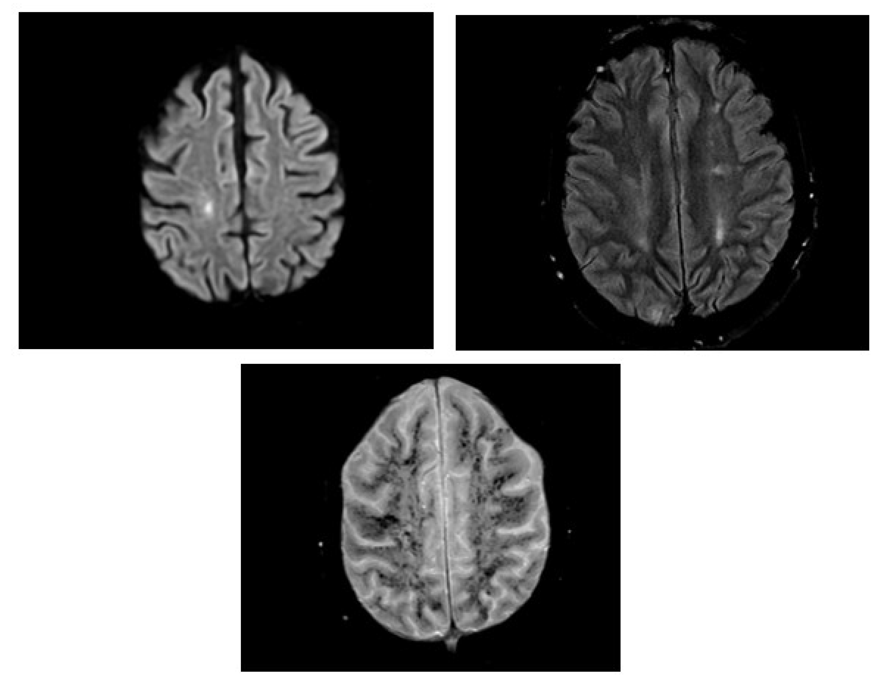

Table 1.
Laboratory investigations from admission until discharge.
| Days | Wbc (103/uL) | Hgb (gm/dL) | Platelet (103/uL) | T. bilirubin (mg/dL) | D. Bilirubin (mg/dL) | LDH (U/L) |
| 1 | 6.1 | 10.9 | 140 | 2.3 | 0.8 | 321 |
| 2 | 12.7 | 7.7 | 84 | 7.9 | 0.63 | 2838 |
| 7 | 3.7 | 7.5 | 36 | 1.3 | 0.47 | 1747 |
| 10 | 4.1 | 8.1 | 58 | 1.7 | 0.69 | 622 |
| Discharge | 5.9 | 8.5 | 238 | 1 | 0.44 | 292 |
Wbc: white blood cell count, Hgb: hemoglobin, T.bilirubin: total bilirubin, D.bilirubin: direct bilirubin, LDH: lactat dehydrogenase.
Disclaimer/Publisher’s Note: The statements, opinions and data contained in all publications are solely those of the individual author(s) and contributor(s) and not of MDPI and/or the editor(s). MDPI and/or the editor(s) disclaim responsibility for any injury to people or property resulting from any ideas, methods, instructions or products referred to in the content. |
© 2026 by the author. Licensee MDPI, Basel, Switzerland. This article is an open access article distributed under the terms and conditions of the Creative Commons Attribution (CC BY) license.
Copyright: This open access article is published under a Creative Commons CC BY 4.0 license, which permit the free download, distribution, and reuse, provided that the author and preprint are cited in any reuse.