

Submitted:
07 September 2026
Posted:
08 September 2026
You are already at the latest version
Abstract
Dynamic knee valgus (DKV) is commonly used in functional screening related to anterior cruciate ligament (ACL) injury risk; however, assessments often rely on peak valgus alone despite the combined influence of knee valgus and flexion on knee loading. This study compared knee kinematics and vertical ground reaction forces (GRFs) between single-leg landing (SLL) and single-leg countermovement jump (SL-CMJ), focusing on peak knee valgus, knee flexion at peak valgus, valgus angular velocity, knee flexion-normalized valgus, and GRFs. Physically active, asymptomatic female participants (n = 30) performed both the SLL and SL-CMJ tasks. Knee kinematics and GRFs were recorded using motion-tracking sensors and a force platform. Peak knee valgus did not differ between tasks. Compared with SL-CMJ, SLL produced 24% less knee flexion at peak valgus, 17% greater knee flexion-normalized valgus, 254% higher valgus angular velocity, and 124% higher GRFs (all p < 0.05). These findings demonstrate task-dependent differences in landing mechanics that are not captured by peak knee valgus alone. Assessing knee valgus together with knee flexion and its temporal development may provide a more comprehensive characterization of potentially relevant knee-loading mechanics. The high valgus angular velocities observed during SLL may also inform the development of velocity-specific neuromuscular training strategies.
Keywords:
GRF
; knee flexion
; knee valgus
; knee valgus angular velocity
; normalized knee valgus
1. Introduction
Rapid decelerations, changes of direction, and landings are associated with ~70% of non-contact anterior cruciate ligament (ACL) injuries [1,2,3,4,5,6,7,8,9,10,11]. Such movements are typically characterized by increased knee valgus [12]. A critical element of ACL injury prevention is risk assessment [13,14,15]. This process involves the administration of tests executed on one or two legs such as various forms of squats, countermovement jumps (CMJ), and landings in an effort to mimic sport-specific situations where many ACL injuries occur. ACL injury risk is then inferred from these tests based on the magnitude of dynamic knee valgus (DKV). Its simplicity has made the squat test popular [16,17,18,19,20,21], however numerous researchers have pointed out the limitation that the squat test does not resemble the velocity profile of real-life sport movements, potentially reducing its sensitivity to predict ACL injuries. Moreover, the bilateral execution of the squat test further limits its utility, as knee injury risk is known to increase predominantly during unilateral weight-bearing tasks, such as single-leg jumps, cutting maneuvers, and changes of direction[1,9].
The single-leg counter-movement jump (SL-CMJ) test is emerging as a simple and clinically perhaps more valid test than the squat test to screen for ACL risk by measuring DKV. Its potential advantage arises from the high loads on the knee and its resemblance to knee joint kinematics observed during on-field movements. DKV was also higher in tasks performed with one compared with two legs (landing, drop jump, squat) [22,23]. Still, both the SL-CMJ and the squat tests can be inaccurate and often fail to identify those with high risks for a future ACL injury [24,25]. DKV is a reasonably accurate predictor of future ACL injuries [13,26]. The biomechanical basis of this prediction is that ACL loading increases with DKV [27,28,29,30]. Biomechanical studies reported that DKV tends to be higher in women than men [23]. Because DKV is known to increase ACL strain, it may partly explain the 2–9-fold higher incidence of non-contact ACL injuries in females compared with males [1,31,32].
In addition to knee valgus, knee flexion magnitude also affects ACL strain, which is highest at low knee flexion angles and progressively decreases as knee flexion increases [30,33,34,35]. Recent in vivo imaging evidence indicates that ACL strain during single-leg jumping tasks is more strongly associated with sagittal-plane knee mechanics, with higher strain observed when the knee remains relatively extended during task execution [36]. Modeling studies suggest that sagittal-plane kinematics and lower-limb muscle actions jointly influence ACL strain during single-leg landing (SLL), consistent with a task-dependent mechanism of ACL loading [37] . Consistent with these findings, video analyses of confirmed non-contact ACL injuries also reveal a tendency toward smaller knee flexion angles during ground contact in injury cases, although not always reaching statistical significance[4].
Taken together, these findings suggest that the combined assessment of DKV magnitude and knee flexion magnitude is warranted, as both reduced knee flexion and increased DKV have been shown to cumulatively increase ACL strain. Specifically, the same peak valgus may be less concerning if it occurs at a greater knee flexion, potentially indicating a lower-risk for an ACL injury. Accordingly, incorporating knee flexion magnitude into the interpretation of valgus-based metrics — such as by normalizing peak knee valgus to peak knee flexion during functional tasks — may provide a more context-sensitive estimation of ACL injury risk. Therefore, the present study examined the dynamic interplay between knee valgus and knee flexion during SLL and SL-CMJ.
SLL and SL-CMJ are candidate tests for ACL risk prediction based on knee kinematics. In particular, SLL might be preferred not only to the squat but even to the SL-CMJ because knee kinematics more closely resembles those observed during on-field tasks [38]. Critically, peak knee flexion and peak knee valgus and the corresponding angular velocities may be closer to on-field conditions during SLL compared with SL-CMJ. This may be because, although peak knee valgus is comparable between SL-CMJ and SLL, peak knee flexion is smaller and knee valgus angular velocity could be greater during the SLL task.
The purpose of the present study was to compare knee kinematics and external loading characteristics between SLL and SL-CMJ tasks, with a specific focus on peak knee valgus, knee flexion, knee valgus angular velocity, and ground reaction forces (GRF). We hypothesized that: 1. peak knee valgus would not differ between SLL and SL-CMJ; 2. during SLL compared with SL-CMJ, knee flexion at peak valgus would be smaller, and 3. knee valgus angular velocity would be higher during SLL than during SL-CMJ. We also performed exploratory correlation analyses among anthropometric measures and knee flexion and knee valgus magnitude and velocity.
2. Materials and Methods
2.1. Participants
We recruited asymptomatic, physically active female college students who were free from injury but demonstrated high DKV (≥ 10°) during the screening procedures (see below). The inclusion criteria were female sex, age between 18 and 30 years and DKV ≥ 10°. Exclusion criteria were current injuries and past surgeries in the spine, hip, knee, and ankle joints, or pain of orthopedic origin. Based on these criteria, 30 participants were ultimately enrolled in the study (age: 21.5 ± 2.21 years; height: 168.8 ± 6.97 cm; mass: 62.6 ± 8.89 kg; exercise training history: 15.1 ± 3.36 years). Participants pursued ground-contact sports 6.2 ± 2.93 hours per week at the club level, but none of them competed at or above national levels. Participants received verbal and written explanations of the experimental procedures and were informed about the potential risks. The study was approved by the University Ethics Committee (approval number: 7961-PTE2019) and was conducted in accordance with the Declaration of Helsinki. Written informed consent was obtained from all participants prior to participation. Written informed consent was obtained from the study participant depicted in the figures for publication of the images in an online open-access scientific journal.
2.2. Sample Size Calculation
We performed an a priori sample size calculation using G*Power 3.1.9.7, targeting a medium effect size (Cohen’s d ≈ 0.5), α = 0.05, and statistical power (1–β) = 0.80. This analysis indicated that a minimum of 27 participants would be required. To increase statistical power and account for potential dropouts, the final sample included 30 participants.
2.3. Experimental Procedure
One week prior to the experimental session, participants visited the laboratory for a familiarization session to ensure that they were comfortable with and able to correctly execute the experimental tasks, including SLL and SL-CMJ. During this session, they also performed a single-leg squat for screening purposes to identify the leg exhibiting DKV ≥ 10°.
The main experimental session lasted approximately 30 minutes. Participants completed a 5-minute self-paced warm-up on a cycle ergometer, followed by general and lower-extremity specific stretching. Prior to the test trials, participants stood quietly on the force plate while body weight was measured and recorded for normalization of GRF data. They then executed the experimental tests in a fixed order: SL-CMJ followed by SLL, with 3 minutes of inter-trial rest to minimize fatigue and maintain consistent jumping and landing performance.
2.4. Single-Leg CMJ
Participants executed the SL-CMJs on a force platform (Tenzi Ltd., Pilisvörösvár, Hungary) using the leg previously identified as exhibiting greater DKV (Figure 1). Participants stood on one leg with arms akimbo and the non-supporting knee slightly flexed. They were instructed to jump as high as possible while keeping their hands on the hips; no additional instructions regarding jump strategy were provided. Each participant performed three trials, with a 3-minute inter-trial rest period to minimize fatigue and ensure consistent performance. Vertical GRF was recorded at a sampling rate of 420 Hz and normalized to the participant’s body weight measured on the force plate prior to testing, resulting in dimensionless values expressed as multiples of body weight (×BW).
2.5. Single-Leg Landing
Participants executed the SLLs from a 0.46-m-high plyometric box (Capital Sports Plyo Crew Box) using the lower limb previously identified as exhibiting greater DKV (Figure 2). They were instructed to step off the box without jumping, land on the designated leg, and maintain the landing position for 5 s. Each participant completed three trials, with a 3-minute inter-trial rest period to minimize fatigue and ensure consistent performance. Vertical GRF was recorded, and data were processed similarly to that described during SL-SMJ).
2.6. Knee JOINT KINEMATICS
A 3D motion tracking system (Noraxon, Scottsdale, USA) with a sampling frequency of 100 Hz was used to analyze the kinematics of the SL-CMJ and SLL. Inertial sensors comprising accelerometer, gyroscope, and magnetometer units were affixed with Velcro straps on the shank and thigh according to the manufacturer’s recommendations for sensor placement and calibration. Using the sensor’s segmental orientation angle-time data, peak knee valgus angle was considered as the frontal-plane thigh orientation angle. Knee flexion angle at the time point of peak knee valgus was determined during both SL-CMJ and SLL tasks, using the sagittal-plane shank and thigh orientation angle data. To account for the combined contribution of knee valgus and knee flexion to ACL loading, peak knee valgus was normalized to the corresponding knee flexion angle, representing a novel normalization approach in ACL risk assessment. During this procedure, peak knee valgus was divided by the knee flexion angle measured at the time of peak knee valgus, yielding a knee valgus value normalized to knee flexion.
2.7. Data Processing and Statistical Analyses
Data were exported from the measurement systems (force plate and the Noraxon MyoMotion system) and analyzed in MATLAB [39], where the datasets were screened and corrected for technical or transfer errors prior to statistical analysis. The cleaned numerical data were then transferred to Origin for statistical analyses. We present the data as mean ± standard deviation (SD) for normally distributed variables and as median [interquartile range, IQR] for variables deviating from normality. Normality was assessed using the Shapiro–Wilk test. For normally distributed outcomes, differences between the two test conditions (SL-CMJ vs SLL) were evaluated using paired-samples t-tests. When the assumption of normality was violated, paired comparisons were performed using the Wilcoxon signed-rank test. Effect sizes were calculated as Cohen’s d for paired samples (for t-tests) and as r = |Z|/√N for Wilcoxon tests. Statistical significance was set at p < 0.05. All analyses were performed in Origin (OriginLab, Northampton, MA, USA).
3. Results
All variables were normally distributed except knee valgus angular velocity during the single-leg landing task and vertical GRF during the SL-CMJ.
3.1. Comparisons Between SLL and SL-CMJ
Figure 3 shows a typical example of knee joint kinematics in the two tasks, including knee flexion and knee valgus angles and their corresponding angular velocities.
Compared with SL-CMJ, SLL was characterized by lower knee flexion at peak DKV but higher vertical GRF and faster movement dynamics (Table 1). Knee flexion angle at peak DKV was 14.8° (-24%) lower during SLL than during SL-CMJ (p < 0.001, d = 1.69). Peak DKV angle did not differ statistically between tasks (Δ = 1.2°, 6.6%; p > 0.05, d = 0.21). Despite similar peak DKV, SLL showed higher DKV normalized to knee flexion (+0.045; +17%) (p < 0.05, d = 0.42), higher knee flexion angular velocity (300.5°/s; +153%) (p < 0.001, d = 1.95), and higher DKV angular velocity (137.3 °/s; +254%) (p < 0.001, r = 0.87) than SL-CMJ. Vertical GRF was 2.05xBW higher during SLL (+124%) (p < 0.001, r = 0.87).
3.2. Correlation Analyses
Figure 4 shows the Pearson correlation between body weight and the magnitude of DKV during SLL (r = 0.37, p = 0.047); however, no significant correlation was observed between body weight and DKV SL-CMJ (p > 0.05).
Of the four knee flexion and valgus outcomes, the correlation (Figure 5) between body height and knee valgus during SLL approached significance (r = 0.33, p = 0.075). No other correlations between height and knee kinematics were significant (all p >0.05).
4. Discussion
The primary findings of the present study were fourfold. First, DKV magnitude did not differ between the SLL and SL-CMJ tasks. Second, knee flexion at peak knee valgus was significantly smaller during SLL compared with SL-CMJ. Third, when DKV was interpreted in relation to knee flexion—by normalizing DKV magnitude to knee flexion—significantly greater values were observed during SLL. Fourth, the temporal characteristics of DKV development differed markedly between tasks, with DKV evolving at a substantially higher angular velocity during SLL vs. SL-CMJ. Collectively, these findings suggest that although peak DKV magnitude alone appears similar between tasks, SLL is characterized by a biomechanical context involving reduced knee flexion and more rapid DKV development, which may influence how valgus-based screening metrics are interpreted [24,25].
In line with our first hypothesis, DKV magnitude did not differ significantly between SLL and SL-CMJ (Figure 3). This finding is consistent with previous reports showing that peak DKV alone is often insufficient to discriminate between tasks or individuals with differing ACL injury risk [24,25]. Although DKV has been identified as a reasonably accurate predictor of future ACL injury [13,26], several studies have highlighted that reliance on peak valgus magnitude alone may be insufficient for assessment of risk for an ACL injury and mask important biomechanical differences in task execution [25].
Consistent with our second hypothesis, knee flexion angle at peak valgus was significantly smaller during SLL compared with SL-CMJ (Figure 3). This observation is relevant given the well-established relationship between knee flexion magnitude and ACL loading, as ACL strain is highest at relatively small knee flexion angles and progressively decreases as knee flexion increases [30,33,34,35,36]. Accordingly, the smaller knee flexion angles observed during SLL suggest a biomechanically less favorable joint configuration with respect to ACL loading compared with SL-CMJ and enhance the sensitivity of SLL to identify those with an increased risk for an ACL injury.
A key novel finding of the present study was that knee valgus normalized to knee flexion was significantly greater during SLL compared with SL-CMJ. This result directly supports the conceptual framework outlined in the Introduction, which proposed that the ACL strain associated with a given valgus magnitude may be attenuated when valgus occurs at greater knee flexion angles [30,33,34,36]. By integrating valgus and flexion into a single metric, the present findings provide a more context-sensitive interpretation of frontal-plane knee mechanics with respect to screening for ACL injury risks. This approach aligns with emerging evidence suggesting that isolated peak metrics may underestimate ACL injury risk if not interpreted within the broader kinematic context of task execution [36,37].
In agreement with our third hypothesis, knee valgus angular velocity was markedly higher during SLL than during SL-CMJ (Figure 3). This finding highlights the importance of considering not only the magnitude but also the rate at which knee valgus develops, consistent with recent evidence emphasizing the role of neuromuscular timing in female soccer players [40]. Rapid development of valgus has been proposed as a critical factor in ACL injury mechanisms, as high loading rates may exceed the capacity of neuromuscular control to stabilize the knee joint [5].
The substantially higher valgus angular velocity observed during SLL supports previous assertions that landing tasks more closely resemble the high-speed, high-load conditions encountered during on-field injury scenarios than jump-based screening tasks [9,38]. This finding is particularly relevant given prior criticisms that commonly used screening tasks, such as squats and CMJ-based tests, do not adequately reflect the velocity profiles of real-world sporting movements [15,16,17]. Previous evidence suggests that eccentric-only training elicits significantly greater increases in eccentric maximal strength than concentric-only training (≈27% vs. ≈10%) [41]. Consistent with this observation, our previous work demonstrated that high-intensity eccentric strengthening of the hip abductor musculature, and the resulting increases in eccentric hip abduction torque, led to greater reductions in knee valgus during functional tasks (jumping and landing) following a four-week intervention compared with concentric training [42]. The literature consistently indicates that training involving high-velocity contractions is more effective at enhancing rapid force production than low-velocity or isometric strength training [43,44,45,46]. In the present study, the high knee valgus angular velocities observed during the SLL task (~200°/s) suggest that, during landing, the hip abductors must control valgus development through rapid eccentric contractions, underscoring the importance of velocity-specific training considerations in ACL injury prevention.
This finding is biomechanically intuitive, as the landing phase of the SLL occurs at a greater angular velocity than the propulsion phase of the SL-CMJ. Consequently, ground reaction forces were more than twofold greater during SLL than during SL-CMJ propulsion. These results suggest that, during landing, the hip abductor muscles must generate faster eccentric contractions while simultaneously resisting greater external forces to control excessive valgus positioning and potentially reduce ACL injury risk.
Taken together, the present findings highlight two key implications for ACL injury risk assessment and prevention. First, peak knee valgus magnitude should not be interpreted in isolation when screening for ACL injury risk. Rather, valgus magnitude should be considered in conjunction with the knee flexion angle at peak valgus. Normalizing knee valgus to knee flexion may provide a more biomechanically meaningful representation of ACL loading than valgus position alone, as valgus and knee flexion jointly determine the strain experienced by the ligament. Second, the substantially higher knee valgus angular velocity observed during the more mechanically demanding SLL task suggests that rapid frontal-plane femoral motion is an important characteristic of potentially injurious movement patterns. Consequently, conditioning of the hip abductor musculature—which plays a key role in resisting medial femoral displacement—may benefit from incorporating higher-velocity contractions. In accordance with the principle of mode specificity, training at contraction velocities that more closely resemble those observed during high-risk landing tasks may be more effective in improving rapid movement control, although this requires confirmation by randomized trials.
5. Conclusions
ACL injury risk assessment should extend beyond the measurement of peak valgus alone and incorporate valgus normalization to knee flexion. The high valgus angular velocities (~200°/s) observed during SLL call for velocity-specific hip abductor training to reduce risks for an ACL injury. Consequently, conditioning of the hip abductor musculature—which plays a key role in resisting medial femoral displacement—may benefit from incorporating higher-velocity contractions. In accordance with the principle of mode specificity, training at contraction velocities that more closely resemble those observed during high-risk landing tasks may be more effective in improving rapid movement control, although this requires confirmation by randomized trials. These findings may complement existing neuromuscular warm-up and injury-prevention programmes, such as FIFA 11+, PEP, and HarmoKnee, which have been shown to influence phase-specific CMJ performance and neuromuscular activation patterns in youth soccer players [40,47,48,49].
Author Contributions
Á.F. conceptualized the study, developed the methodology, designed the biomechanical protocol and experimental procedures, performed data curation and statistical analyses, created the figures, and wrote the original manuscript. L.B., I.M., J.P., and P.I. contributed to participant recruitment, data collection, and measurements. B.S. contributed to the development of the measurement protocol, assisted with the experimental setup, participated in data collection, contributed to visualization, and critically revised the manuscript. B.G. contributed to study design visualization, data collection, and measurements. T.H. and M.V. contributed to study conception and hypothesis development, provided supervision, contributed to methodology development, critically revised the manuscript, and supported the interpretation of the results. All authors reviewed and approved the final version of the manuscript.
Funding
Á.F. was supported by the University Research Scholarship Programme (EKÖP-25-3-I-PTE-484), funded by the Ministry of Culture and Innovation from the source of the National Research, Development and Innovation Fund. The remaining authors declare no funding.
Institutional Review Board Statement
The study was conducted in accordance with the Declaration of Helsinki and approved by the University of Pécs Regional and Institutional Committee of Science and Research Ethics (approval number: 7961-PTE2019).
Informed Consent Statement
Written informed consent was obtained from all participants prior to participation. Written informed consent was also obtained from the study participant depicted in the figures for publication of the images in an online open-access scientific journal.
Data Availability Statement
All relevant data supporting the conclusions of this article have been deposited in the Zenodo repository and are available at https://zenodo.org/records/18922773.
Acknowledgments
The authors thank all participants for their involvement in the study. No companies, manufacturers, or outside organizations provided technical or equipment support for this study.
Conflicts of Interest
The authors declare that they have no conflicts of interest. The authors have no professional relationships with companies or manufacturers that could benefit from the results of the present study. The results of the study are presented clearly, honestly, and without fabrication, falsification, or inappropriate data manipulation. The results of the present study do not constitute endorsement by the American College of Sports Medicine.
Abbreviations
The following abbreviations are used in this manuscript:
| DKV | dynamic knee valgus |
| ACL | anterior cruciate ligament |
| SL-CMJ | single-leg countermovement jump |
| SLL | single-leg landing |
| GRF | ground reaction force |
| BW | body weight |
References
- Agel, J.; Arendt, E.A.; Bershadsky, B. Anterior cruciate ligament injury in national collegiate athletic association basketball and soccer: a 13-year review. Am. J. Sports Med.;PubMed 2005, 33(4), 524–30. [Google Scholar] [CrossRef] [PubMed]
- Besier, T.F.; Lloyd, D.G.; Cochrane, J.L.; Ackland, T.R. External loading of the knee joint during running and cutting maneuvers. Med. Sci. Sports Exerc.;PubMed 2001, 33(7), 1168–75. [Google Scholar] [CrossRef] [PubMed]
- Boden, B.P.; Dean, G.S.; Feagin, J.A.; Garrett, W.E. Mechanisms of anterior cruciate ligament injury. Orthopedics;PubMed 2000, 23(6), 573–8. [Google Scholar] [CrossRef] [PubMed]
- Boden, B.P.; Torg, J.S.; Knowles, S.B.; Hewett, T.E. Video Analysis of Anterior Cruciate Ligament Injury: Abnormalities in Hip and Ankle Kinematics. Am. J. Sports Med. 2009, 37(2), 252–9. [Google Scholar] [CrossRef] [PubMed]
- Boden, B.P.; Sheehan, F.T. Mechanism of non-contact ACL injury: OREF Clinical Research Award 2021. In J Orthop Res Off Publ Orthop Res Soc.;PubMed; PubMed Central, Mar 2022; Volume 40, 3, pp. 531–40. [Google Scholar] [CrossRef] [PubMed] [PubMed Central]
- Chia, L.; De Oliveira Silva, D.; Whalan, M.; McKay, M.J.; Sullivan, J.; Fuller, C.W.; et al. Non-contact Anterior Cruciate Ligament Injury Epidemiology in Team-Ball Sports: A Systematic Review with Meta-analysis by Sex, Age, Sport, Participation Level, and Exposure Type. Sports Med. 2022, 52(10), 2447–67. [Google Scholar] [CrossRef] [PubMed]
- Childers, J.; Eng, E.; Lack, B.; Lin, S.; Knapik, D.M.; Kaplan, D.J.; et al. Reported Anterior Cruciate Ligament Injury Incidence in Adolescent Athletes Is Greatest in Female Soccer Players and Athletes Participating in Club Sports: A Systematic Review and Meta-analysis. Arthrosc. J. Arthrosc. Relat. Surg. 2025, 41(3), 774–784.e2. [Google Scholar] [CrossRef] [PubMed]
- Crotti, M.; Heering, T.; Lander, N.; Fox, A.; Barnett, L.M.; Duncan, M.J. Extrinsic Risk Factors for Primary Noncontact Anterior Cruciate Ligament Injury in Adolescents Aged between 14 and 18 years: A Systematic Review. Sports Med. 2024, 54(4), 875–94. [Google Scholar] [CrossRef] [PubMed]
- Dai, B.; Mao, M.; Garrett, W.E.; Yu, B. Biomechanical characteristics of an anterior cruciate ligament injury in javelin throwing. J. Sport Health Sci. 2015, 4(4), 333–40. [Google Scholar] [CrossRef]
- Lewis, D.A.; Kirkbride, B.; Vertullo, C.J.; Gordon, L.; Comans, T.A. Comparison of four alternative national universal anterior cruciate ligament injury prevention programme implementation strategies to reduce secondary future medical costs. Br. J. Sports Med. 2018, 52(4), 277–82. [Google Scholar] [CrossRef] [PubMed]
- Motififard, M.; Akbari Aghdam, H.; Ravanbod, H.; Jafarpishe, M.S.; Shahsavan, M.; Daemi, A.; et al. Demographic and Injury Characteristics as Potential Risk Factors for Anterior Cruciate Ligament Injuries: A Multicentric Cross-Sectional Study. J. Clin. Med. 2024, 13(17), 5063. [Google Scholar] [CrossRef] [PubMed]
- Mauntel, T.C.; Frank, B.S.; Begalle, R.L.; Blackburn, J.T.; Padua, D.A. Kinematic differences between those with and without medial knee displacement during a single-leg squat. J. Appl. Biomech. 2014, 30(6), 707–12. [Google Scholar] [CrossRef] [PubMed]
- Hewett, T.E.; Myer, G.D.; Ford, K.R.; Heidt, R.S.; Colosimo, A.J.; McLean, S.G.; et al. Biomechanical Measures of Neuromuscular Control and Valgus Loading of the Knee Predict Anterior Cruciate Ligament Injury Risk in Female Athletes: A Prospective Study. Am. J. Sports Med. 2005, 33(4), 492–501. [Google Scholar] [CrossRef] [PubMed]
- Hewett, T.E.; Di Stasi, S.L.; Myer, G.D. Current Concepts for Injury Prevention in Athletes After Anterior Cruciate Ligament Reconstruction. Am. J. Sports Med. 2013, 41(1), 216–24. [Google Scholar] [CrossRef] [PubMed]
- Padua, D.A.; Marshall, S.W.; Boling, M.C.; Thigpen, C.A.; Garrett, W.E.; Beutler, A.I. The Landing Error Scoring System (LESS) Is a valid and reliable clinical assessment tool of jump-landing biomechanics: The JUMP-ACL study. Am. J. Sports Med.;PubMed 2009, 37(10), 1996–2002. [Google Scholar] [CrossRef] [PubMed]
- Avcı, E.E.; Kartal, G.; Çakır, F.; Polat, M.G.; Demirbüken, İ. Squat task as an assessment tool for detecting injury risk: A systematic review. J. Bodyw. Mov. Ther. 2026, 46, 476–82. [Google Scholar] [CrossRef] [PubMed]
- Bunn, P.D.S.; Silva, E.B.D. Dynamic Movement Assessment and Functional Movement Screening for injury prediction: a systematic review. Fisioter. E Pesqui. 2018, 25(3), 352–61. [Google Scholar] [CrossRef]
- Butowicz, C.M.; Pontillo, M.; Ebaugh, D.; Silfies, S.P. Comprehensive movement system screening tool (MSST) for athletes: Development and measurement properties. Braz. J. Phys. Ther. 2020, 24(6), 512–23. [Google Scholar] [CrossRef] [PubMed]
- Cook, G.; Burton, L.; Hoogenboom, B.J.; Voight, M. Functional movement screening: the use of fundamental movements as an assessment of function - part 1. In Int J Sports Phys Ther.; PubMed; PubMed Central, May 2014; Volume 9, 3, pp. 396–409. [Google Scholar] [PubMed] [PubMed Central]
- Eckard, T.; Padua, D.; Mauntel, T.; Frank, B.; Pietrosimone, L.; Begalle, R.; et al. Association between double-leg squat and single-leg squat performance and injury incidence among incoming NCAA Division I athletes: A prospective cohort study. Phys. Ther. Sport. 2018, 34, 192–200. [Google Scholar] [CrossRef] [PubMed]
- Lisman, P.J.; Wilder, J.N.; Hansberger, B.L.; Zabriskie, H.A. Examination of overhead and single-leg squat kinematics and lower extremity injury in female collegiate athletes. J. Sports Med. Phys. Fitness.;PubMed 2023, 63(7), 787–96. [Google Scholar] [CrossRef] [PubMed]
- Munro, A.; Herrington, L.; Comfort, P. The Relationship Between 2-Dimensional Knee-Valgus Angles During Single-Leg Squat, Single-Leg-Land, and Drop-Jump Screening Tests. J. Sport Rehabil.;PubMed 2017, 26(1), 72–7. [Google Scholar] [CrossRef] [PubMed]
- Pappas, E.; Hagins, M.; Sheikhzadeh, A.; Nordin, M.; Rose, D. Biomechanical Differences Between Unilateral and Bilateral Landings From a Jump: Gender Differences. Clin. J. Sport Med. 2007, 17(4), 263–8. [Google Scholar] [CrossRef] [PubMed]
- Bates, N.A.; Myer, G.D.; Hale, R.F.; Schilaty, N.D.; Hewett, T.E. Prospective Frontal Plane Angles Used to Predict ACL Strain and Identify Those at High Risk for Sports-Related ACL Injury. In Orthop J Sports Med.; PubMed Central, Oct 2020; Volume 8, 10, p. 2325967120957646. [Google Scholar] [CrossRef] [PubMed] [PubMed Central]
- Petushek, E.; Nilstad, A.; Bahr, R.; Krosshaug, T. Drop Jump? Single-Leg Squat? Not if You Aim to Predict Anterior Cruciate Ligament Injury From Real-Time Clinical Assessment: A Prospective Cohort Study Involving 880 Elite Female Athletes. J. Orthop. Sports Phys. Ther. 2021, 51(7), 372–8. [Google Scholar] [CrossRef] [PubMed]
- Numata, H.; Nakase, J.; Kitaoka, K.; Shima, Y.; Oshima, T.; Takata, Y.; et al. Two-dimensional motion analysis of dynamic knee valgus identifies female high school athletes at risk of non-contact anterior cruciate ligament injury. Knee Surg. Sports Traumatol. Arthrosc. Off. J. ESSKA;PubMed 2018, 26(2), 442–7. [Google Scholar] [CrossRef] [PubMed]
- Fukuda, Y.; Woo, S.L.Y.; Loh, J.C.; Tsuda, E.; Tang, P.; McMahon, P.J.; et al. A quantitative analysis of valgus torque on the ACL: A human cadaveric study. J. Orthop. Res. 2003, 21(6), 1107–12. [Google Scholar] [CrossRef] [PubMed]
- Kiapour, A.M.; Kiapour, A.; Goel, V.K.; Quatman, C.E.; Wordeman, S.C.; Hewett, T.E.; et al. Uni-directional coupling between tibiofemoral frontal and axial plane rotation supports valgus collapse mechanism of ACL injury. J. Biomech. 2015, 48(10), 1745–51. [Google Scholar] [CrossRef] [PubMed]
- Markolf, K.L.; Burchfield, D.M.; Shapiro, M.M.; Shepard, M.F.; Finerman, G.A.M.; Slauterbeck, J.L. Combined knee loading states that generate high anterior cruciate ligament forces. J. Orthop. Res. 1995, 13(6), 930–5. [Google Scholar] [CrossRef] [PubMed]
- Quatman, C.E.; Kiapour, A.M.; Demetropoulos, C.K.; Kiapour, A.; Wordeman, S.C.; Levine, J.W.; et al. Preferential Loading of the ACL Compared With the MCL During Landing: A Novel In Sim Approach Yields the Multiplanar Mechanism of Dynamic Valgus During ACL Injuries. Am. J. Sports Med. 2014, 42(1), 177–86. [Google Scholar] [CrossRef] [PubMed]
- Ellison, T.M.; Flagstaff, I.; Johnson, A.E. Sexual Dimorphisms in Anterior Cruciate Ligament Injury: A Current Concepts Review. Orthop. J. Sports Med. 2021, 9(12), 23259671211025304. [Google Scholar] [CrossRef] [PubMed]
- Prodromos, C.C.; Han, Y.; Rogowski, J.; Joyce, B.; Shi, K. A Meta-analysis of the Incidence of Anterior Cruciate Ligament Tears as a Function of Gender, Sport, and a Knee Injury–Reduction Regimen. Arthrosc. J. Arthrosc. Relat. Surg. 2007, 23(12), 1320–1325.e6. [Google Scholar] [CrossRef] [PubMed]
- Butler, D.L.; Noyes, F.R.; Grood, E.S. Ligamentous restraints to anterior-posterior drawer in the human knee. A biomechanical study. In J Bone Joint Surg Am; PubMed, Mar 1980; Volume 62, 2, pp. 259–70. [Google Scholar] [PubMed]
- Escamilla, R.F.; Macleod, T.D.; Wilk, K.E.; Paulos, L.; Andrews, J.R. ACL Strain and Tensile Forces for Weight Bearing and Non—Weight-Bearing Exercises After ACL Reconstruction: A Guide to Exercise Selection. J. Orthop. Sports Phys. Ther. 2012, 42(3), 208–20. [Google Scholar] [CrossRef] [PubMed]
- Herzog, W.; Read, L.J. Lines of action and moment arms of the major force-carrying structures crossing the human knee joint. In J Anat.; PubMed; PubMed Central, Apr 1993; Volume 182, Pt 2)(Pt 2, pp. 213–30. [Google Scholar] [PubMed] [PubMed Central]
- Foody, J.N.; Bradley, P.X.; Spritzer, C.E.; Wittstein, J.R.; DeFrate, L.E.; Englander, Z.A. Elevated In Vivo ACL Strain Is Associated With a Straight Knee in Both the Sagittal and the Coronal Planes. Am. J. Sports Med. 2023, 51(2), 422–8. [Google Scholar] [CrossRef] [PubMed]
- Rao, H.; Bakker, R.; McLachlin, S.; Chandrashekar, N. Computational study of extrinsic factors affecting ACL strain during single-leg jump landing. BMC Musculoskelet. Disord. 2024, 25(1), 318. [Google Scholar] [CrossRef] [PubMed]
- Brown, S.R.; Brughelli, M.; Hume, P.A. Knee mechanics during planned and unplanned sidestepping: a systematic review and meta-analysis. Sports Med. Auckl. NZ;PubMed 2014, 44(11), 1573–88. [Google Scholar] [CrossRef] [PubMed]
- Hunter, J.D. Matplotlib: A 2D Graphics Environment. Comput Sci. Eng. 2007, 9(3), 90–5. [Google Scholar] [CrossRef]
- Köklü, Ö. Wearable sensor assessment of neuromuscular latency: revealing the strength-timing trade-off in female soccer. BMC Sports Sci. Med. Rehabil. 2026. [Google Scholar] [CrossRef] [PubMed]
- Spudić, D.; Nosaka, K. Eccentric-Only Versus Concentric-Only Isokinetic Strength Training Effects on Maximal Voluntary Eccentric, Concentric and Isometric Contraction Strength: A Systematic Review and Meta-analysis. Sports Med.-Open 2025, 11(1), 95. [Google Scholar] [CrossRef] [PubMed]
- Fésüs, Á.; Sebesi, B.; Murlasits, Z.; Ivusza, P.; Prókai, J.; Vadász, K.; et al. High-intensity eccentric hip abductor strength training improves dynamic knee valgus in a task-dependent manner in asymptomatic young women: A pilot study. In PLOS One; Espada, M., Ed.; 5 May 2026; Volume 21, 5. [Google Scholar] [CrossRef] [PubMed]
- Balshaw, T.G.; Massey, G.J.; Maden-Wilkinson, T.M.; Tillin, N.A.; Folland, J.P. Training-specific functional, neural, and hypertrophic adaptations to explosive- vs. sustained-contraction strength training. J. Appl. Physiol. 2016, 120(11), 1364–73. [Google Scholar] [CrossRef] [PubMed]
- Behm, D.G.; Sale, D.G. Velocity specificity of resistance training. Sports Med.;PubMed 1993, 15(6), 374–88. [Google Scholar] [CrossRef] [PubMed]
- González-Badillo, J.J.; Rodríguez-Rosell, D.; Sánchez-Medina, L.; Gorostiaga, E.M.; Pareja-Blanco, F. Maximal intended velocity training induces greater gains in bench press performance than deliberately slower half-velocity training. Eur. J. Sport Sci. 2014, 14(8), 772–81. [Google Scholar] [CrossRef] [PubMed]
- Napoli, N.J.; Mixco, A.R.; Bohorquez, J.E.; Signorile, J.F. An EMG comparative analysis of quadriceps during isoinertial strength training using nonlinear scaled wavelets. Hum. Mov. Sci. 2015, 40, 134–53. [Google Scholar] [CrossRef] [PubMed]
- Gao, X.; Li, J.; Cui, X.; Liu, C.; Wang, P. Impact of FIFA 11+ and Harmoknee warm-up protocols on coordination and change of direction performance in football athletes. BMC Sports Sci. Med. Rehabil. 2026, 18(1), 263. [Google Scholar] [CrossRef] [PubMed]
- Gilchrist, J.; Mandelbaum, B.R.; Melancon, H.; Ryan, G.W.; Silvers, H.J.; Griffin, L.Y.; et al. A Randomized Controlled Trial to Prevent Noncontact Anterior Cruciate Ligament Injury in Female Collegiate Soccer Players. Am. J. Sports Med. 2008, 36(8), 1476–83. [Google Scholar] [CrossRef] [PubMed]
- Soligard, T.; Myklebust, G.; Steffen, K.; Holme, I.; Silvers, H.; Bizzini, M.; et al. Comprehensive warm-up programme to prevent injuries in young female footballers: cluster randomised controlled trial. BMJ 2008, 337(dec09 2), a2469–a2469. [Google Scholar] [CrossRef] [PubMed]
Figure 1.
Motion tracking during single-leg counter-movement jump performed on a force platform. Initial standing position (A), countermovement phase characterized by center-of-mass lowering (B), and take-off phase (C).
Figure 1.
Motion tracking during single-leg counter-movement jump performed on a force platform. Initial standing position (A), countermovement phase characterized by center-of-mass lowering (B), and take-off phase (C).
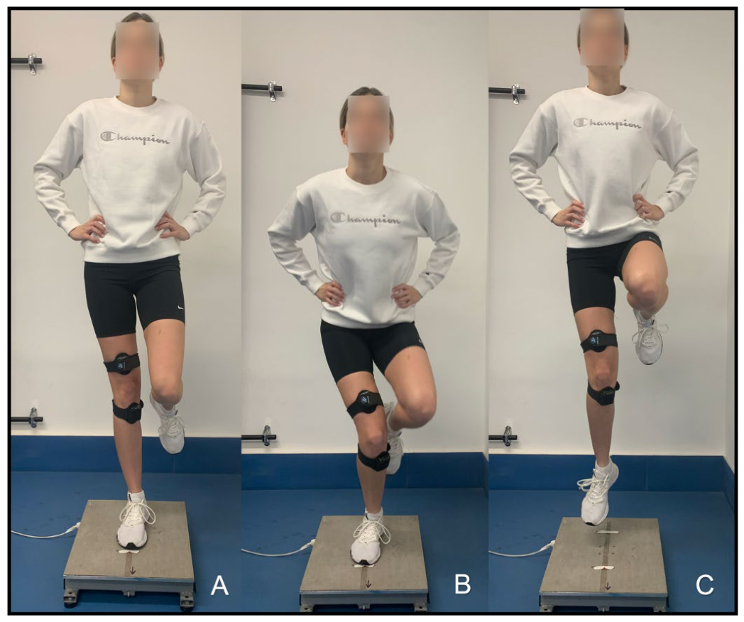
Figure 2.
Motion tracking during single-leg drop landing performed from a plyometric box set at a height of 0.46 m onto a force plate. Initial starting position on the box (A), flight phase following box drop (B), and ground contact with subsequent stabilization phase (C).
Figure 2.
Motion tracking during single-leg drop landing performed from a plyometric box set at a height of 0.46 m onto a force plate. Initial starting position on the box (A), flight phase following box drop (B), and ground contact with subsequent stabilization phase (C).
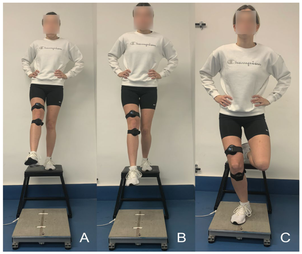
Figure 3.
Knee joint angular position and angular velocity in the sagittal and frontal planes recorded during single-leg counter-movement jump (SL-CMJ, left panels) and single-leg landing (SLL, right panels) in one representative healthy female participant. Knee joint angular position data are presented in the upper panels, while the corresponding angular velocity data are shown in the lower panels, as angular velocity was calculated by temporal differentiation of joint position. Within both conditions, knee valgus angle (A, B) and knee flexion angle (C, D) are shown in the upper panels, followed by knee valgus angular velocity (E, F) and knee flexion angular velocity (G, H) in the lower panels. Positive values denote knee flexion and medial deviation (valgus). On the horizontal axis, time zero corresponds to the start of data acquisition. The red highlighted field in each panel denotes the analysis window.
Figure 3.
Knee joint angular position and angular velocity in the sagittal and frontal planes recorded during single-leg counter-movement jump (SL-CMJ, left panels) and single-leg landing (SLL, right panels) in one representative healthy female participant. Knee joint angular position data are presented in the upper panels, while the corresponding angular velocity data are shown in the lower panels, as angular velocity was calculated by temporal differentiation of joint position. Within both conditions, knee valgus angle (A, B) and knee flexion angle (C, D) are shown in the upper panels, followed by knee valgus angular velocity (E, F) and knee flexion angular velocity (G, H) in the lower panels. Positive values denote knee flexion and medial deviation (valgus). On the horizontal axis, time zero corresponds to the start of data acquisition. The red highlighted field in each panel denotes the analysis window.
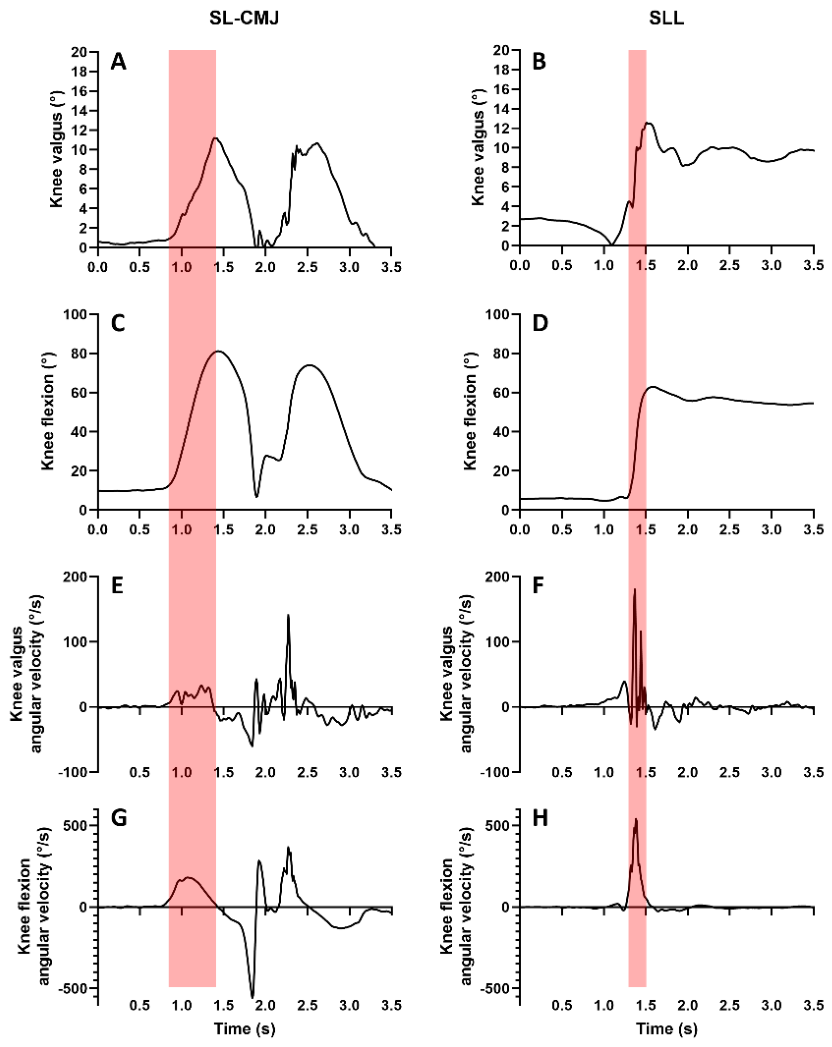
Figure 4.
Correlation between body weight and knee valgus magnitude during single-leg landing (SLL). The solid line represents the linear regression (y = 0.3908x + 161.6). The correlation coefficient was r = 0.37 (p = 0.047). Dashed lines represent the 95% confidence intervals of the regression line.
Figure 4.
Correlation between body weight and knee valgus magnitude during single-leg landing (SLL). The solid line represents the linear regression (y = 0.3908x + 161.6). The correlation coefficient was r = 0.37 (p = 0.047). Dashed lines represent the 95% confidence intervals of the regression line.
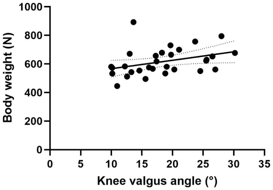
Figure 5.
Correlation between body height and knee valgus magnitude during single-leg landing (SLL). The solid line represents the linear regression (y = 5.914x + 507.7). The correlation coefficient was r = 0.33 (p = 0.075). Dashed lines represent the 95% confidence intervals of the regression line.
Figure 5.
Correlation between body height and knee valgus magnitude during single-leg landing (SLL). The solid line represents the linear regression (y = 5.914x + 507.7). The correlation coefficient was r = 0.33 (p = 0.075). Dashed lines represent the 95% confidence intervals of the regression line.
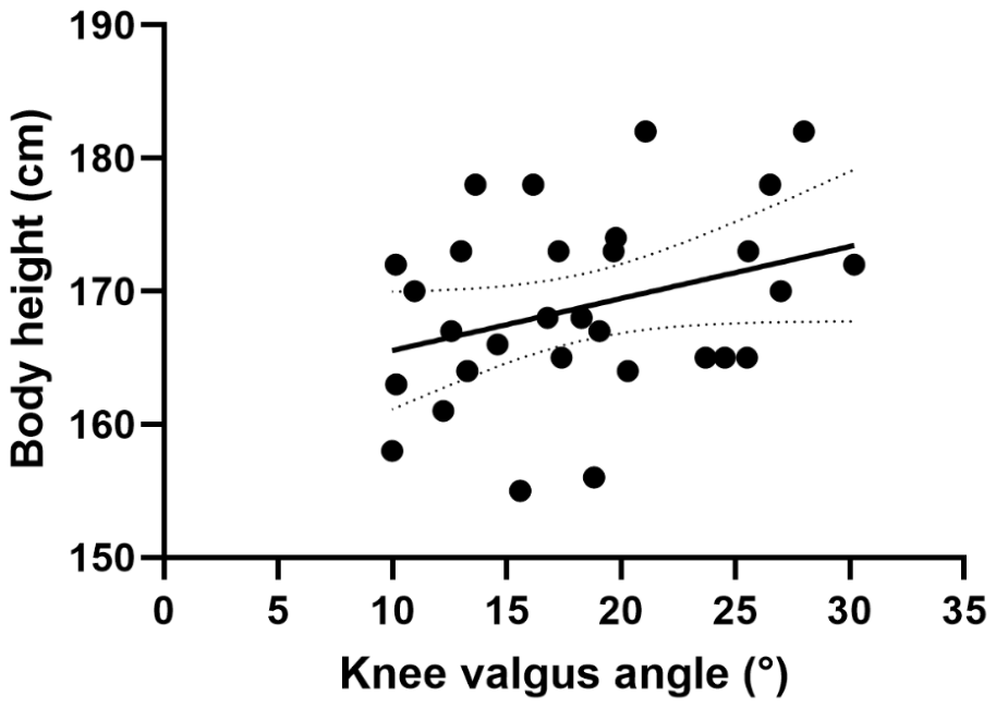

Table 1.
Comparison of the kinematics and kinetics between single-leg countermovement jump (SL-CMJ) and single-leg landing (SLL) tasks (n = 30).
Table 1.
Comparison of the kinematics and kinetics between single-leg countermovement jump (SL-CMJ) and single-leg landing (SLL) tasks (n = 30).
Values are presented as mean ± SD for normally distributed variables and as median [interquartile range] for non-normally distributed variables. Peak values are reported for all variables; however, for knee flexion, values correspond to the knee flexion angle observed at the time of peak knee valgus. Effect sizes are reported as Cohen’s d for paired t-tests and r for Wilcoxon signed-rank tests. Comparisons between tasks were performed using paired t-tests (t values) or Wilcoxon signed-rank tests (Z values), depending on data distribution. a.u., arbitrary unit; BW, body weight.
Disclaimer/Publisher’s Note: The statements, opinions and data contained in all publications are solely those of the individual author(s) and contributor(s) and not of MDPI and/or the editor(s). MDPI and/or the editor(s) disclaim responsibility for any injury to people or property resulting from any ideas, methods, instructions or products referred to in the content. |
© 2026 by the authors. Licensee MDPI, Basel, Switzerland. This article is an open access article distributed under the terms and conditions of the Creative Commons Attribution (CC BY) license.
Copyright: This open access article is published under a Creative Commons CC BY 4.0 license, which permit the free download, distribution, and reuse, provided that the author and preprint are cited in any reuse.