

Submitted:
08 September 2026
Posted:
08 September 2026
You are already at the latest version
Abstract
Background/Objectives: Early corneal changes after phacoemulsification reflect both surgery-related stress and individual tissue susceptibility. The contribution of preoperative corneal hysteresis (CH) to this variability remains uncertain. To test the interaction between preoperative CH and cumulative dissipated energy (CDE) in relation to early postoperative central corneal thickness (CCT), and to characterize postoperative pachymetric and biomechanical changes. Methods: This prospective study included 89 eyes of 80 patients undergoing uncomplicated phacoemulsification. CH and CCT were measured preoperatively, on postoperative day 1, and at 1 month; CDE was recorded intraoperatively. Early change in CCT (ΔCCT) was the primary outcome. Multivariable regression assessed the CH×CDE interaction adjusted for baseline CCT and age, IOPcc and IOPg. Longitudinal and biometric associations were also evaluated. Results: CCT increased by 149.0 ± 112.2 µm and CH decreased by 1.60 ± 1.98 mmHg on postoperative day one. CH significantly modified the CDE–ΔCCT association (β=1.99, 95% CI: 0.08–3.90; p=0.041): higher CH was associated with less predicted thickening at low-to-moderate CDE, with attenuation as CDE increased. CDE was strongly associated with ΔCCT but only weakly with CH reduction. At late follow-up, CCT was no longer significantly different from baseline, whereas CH remained reduced (MD=−0.68 mmHg, 95% CI:−1.30, −0.06; p=0.025). Eyes with ACD ≤2.5 mm showed a greater CCT response to CDE (β=7.70; p=0.028). Conclusions: Preoperative CH showed a moderating effect on the relationship between phacoemulsification energy and early corneal thickening, supporting a contribution of baseline biomechanics to individual corneal susceptibility.
Keywords:
corneal hysteresis
; cumulative dissipated energy
; phacoemulsification
; Ocular Response Analyzer
; corneal biomechanics
; central corneal thickness
; cataract surgery
1. Introduction
Cataract surgery is associated with early central corneal thickness (CCT) increase [1,2,3], endothelial cell loss [4,5,6], and transient alterations in corneal biomechanics [1,7,8,9]. Early postoperative corneal edema appears to reflect, at least in part, endothelial injury, as greater CCT increase has been associated with subsequent endothelial cell loss [10,11]. Both postoperative CCT increase and endothelial cell loss have been associated with several intraoperative parameters, such as effective phacoemulsification time, ultrasound exposure, cumulative dissipated energy (CDE), operative duration, and irrigation volume [4,12,13,14,15,16]. In contrast, the determinants of postoperative CH alterations are less well characterized. Kandarakis et al. [1] found that effective phacoemulsification time correlated with postoperative CCT increase but not with CH reduction, suggesting that the biomechanical response reflects broader effects of cataract surgery rather than ultrasound exposure alone. Moreover, Corvis ST studies have reported postoperative biomechanical alterations that were not fully accounted for by concurrent changes in pachymetry and intraocular pressure, further supporting only partial overlap between pachymetric and biomechanical responses [17,18].
The magnitude of postoperative corneal injury also varies according to individual ocular susceptibility. Age, endothelial reserve, cataract density, and anterior chamber depth (ACD), have been associated with endothelial loss or postoperative corneal edema [14,19]. Preoperative CH may represent an additional dimension of this susceptibility by providing biomechanical information complementary to conventional structural and biometric parameters.
Among intraoperative parameters, cumulative dissipated energy (CDE) is particularly relevant because it provides an integrated measure of ultrasound energy delivery during phacoemulsification, incorporating both the magnitude and duration of ultrasound application. In contrast, phacoemulsification time, operative duration, and irrigation volume capture only individual components of the surgical procedure and may not directly reflect the total ultrasonic energy applied. CDE has been widely used as a quantitative measure of ultrasound energy exposure during phacoemulsification [20,21] and has been associated with endothelial cell loss and early postoperative corneal edema [13,14,15].
However, the relationship between preoperative CH and the corneal response to CDE remains largely unexplored. We therefore hypothesized that the degree of postoperative corneal thickening associated with increasing CDE would vary according to the preoperative biomechanical state of the cornea, as reflected by CH. Clarifying this relationship may help define the potential value of CH as a preoperative indicator of individual susceptibility to phacoemulsification-related stress beyond structural and biometric parameters.
Given the apparent divergence between pachymetric and biomechanical responses after cataract surgery, further evaluation of their postoperative course and interrelationship is also needed.
To address these gaps, the primary aim of the present study was to determine whether preoperative CH modifies the association between CDE and early postoperative corneal thickening following uncomplicated phacoemulsification. Secondary objectives were to characterize postoperative changes in CH and CCT, assess their temporal recovery, and explore whether ocular biometric and clinical characteristics were associated with differences in corneal response.
2. Materials and Methods
2.1. Study Design and Participants
This prospective study included 89 eyes of 80 patients undergoing phacoemulsification cataract surgery and intraocular lens (IOL) implantation at Mureș County Hospital, Ophthalmology Clinic in Târgu Mureș, Romania between March-July 2026. Patients were eligible if complete preoperative and early postoperative measurements of CCT and CH, together with CDE, were available, and only eyes undergoing uncomplicated phacoemulsification cataract surgery were included. Eyes with pseudoexfoliation syndrome (PEX), glaucoma, or corneal guttata/ Fuchs endothelial corneal dystrophy (FECD) were retained for exploratory diagnosis-specific analyses. Exclusion criteria were ocular infection, central corneal opacities, keratoconus, previous corneal or refractive surgery, and current topical ocular or systemic corticosteroid treatment. Demographic data, corneal biomechanical parameters, and ocular biometric characteristics were collected for all participants.
2.2. Clinical Assessment and Outcome Measures
Preoperative evaluation included demographic data, central corneal thickness (CCT), corneal hysteresis (CH), corneal-compensated intraocular pressure (IOPcc), Goldmann-correlated intraocular pressure (IOPg), anterior chamber depth (ACD), and axial length (AL). CH was measured using the Ocular Response Analyzer (ORA; Reichert Technologies, Depew, NY, USA), and CCT using anterior pachymetry with spectral-domain optical coherence tomography (RTVue XR Avanti, Optovue Inc., Fremont, CA, USA). ACD and AL were measured by contact applanation ultrasonography using the OcuScan RxP Ophthalmic Ultrasound System (Alcon Laboratories, Inc., Fort Worth, TX, USA). All measurements were performed by the same experienced investigator using standardized central probe positioning and minimal corneal contact to minimize variation in probe alignment and contact pressure. The device-generated mean of 10 accepted measurements was used for analysis.
Pre-existing ocular conditions were documented from medical history and ophthalmic examination. Eyes were classified as controls or as having glaucoma, pseudoexfoliation syndrome (PEX), or corneal guttata; control eyes had none of these conditions.
CCT and CH were reassessed on postoperative day 1, and at one month. The primary outcome was the absolute early postoperative change in CCT (ΔCCT). Postoperative change in CH (ΔCH) and longitudinal changes in CCT and CH were evaluated as secondary outcomes.
2.3. Surgical Procedure
All patients underwent standard phacoemulsification cataract surgery using the CONSTELLATION Vision System (Alcon Laboratories, Inc., Fort Worth, TX, USA). All procedures were performed by a single experienced surgeon (KH) according to a standardized surgical protocol. A 2.2-mm clear-corneal main incision and two 1.2-mm side-port incisions were created. Dispersive and cohesive ophthalmic viscosurgical devices were used in a soft-shell technique to protect the corneal endothelium and anterior chamber structures. Phacoemulsification was performed using a vertical chop technique. CDE was automatically recorded by the phacoemulsification system and used as a measure of ultrasound energy delivered during surgery. At the end of the procedure, intracameral cefuroxime was administered for endophthalmitis prophylaxis.
2.4. Statistical Analysis
The primary analysis used multivariable linear regression with early postoperative ΔCCT as the dependent variable. Preoperative CH, CDE, baseline CCT, and age were entered as continuous predictors and mean-centered before model fitting. To test if the association between phacoemulsification energy and postoperative corneal thickening varied according to preoperative corneal biomechanics, a CH-CDE interaction term was included. The interaction was evaluated using its regression coefficient and post-estimation testing. Marginal predictions were subsequently estimated across representative preoperative CH values to illustrate how the association between CDE and predicted ΔCCT varied according to baseline CH.
In eyes with available biometric measurements, ACD was added to the multivariable model to assess its independent association with early postoperative ΔCCT after adjustment for CDE, preoperative CH, baseline CCT, and age. ACD was also evaluated as a continuous modifier of the CDE–ΔCCT association. An exploratory threshold-based analysis compared eyes with very shallow anterior chambers (ACD ≤2.5 mm) with those with ACD >2.5 mm, focusing on potential effect modification of the CDE–ΔCCT relationship at very low ACD. HC3 heteroskedasticity-robust standard errors were used because of the small subgroup size and potential influence of high-leverage observations.
Diagnostic subgroup analyses examined early postoperative corneal responses according to pre-existing ocular pathology. Eyes were classified as controls, glaucoma, PEX, or corneal guttata. Multivariable linear regression compared early postoperative ΔCCT with controls after adjustment for CDE, preoperative CH, baseline CCT, and age. CDE exposure across diagnostic groups was compared using the Kruskal–Wallis test. Longitudinal CH changes were evaluated relative to controls using mixed-effects models with diagnosis-by-time interaction terms. Given the small subgroup sizes, these analyses were considered exploratory.
Because CH is influenced by intraocular pressure and CH, IOPcc, and IOPg are derived from the same ORA measurement, sensitivity analyses were performed to assess whether the observed CH-CDE interaction was dependent on baseline IOP. Baseline IOPcc and IOPg were entered separately into the primary multivariable model including preoperative CH, CDE, the CH-CDE interaction, baseline CCT, and age. The two IOP parameters were not included simultaneously because of their strong intercorrelation and shared derivation from the ORA signal. In an additional exploratory model examining early postoperative CH change, baseline IOPcc was included together with CDE, baseline CH, baseline CCT, and age.
Longitudinal changes in CCT and CH were assessed using mixed-effects models. Pairwise comparisons between time points were derived from estimated marginal means and adjusted for multiple testing using the Bonferroni method. Model assumptions were assessed using residual diagnostics; multicollinearity, heteroskedasticity, and potentially influential observations were evaluated for the linear regression models. All tests were two-sided, and p<0.05 was considered statistically significant. Statistical analyses were performed using Stata, version 17 (StataCorp LLC, College Station, TX, USA).
2.5. Ethics Approval
The study was conducted in accordance with the Declaration of Helsinki and approved by the ethics committee of the hospital where the study took place (2374/18.03.2026). Written informed consent was obtained from all subjects before procedures.
3. Results
A total of 89 eyes were included in the analysis. Baseline demographic, corneal, and biometric characteristics are summarized in Table 1. Data are presented as mean ± standard deviation, median [interquartile range] or n (%), as appropriate.
3.1. Early Postoperative Corneal Response and CH–CDE Interaction
On postoperative day one, mean CCT was 687.1 ± 121.1 µm, corresponding to an absolute increase of 149.0 ± 112.2 µm and a relative increase of 27.6 ± 20.6%. Mean CH decreased to 9.28 ± 2.01 mmHg, with a mean change of −1.60 ± 1.98 mmHg from baseline.
A significant interaction between preoperative CH and CDE was observed in relation to early postoperative ΔCCT (Table 2). At mean CDE exposure, the model estimated a smaller postoperative CCT increase with higher CH, with the CH simple effect showing a trend toward statistical significance. The association between higher CH and lower predicted ΔCCT progressively attenuated as CDE increased (Figure 1).
The CH-CDE interaction remained unchanged after additional adjustment for baseline IOPcc or IOPg (β = 2.00, 95% CI [0.08, 3.92]; p = 0.041). Neither baseline IOPcc nor IOPg was independently associated with postoperative ΔCCT (p = 0.778 and p = 0.763, respectively).
3.2. Biometric and Diagnostic Subgroup Analyses
In the subset with available biometric measurements (n=67), neither ACD (β=4.2, 95% CI [−49.97, 58.52]; p=0.875) nor AL (β=−9.56 µm/mm, 95% CI [−26.27, 7.16]; p=0.257) was independently associated with early postoperative ΔCCT after adjustment for CDE, preoperative CH, baseline CCT, and age. ACD also did not modify the association between CDE and ΔCCT when analyzed continuously across the full biometric range (β=−2.35, 95% CI [−10.74, 6.03]; p=0.576). However, in the threshold-based analysis, shallow anterior chambers (ACD ≤2.5 mm; n=10) significantly modified the association between CDE and early postoperative ΔCCT (β=7.70, 95% CI [0.84, 14.56]; p=0.028). The estimated CDE–ΔCCT slope was 7.36 µm per CDE unit in eyes with ACD >2.5 mm and 15.06 µm per CDE unit in eyes with ACD ≤2.5 mm. CDE exposure was comparable between eyes with ACD ≤2.5 mm and those with deeper anterior chambers (p=0.94).
Early postoperative corneal responses varied according to diagnosis. Eyes with PEX or corneal guttata showed a tendency toward greater adjusted ΔCCT than controls (+45.4 µm; p=0.074), whereas glaucomatous eyes showed a tendency toward lower ΔCCT (−43.6 µm; p=0.083). CDE exposure did not differ significantly across diagnostic groups (p=0.154). In contrast, glaucomatous eyes exhibited a greater early postoperative CH decrease than controls (p=0.039), whereas the CH trajectory in eyes with PEX or corneal guttata did not differ significantly from controls (p=0.540).
3.3. Postoperative CH Response and CCT–CH Relationship
Higher CDE was weakly associated with a greater early postoperative reduction in CH (r=−0.222, p=0.036). After adjustment for baseline CCT and age, the effect remained small and showed only a trend toward statistical significance (β=−0.057 mmHg per CDE unit; p=0.054). In contrast, the magnitude of early postoperative CCT increase was not associated with CH reduction (r=−0.155, p=0.146) and remained unrelated to ΔCH after adjustment for CDE, baseline CH, baseline CCT, and age (p=0.786).
The association between CCT and CH was positive preoperatively (β=0.0094 mmHg/µm; p=0.047) but became significantly inverse on postoperative day 1 (β=−0.0027 mmHg/µm; p=0.034). At late follow-up, the association was again positive, with only a trend toward statistical significance (β=0.0122 mmHg/µm; p=0.085).
Follow-up measurements were available for 40 eyes. CCT was not significantly different from the preoperative value (MD=−14.63 µm, 95% CI [−48.45, 19.20]; p=0.902). CH showed substantial recovery from the early postoperative decrease but remained slightly lower than preoperatively (MD=−0.68 mmHg, 95% CI [−1.30, −0.06]; p=0.025).
4. Discussion
The main findings of the present study can be summarized as follows: (i) preoperative CH significantly modified the association between phacoemulsification energy and early postoperative corneal thickening, with higher CH being associated with a smaller predicted CCT increase at low-to-moderate CDE levels and this moderating effect progressively diminishing as CDE increased; (ii) although CDE was a strong determinant of postoperative CCT increase, its association with CH reduction was substantially weaker, while the magnitude of corneal swelling itself was not associated with CH decrease, suggesting that additional factors contribute to the early biomechanical response; and (iii) CCT returned toward preoperative values earlier than CH, indicating only partial overlap between pachymetric and biomechanical recovery. In secondary analyses, the increase in postoperative CCT per unit of CDE was approximately twice as large in eyes with shallow anterior chambers as in those with deeper chambers, suggesting greater sensitivity to phacoemulsification energy.
The interaction between preoperative CH and CDE indicates that the corneal response to phacoemulsification energy varies according to the preoperative biomechanical state of the cornea. At low-to-moderate CDE levels, higher CH was associated with a smaller predicted postoperative CCT increase, whereas this association progressively attenuated and became negligible at higher CDE levels. The interaction remained unchanged after adjustment for baseline CCT, age and baseline IOPcc or IOPg, supporting an independent association. Nevertheless, it should be interpreted as evidence of differential susceptibility rather than as a clinically meaningful threshold.
A related finding was that CDE was strongly associated with early postoperative CCT increase but only weakly with CH reduction, consistent with previous evidence of a partial dissociation between pachymetric and biomechanical responses to phacoemulsification [1]. To date, the relationship between intraoperative energy exposure and postoperative CH change remains poorly characterized. Our findings therefore add to limited evidence suggesting the involvement of additional determinants beyond ultrasound energy exposure and concurrent corneal edema.
Perioperative changes in ocular mechanical loading may represent one such influence. During phacoemulsification, irrigation and aspiration generate substantial transient variations in anterior chamber pressure, resulting in marked intraoperative IOP fluctuations [22,23,24]. Higher fluidic settings and intraoperative pressure have been associated with greater early postoperative corneal thickening, whereas lower target IOP with active-fluidics systems has been associated with smaller early CCT increases [25,26,27]. Although a direct effect of these pressure dynamics on postoperative CH has not been established, their contribution is plausible given the known dependence of corneal deformation and CH on mechanical loading and IOP [28,29,30].
Incision-related structural changes may also contribute to postoperative biomechanical alterations, although evidence regarding the influence of incision characteristics on the magnitude and recovery of these changes remains mixed [18,31,32]. However, because a standardized 2.2-mm clear-corneal incision was used in all eyes in the present cohort, incision size cannot explain the interindividual variability in CH change. Rather, the incision likely represents a common biomechanical perturbation, while differences in ocular loading and tissue hydration may contribute to variability in CH recovery [29,30,33].
The different temporal behavior of CCT and CH provides further evidence that pachymetric and biomechanical recovery only partially overlap. In the present study, CCT was no longer significantly different from its preoperative value at late follow-up, whereas CH, despite substantial recovery from postoperative day one, remained slightly but significantly reduced. Previous studies [3] have generally shown a transient reduction in CH after cataract surgery, although reported recovery ranges from normalization within one week [8] to approximately one month [9] while Corvis ST data suggest that altered biomechanical deformation may persist for up to three months even after accounting for IOP and pachymetric changes [18]. This difference is biologically plausible because CCT primarily reflects corneal thickness and hydration, whereas CH reflects the dynamic response of the cornea to loading and is only partly related to CCT [29,30,33]. Thus, pachymetric normalization should not necessarily be interpreted as complete biomechanical recovery. Persistent biomechanical alteration after pachymetric normalization may also be relevant when interpreting postoperative applanation-based IOP measurements, particularly in glaucoma patients [18,34].
ACD was not independently associated with early postoperative corneal thickening and did not significantly modify the effect of CDE when analyzed continuously. However, in eyes with shallow anterior chambers, the increase in postoperative CCT per unit of CDE was approximately twice that observed in deeper chambers, indicating greater susceptibility to phacoemulsification energy. Previous studies have reported greater endothelial injury in shallow anterior chambers, particularly in eyes with denser cataracts or under conditions of greater surgical stress, although findings have varied according to surgical technique and other operative factors [19,35,36]. Our findings extend these observations by indicating that ACD may influence the corneal response to a given amount of CDE rather than merely identifying susceptibility under high-energy conditions. The steeper CDE–CCT relationship in shallow chambers was evident across the observed CDE range, including at low-to-moderate levels, suggesting that increased susceptibility may also be relevant at lower energy exposures. This threshold-based finding should nevertheless be considered hypothesis-generating, as only 10 eyes had ACD ≤2.5 mm, and requires confirmation in larger cohorts.
Diagnosis-specific responses also supported a partial dissociation between pachymetric and biomechanical changes. Glaucoma, PEX, and corneal guttata/FECD have been associated with altered corneal biomechanics [37,38], although postoperative CH data remain limited [39,40]. In our cohort, glaucomatous eyes showed a greater early CH decrease despite a tendency toward less corneal thickening, whereas PEX or corneal guttata tended toward greater ΔCCT without a significantly different CH trajectory. These findings are consistent with previous reports of greater endothelial injury in PEX and increased postoperative edema susceptibility in corneal guttata [41,42]. Given the small subgroups, these results require confirmation in larger prospective studies.
From a clinical perspective, these findings support the potential value of CH as a complementary preoperative marker of individual corneal susceptibility to phacoemulsification-related stress. An integrated assessment incorporating biomechanical, structural, and biometric characteristics may allow a more nuanced characterization of corneal response and postoperative recovery.
Several limitations should be acknowledged. First, although the CH–CDE interaction reached statistical significance, the effect was modest and was identified in a relatively small cohort of 89 eyes; therefore, it should be interpreted cautiously and confirmed in larger independent samples. Second, late follow-up measurements were available in only 40 eyes, limiting the precision of conclusions regarding biomechanical recovery. Biometric measurements were available only in a subset of eyes, and the shallow-ACD subgroup was small. In addition, ACD was measured by contact applanation ultrasonography, which may introduce some underestimation through corneal indentation. However, all measurements were obtained by the same experienced investigator using a standardized technique to minimize measurement variability. CDE provides an integrated estimate of ultrasound energy exposure but does not capture other components of intraoperative mechanical stress, including pressure fluctuations and fluidic dynamics, which were not directly measured. Diagnosis-specific analyses involved small subgroups and should therefore be regarded as exploratory. Finally, only uncomplicated cataract procedures were included; although this improves internal consistency, the findings may not generalize to eyes experiencing substantial intraoperative endothelial trauma.
Author Contributions
K.U.H, E.A.M., and F.V. contributed to the conceptualization and study design. E.A.M. performed data collection, data analysis, preparation of the original manuscript draft. K.U.H. and F.V. contributed to manuscript review and editing and provided supervision. All authors reviewed and approved the final version of the manuscript.
Funding
This work was supported by the George Emil Palade University of Medicine, Pharmacy, Science and Technology of Târgu Mureș, Research Grant number 795/3/22.01.2025.
Institutional Review Board Statement
The study was conducted in accordance with the Declaration of Helsinki and approved by the Ethics Committee of Mureș County Clinical Hospital (protocol code 2374, 18 March 2026).
Informed Consent Statement
Informed consent was obtained from all subjects involved in the study.
Data Availability Statement
Anonymized individual-level data and analytic materials underlying the findings of this study will be made available upon reasonable request to the corresponding author, subject to institutional approval and applicable ethical and data protection requirements.
Acknowledgments
This work was supported by the George Emil Palade University of Medicine, Pharmacy, Science and Technology of Târgu Mureș, Research Grant number 795/3/22.01.2025.
Conflicts of Interest
The authors declare no conflicts of interest.
Abbreviations
The following abbreviations are used in this manuscript:
| ACD | Anterior chamber depth |
| AL | Axial length |
| CCT | Central corneal thickness |
| CDE | Cumulative dissipated energy |
| CH | Corneal hysteresis |
| FECD | Fuchs endothelial corneal dystrophy |
| IOPcc | Corneal compensated intraocular pressure |
| IOPg | Goldman correlated intraocular pressure |
| ORA | Ocular response analyzer |
| PEX | Pseudoexfoliation syndrome |
References
- Kandarakis, A.; Soumplis, V.; Karampelas, M.; Koutroumanos, I.; Panos, C.; Kandarakis, S.; Karagiannis, D. Response of corneal hysteresis and central corneal thickness following clear corneal cataract surgery. Acta Ophthalmol. 2012, 90, 526–529. [Google Scholar] [CrossRef] [PubMed]
- Kongsap, P. Central corneal thickness changes following manual small incision cataract surgery versus phacoemulsification for white cataract. Rom. J. Ophthalmol. 2019, 63, 61–67. [Google Scholar] [CrossRef]
- Li, X.; Li, X.; Tan, L.; Chen, H.; Lai, Y. The corneal biomechanical changes of phacoemulsification in cataract patients: a systematic review and meta-analysis. PLoS ONE 2025, 20, e0317179. [Google Scholar] [CrossRef]
- Ghiță, A.M.; Iliescu, D.A.; Ilie, L.A.; Ghiță, A.C. Endothelial cell loss after phacoemulsification in a Romanian cohort: early outcomes and associated risk factors. Diagnostics 2026, 16, 1468. [Google Scholar] [CrossRef] [PubMed]
- Sato, T. Corneal endothelial changes after phacoemulsification using the eight-chop technique in diabetic eyes. J. Pers. Med. 2025, 15, 209. [Google Scholar] [CrossRef] [PubMed]
- Upasani, D.; Daigavane, S. Phacoemulsification techniques and their effects on corneal endothelial cells and visual acuity: a review of “direct-chop” and “stop-and-chop” approaches under topical anesthesia. Cureus 2024, 16, e66587. [Google Scholar] [CrossRef] [PubMed]
- Čulina, K.; Tomić, M.; Bulum, T.; Medić, A.; Šoša, I.; Ivanišević, K.; Jukić, T. Corneal biomechanics and other factors associated with postoperative astigmatism after cataract surgery. Life 2024, 14, 655. [Google Scholar] [CrossRef] [PubMed]
- Kamiya, K.; Shimizu, K.; Ohmoto, F.; Amano, R. Time course of corneal biomechanical parameters after phacoemulsification with intraocular lens implantation. Cornea 2010, 29, 1256–1260. [Google Scholar] [CrossRef] [PubMed]
- Kucumen, R.B.; Yenerel, N.M.; Gorgun, E.; Kulacoglu, D.N.; Oncel, B.; Kohen, M.C.; Alimgil, M.L. Corneal biomechanical properties and intraocular pressure changes after phacoemulsification and intraocular lens implantation. J. Cataract Refract Surg. 2008, 34, 2096–2098. [Google Scholar] [CrossRef] [PubMed]
- Chen, S.; Li, X.; Huang, L.; Feng, Q.; Lu, H.; Mu, J. Correlation between early corneal edema and endothelial cell loss after phacoemulsification cataract surgery. Front Med. 2025, 12, 1562717. [Google Scholar] [CrossRef] [PubMed]
- Lundberg, B.; Jonsson, M.; Behndig, A. Postoperative corneal swelling correlates strongly to corneal endothelial cell loss after phacoemulsification cataract surgery. Am. J. Ophthalmol. 2005, 139, 1035–1041. [Google Scholar] [CrossRef] [PubMed]
- Doors, M.; Berendschot, T.T.J.M.; Touwslager, W.; Webers, C.A.; Nuijts, R.M.M.A. Phacopower modulation and the risk for postoperative corneal decompensation: a randomized clinical trial. JAMA Ophthalmol. 2013, 131, 1443–1450. [Google Scholar] [PubMed]
- Fu, J.; Shi, Y.; Yang, Y.; Wang, T.; Du, W.; Mo, L. Predicting early postoperative corneal edema after phacoemulsification: a retrospective observational study. Medicine 2026, 105, e49989. [Google Scholar] [CrossRef] [PubMed]
- Lee, N.S.Y.; Ong, K. Risk factors for corneal endothelial cell loss after phacoemulsification. Taiwan J. Ophthalmol. 2024, 14, 83–87. [Google Scholar] [CrossRef] [PubMed]
- Sato, T. Minimizing endothelial cell loss in hard nucleus cataract surgery: efficacy of the eight-chop technique. J. Clin. Med. 2025, 14, 2576. [Google Scholar] [CrossRef] [PubMed]
- Wong, M.M.; Shukla, A.N.; Munir, W.M. Correlation of corneal thickness and volume with intraoperative phacoemulsification parameters using Scheimpflug imaging and optical coherence tomography. J. Cataract Refract Surg. 2014, 40, 2067–2075. [Google Scholar] [CrossRef] [PubMed]
- Hirasawa, K.; Nakakura, S.; Nakao, Y.; Fujino, Y.; Matsuura, M.; Murata, H.; Kiuchi, Y.; Asaoka, R. Changes in corneal biomechanics and intraocular pressure following cataract surgery. Am. J. Ophthalmol. 2018, 195, 26–35. [Google Scholar] [CrossRef] [PubMed]
- Wallace, H.B.; Misra, S.L.; Li, S.S.; McKelvie, J. Biomechanical changes in the cornea following cataract surgery: a prospective assessment with the Corneal Visualisation Scheimpflug Technology. Clin. Exp. Ophthalmol. 2019, 47, 461–468. [Google Scholar] [CrossRef] [PubMed]
- Hwang, H.B.; Lyu, B.; Yim, H.B.; Lee, N.Y. Endothelial cell loss after phacoemulsification according to different anterior chamber depths. J. Ophthalmol. 2015, 2015, 210716. [Google Scholar] [CrossRef] [PubMed]
- Fernández-Muñoz, E.; Chávez-Romero, Y.; Rivero-Gómez, R.; Aridjis, R.; Gonzalez-Salinas, R. Cumulative dissipated energy (CDE) in three phaco-fragmentation techniques for dense cataract removal. Clin. Ophthalmol. 2023, 17, 2405–2412. [Google Scholar] [CrossRef] [PubMed]
- Ilhan, C.; Kocak Altintas, A.G. Cumulative dissipated energy in eyes with and without corneal opacity. Rom. J. Ophthalmol. 2022, 66, 233–239. [Google Scholar] [CrossRef] [PubMed]
- Grinbaum, A.; Blumenthal, M.; Assia, E. Comparison of intraocular pressure profiles during cataract surgery by phacoemulsification and extracapsular cataract extraction. Ophthalmic Surg. Lasers Imaging 2003, 34, 182–186. [Google Scholar] [CrossRef]
- Vasavada, V.; Raj, S.M.; Praveen, M.R.; Vasavada, A.R.; Henderson, B.A.; Asnani, P.K. Real-time dynamic intraocular pressure fluctuations during microcoaxial phacoemulsification using different aspiration flow rates and their impact on early postoperative outcomes: a randomized clinical trial. J. Refract Surg. 2014, 30, 534–540. [Google Scholar] [CrossRef] [PubMed]
- Vasavada, V.; Vasavada, A.R.; Vasavada, V.A.; Vasavada, S.A.; Bhojwani, D. Real-time dynamic changes in intraocular pressure after occlusion break: comparing 2 phacoemulsification systems. J. Cataract Refract Surg. 2021, 47, 1205–1209. [Google Scholar] [CrossRef] [PubMed]
- Liu, Y.; Hong, J.; Chen, X. Comparisons of the clinical outcomes of Centurion active fluidics system with a low IOP setting and gravity fluidics system with a normal IOP setting for cataract patients with low corneal endothelial cell density. Front Med. 2023, 10, 1294808. [Google Scholar] [CrossRef] [PubMed]
- Qiao, Y.; Ye, Y.; Liu, F.; Luo, L.; Huang, X.; Liang, B.; Xi, W.; Liu, X.; Zeng, K. Active fluidics at lower intraocular pressure reduces intraoperative discomfort during phacoemulsification: a propensity score-matched cohort study. Front Med. 2026, 13, 1783229. [Google Scholar] [CrossRef] [PubMed]
- Rauen, M.P.; Joiner, H.; Kohler, R.A.; O’Connor, S. Phacoemulsification using an active fluidics system at physiologic vs high intraocular pressure: impact on anterior and posterior segment physiology. J. Cataract Refract Surg. 2024, 50, 822–827. [Google Scholar] [CrossRef] [PubMed]
- Katiyar, S.; Tong, J.; Pensyl, D.; Sullivan-Mee, M. Corneal biomechanical changes caused by acute elevation of IOP in eyes with and without glaucoma. Optom. Vis. Sci. 2021, 98, 367–373. [Google Scholar] [CrossRef] [PubMed]
- McMonnies, C.W. Assessing corneal hysteresis using the Ocular Response Analyzer. Optom. Vis. Sci. 2012, 89, E343–E349. [Google Scholar] [CrossRef] [PubMed]
- Terai, N.; Raiskup, F.; Haustein, M.; Pillunat, L.E.; Spoerl, E. Identification of biomechanical properties of the cornea: the Ocular Response Analyzer. Curr. Eye Res. 2012, 37, 553–562. [Google Scholar] [CrossRef] [PubMed]
- Alió, J.L.; Agdeppa, M.C.C.; Rodríguez-Prats, J.L.; Amparo, F.; Piñero, D.P. Factors influencing corneal biomechanical changes after microincision cataract surgery and standard coaxial phacoemulsification. J. Cataract Refract Surg. 2010, 36, 890–897. [Google Scholar] [CrossRef] [PubMed]
- Zhang, Z.; Yu, H.; Dong, H.; Wang, L.; Jia, Y.D.; Zhang, S.H. Corneal biomechanical properties changes after coaxial 2.2-mm microincision and standard 3.0-mm phacoemulsification. Int. J. Ophthalmol. 2016, 9, 230–234. [Google Scholar] [CrossRef] [PubMed]
- Huseynova, T.; Waring, G.O., 4th; Roberts, C.; Krueger, R.R.; Tomita, M. Corneal biomechanics as a function of intraocular pressure and pachymetry by dynamic infrared signal and Scheimpflug imaging analysis in normal eyes. Am. J. Ophthalmol. 2014, 157, 885–893. [Google Scholar] [CrossRef] [PubMed]
- Aoki, S.; Asaoka, R.; Fujino, Y.; Nakakura, S.; Murata, H.; Kiuchi, Y. Comparing corneal biomechanic changes among solo cataract surgery, microhook ab interno trabeculotomy and iStent implantation. Sci. Rep. 2023, 13, 19148. [Google Scholar] [CrossRef] [PubMed]
- Cho, Y.K.; Chang, H.S.; Kim, M.S. Risk factors for endothelial cell loss after phacoemulsification: comparison in different anterior chamber depth groups. Korean J. Ophthalmol. 2010, 24, 10–15. [Google Scholar] [CrossRef] [PubMed]
- Sato, T. Corneal endothelial cell loss in shallow anterior chamber eyes after phacoemulsification using the eight-chop technique. J. Clin. Med. 2025, 14, 3045. [Google Scholar] [CrossRef] [PubMed]
- Clemmensen, K.; Hjortdal, J. Intraocular pressure and corneal biomechanics in Fuchs’ endothelial dystrophy and after posterior lamellar keratoplasty. Acta Ophthalmol. 2014, 92, 350–354. [Google Scholar] [CrossRef] [PubMed]
- Sit, A.J.; Chen, T.C.; Takusagawa, H.L.; Rosdahl, J.A.; Hoguet, A.; Chopra, V.; Richter, G.M.; Ou, Y.; Kim, S.J.; WuDunn, D. Corneal hysteresis for the diagnosis of glaucoma and assessment of progression risk: a report by the American Academy of Ophthalmology. Ophthalmology 2023, 130, 433–442. [Google Scholar] [PubMed]
- Siggel, R.; Christofi, E.; Giasoumi, F.; Adler, W.; Siebelmann, S.; Bachmann, B.; Cursiefen, C.; Heindl, L.M. Changes in corneal biomechanical properties after Descemet membrane endothelial keratoplasty. Cornea 2019, 38, 964–969. [Google Scholar] [CrossRef] [PubMed]
- Zehavi-Dorin, T.; Kutzscher, A.E.; Badr, M.; Mora, M.; Shyu, Y.; Porco, T.C.; Stamper, R.L. Repeatability of corneal hysteresis measurements in glaucoma patients during routine follow up and after cataract surgery. J. Glaucoma 2022, 31, 590–594. [Google Scholar] [CrossRef] [PubMed]
- Hayashi, K.; Manabe, S.I.; Yoshimura, K.; Kondo, H. Corneal endothelial damage after cataract surgery in eyes with pseudoexfoliation syndrome. J. Cataract Refract Surg. 2013, 39, 881–887. [Google Scholar] [CrossRef] [PubMed]
- Liesenborghs, L.F.; Hennekes, R. Phacoemulsification and IOL implantation in eyes with cornea guttata. Bull. Soc. Belge. Ophtalmol. 1991, 242, 11–18. [Google Scholar] [PubMed]
Figure 1.
Predicted early postoperative change in central corneal thickness according to cumulative dissipated energy at selected levels of preoperative corneal hysteresis (CH). Blue, red, and green lines represent CH values of 9, 11, and 13 mmHg, respectively. Predicted values are shown with 95% confidence intervals.
Figure 1.
Predicted early postoperative change in central corneal thickness according to cumulative dissipated energy at selected levels of preoperative corneal hysteresis (CH). Blue, red, and green lines represent CH values of 9, 11, and 13 mmHg, respectively. Predicted values are shown with 95% confidence intervals.
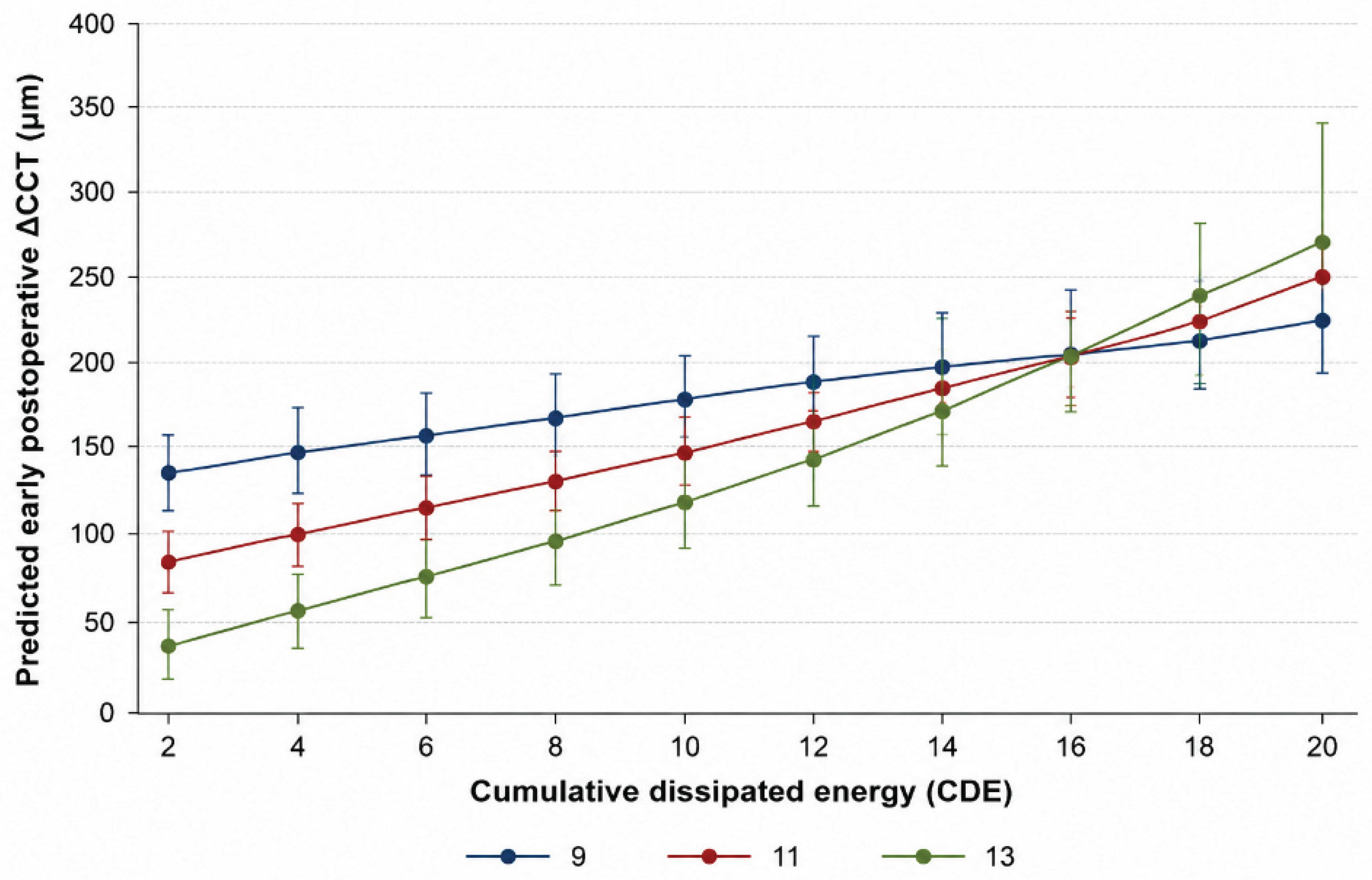

Table 1.
Demographic, preoperative corneal, intraoperative, and ocular biometric characteristics of the study population. CH: corneal hysteresis; CCT: central corneal thickness; CDE: cumulative dissipated energy; ACD: anterior chamber depth; AL: axial length. IOPcc: corneal compensated intraocular pressure; IOPg: Goldmann-correlated intraocular pressure. PEX: pseudoexfoliation syndrome.
Table 1.
Demographic, preoperative corneal, intraoperative, and ocular biometric characteristics of the study population. CH: corneal hysteresis; CCT: central corneal thickness; CDE: cumulative dissipated energy; ACD: anterior chamber depth; AL: axial length. IOPcc: corneal compensated intraocular pressure; IOPg: Goldmann-correlated intraocular pressure. PEX: pseudoexfoliation syndrome.
| Variable | N | Value | Range |
|---|---|---|---|
| Age, years | 89 | 73.52 ± 5.77 | 58–88 |
| Sex, (%) | 89 | ||
| Female | 50 (56.2%) | ||
| Male | 39 (43.8%) | ||
| Preoperative CH, mmHg | 89 | 10.89 ± 1.13 | 8.60–13.30 |
| Preoperative CH, mmHg | 89 | 10.89 ± 1.13 | 8.60–13.30 |
| CDE | 89 | 6.00 [2.99–8.83] | 1.26–44.56 |
| ACD, mm | 67 | 2.96 ± 0.40 | 2.05–3.98 |
| AL, mm | 67 | 23.21 ± 1.32 | 20.87–29.21 |
| IOPcc, mmHg | 89 | 17.41 ± 3.44 | 10-25.9 |
| IOPg, mmHg | 89 | 17.77 ± 3.58 | 9.5–26.6 |
| Pre-existing ocular diagnosis, (%) | |||
| Control | 63 (70.8%) | ||
| Glaucoma | 13 (14.6%) | ||
| PEX | 7 (7.9%) | ||
| Cornea guttata | 6 (6.7%) |
Table 2.
Multivariable linear regression of early postoperative ΔCCT. N=89; R²=0.515; adjusted R²=0.486; F(5,83) =17.65; p<0.001. CH and CDE were mean-centered. Accordingly, the main effect of CH represents its association with ΔCCT at mean CDE, whereas the main effect of CDE represents its association with ΔCCT at mean CH. CH: corneal hysteresis; CDE: cumulative dissipated energy; CCT: central corneal thickness.
Table 2.
Multivariable linear regression of early postoperative ΔCCT. N=89; R²=0.515; adjusted R²=0.486; F(5,83) =17.65; p<0.001. CH and CDE were mean-centered. Accordingly, the main effect of CH represents its association with ΔCCT at mean CDE, whereas the main effect of CDE represents its association with ΔCCT at mean CH. CH: corneal hysteresis; CDE: cumulative dissipated energy; CCT: central corneal thickness.
| Predictor | β | 95% CI | p |
|---|---|---|---|
| Preoperative CH | −15.26 | −31.61, 1.09 | 0.067 |
| CDE | 9.38 | 6.83, 11.92 | <0.001 |
| CH × CDE | 1.99 | 0.08, 3.90 | 0.041 |
| Baseline CCT | 0.79 | 0.23, 1.35 | 0.007 |
| Age | 2.69 | −0.68, 6.06 | 0.116 |
Disclaimer/Publisher’s Note: The statements, opinions and data contained in all publications are solely those of the individual author(s) and contributor(s) and not of MDPI and/or the editor(s). MDPI and/or the editor(s) disclaim responsibility for any injury to people or property resulting from any ideas, methods, instructions or products referred to in the content. |
© 2026 by the authors. Licensee MDPI, Basel, Switzerland. This article is an open access article distributed under the terms and conditions of the Creative Commons Attribution (CC BY) license (http://creativecommons.org/licenses/by/4.0/).
Copyright: This open access article is published under a Creative Commons CC BY 4.0 license, which permit the free download, distribution, and reuse, provided that the author and preprint are cited in any reuse.