

Submitted:
06 September 2026
Posted:
08 September 2026
You are already at the latest version
Abstract
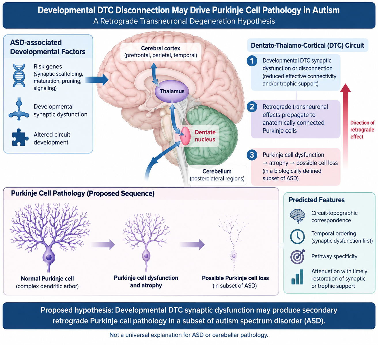
Purkinje cell abnormalities, including reduced cell size and lower cell density in some postmortem series, are among the most frequently discussed cerebellar findings in autism spectrum disorder (ASD), yet their mechanistic origin and causal significance remain unresolved. We propose a circuit-based hypothesis in which developmental synaptic dysfunction or disconnection within the dentato-thalamo-cortical (DTC) network produces retrograde transneuronal changes in anatomically connected Purkinje cells, ranging from functional impairment and atrophy to cell loss. The model integrates four observations: heterogeneous Purkinje cell pathology in ASD; the participation of posterolateral cerebellar territories in non-motor cerebro-cerebellar circuits; structural and functional abnormalities reported at multiple nodes of DTC networks; and the enrichment of ASD-associated genes in pathways regulating synaptic scaffolding, maturation, pruning, and intracellular signaling. We distinguish three competing models: primary Purkinje cell pathology causing downstream network dysfunction; a shared developmental defect producing parallel abnormalities across cerebellar and cerebral structures; and our proposed model, in which distal or intermediate DTC synaptic dysfunction produces secondary retrograde Purkinje cell pathology. The hypothesis predicts circuit-topographic correspondence, temporal ordering from synaptic dysfunction to upstream neuronal change, pathway specificity, and attenuation of Purkinje cell pathology after timely restoration of synaptic or trophic support. Longitudinal multimodal imaging, postmortem connectomic-pathological correlation, and pathway-specific developmental models can directly test these predictions. Retrograde transneuronal degeneration (RTD) is proposed as a potential mechanism in a biologically defined subset of ASD, not as a universal explanation for ASD or cerebellar pathology.
Keywords:
autism spectrum disorder
; Purkinje cell atrophy
; cerebellum
; dentato-thalamo-cortical pathway
; developmental disconnection
; retrograde transneuronal degeneration
; synaptic scaffolding
1. Introduction
Autism spectrum disorder (ASD) is a heterogeneous neurodevelopmental condition in which genetic, molecular, cellular, and circuit-level abnormalities may converge on overlapping behavioral phenotypes. No single anatomical lesion accounts for ASD. Nevertheless, neuropathological studies have repeatedly drawn attention to the cerebellum, particularly to abnormalities and reduced numbers of Purkinje cells. The biological meaning of this observation remains unsettled: Purkinje cell loss may be primary, secondary, developmental, acquired, regionally selective, or present only in a subset of ASD.
The key question addressed here is therefore not whether cerebellar pathology alone causes autism. Instead, we ask whether the topography of reported Purkinje cell abnormalities can be partly explained by the circuitry in which those cells participate. We propose that developmental synaptic dysfunction within the dentato-thalamo-cortical (DTC) system may, under some circumstances, lead to secondary retrograde transneuronal effects on upstream cerebellar neurons. This hypothesis links three bodies of literature that are often considered separately: selective Purkinje cell pathology, DTC/cerebro-cerebellar connectivity abnormalities, and ASD-associated synaptic genes.
The model is deliberately restricted. It does not imply that all individuals with ASD have Purkinje cell loss, that DTC dysfunction is unique to ASD, or that retrograde degeneration is the sole explanation for cerebellar pathology. Its value lies in producing a falsifiable circuit-level explanation for a recurrent pathological observation.
2. The Observation to Be Explained: Purkinje Cell Abnormalities in ASD
Purkinje cells are the sole output neurons of the cerebellar cortex and have exceptionally complex dendritic and synaptic architecture. Human postmortem findings in ASD are heterogeneous: some series report lower Purkinje cell density or number, while others describe reduced Purkinje cell size without reduced density (Bauman and Kemper, 1985; Ritvo et al., 1986; Bailey et al., 1998; Kemper and Bauman, 1998; Fatemi et al., 2002; Whitney et al., 2009). A 2024 reassessment of the cerebellar neuropathology literature emphasized this heterogeneity and questioned whether cerebellar pathology should be interpreted as a primary cause of ASD (Baizer, 2024). This uncertainty is central to the present hypothesis, not a weakness to be dismissed.
The regional distribution is also not uniform across studies. Several classic series described marked abnormalities in lateral, inferior, or posterolateral cerebellar territories, whereas others reported more diffuse changes (Bauman and Kemper, 1985; Ritvo et al., 1986; Bailey et al., 1998). We therefore do not assume that Purkinje cell loss is uniformly localized. Instead, we ask whether, in a subset of cases, regional Purkinje cell atrophy or loss follows the topology of affected cerebro-cerebellar circuits.
The distinction between atrophy and cell loss may be mechanistically important. Fatemi et al. (2002) reported markedly smaller Purkinje cell size in ASD without a significant reduction in density. A transneuronal process need not begin with neuronal death: impaired connectivity may first produce physiological change, altered trophic signaling, and cellular atrophy, with cell loss representing a more severe or later endpoint. We therefore use the broader term 'Purkinje cell pathology' to include functional change, atrophy, and loss.
3. Why Consider the Dentato-Thalamo-Cortical Circuit?
The lateral cerebellar hemispheres participate in reciprocal cerebro-cerebellar networks involved not only in movement but also in executive, linguistic, visuospatial, and affective functions (Schmahmann and Pandya, 1995; Middleton and Strick, 2000). Purkinje cells inhibit neurons of the deep cerebellar nuclei; output from the dentate nucleus travels through the superior cerebellar peduncle, crosses, and reaches thalamic and cortical targets, including prefrontal and motor territories. The DTC system therefore provides an anatomical framework through which abnormalities distant from the cerebellar cortex could influence cerebellar function.
Multiple observations in ASD are compatible with disruption at different points in this network. Pathological studies have described abnormalities of cerebellar nuclei and altered GAD65 expression in the dentate nucleus (Yip et al., 2009). Diffusion imaging studies have reported altered microstructural integrity of superior cerebellar peduncles, while functional imaging has identified altered activation or metabolism in dentate, thalamic, and prefrontal regions during cognitive or language paradigms (Chugani et al., 1997; Müller et al., 1998; Sivaswamy et al., 2010). None of these findings proves a single directional degenerative process. Taken together, however, they establish that the relevant circuit can be abnormal at more than one anatomical level.
Clinical observations outside ASD show that disruption of cerebello-cerebral pathways can produce remote structural or functional effects. Crossed cerebellar atrophy following cerebral lesions and DTC abnormalities associated with posterior fossa/cerebellar mutism syndrome illustrate the broader principle that injury within a distributed network may have consequences remote from the initiating lesion (Chung, 1985; Morris et al., 2009; Law et al., 2012). These disorders are not models of ASD, but they demonstrate biological plausibility for transneuronal effects within connected systems.
4. Retrograde Transneuronal Degeneration: The Proposed Mechanism
Retrograde transneuronal degeneration (RTD) refers to structural and biochemical changes in neurons that occur after loss or severe dysfunction of their postsynaptic targets (Pinching and Powell, 1971; Chung, 1985). During development, stable synaptic contact and target-derived signals contribute to neuronal maturation and survival. Failure of a target population to develop normally, or severe impairment of synaptic connectivity, can therefore affect neurons located upstream in the circuit.
We propose the following sequence for a subset of ASD: (1) a genetic or developmental perturbation impairs synapse formation, stabilization, or signaling at one or more nodes of a non-motor DTC circuit; (2) effective connectivity and target-derived support are reduced during a vulnerable developmental window; (3) chronically disconnected upstream neurons undergo functional downregulation, atrophy, or, in more severe cases, cell loss; and (4) because connectivity is topographically organized, the resulting Purkinje cell abnormality is regionally selective rather than diffuse.
This sequence is a hypothesis, not an established causal pathway in ASD. In particular, existing cross-sectional imaging and postmortem studies cannot determine the direction of causality. The same observations could arise from primary Purkinje cell pathology causing downstream DTC abnormalities, from a common developmental insult affecting several nodes simultaneously, or from independent abnormalities within a broadly vulnerable network. These alternatives are essential competing models.
5. Synaptic Genes Provide a Plausible Initiating Substrate
A substantial group of ASD-associated genes encode proteins involved in synaptic adhesion, scaffolding, signaling, dendritic integration, and developmental circuit refinement. Neuroligins and neurexins participate in trans-synaptic organization; SHANK proteins organize postsynaptic scaffolds; SYNGAP1 regulates synaptic maturation; and PTEN and TSC1/2 influence growth and cellular homeostasis (Kim et al., 2008; Durand et al., 2007; Clement et al., 2012; Kwon et al., 2006; Tsai et al., 2012). A recent review focused specifically on ASD-associated genes and signaling pathways in the cerebellum, emphasizing Purkinje neurons and synaptic dysfunction as important points of convergence (D’Mello, 2025).
Developmental synapse refinement provides an especially relevant biological context. Watanabe and Kano (2024) reviewed the molecular mechanisms of climbing-fiber synapse elimination in developing Purkinje cells and the involvement of ASD-related genes. This literature shows that ASD-associated molecular perturbations can alter the developmental selection, strengthening, and elimination of cerebellar synapses. Such abnormalities do not prove RTD, but they provide a plausible substrate through which circuit maturation and effective connectivity could be disturbed during a vulnerable developmental period.
The scaffolding concept also continues to evolve. A 2026 framework integrating ANK2, SCN2A, and SHANK-family biology proposed that distinct dendritic scaffold systems can converge on impaired dendritic integration and synaptic maturation across cortico-subcortical circuits (Cacciato Salcedo et al., 2026). Although that model does not address DTC-mediated Purkinje cell degeneration, it reinforces the broader principle that molecularly distinct ASD risk pathways can converge on circuit-level synaptic instability.
The present hypothesis therefore does not require a single molecular lesion. Different genetic or developmental perturbations could impair different nodes of a DTC circuit. Circuit topology, developmental timing, severity, and compensatory capacity would then determine whether the consequence is altered physiology alone, cellular atrophy, or eventual neuronal loss.
6. What Is Novel About This Hypothesis?
The broader concepts of cerebellar involvement, cerebro-cerebellar dysconnectivity, and synaptic dysfunction in ASD are not new. Indeed, a 2024 analysis argued that cerebellar abnormalities may develop in parallel with abnormalities in structures more directly responsible for ASD rather than constituting its primary cause (Baizer, 2024). We agree that Purkinje cell pathology should not automatically be interpreted as the initiating cause of ASD. We propose a third possibility that is mechanistically distinct from both a primary-cerebellar model and a purely parallel-development model.
Model A - primary Purkinje cell pathology: a cell-autonomous or local cerebellar abnormality occurs first and produces downstream dysfunction of deep cerebellar nuclei, thalamus, and cerebral targets. Model B - shared developmental pathology: the same genetic or developmental perturbation independently affects cerebellar and cerebral structures, producing parallel abnormalities without a directional transneuronal relationship. Model C - the present hypothesis: developmental synaptic dysfunction at distal or intermediate nodes of a DTC network reduces effective connectivity and target-derived support, producing secondary retrograde transneuronal changes in anatomically connected Purkinje cells.
To our knowledge, retrograde transneuronal degeneration secondary to developmental DTC synaptic disconnection has not been explicitly formulated as a mechanism linking synaptic pathology to Purkinje cell atrophy and loss in ASD. This is a qualified novelty claim rather than a claim that the individual components of the model are new.
The proposed model is distinguishable experimentally. It predicts a direction and temporal order, anatomical correspondence between affected circuit nodes and cerebellar territories, and a severity relationship in which earlier or more profound disconnection produces greater upstream structural change. These predictions allow Model C to be tested against Models A and B.
7. Testable Predictions and Experiments
A useful hypothesis must be falsifiable. The proposed model generates several predictions.
Prediction 1 - Circuit-topographic correspondence. Individuals with the greatest structural or functional disruption of non-motor DTC pathways should show the strongest abnormalities in the corresponding lateral cerebellar territories. A diffuse or unrelated distribution of Purkinje cell pathology would argue against the model.
Prediction 2 - Temporal ordering. In longitudinal developmental models, synaptic dysfunction or target abnormality should precede Purkinje cell atrophy or loss. Demonstration that Purkinje cell degeneration consistently precedes distal circuit abnormalities would favor the reverse-causality model.
Prediction 3 - Pathway specificity. Experimental disruption of synaptic function at selected DTC targets should produce greater changes in anatomically connected cerebellar regions than in unconnected control territories.
Prediction 4 - Molecular rescue. Restoring synaptic function or trophic signaling during an appropriate developmental window should attenuate subsequent upstream Purkinje cell abnormalities, even if the initiating molecular defect is not located within Purkinje cells.
Prediction 5 - Human multimodal correlation. Postmortem studies combining regional Purkinje cell quantification with tract-specific markers, deep-nuclear pathology, thalamic/cortical pathology, and ideally premortem imaging should show coherent abnormalities along the same network.
These predictions can be tested using pathway-specific genetic manipulation, viral tracing, longitudinal diffusion and functional imaging, spatial transcriptomics, quantitative neuropathology, and developmental rescue experiments.
8. Alternative Explanations and Limitations
Several alternative mechanisms remain at least as plausible as RTD on current evidence. Purkinje cells may be directly vulnerable to ASD-associated genetic abnormalities, excitatory/inhibitory imbalance, mitochondrial dysfunction, oxidative or metabolic stress, or developmental insults. Primary Purkinje cell dysfunction could itself alter downstream DTC development. Alternatively, a common molecular defect could independently affect Purkinje cells and their connected targets without a transneuronal causal relationship.
The 2024 critique by Baizer is particularly relevant: Purkinje cell abnormalities are neither uniform across ASD nor sufficient to explain ASD, and conditions producing cerebellar or Purkinje cell pathology do not invariably produce autism. Our model does not contradict that observation because it does not assign Purkinje cell pathology a necessary or sufficient causal role in ASD. Rather, it treats Purkinje cell pathology as a potential downstream signature of abnormal circuit development in a subset of cases.
Human neuropathological studies are constrained by small samples, variable ages and comorbidities, differences in sampling and cell-counting methods, and ASD heterogeneity. Cross-sectional imaging cannot establish causal direction. Recent functional-connectivity work continues to show atypical cerebellar relationships with thalamic and cortical regions in ASD, including altered thalamic connectivity associated with sensory phenotypes (Cakar et al., 2024), but such findings demonstrate network abnormality rather than RTD. Evidence for RTD after acquired adult lesions also cannot be simply extrapolated to the developing brain, where plasticity may lead to altered maturation rather than neuronal death.
Accordingly, the model should be regarded as a falsifiable explanation for a subset of Purkinje cell pathology, not as a unifying theory of ASD.
9. Implications
If supported, this model would shift interpretation of Purkinje cell loss from an isolated cerebellar lesion toward a marker of abnormal development within a distributed circuit. It could also encourage stratification of ASD according to circuit-level biology rather than assuming a single neuropathological mechanism. The most important implication is experimental: studies should measure connected nodes together and establish temporal order, rather than examining the cerebellum, thalamus, or cortex in isolation.
The model may also have therapeutic relevance in principle. If secondary degeneration follows a period of dysfunctional but potentially reversible connectivity, developmental timing could create a window in which restoration of synaptic function or network activity limits later structural consequences. This remains speculative and should not be interpreted as evidence for a current treatment strategy.
10. Conclusion
Purkinje cell pathology in ASD is recurrent but heterogeneous and may include altered physiology, cellular atrophy, and reduced cell number. We propose that, in a biologically defined subset of ASD, developmental synaptic dysfunction within the DTC network may cause secondary retrograde transneuronal changes in anatomically connected Purkinje cells. This model occupies a distinct position between two established alternatives: primary Purkinje cell pathology driving downstream dysfunction and a shared developmental defect producing parallel abnormalities across multiple brain regions.
Existing neuropathological, genetic, developmental-synapse, and connectivity findings make the proposed mechanism plausible but do not establish it. Its value lies in testable predictions concerning topology, temporal sequence, pathway specificity, severity, and rescue. Demonstrating those features would support RTD as one mechanism linking developmental synaptic dysfunction to cerebellar pathology; failure to demonstrate them would favor primary Purkinje cell pathology or a shared developmental cause.
Funding
The authors extend their appreciation to the King Salman Center for Disability Research for funding this work through Research Group No. KSRG-2024-302.
Acknowledgments
The authors have no acknowledgments to report.
Conflict of Interest
The authors declare no conflicts of interest related to this work.
References
- Bailey, A.; Luthert, P.; Dean, A.; Harding, B.; Janota, I.; Montgomery, M.; et al. A clinicopathological study of autism. Brain 1998, 121 Pt 5, 889–905. [Google Scholar] [CrossRef]
- Baizer, J.S. Neuroanatomy of autism: what is the role of the cerebellum? Cerebral Cortex 2024, 34(13), 94–103. [Google Scholar] [CrossRef]
- Bauman, M.; Kemper, T.L. Histoanatomic observations of the brain in early infantile autism. Neurology 1985, 35(6), 866–74. [Google Scholar] [CrossRef]
- Cacciato Salcedo, S.; Lao Rodriguez, A.B.; Petrinovic, M.M.; Malmierca, M.S. Subcortical Dendritic Scaffolding in Autism Spectrum Disorder: A Testable ANK2-SCN2A-SHANK Framework. Int J Mol Sci. 2026, 27(13), 5979. [Google Scholar] [CrossRef]
- Cakar, M.E.; Okada, N.J.; Cummings, K.K.; Jung, J.; Bookheimer, S.Y.; Dapretto, M.; Green, S.A. Functional connectivity of the sensorimotor cerebellum in autism: associations with sensory over-responsivity. Front Psychiatry 2024, 15, 1337921. [Google Scholar] [CrossRef]
- Chugani, D.C.; Muzik, O.; Rothermel, R.; Behen, M.; Chakraborty, P.; Mangner, T.; et al. Altered serotonin synthesis in the dentatothalamocortical pathway in autistic boys. Ann Neurol. 1997, 42(4), 666–9. [Google Scholar] [CrossRef]
- Chung, H.D. Retrograde crossed cerebellar atrophy. Brain 1985, 108 Pt 4, 881–95. [Google Scholar] [CrossRef]
- Clement, J.P.; Aceti, M.; Creson, T.K.; Ozkan, E.D.; Shi, Y.; Reish, N.J.; et al. Pathogenic SYNGAP1 mutations impair cognitive development by disrupting maturation of dendritic spine synapses. Cell 2012, 151(4), 709–723. [Google Scholar] [CrossRef]
- Durand, C.M.; Betancur, C.; Boeckers, T.M.; Bockmann, J.; Chaste, P.; Fauchereau, F.; et al. Mutations in the gene encoding the synaptic scaffolding protein SHANK3 are associated with autism spectrum disorders. Nat Genet. 2007, 39(1), 25–27. [Google Scholar] [CrossRef]
- D’Mello, S.R. Autism Spectrum Disorder: The Cerebellum, Genes, and Pathways. Neurol Int. 2025, 17(10), 173. [Google Scholar] [CrossRef]
- Fatemi, S.H.; Halt, A.R.; Realmuto, G.; Earle, J.; Kist, D.A.; Thuras, P.; et al. Purkinje cell size is reduced in cerebellum of patients with autism. Cell Mol Neurobiol. 2002, 22(2), 171–5. [Google Scholar] [CrossRef]
- Kemper, T.L.; Bauman, M. Neuropathology of infantile autism. J Neuropathol Exp Neurol. 1998, 57(7), 645–52. [Google Scholar] [CrossRef]
- Kim, H.G.; Kishikawa, S.; Higgins, A.W.; Seong, I.S.; Donovan, D.J.; Shen, Y.; et al. Disruption of neurexin 1 associated with autism spectrum disorder. Am J Hum Genet. 2008, 82(1), 199–207. [Google Scholar] [CrossRef]
- Kwon, C.H.; Luikart, B.W.; Powell, C.M.; Zhou, J.; Matheny, S.A.; Zhang, W.; et al. Pten regulates neuronal arborization and social interaction in mice. Neuron 2006, 50(3), 377–388. [Google Scholar] [CrossRef]
- Law, N.; Greenberg, M.; Bouffet, E.; Taylor, M.D.; Laughlin, S.; Strother, D.; Fryer, C.; McConnell, D.; Hukin, J.; Kaise, C.; Wang, F.; Mabbott, D.J. Clinical and neuroanatomical predictors of cerebellar mutism syndrome. Neuro Oncol. 2012, 14(10), 1294–303. [Google Scholar] [CrossRef] [PubMed] [PubMed Central]
- Middleton, F.A.; Strick, P.L. Cerebellar projections to the prefrontal cortex of the primate. J Neurosci. 2000, 20(17), RC107. [Google Scholar]
- Morris, E.B.; Phillips, N.S.; Laningham, F.H.; Patay, Z.; Gajjar, A.; Wallace, D.; Boop, F.; Sanford, R.; Ness, K.K.; Ogg, R.J. Proximal dentatothalamocortical tract involvement in posterior fossa syndrome. Brain 2009, 132 Pt 11, 3087–95. [Google Scholar] [CrossRef] [PubMed] [PubMed Central]
- Muller, R.A.; Chugani, D.C.; Behen, M.E.; Rothermel, R.D.; Muzik, O.; Chakraborty, P.K.; et al. Impairment of dentato-thalamo-cortical pathway in autistic men: language activation data from positron emission tomography. Neurosci Lett. 1998, 245(1), 1–4. [Google Scholar] [CrossRef]
- Pinching, A.J.; Powell, T.P.S. Ultrastructural features of transneuronal cell degeneration in the olfactory system. Journal of Cell Science 1971, 8(1), 253–87. [Google Scholar] [CrossRef]
- Ritvo, E.R.; Freeman, B.J.; Scheibel, A.B.; Duong, T.; Robinson, H.; Guthrie, D.; et al. Lower Purkinje cell counts in the cerebella of four autistic subjects: initial findings of the UCLA-NSAC Autopsy Research Report. Am J Psychiatry 1986, 143(7), 862–6. [Google Scholar] [CrossRef]
- Schmahmann, J.D.; Pandya, D.N. Prefrontal cortex projections to the basilar pons in rhesus monkey: implications for the cerebellar contribution to higher function. Neurosci Lett. 1995, 199(3), 175–8. [Google Scholar] [CrossRef]
- Sivaswamy, L.; Kumar, A.; Rajan, D.; Behen, M.; Muzik, O.; Chugani, D.; et al. A diffusion tensor imaging study of the cerebellar pathways in children with autism spectrum disorder. J Child Neurol. 2010, 25(10), 1223–31. [Google Scholar] [CrossRef]
- Tsai, P.T.; Hull, C.; Chu, Y.; Greene-Colozzi, E.A.; Sadowski, A.R.; Leech, J.M.; et al. Autistic-like behaviour and cerebellar dysfunction in Purkinje cell Tsc1 mutant mice. Nature 2012, 488(7413), 647–651. [Google Scholar] [CrossRef]
- Watanabe, T.; Kano, M. Molecular and cellular mechanisms of developmental synapse elimination in the cerebellum: Involvement of autism spectrum disorder-related genes. Proc Jpn Acad Ser B Phys Biol Sci. 2024, 100(9), 508–523. [Google Scholar] [CrossRef]
- Whitney, E.R.; Kemper, T.L.; Rosene, D.L.; Bauman, M.L.; Blatt, G.J. Density of cerebellar basket and stellate cells in autism: evidence for a late developmental loss of Purkinje cells. J Neurosci Res. 2009, 87(10), 2245–54. [Google Scholar] [CrossRef]
- Yip, J.; Soghomonian, J.J.; Blatt, G.J. Decreased GAD65 mRNA levels in select subpopulations of neurons in the cerebellar dentate nuclei in autism: an in situ hybridization study. Autism Res. 2009, 2(1), 50–9. [Google Scholar] [CrossRef]
Disclaimer/Publisher’s Note: The statements, opinions and data contained in all publications are solely those of the individual author(s) and contributor(s) and not of MDPI and/or the editor(s). MDPI and/or the editor(s) disclaim responsibility for any injury to people or property resulting from any ideas, methods, instructions or products referred to in the content. |
© 2026 by the authors. Licensee MDPI, Basel, Switzerland. This article is an open access article distributed under the terms and conditions of the Creative Commons Attribution (CC BY) license.
Copyright: This open access article is published under a Creative Commons CC BY 4.0 license, which permit the free download, distribution, and reuse, provided that the author and preprint are cited in any reuse.