

Submitted:
06 September 2026
Posted:
07 September 2026
You are already at the latest version
Abstract
Topical vitamin D analogs are well-established therapies for psoriasis, but their broader dermatologic potential increasingly depends not only on their biological activity but also on the ability of topical formulations to achieve effective and localized cutaneous delivery. This narrative review critically examines the emerging use of calcipotriol, tacalcitol, and calcitriol beyond psoriasis, with particular emphasis on formulation strategies, cutaneous drug delivery, and their relationship with clinical performance. Conventional topical dosage forms, including ointments, creams, and solutions, remain the principal delivery systems used in clinical studies. Their therapeutic performance is influenced by vehicle composition, skin-barrier integrity, local retention, application conditions, and the physi-cochemical properties of the vitamin D compound, which collectively determine cutaneous penetration, tolerability, and systemic exposure. Across the reviewed indications, formula-tions and treatment protocols vary considerably, contributing to heterogeneity in clinical outcomes. Vitiligo currently presents the strongest clinical evidence, particularly when topical vitamin D analogs are combined with narrowband ultraviolet B phototherapy, whereas encouraging results have been reported for morphea, acne vulgaris, and inherited ichthyoses. Evidence for atopic dermatitis, alopecia areata, and basal cell carcinoma re-mains limited or inconsistent. Beyond conventional vehicles, emerging delivery technolo-gies including liposomes, nanoemulsions, solid lipid nanoparticles, nanostructured lipid carriers, polymeric nanocarriers, hydrogels, and microneedle-assisted systems offer op-portunities to enhance skin targeting, improve local drug retention and reduce systemic exposure. However, evidence specifically evaluating these advanced systems for vitamin D analogs remains predominantly preclinical. Future research should integrate clinical evaluation with formulation optimization, cutaneous pharmacokinetics, and comparative delivery studies, as accumulating evidence suggests that formulation-dependent skin de-livery may be a critical determinant of therapeutic success beyond psoriasis.
Keywords:
vitamin D analogs
; topical drug delivery
; cutaneous delivery
; topical formulations
; calcipotriol
; tacalcitol
; calcitriol
; skin targeting
1. Introduction
Vitamin D is increasingly recognized as a multifunctional signaling molecule whose biological activity extends far beyond its classical role in calcium and phosphate homeostasis. The discovery that the vitamin D receptor (VDR) is widely expressed in both immune and non-immune tissues has transformed our understanding of vitamin D from a regulator of mineral metabolism into a pleiotropic mediator of cellular proliferation, differentiation, apoptosis, and immune homeostasis. Among human organs, the skin occupies a unique position because it functions simultaneously as the principal site of endogenous vitamin D synthesis and as a major target of vitamin D signaling. Following ultraviolet B (UVB) exposure, epidermal 7-dehydrocholesterol is converted to vitamin D₃, which subsequently undergoes sequential hydroxylation to form the biologically active metabolite, 1,25-dihydroxyvitamin D₃ (calcitriol). In addition to this classical endocrine pathway, keratinocytes possess the enzymatic machinery required for local activation and degradation of vitamin D, establishing an autonomous cutaneous vitamin D system capable of regulating epidermal homeostasis independently of circulating vitamin D concentrations. Ligand binding to the VDR induces heterodimerization with the retinoid X receptor (RXR) and subsequent interaction with vitamin D response elements (VDREs), regulating genes involved in epidermal differentiation, immune homeostasis, oxidative stress responses, tissue remodeling, and cell survival. Consequently, VDR signaling influences virtually every major cutaneous cell population including keratinocytes, melanocytes, sebocytes, dermal fibroblasts, and resident immune cells providing a strong biological rationale for therapeutic intervention across a broad spectrum of inflammatory, pigmentary, hyperproliferative, fibrotic, and neoplastic skin diseases [1,2,3,4,5]. The recognition that vitamin D signaling regulates epidermal proliferation and differentiation prompted the development of synthetic vitamin D analogs specifically designed for topical dermatologic use. Although calcitriol, the naturally active form of vitamin D₃, demonstrated potent biological activity in early experimental studies, its therapeutic application was limited by the risk of systemic hypercalcemia when administered at clinically relevant doses. Consequently, considerable efforts were directed toward developing synthetic analogs capable of preserving VDR-mediated biological activity while minimizing calcemic effects and improving pharmacokinetic properties suitable for long-term topical administration. These developments led to the clinical use of three principal topical vitamin D compounds in dermatology: the synthetic analogs calcipotriol (calcipotriene) and tacalcitol, together with calcitriol, the naturally active form of vitamin D₃. Importantly, the therapeutic performance of topical vitamin D analogs is determined not only by their intrinsic pharmacological activity but also by the formulation and delivery system through which they are administered. Conventional formulations used clinically or investigated in dermatologic studies include ointments, creams, gels, lotions, solutions, and foams, each of which differs in drug release, occlusivity, skin hydration, cosmetic acceptability, and capacity to promote penetration across the stratum corneum. Ointment-based formulations may enhance local drug retention and penetration through their occlusive properties, whereas creams, gels, lotions, solutions, and foams may offer improved spreadability and patient acceptability, particularly on the face, scalp, or hair-bearing areas. Beyond conventional vehicles, advanced delivery platforms, including liposomes, nanoemulsions, solid lipid nanoparticles (SLNs), nanostructured lipid carriers (NLCs), polymeric nanoparticles, and hydrogels, are increasingly being investigated to improve cutaneous targeting, drug stability, controlled release, and local bioavailability while minimizing systemic exposure [6,7,8,9]. These formulation considerations are particularly relevant to the repurposing of vitamin D analogs because the desired site of drug delivery varies according to disease pathophysiology, ranging from the pilosebaceous unit in acne and hair follicle in alopecia areata to the epidermal melanocyte compartment in vitiligo and deeper dermal tissue in morphea. Accordingly, formulation design and cutaneous drug delivery represent important determinants of therapeutic performance and should be considered alongside pharmacological activity when interpreting existing clinical evidence and developing future therapeutic strategies. Despite sharing a common mechanism of action through VDR activation, these molecules differ in chemical structure, receptor affinity, metabolic stability, local tolerability, and systemic calcemic potential. Such pharmacological differences may influence their suitability for specific anatomical sites, treatment duration, and disease indications. Although all three agents are approved primarily for the treatment of plaque psoriasis, their biological activity extends well beyond the regulation of keratinocyte proliferation, suggesting therapeutic potential across a much broader spectrum of skin diseases [4,5,10]. Over the past two decades, advances in cutaneous immunology and molecular dermatology have substantially broadened our understanding of VDR biology. Beyond its well-established effects on keratinocyte proliferation, VDR signaling has been implicated in the regulation of innate and adaptive immune responses, epidermal barrier integrity, oxidative stress, melanocyte survival, sebocyte differentiation, extracellular matrix remodeling, and tumor cell biology. These diverse biological functions suggest that topical vitamin D analogs may offer therapeutic benefits across a spectrum of inflammatory, pigmentary, hyperproliferative, fibrosing, and neoplastic skin disorders that extend far beyond psoriasis. Accordingly, topical vitamin D analogs have been investigated in conditions including acne vulgaris, atopic dermatitis, vitiligo, basal cell carcinoma, inherited ichthyoses, morphea, alopecia areata, and several other less common dermatoses. The biological rationale varies according to disease pathogenesis, ranging from modulation of antimicrobial peptide expression and sebocyte activity in acne to regulation of melanocyte survival in vitiligo, suppression of transforming growth factor-β-driven fibrosis in morphea, and inhibition of oncogenic Hedgehog signaling in basal cell carcinoma. Collectively, these findings position topical vitamin D analogs as promising candidates for therapeutic repurposing in modern dermatology [1,2,11,12,13]. Despite increasing interest in the therapeutic potential of topical vitamin D analogs beyond psoriasis, the available evidence remains fragmented across diverse dermatologic conditions and study designs. Most published reviews have focused on individual diseases, systemic vitamin D supplementation, or the role of vitamin D deficiency in skin disorders, while comprehensive evaluations specifically addressing topical vitamin D analogs across multiple non-psoriatic indications remain lacking. Moreover, calcipotriol, tacalcitol, and calcitriol are frequently considered interchangeable despite important differences in their pharmacological characteristics, local tolerability, and potential clinical applications. Consequently, clinicians currently have limited guidance regarding which analog may be most appropriate for specific diseases or anatomical sites. In this context, the present narrative review, Figure 1, provides a comprehensive and critical synthesis of the available evidence regarding topical vitamin D analogs in non-psoriatic skin diseases. We summarize the mechanistic rationale supporting their therapeutic use, critically evaluate the clinical evidence across individual dermatologic conditions, compare the pharmacological characteristics of the three currently available topical analogs, and discuss practical considerations, current limitations, and future research priorities. By integrating molecular mechanisms with clinical evidence, this review aims to provide a practical framework to guide both future research and the rational clinical application of topical vitamin D analogs beyond psoriasis [1,2,13,14].
2. Biology and Pharmacology of Topical Vitamin D Analogs
2.1. Molecular Structure
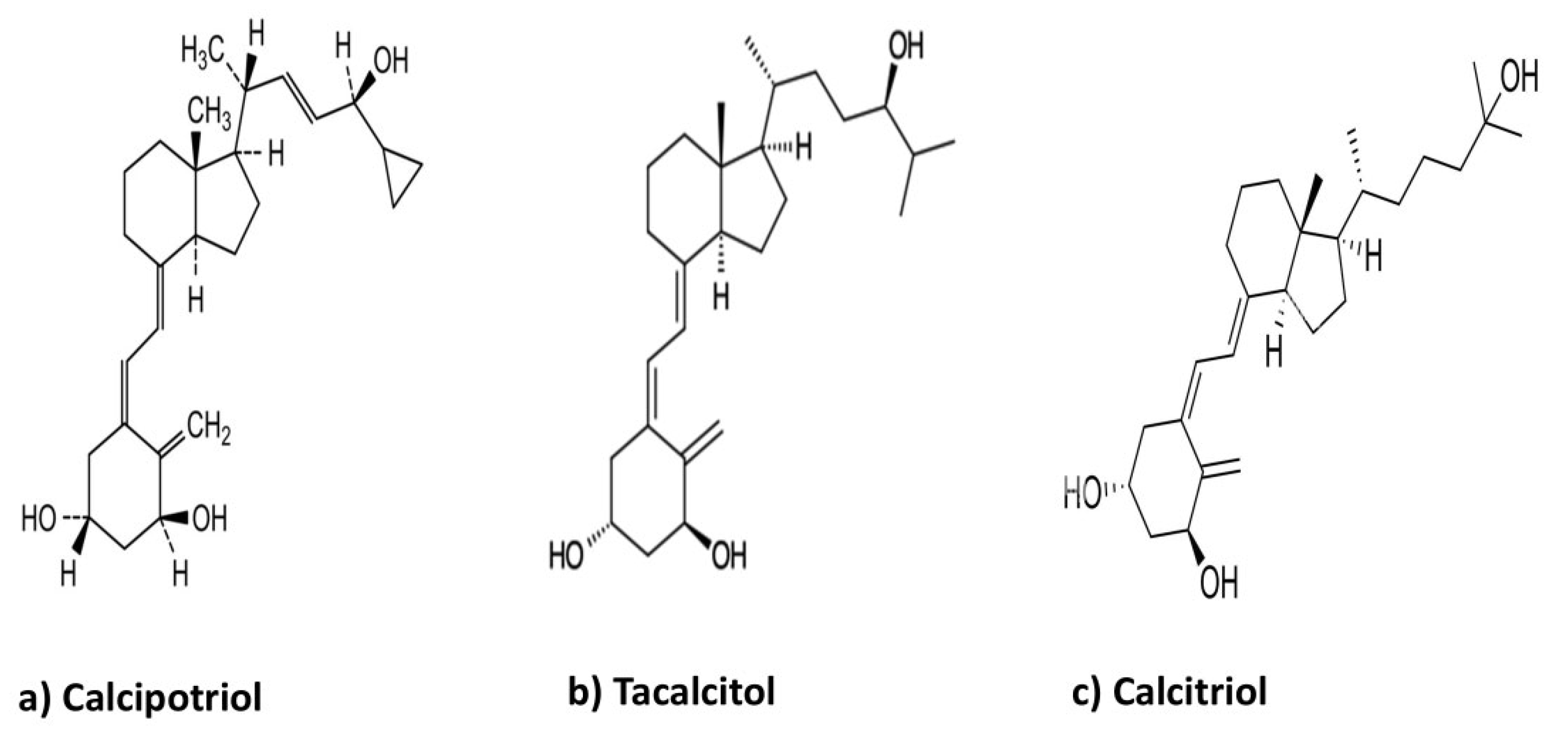
Calcitriol (1,25-dihydroxyvitamin D₃) is the biologically active form of vitamin D₃ and serves as the structural and pharmacological reference for synthetic vitamin D analogs. Calcipotriol and tacalcitol retain the characteristic secosteroid scaffold of calcitriol but incorporate structural modifications designed to preserve local VDR-mediated activity while modifying metabolic stability and reducing systemic calcemic effects [4,5]. Calcipotriol contains modifications in the side chain that alter its metabolic profile and contribute to rapid systemic inactivation, whereas tacalcitol is a hydroxylated vitamin D₃ derivative with high VDR activity and favorable topical tolerability [4,5,17]. These structural differences contribute to variations in pharmacological behavior among the three compounds and provide the basis for their different therapeutic and safety profiles following topical administration.
Figure 2.
Chemical structures of calcitriol, calcipotriol, and tacalcitol.
2.2. Pharmacological Characteristics
Despite sharing a common mechanism of action, calcipotriol, tacalcitol, and calcitriol demonstrate clinically relevant pharmacological differences. Calcipotriol is the most extensively investigated topical vitamin D analog and accounts for the majority of clinical evidence concerning both approved and off-label dermatologic applications. It exhibits high VDR affinity while undergoing rapid hepatic metabolism, resulting in minimal systemic calcium effects despite potent local biological activity. Its favorable balance between efficacy and safety explains its predominance within the clinical literature [4,5]. Tacalcitol possesses prolonged receptor occupancy and greater metabolic stability than calcipotriol, permitting once-daily administration. Clinical studies in psoriasis have reported excellent local tolerability, making tacalcitol particularly attractive for long-term therapy, although considerably fewer studies have evaluated its use in non-psoriatic skin diseases [17,24]. Calcitriol, the naturally active form of vitamin D₃, demonstrates lower irritancy than synthetic analogs and is generally preferred for sensitive anatomical sites, including the face, flexural regions, and intertriginous skin. However, because systemic absorption of calcitriol has a greater potential to influence calcium metabolism, prolonged treatment of extensive body surface areas requires greater caution than treatment with synthetic analogs [4,24,77]. Although these pharmacological differences are well recognized, virtually no randomized studies have directly compared the three analogs in non-psoriatic skin diseases. Consequently, current therapeutic selection relies primarily on extrapolation from psoriasis studies, clinician experience, and anatomical considerations rather than comparative evidence [4,5,17,24,25].

Table 1.
Pharmacological and pharmaceutical characteristics of topical vitamin D compounds.
| Property | Calcipotriol | Tacalcitol | Calcitriol | References |
|---|---|---|---|---|
| VDR activity | High | High | Native VDR ligand | [4,5,17] |
| Calcemic potential | Low | Low | Higher relative potential | [4,5,17,77] |
| Common topical concentration | 50 µg/g (0.005%) | 4 µg/g | 3 µg/g | [4,17,77] |
| Application frequency | Once or twice daily | Once daily | Twice daily | [4,17,77] |
| Systemic calcium considerations | Low at recommended topical exposure | Low at recommended topical exposure | Greater caution with extensive/prolonged application | [4,10,17,24,77]. |
| Local tolerability (face/flexures) | Irritation may occur, particularly at sensitive sites | Generally, well tolerated | Generally favorable at sensitive/flexural sites | [4,17,77] |
| Principal established dermatologic indication | Plaque psoriasis | Plaque psoriasis | Plaque psoriasis | [4,17,77] |
| Common topical dosage forms | Cream, ointment, solution, and other topical vehicles | Ointment | Ointment | [4,10,17,77] |
2.3. Pharmacodynamics and Vitamin D Receptor Signaling
The pharmacological activity of topical vitamin D analogs is mediated primarily through activation of the vitamin D receptor (VDR), a member of the nuclear receptor superfamily that functions as a ligand-activated transcription factor. Following topical application and penetration into viable epidermal layers, vitamin D analogs bind to intracellular VDR expressed in keratinocytes, melanocytes, sebocytes, dermal fibroblasts, and multiple immune cell populations. Ligand binding induces a conformational change in the receptor, facilitating heterodimerization with the retinoid X receptor (RXR). The activated VDR–RXR complex subsequently translocates to the nucleus, where it recognizes vitamin D response elements (VDREs) within promoter regions of target genes, thereby regulating transcriptional programs involved in epidermal homeostasis, cellular differentiation, immune regulation, and tissue remodeling [2,3,18]. Genomic activation of VDR influences the expression of hundreds of genes involved in skin physiology. In keratinocytes, vitamin D signaling suppresses excessive proliferation while promoting terminal differentiation through increased expression of structural proteins such as involucrin, loricrin, filaggrin, and transglutaminase. Simultaneously, VDR activation regulates genes controlling cell-cycle progression, apoptosis, and maintenance of epidermal barrier integrity. These coordinated effects restore physiological epidermal architecture while limiting hyperproliferative responses that characterize several inflammatory and inherited skin disorders [3,21]. Beyond its effects on epidermal differentiation, VDR activation exerts profound immunomodulatory activity. Vitamin D signaling suppresses pro-inflammatory T-helper 1 (Th1) and T-helper 17 (Th17) immune responses while promoting regulatory T-cell differentiation and anti-inflammatory cytokine production. In parallel, antigen presentation by dendritic cells is attenuated, resulting in reduced production of inflammatory mediators including interleukin (IL)-2, IL-6, IL-17, IL-23, interferon-γ, and tumor necrosis factor-α. These immunological effects provide a mechanistic basis for the investigation of topical vitamin D analogs in immune-mediated skin disorders such as vitiligo, morphea, alopecia areata, and atopic dermatitis [3,22,23,26,27,28]. Vitamin D signaling also enhances innate immune function through transcriptional regulation of antimicrobial peptides, particularly cathelicidin (LL-37) and β-defensins. These peptides contribute not only to antimicrobial defense against bacterial, viral, and fungal pathogens but also to wound repair, immune surveillance, and maintenance of cutaneous barrier function. Such effects are particularly relevant in acne vulgaris, where modulation of host inflammatory responses may complement conventional antimicrobial therapy [29,30,31,32]. Accumulating evidence further suggests that vitamin D analogs may induce rapid non-genomic responses independent of direct gene transcription. These effects have been associated with modulation of intracellular calcium signaling, activation of phospholipase C, protein kinase C, mitogen-activated protein kinase (MAPK), and phosphatidylinositol-3 kinase (PI3K)/Akt pathways. Although the precise contribution of these rapid signaling mechanisms to the clinical efficacy of topical vitamin D analogs remains incompletely understood, they are believed to complement classical genomic actions by influencing cellular migration, survival, and inflammatory responses [19,23]. Importantly, the widespread distribution of VDR throughout the skin explains the broad therapeutic potential of topical vitamin D analogs. In addition to keratinocytes, functional VDR expression has been demonstrated in melanocytes, sebocytes, dermal fibroblasts, dendritic cells, macrophages, mast cells, endothelial cells, and activated T lymphocytes. Consequently, activation of a single receptor system simultaneously influences multiple interconnected biological pathways involved in epidermal differentiation, pigmentation, innate and adaptive immunity, fibrosis, wound healing, and tumor biology. This pleiotropic pharmacodynamic profile distinguishes topical vitamin D analogs from therapies targeting individual inflammatory mediators and provides the biological basis for their therapeutic repurposing across a wide spectrum of non-psoriatic dermatologic disorders [1,2,3,20].
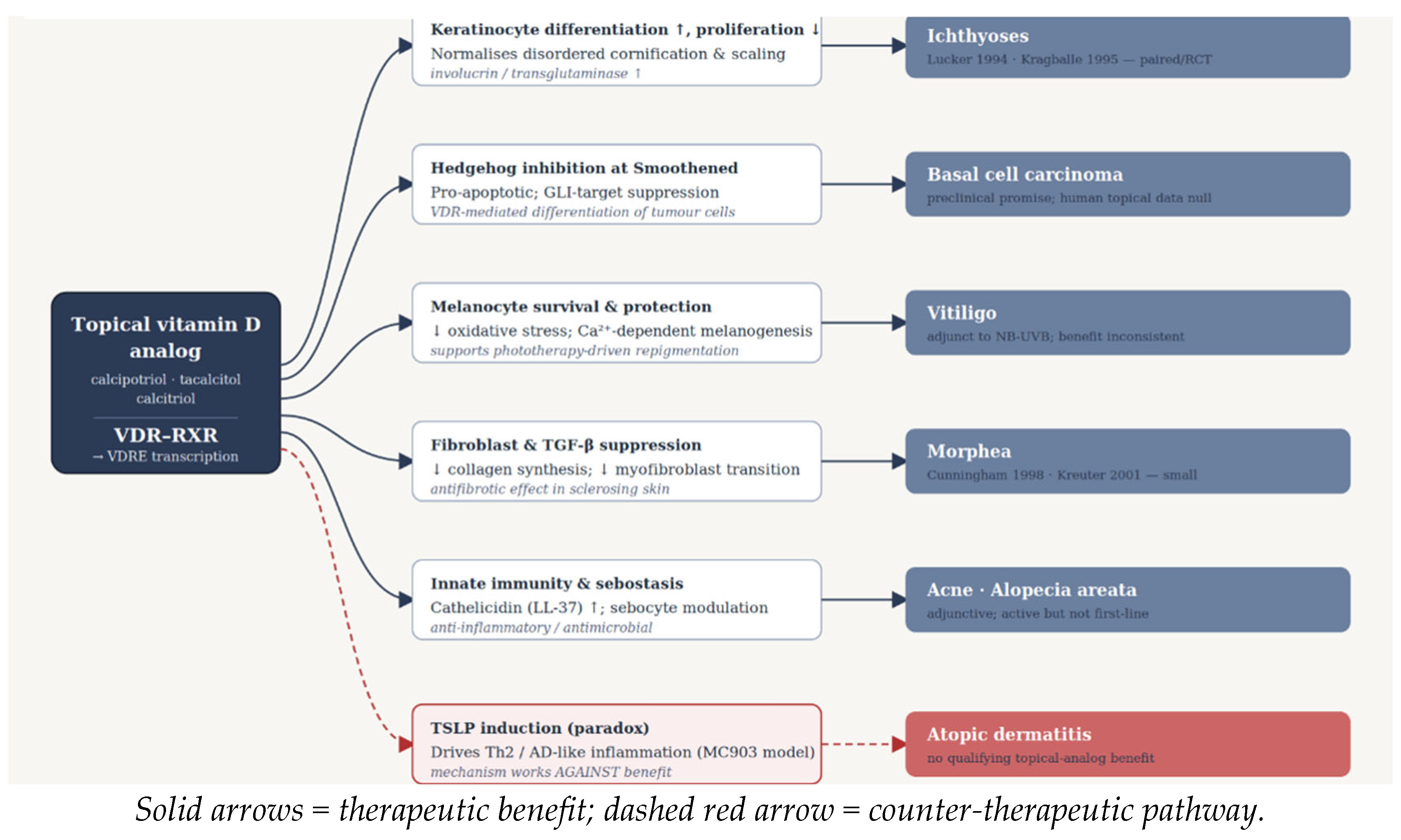
Figure 3.
Mechanisms linking activation of the vitamin D receptor (VDR) to the therapeutic effects of topical vitamin D analogs in non-psoriatic skin diseases. Ligand binding activates the VDR/RXR complex, regulating lineage-specific pathways involved in epidermal differentiation, immune modulation, melanocyte biology, fibrosis, and tumor suppression.
Figure 3.
Mechanisms linking activation of the vitamin D receptor (VDR) to the therapeutic effects of topical vitamin D analogs in non-psoriatic skin diseases. Ligand binding activates the VDR/RXR complex, regulating lineage-specific pathways involved in epidermal differentiation, immune modulation, melanocyte biology, fibrosis, and tumor suppression.
2.4. Pharmacokinetics of Topical Vitamin D Analogs
Following topical administration, vitamin D analogs penetrate the stratum corneum and diffuse into the viable epidermis, where they exert predominantly local pharmacological effects through activation of the vitamin D receptor. Their cutaneous bioavailability is influenced by several factors, including the physicochemical properties of the individual analog, formulation type, anatomical site of application, skin barrier integrity, and the extent of the treated body surface area. Under recommended therapeutic conditions, systemic absorption is generally low, allowing high local efficacy while minimizing alterations in calcium homeostasis. Nevertheless, prolonged treatment of extensive skin surfaces or use under occlusive conditions may increase systemic exposure, particularly with calcitriol, necessitating careful consideration of dose and duration in susceptible patients. Differences in metabolic stability and calcemic activity among calcipotriol, tacalcitol, and calcitriol further contribute to their distinct safety profiles and clinical applications [4,5,10,17,24].
2.5. Pharmaceutical Formulations and Topical Drug Delivery
The clinical performance of topical vitamin D analogs depends not only on their pharmacological activity but also on the characteristics of the delivery system. Commercially available formulations include ointments, creams, gels, foams, and scalp solutions, each designed to optimize cutaneous drug delivery while minimizing systemic exposure. Formulation selection influences drug release, penetration through the stratum corneum, patient acceptability, and treatment adherence, all of which may ultimately affect therapeutic outcomes [6,7,8,9]. Recent advances in topical drug-delivery research have explored liposomes, nanoemulsions, solid lipid nanoparticles (SLNs), nanostructured lipid carriers (NLCs), polymeric nanocarriers, and microneedle-assisted systems as strategies to improve drug stability, controlled release, cutaneous retention, and skin targeting [6,7,8,9]. These technologies provide a strong pharmaceutical rationale for improving the cutaneous delivery of vitamin D compounds; however, evidence specifically evaluating advanced delivery systems for topical vitamin D analogs remains limited and predominantly preclinical. Such approaches may potentially increase local drug concentrations while limiting systemic exposure, but their clinical advantages over conventional vitamin D formulations remain to be established. Continued optimization of topical formulations, together with advances in nanotechnology and biomaterial-based delivery systems, may substantially expand the therapeutic potential of vitamin D analogs beyond their current indications and improve their integration into personalized dermatologic therapy [6,7,8,9].
2.6. Biological Effects Relevant to Non-Psoriatic Skin Diseases
2.6.1. Regulation of Keratinocyte Proliferation and Differentiation
One of the best-characterized biological functions of vitamin D signaling is the regulation of epidermal homeostasis. Activation of the vitamin D receptor suppresses excessive keratinocyte proliferation while simultaneously promoting terminal differentiation through coordinated regulation of genes involved in cell-cycle control and epidermal maturation. These effects include increased expression of structural proteins such as loricrin, filaggrin, and transglutaminase, together with inhibition of hyperproliferative signaling pathways. Consequently, vitamin D analogs contribute to restoration of epidermal architecture while preserving normal barrier function [21]. Although these mechanisms were originally characterized in psoriasis, they are equally relevant to several non-psoriatic disorders characterized by abnormal epidermal differentiation. In inherited ichthyoses, impaired terminal differentiation represents a central pathogenic event, providing a direct biological rationale for topical vitamin D therapy. Similarly, restoration of physiological differentiation may contribute to limiting uncontrolled keratinocyte proliferation in certain premalignant and neoplastic skin conditions, including basal cell carcinoma. Thus, regulation of keratinocyte differentiation represents one of the fundamental mechanisms through which topical vitamin D analogs may exert therapeutic activity beyond psoriasis [21,34,35].
2.6.2. Immunomodulatory Effects
The immunomodulatory properties of vitamin D analogs extend far beyond suppression of epidermal hyperproliferation and represent one of the principal reasons for their investigation in inflammatory skin diseases. Activation of the VDR influences both innate and adaptive immune responses by modulating cytokine production, antigen presentation, and lymphocyte differentiation. Collectively, these actions promote restoration of immune homeostasis while limiting excessive inflammatory activation [3,22]. Vitamin D signaling suppresses pro-inflammatory T-helper 1 (Th1) and T-helper 17 (Th17) responses while promoting the expansion and functional activity of regulatory T cells (Tregs), thereby contributing to immune tolerance. Simultaneously, production of inflammatory mediators including interleukin (IL)-2, IL-6, IL-17, IL-23, interferon-γ, and tumor necrosis factor-α (TNF-α) is reduced, whereas anti-inflammatory cytokines such as IL-10 may be enhanced. These coordinated effects provide a strong biological rationale for investigating topical vitamin D analogs in disorders characterized by dysregulated immune activation, including atopic dermatitis, vitiligo, morphea, and alopecia areata [3,22,23,27,28]. Unlike conventional topical immunosuppressive agents, vitamin D analogs appear to restore immune balance rather than induce generalized immunosuppression, a characteristic that may partly explain their favorable long-term safety profile. Nevertheless, the extent to which these immunological mechanisms translate into clinically meaningful therapeutic benefit varies substantially among different dermatologic conditions, highlighting the importance of disease-specific pathogenic pathways.
2.6.3. Regulation of Innate Immunity and Antimicrobial Peptides
Beyond its effects on adaptive immunity, vitamin D signaling plays a fundamental role in regulating the skin’s innate immune defense. Activation of the vitamin D receptor stimulates the transcription of several antimicrobial peptides (AMPs), most notably cathelicidin (LL-37) and β-defensins, which constitute essential components of the cutaneous antimicrobial barrier. These peptides exhibit broad-spectrum activity against bacteria, viruses, and fungi while also participating in immune surveillance, wound healing, and maintenance of epidermal integrity [29,31]. The ability of vitamin D analogs to modulate antimicrobial peptide expression has attracted particular interest in acne vulgaris, where dysregulated innate immune responses contribute substantially to disease pathogenesis. Rather than acting solely through direct antimicrobial activity against Cutibacterium acnes, LL-37 influences chemotaxis, cytokine production, angiogenesis, and tissue repair, suggesting that vitamin D signaling may help restore immune homeostasis within the pilosebaceous unit. Experimental studies further demonstrate that vitamin D analogs suppress inflammatory cytokines, including IL-6, IL-8, and TNF-α, in stimulated sebocytes, supporting a combined antimicrobial and anti-inflammatory mechanism of action [30,31,32]. These observations suggest that topical vitamin D analogs may complement, rather than replace, conventional antimicrobial therapies by targeting the host immune response instead of bacterial proliferation alone. Such an approach may become increasingly relevant as concerns regarding antimicrobial resistance continue to influence acne management and encourage the development of non-antibiotic therapeutic strategies.
2.6.4. Effects on Melanocyte Biology and Oxidative Stress
Vitamin D signaling also plays an important role in maintaining melanocyte homeostasis. Melanocytes express functional vitamin D receptors, and activation of these receptors regulates multiple biological processes, including melanocyte survival, differentiation, melanogenesis, and resistance to oxidative stress. Given that oxidative injury and autoimmune destruction are central features of vitiligo pathogenesis, these observations provide a strong biological rationale for investigating topical vitamin D analogs as adjunctive therapies in pigmentary disorders [36,37].Experimental studies have demonstrated that vitamin D enhances melanocyte survival by reducing oxidative stress-induced apoptosis and modulating Wnt/β-catenin signaling, a pathway closely associated with melanocyte regeneration and pigmentation. In parallel, the immunomodulatory actions of vitamin D may attenuate the cytotoxic T-cell responses responsible for melanocyte destruction. These complementary mechanisms are believed to underlie the synergistic effects observed when topical vitamin D analogs are combined with narrowband ultraviolet B (NB-UVB) phototherapy, which stimulates melanocyte proliferation and migration while vitamin D signaling promotes melanocyte survival and stabilization [36,37,38,39]. Although the precise molecular interactions remain incompletely understood, the combined effects on oxidative stress, immune regulation, and melanocyte biology distinguish vitamin D analogs from conventional anti-inflammatory topical therapies and support their investigation as biologically targeted adjuncts for vitiligo management.
2.6.5. Antifibrotic Activity
Beyond their immunomodulatory properties, topical vitamin D analogs exhibit antifibrotic activity through direct effects on dermal fibroblasts and extracellular matrix remodeling. Dermal fibroblasts express functional vitamin D receptors, and activation of VDR signaling suppresses fibroblast proliferation, inhibits myofibroblast differentiation, and reduces excessive collagen synthesis. These effects are mediated primarily through modulation of the transforming growth factor-beta (TGF-β)/SMAD signaling pathway, a central regulator of tissue fibrosis in both cutaneous and systemic fibrotic disorders [40,41,42]. Experimental studies have demonstrated that vitamin D signaling downregulates the expression of profibrotic mediators while attenuating extracellular matrix deposition and fibroblast activation. In addition to these direct effects, the anti-inflammatory properties of vitamin D may further limit fibrosis by reducing the chronic inflammatory stimuli that drive persistent tissue remodeling. Collectively, these mechanisms provide a strong biological rationale for investigating topical vitamin D analogs in localized scleroderma (morphea), where excessive collagen deposition and progressive dermal sclerosis represent the principal pathological features [41,42,43]. Although clinical evidence remains limited, the mechanistic consistency between VDR signaling and established profibrotic pathways suggests that topical vitamin D analogs may represent valuable adjunctive therapies, particularly during the inflammatory phase of disease when fibroblast activation remains potentially reversible.
3. Clinical Applications of Topical Vitamin D Analogs Beyond Psoriasis
3.1. Acne Vulgaris
3.1.1. Disease Background and Biological Rationale
Acne vulgaris is one of the most prevalent inflammatory skin disorders worldwide, affecting approximately 85% of adolescents and a substantial proportion of adults. Its pathogenesis is multifactorial, involving excessive sebum production, follicular hyperkeratinization, colonization by Cutibacterium acnes, and a dysregulated innate and adaptive immune response. While current therapies including topical retinoids, benzoyl peroxide, antibiotics, and hormonal agents are generally effective, treatment failure, antibiotic resistance, local irritation, and poor adherence remain important clinical challenges. Consequently, considerable interest has emerged in identifying adjunctive therapies capable of targeting multiple pathogenic mechanisms simultaneously [30,31,32,44].The biological rationale for topical vitamin D analogs in acne extends beyond their established effects on keratinocyte differentiation. Sebocytes express functional vitamin D receptors, allowing direct regulation of sebocyte proliferation, differentiation, and lipid metabolism. Experimental studies have further demonstrated that vitamin D signaling suppresses the production of pro-inflammatory cytokines, including IL-6, IL-8, and TNF-α, while stimulating antimicrobial peptide expression, particularly cathelicidin (LL-37), thereby enhancing innate immune defense against C. acnes. Collectively, these observations suggest that topical vitamin D analogs may simultaneously modulate follicular hyperkeratinization, sebaceous gland activity, and cutaneous inflammation, providing a biologically plausible basis for therapeutic repurposing in acne vulgaris [30,31,32].
3.1.2. Clinical Evidence
Clinical evaluation of topical vitamin D analogs in acne vulgaris remains limited, with available evidence consisting primarily of small randomized trials and comparative split-face studies. Nevertheless, the published literature consistently suggests that calcipotriol possesses measurable anti-inflammatory activity, although its clinical efficacy appears inferior to that of established topical retinoids. The strongest evidence derives from a triple-blind, randomized, split-face clinical trial involving 60 patients, in which once-daily calcipotriol 0.005% cream significantly reduced both inflammatory and non-inflammatory acne lesions compared with placebo over eight weeks. However, adapalene 0.1% gel produced significantly greater clinical improvement and higher patient satisfaction, indicating that although calcipotriol demonstrates biological activity, it is unlikely to replace first-line topical retinoids [45]. These findings are supported by an earlier prospective split-face study involving 40 patients, which similarly demonstrated significant reductions in acne lesion counts following treatment with calcipotriol. Importantly, both studies suggest that vitamin D analogs possess clinically relevant anti-inflammatory effects while exhibiting acceptable local tolerability, supporting their potential role as adjunctive rather than primary therapeutic agents [46]. Although combination approaches targeting complementary pathogenic pathways are biologically attractive, robust randomized studies evaluating the additive clinical benefit of vitamin D analogs within multimodal acne regimens remain unavailable. From a drug-delivery perspective, the available clinical studies primarily investigated calcipotriol 0.005% in a cream formulation, applied once daily to facial lesions, providing a relatively non-occlusive vehicle suitable for acne-prone skin [45,46].
3.1.3. Critical Appraisal
Although the biological rationale supporting topical vitamin D analogs in acne vulgaris is compelling, current clinical evidence remains insufficient to support routine therapeutic use. Existing studies are limited by small sample sizes, short follow-up periods, heterogeneous outcome measures, and the absence of direct comparisons among different vitamin D analogs. Furthermore, nearly all available trials have evaluated calcipotriol, leaving the potential roles of tacalcitol and calcitriol largely unexplored. From a clinical perspective, topical vitamin D analogs should currently be viewed as investigational adjunctive therapies rather than alternatives to established acne treatments. Their greatest potential may lie in combination regimens that reduce reliance on topical antibiotics or improve inflammatory control in selected patients. Given increasing concerns regarding antimicrobial resistance, future randomized controlled trials evaluating vitamin D analogs within multimodal acne treatment strategies appear justified.
3.1.4. Future Perspectives
Future research should prioritize adequately powered randomized controlled trials comparing different vitamin D analogs both as monotherapy and in combination with established acne treatments. Standardized clinical endpoints, longer follow-up periods, and biomarker-based patient stratification may help identify individuals most likely to benefit from VDR-targeted therapy. Further investigation into the interaction between vitamin D signaling, sebocyte biology, and the cutaneous microbiome may also provide new opportunities for personalized acne management.
3.2. Atopic Dermatitis
3.2.1. Disease Background and Biological Rationale
Atopic dermatitis (AD) is a chronic inflammatory skin disease characterized by epidermal barrier dysfunction, immune dysregulation, and recurrent eczematous lesions. The pathogenesis of AD involves a complex interplay between genetic susceptibility, impaired skin barrier integrity, altered cutaneous microbiota, and a predominantly T-helper 2 (Th2)-driven immune response, particularly during the acute phase of disease. Current treatment strategies rely primarily on emollients, topical corticosteroids, topical calcineurin inhibitors, phosphodiesterase-4 inhibitors, and, in moderate-to-severe disease, targeted biologic therapies and Janus kinase (JAK) inhibitors. Despite these advances, long-term disease control remains challenging for many patients because of recurrent flares, treatment-related adverse effects, and the need for prolonged therapy [48,49,53]. The biological rationale for investigating topical vitamin D analogs in AD is supported by several mechanistic observations. Vitamin D signaling contributes to epidermal barrier maintenance through regulation of keratinocyte differentiation and increased expression of structural proteins, including filaggrin, involucrin, and loricrin. In addition, activation of the vitamin D receptor promotes antimicrobial peptide production, enhances innate immune defense, and modulates adaptive immune responses by suppressing pro-inflammatory cytokine production while promoting regulatory T-cell activity. Collectively, these effects suggest that vitamin D analogs could theoretically improve both barrier function and immune homeostasis in patients with AD [1,2,48,49].
3.2.2. Clinical Evidence
Despite this compelling biological rationale, clinical evidence supporting the use of topical vitamin D analogs in atopic dermatitis remains remarkably limited. To date, no adequately powered randomized controlled trials have demonstrated clear therapeutic benefit in patients with classic AD. The available literature consists primarily of experimental studies, mechanistic investigations, and a small number of clinical reports evaluating chronic hand eczema rather than generalized atopic dermatitis [48,49,50,51,53,55] .One comparative clinical study reported that topical calcipotriol achieved improvements comparable to those of a topical corticosteroid in chronic hand eczema. Although encouraging, these findings cannot be directly extrapolated to AD because chronic hand eczema represents a clinically distinct disorder with a different pathophysiological profile. Furthermore, no studies have established superiority over standard anti-inflammatory therapies or demonstrated additional benefit when vitamin D analogs are combined with established topical treatments [52]. Experimental studies have also produced conflicting findings. While vitamin D signaling promotes epidermal barrier repair and immune regulation, topical calcipotriol has been shown to induce thymic stromal lymphopoietin (TSLP) expression and an AD-like inflammatory phenotype in murine models [51,55]. These observations suggest that the biological effects of vitamin D analogs may differ substantially between healthy and inflamed skin, complicating translation from experimental models to clinical practice [51,55]. The available clinical evidence has predominantly evaluated calcipotriol in an ointment formulation, providing an occlusive vehicle that may enhance stratum corneum hydration and local drug retention in dry and barrier-impaired skin [52].
3.2.3. Critical Appraisal
Among all dermatologic conditions reviewed, atopic dermatitis presents one of the greatest discrepancies between mechanistic plausibility and clinical evidence. Multiple biological pathways support a potential therapeutic role for vitamin D analogs, including enhancement of epidermal barrier function, modulation of adaptive immunity, and stimulation of innate antimicrobial defenses. Nevertheless, these promising mechanisms have not translated into convincing clinical efficacy. Several factors may contribute to this discrepancy. First, the pro-inflammatory effects observed in experimental models raise concerns regarding local irritation and disease exacerbation in barrier-disrupted skin. Second, increased percutaneous absorption in AD may narrow the therapeutic window and increase the likelihood of adverse local reactions. Finally, the absence of adequately designed randomized clinical trials makes it impossible to determine whether the limited clinical use of topical vitamin D analogs reflects genuine therapeutic failure or simply insufficient investigation. Consequently, current evidence does not support routine use of topical vitamin D analogs in atopic dermatitis. At present, these agents should be regarded as investigational therapies whose potential remains biologically intriguing but clinically unproven.
3.2.4. Future Perspectives
Future research should focus on identifying patient subgroups most likely to benefit from topical vitamin D analog therapy, particularly individuals with specific barrier defects or altered VDR signaling. Carefully designed randomized controlled trials evaluating optimized formulations, lower concentrations, or combination regimens with established topical anti-inflammatory agents may clarify whether the favorable mechanistic profile of vitamin D analogs can be translated into meaningful clinical benefit. In parallel, further investigation into the interaction between vitamin D signaling, epidermal barrier biology, and TSLP-mediated inflammation may help explain the apparent disconnect between experimental findings and clinical outcomes.
3.3. Vitiligo
3.3.1. Disease Background and Biological Rationale
Vitiligo is a chronic acquired depigmenting disorder characterized by the selective destruction of epidermal melanocytes, resulting in well-demarcated depigmented macules and patches. Although the precise etiology remains incompletely understood, current evidence supports a multifactorial pathogenesis involving autoimmune mechanisms, oxidative stress, genetic susceptibility, and impaired melanocyte regeneration. Narrowband ultraviolet B (NB-UVB) phototherapy remains the cornerstone of treatment for generalized vitiligo, whereas topical corticosteroids and calcineurin inhibitors are recommended for localized disease. Nevertheless, treatment responses remain variable, and complete repigmentation is achieved in only a subset of patients, highlighting the need for effective adjunctive therapies [36,37,39]. The biological rationale for topical vitamin D analogs in vitiligo is supported by several complementary mechanisms. Functional VDR expression in melanocytes enables vitamin D signaling to regulate melanocyte proliferation, differentiation, and survival while reducing oxidative stress-induced apoptosis. In addition, vitamin D modulates local immune responses by suppressing autoreactive T-cell activity and promoting regulatory immune pathways, thereby addressing two fundamental components of vitiligo pathogenesis. These combined effects provide a strong mechanistic basis for combining topical vitamin D analogs with phototherapy, which primarily stimulates melanocyte proliferation and migration [36,37,38,39].
3.3.2. Clinical Evidence
Among all non-psoriatic indications reviewed, vitiligo possesses the most substantial clinical evidence supporting the use of topical vitamin D analogs. Clinical investigations have consistently focused on their use as adjunctive therapies rather than monotherapy, particularly in combination with NB-UVB phototherapy and psoralen plus ultraviolet A (PUVA).
The highest-quality evidence derives from a 2022 systematic review and meta-analysis including 14 clinical studies, which demonstrated that the addition of calcipotriol or tacalcitol to NB-UVB significantly improved repigmentation compared with NB-UVB alone. Combination therapy was particularly effective for lesions located on the face and neck, whereas acral lesions continued to exhibit limited therapeutic responses. Importantly, no significant increase in adverse events was observed, supporting the favorable safety profile of vitamin D analogs when used alongside phototherapy [39]. Earlier randomized controlled studies evaluating calcipotriol combined with oral PUVA similarly reported enhanced repigmentation while reducing the cumulative UVA dose required to achieve clinical improvement. These findings suggest that vitamin D analogs may potentiate the therapeutic effects of phototherapy while minimizing ultraviolet exposure, an advantage that may have important implications for long-term treatment safety [56]. Not all studies have reported positive outcomes. A randomized right-left comparative trial by Khullar et al. found no significant difference between NB-UVB alone and NB-UVB combined with calcipotriol. However, this isolated negative finding is generally considered within the context of substantial heterogeneity among published studies, including differences in disease duration, lesion location, treatment protocols, and patient selection [58]. Overall, the current body of evidence consistently supports topical vitamin D analogs as adjunctive rather than stand-alone therapies in vitiligo, with the greatest benefit observed when combined with NB-UVB phototherapy. In vitiligo studies, calcipotriol and tacalcitol have been administered predominantly as topical ointments, either as monotherapy or in combination with phototherapy, including NB-UVB and PUVA [56,57,58,59].
3.3.3. Critical Appraisal
Vitiligo represents the strongest clinical indication for therapeutic repurposing of topical vitamin D analogs beyond psoriasis. Unlike several other dermatologic disorders reviewed in this article, the biological rationale is supported by multiple randomized clinical studies and reinforced by systematic review evidence. The consistency between mechanistic findings and clinical outcomes strengthens confidence that vitamin D signaling contributes meaningfully to melanocyte preservation and repigmentation. Nevertheless, important limitations remain. Most studies have evaluated calcipotriol, with comparatively little evidence available for tacalcitol and calcitriol. Treatment protocols vary considerably with respect to phototherapy schedules, treatment duration, and outcome assessment, limiting direct comparisons across studies. Furthermore, durable long-term repigmentation and relapse rates remain insufficiently characterized. Based on the currently available evidence, topical vitamin D analogs should not be considered substitutes for established vitiligo therapies. Rather, they appear to function as effective adjunctive agents capable of enhancing phototherapy outcomes while maintaining an excellent safety profile.
3.3.4. Future Perspectives
Future clinical research should prioritize large multicenter randomized controlled trials directly comparing individual vitamin D analogs in combination with modern vitiligo therapies, including topical Janus kinase inhibitors and emerging biologic treatments. Biomarker-guided patient selection based on VDR expression, oxidative stress profiles, or immune signatures may further optimize treatment responses. In addition, mechanistic studies investigating the interaction between vitamin D signaling, melanocyte stem cell activation, and regenerative pathways could help identify novel combination strategies capable of improving long-term repigmentation and reducing disease recurrence.
3.4. Basal Cell Carcinoma
3.4.1. Disease Background and Biological Rationale
Basal cell carcinoma (BCC) is the most common human malignancy, accounting for approximately 80% of all non-melanoma skin cancers. Although metastatic disease is exceedingly rare, untreated BCC may cause significant local tissue destruction and functional impairment. Surgical excision remains the standard treatment for most lesions, while topical therapies, including imiquimod and 5-fluorouracil, photodynamic therapy, and Hedgehog pathway inhibitors, are reserved for selected superficial or locally advanced tumors. Nevertheless, recurrence, treatment-related morbidity, and the increasing incidence of BCC continue to stimulate interest in novel adjunctive therapeutic approaches [34,35]. The biological rationale for investigating topical vitamin D analogs in BCC extends beyond their established antiproliferative effects. Aberrant activation of the Hedgehog signaling pathway represents the principal molecular driver of BCC, and accumulating experimental evidence suggests that vitamin D signaling can modulate this pathway independently of classical VDR-mediated genomic activity. In addition, vitamin D analogs regulate keratinocyte differentiation, promote apoptosis, enhance DNA repair mechanisms, and suppress inflammatory pathways associated with tumor progression. Collectively, these mechanisms provide a compelling theoretical basis for therapeutic repurposing in cutaneous oncology [34,35,62,63].
3.4.2. Clinical Evidence
Despite encouraging experimental findings, clinical evidence supporting topical vitamin D analogs in BCC remains limited. Most published studies have focused on mechanistic investigations or preclinical animal models rather than randomized clinical trials.The strongest available clinical evidence comes from a Phase II randomized controlled trial comparing topical calcitriol with diclofenac in patients with superficial and nodular BCC. Although diclofenac demonstrated significant histological regression in superficial lesions, calcitriol monotherapy failed to produce meaningful clinical or histopathological improvement in either BCC subtype. These findings suggest that the impressive antiproliferative effects observed in experimental models are insufficient, when used alone, to achieve clinically relevant tumor regression [60]. Experimental studies nevertheless continue to support the biological activity of vitamin D analogs. In murine Patched-mutant models, calcitriol inhibited tumor growth through suppression of Hedgehog signaling, while in vitro studies demonstrated greater antiproliferative activity than cyclopamine under certain experimental conditions. Additional investigations have suggested interactions with Wnt/β-catenin signaling and NF-κB-mediated inflammatory pathways, further supporting a multifaceted antitumor mechanism [62,63].Overall, the available evidence indicates a clear discrepancy between strong mechanistic plausibility and limited clinical efficacy. In the BCC trial, calcitriol 3 μg/g was administered as an ointment under occlusion [60]. Adjacent evidence from actinic keratosis has evaluated calcipotriol 0.005% combined with 5-fluorouracil 5% cream [61], illustrating a combination-based topical strategy in keratinocyte carcinogenesis rather than direct BCC treatment.
3.4.3. Critical Appraisal
Basal cell carcinoma illustrates an important principle in translational dermatology: convincing molecular mechanisms do not necessarily translate into successful clinical therapies. Although vitamin D analogs influence several pathways involved in tumor biology, including Hedgehog signaling, apoptosis, and cellular differentiation, these effects have yet to produce consistent therapeutic benefit in patients. Current evidence does not support topical vitamin D analog monotherapy as a treatment for BCC. However, this conclusion should not be interpreted as evidence against the biological relevance of VDR signaling. Rather, it suggests that vitamin D analogs are unlikely to be sufficiently potent as single agents and may instead require combination with established topical therapies or targeted molecular inhibitors. Accordingly, vitamin D analogs should currently be regarded as investigational adjuncts in cutaneous oncology rather than clinically established antitumor agents.
3.4.4. Future Perspectives
Future research should evaluate combination strategies integrating topical vitamin D analogs with established treatments such as imiquimod, 5-fluorouracil, photodynamic therapy, or Hedgehog inhibitors. Greater understanding of the interaction between VDR signaling and oncogenic pathways, particularly Hedgehog and Wnt/β-catenin, may identify biomarkers predictive of therapeutic response. Well-designed translational studies linking molecular pathway modulation with clinical outcomes will be essential before vitamin D analogs can be considered viable adjunctive therapies in non-melanoma skin cancer.
3.5. Morphea (Localized Scleroderma)
3.5.1. Disease Background and Biological Rationale
Morphea, or localized scleroderma, is a chronic inflammatory connective tissue disorder characterized by excessive collagen deposition, dermal fibrosis, and progressive skin sclerosis. Although its pathogenesis is incompletely understood, current evidence implicates immune dysregulation, endothelial injury, and persistent activation of fibroblasts through transforming growth factor-beta (TGF-β)-mediated signaling. Clinical management depends on disease extent and activity, ranging from topical therapies for limited superficial plaques to systemic immunosuppressive agents and phototherapy for more extensive or rapidly progressive disease [41,42]. The biological rationale for topical vitamin D analogs in morphea is particularly compelling because vitamin D signaling directly targets several pathways involved in fibrosis. Activation of the vitamin D receptor suppresses fibroblast proliferation, inhibits myofibroblast differentiation, and downregulates TGF-β/SMAD signaling, thereby reducing collagen synthesis and extracellular matrix deposition. In addition, the anti-inflammatory properties of vitamin D may limit the persistent immune activation that contributes to progressive tissue remodeling. Together, these mechanisms provide a strong scientific basis for investigating topical vitamin D analogs as antifibrotic therapies in localized scleroderma [40,41,42,43].
3.5.2. Clinical Evidence
Clinical evidence remains limited but is remarkably consistent. Published studies have primarily evaluated topical calcipotriol, either under occlusion or in combination with ultraviolet A1 (UVA1) phototherapy, with most reporting improvements in lesion activity and skin sclerosis. One open-label study demonstrated that twice-daily application of calcipotriol under occlusion significantly improved skin extensibility and reduced plaque induration in patients with plaque-type morphea. Although the study included a limited number of participants and lacked a control group, the observed clinical improvements were supported by the known antifibrotic effects of VDR activation [43]. Additional evidence comes from combination therapy studies. In patients receiving low-dose UVA1 phototherapy, the addition of topical calcipotriol produced a 67% reduction in clinical severity scores, suggesting that vitamin D analogs may enhance the antifibrotic effects of phototherapy. These findings are biologically plausible because UVA1 reduces fibroblast activity while vitamin D signaling simultaneously suppresses TGF-β-mediated collagen production, resulting in complementary mechanisms of action [68].Importantly, the 2024 German S2k guideline for localized scleroderma recognizes topical vitamin D analogs as an optional therapeutic approach for limited superficial inflammatory lesions, reflecting cautious but positive expert consensus despite the limited evidence base [69]. In morphea, calcipotriol/calcipotriene 0.005% has predominantly been delivered as an ointment, either under nighttime occlusion or without occlusion in combination with UVA1 phototherapy, highlighting the potential importance of formulation and application conditions for achieving adequate cutaneous exposure [43,68].
3.5.3. Critical Appraisal
Morphea represents one of the most biologically coherent indications for topical vitamin D analog therapy. Unlike several other non-psoriatic conditions, the proposed mechanism of action directly targets a central pathogenic pathway fibroblast activation mediated by TGF-β signaling and available clinical studies consistently report improvements in disease activity. Nevertheless, enthusiasm should be tempered by the limited quantity of evidence. Published studies involve small patient populations, are predominantly open-label, and frequently combine vitamin D analogs with phototherapy, making it difficult to determine the independent contribution of topical treatment. Furthermore, no randomized controlled trials have directly compared vitamin D analogs with established topical therapies such as corticosteroids or tacrolimus. Consequently, current evidence supports topical vitamin D analogs as adjunctive therapies for carefully selected patients with superficial inflammatory morphea rather than as first-line treatment. Their greatest value may lie in combination with phototherapy or during the early inflammatory phase of disease, when fibrosis remains potentially reversible.
3.5.4. Future Perspectives
Future studies should prioritize multicenter randomized controlled trials comparing topical vitamin D analogs with standard topical therapies and evaluating their additive benefit when combined with UVA1 phototherapy. Incorporation of objective outcome measures, including ultrasound imaging and validated disease activity scores, would improve comparability across studies. In addition, mechanistic investigations examining the interaction between vitamin D signaling, fibroblast biology, and extracellular matrix remodeling may help identify biomarkers predictive of treatment response and facilitate more personalized therapeutic approaches.
3.6. Inherited Ichthyoses
3.6.1. Disease Background and Biological Rationale
Inherited ichthyoses comprise a heterogeneous group of genetic disorders characterized by abnormal epidermal differentiation, defective cornification, and excessive scaling resulting from impaired skin barrier formation. Although individual subtypes differ genetically, disruption of keratinocyte maturation and terminal differentiation represents a common pathogenic feature. Current management remains largely symptomatic, relying on emollients, keratolytic agents, topical retinoids, and systemic retinoids in more severe cases. However, long-term treatment is often limited by incomplete efficacy and treatment-related adverse effects, highlighting the need for alternative therapeutic approaches [33,65]. Topical vitamin D analogs provide a biologically plausible therapeutic strategy because they directly regulate keratinocyte proliferation and terminal differentiation through activation of the vitamin D receptor. Experimental studies have demonstrated that VDR signaling increases the expression of structural proteins involved in epidermal maturation while suppressing excessive keratinocyte proliferation. These mechanisms closely target the fundamental pathological abnormalities underlying inherited ichthyoses and therefore represent one of the most direct examples of disease-specific therapeutic repurposing beyond psoriasis [33,65].
3.6.2. Clinical Evidence
Clinical evaluation of topical vitamin D analogs in inherited ichthyoses has primarily focused on calcipotriol. Available randomized and vehicle-controlled studies consistently demonstrate improvements in scaling, epidermal thickness, and overall skin appearance following topical treatment. In one placebo-controlled clinical trial, calcipotriol significantly reduced scaling severity compared with vehicle treatment while simultaneously improving histological markers of epidermal differentiation. These clinical findings were supported by normalization of keratinocyte maturation and reduced epidermal hyperproliferation, confirming that the observed therapeutic effects reflect correction of the underlying biological abnormality rather than simple symptomatic improvement [66,67]. Despite these encouraging findings, treatment outcomes have been variable across different ichthyosis subtypes, reflecting the considerable genetic heterogeneity of these disorders. Furthermore, most available studies enrolled relatively small patient populations and evaluated short-term treatment responses, limiting conclusions regarding sustained clinical benefit. Clinical studies in ichthyosis have primarily employed calcipotriol 0.005% (50 μg/g) ointment, an occlusive formulation well suited to the hyperkeratotic and xerotic characteristics of these disorders [66,67].
3.6.3. Critical Appraisal
Among the dermatologic disorders reviewed, inherited ichthyoses represent one of the strongest examples of concordance between disease pathophysiology and the biological actions of topical vitamin D analogs. The correction of abnormal keratinocyte differentiation directly addresses a central pathogenic mechanism, and available clinical studies consistently demonstrate measurable improvements in scaling and epidermal maturation. Nevertheless, clinical application is constrained by an important safety consideration. Unlike psoriasis, ichthyoses frequently involve extensive body surface areas, increasing the potential for systemic absorption during prolonged treatment. Cases of hypercalcemia have been reported following widespread application of topical vitamin D analogs, particularly in children and in patients with severe generalized disease. Consequently, careful monitoring of cumulative dose, treatment duration, and serum calcium concentrations is recommended when large treatment areas are involved [33]. Accordingly, topical vitamin D analogs should currently be considered adjunctive therapies for carefully selected patients with localized or moderately extensive disease rather than routine treatment for generalized ichthyosis.
3.6.4. Future Perspectives
Future clinical studies should evaluate whether lower-concentration formulations, intermittent dosing schedules, or combination regimens with topical retinoids and barrier-repair therapies can maintain therapeutic efficacy while reducing systemic calcium exposure. Given the marked genetic heterogeneity among inherited ichthyoses, genotype-specific treatment strategies and biomarker-guided patient selection may further optimize clinical outcomes. Longer-term studies evaluating both efficacy and systemic safety will be essential before broader clinical recommendations can be established.
3.7. Alopecia Areata
3.7.1. Disease Background and Biological Rationale
Alopecia areata (AA) is an autoimmune, non-scarring hair loss disorder characterized by T-cell-mediated destruction of anagen hair follicles and collapse of the immune privilege normally maintained within the follicular microenvironment. Current therapies include topical and intralesional corticosteroids, topical immunotherapy, and Janus kinase (JAK) inhibitors, although treatment responses remain variable. The biological rationale for topical vitamin D analogs is supported by evidence that hair follicles express functional vitamin D receptors (VDR), which are essential for normal hair cycling and follicular differentiation. In addition, vitamin D signaling modulates immune responses by promoting regulatory T-cell activity and suppressing inflammatory cytokines implicated in AA pathogenesis [3,22,23,71,72]. These observations provide a mechanistic basis for investigating topical vitamin D analogs as adjunctive therapies in alopecia areata [71,72,73,74,75].
3.7.2. Clinical Evidence
Clinical evidence supporting topical vitamin D analogs in AA remains limited and heterogeneous. Published studies consist primarily of small prospective trials, observational studies, and case reports evaluating topical calcipotriol, either alone or in combination with established therapies.Several small clinical studies have reported partial hair regrowth following topical calcipotriol treatment, particularly in patients with localized patch-type alopecia areata. In some comparative studies, combination therapy with topical corticosteroids appeared to produce greater clinical improvement than either treatment alone, suggesting a potential synergistic effect between immunosuppressive therapy and VDR-mediated immune modulation [71,72,73,74,75]. However, clinical responses have been inconsistent, and the available studies are limited by small sample sizes, short treatment duration, and heterogeneous outcome measures. To date, no adequately powered multicenter randomized controlled trial has established the efficacy of topical vitamin D analogs in alopecia areata. Topical calcipotriol 0.005% has been investigated in several conventional formulations in alopecia areata, including ointment, cream, and lotion, reflecting the need for vehicles with appropriate spreadability and follicular delivery characteristics in hair-bearing areas [71,72,73,74,75].
3.7.3. Critical Appraisal
The available evidence suggests that topical vitamin D analogs possess an interesting biological rationale but only modest clinical support for use in alopecia areata. Unlike vitiligo, where mechanistic and clinical findings are largely concordant, evidence in AA remains preliminary and insufficient to define a clear therapeutic role. An additional limitation is the uncertainty regarding the mechanism responsible for the reported clinical improvements. It remains unclear whether treatment responses primarily reflect direct modulation of hair follicle biology, restoration of immune privilege, suppression of autoimmune inflammation, or a combination of these effects. Furthermore, most available studies have evaluated calcipotriol, leaving the potential role of tacalcitol and calcitriol almost entirely unexplored. At present, topical vitamin D analogs should therefore be regarded as experimental adjunctive therapies rather than evidence-based treatment options for alopecia areata.
3.7.4. Future Perspectives
Future investigations should focus on well-designed randomized controlled trials comparing vitamin D analogs with current standard therapies and evaluating their potential role in combination with topical corticosteroids or JAK inhibitors. Greater understanding of VDR signaling within the hair follicle stem cell niche, together with biomarker-guided patient stratification based on vitamin D receptor expression or immune signatures, may help identify patients most likely to benefit from VDR-targeted therapy. Such studies will be essential to determine whether the promising mechanistic rationale can be translated into clinically meaningful improvements in hair regrowth and disease remission.
3.8. Emerging Clinical Applications
Although most research has focused on acne, vitiligo, morphea, and inherited ichthyoses, topical vitamin D analogs have also been investigated in other dermatologic conditions, including cutaneous lichen planus [76]. Additional potential applications in wound healing, tissue repair, and scar modulation have been proposed on mechanistic or preliminary experimental grounds; however, clinical evidence remains insufficient to establish their therapeutic role. Across these conditions, the proposed therapeutic effects are largely attributed to the pleiotropic actions of vitamin D signaling, including modulation of cutaneous inflammation, enhancement of epidermal differentiation, regulation of immune responses, and promotion of tissue repair. Although these biological mechanisms provide a reasonable scientific rationale, current evidence remains insufficient to support routine clinical use outside carefully selected cases or research settings [1,2,3]. Future investigations should prioritize adequately powered randomized controlled trials, standardized clinical outcome measures, and biomarker-based patient selection to determine whether these preliminary observations can be translated into clinically meaningful therapeutic applications. Until such evidence becomes available, the use of topical vitamin D analogs in these emerging indications should be regarded as investigational.
4. Comparative Analysis of Clinical Evidence
The available literature demonstrates substantial heterogeneity in both the biological rationale and the quality of clinical evidence supporting the use of topical vitamin D analogs beyond psoriasis. Although activation of the vitamin D receptor influences multiple pathogenic pathways, including epidermal differentiation, immune regulation, oxidative stress, fibrosis, and tumor biology, translation of these molecular mechanisms into consistent clinical benefit varies considerably among different dermatologic disorders. Consequently, therapeutic recommendations should be guided not only by mechanistic plausibility but also by the strength and consistency of available clinical evidence [1,2,3,4]. Among all indications reviewed, vitiligo currently possesses the most robust evidence base. Multiple randomized controlled trials, supported by a recent systematic review and meta-analysis, demonstrate that topical calcipotriol and tacalcitol enhance repigmentation when combined with narrowband ultraviolet B (NB-UVB) phototherapy, with no significant increase in adverse effects. These findings suggest that vitamin D analogs function most effectively as adjunctive rather than stand-alone therapies, potentiating the benefits of phototherapy through complementary effects on melanocyte survival, oxidative stress, and immune regulation [36,37,38,39,56,57,58,59]. Morphea also demonstrates a favorable balance between mechanistic rationale and clinical outcomes. Although evidence is limited to small prospective studies, improvements in skin induration, extensibility, and inflammatory activity have been consistently reported, particularly when topical calcipotriol is combined with UVA1 phototherapy. The inclusion of topical vitamin D analogs as an optional therapeutic approach in the recent German S2k guideline further supports their potential role in carefully selected patients with localized superficial inflammatory disease [41,42,43,68,69]. For acne vulgaris and inherited ichthyoses, the available evidence is encouraging but remains moderate. In acne, topical vitamin D analogs demonstrate measurable anti-inflammatory activity and improvements in lesion counts; however, existing studies consistently show inferior efficacy compared with topical retinoids, supporting their role primarily as adjunctive therapies rather than first-line treatment [30,31,32,45,46]. In inherited ichthyoses, the close relationship between VDR signaling and keratinocyte differentiation provides one of the strongest mechanistic rationales among all reviewed conditions. Nevertheless, concerns regarding systemic calcium absorption during treatment of extensive body surface areas continue to limit widespread clinical application despite favorable efficacy results [33,65,66,67]. Conversely, atopic dermatitis, alopecia areata, and basal cell carcinoma illustrate the challenges of translating promising experimental findings into clinical practice. In atopic dermatitis, the paradoxical induction of TSLP-mediated inflammation by topical vitamin D analogs in experimental models raises questions regarding their therapeutic role [51,55]. Clinical evidence in alopecia areata remains limited to relatively small studies with heterogeneous designs and outcomes [71,72,73,74,75]. Similarly, in basal cell carcinoma, clinical investigation has failed to reproduce fully the encouraging antitumor activity observed in preclinical models [60,62,63].An additional observation emerging from this review is the overwhelming predominance of calcipotriol in the published literature. Comparatively few studies have evaluated tacalcitol or calcitriol, making it difficult to determine whether observed differences in clinical outcomes reflect genuine pharmacological variation or simply disparities in research activity. Likewise, no adequately powered head-to-head comparative trials have evaluated the three available analogs across non-psoriatic indications. Consequently, current analog selection continues to rely largely on pharmacological characteristics, anatomical treatment site, local tolerability, and clinician experience rather than comparative clinical evidence [4,5,17,24,25,77]. Overall, the available evidence supports topical vitamin D analogs primarily as adjunctive therapies capable of complementing established treatment strategies rather than replacing standard-of-care interventions. Their greatest therapeutic potential appears to lie in combination regimens that exploit their pleiotropic biological effects while maintaining the favorable safety profile established during decades of psoriasis management. Future clinical development should therefore prioritize disease-specific randomized controlled trials, direct comparisons among available vitamin D analogs, standardized outcome measures, and biomarker-guided patient selection to facilitate more personalized therapeutic approaches [38,45,52,67].
Table 2.
Summary of clinical evidence for topical vitamin D analogs in non-psoriatic dermatologic conditions.
Table 2.
Summary of clinical evidence for topical vitamin D analogs in non-psoriatic dermatologic conditions.
| Condition | Analog | Design | N | Formulation/Protocol | Comparator | Main Outcome | Key Result | Ref |
|---|---|---|---|---|---|---|---|---|
| Acne vulgaris | Calcipotriol | Triple-blind split-face RCT | 60 | 0.005% cream OD × 8 wk to one facial side vs placebo | Adapalene 0.1% gel vs placebo (parallel split-face groups) | Inflammatory & non-inflammatory lesion count; Physician & Patient GA | Significant lesion reduction with calcipotriol; adapalene superior (P =0.001); highest patient satisfaction with adapalene | [45] |
| Acne vulgaris | Calcipotriol | Prospective split-face study | 40 | 0.005% cream × 2 months; once daily application | Adapalene 0.1% gel (contralateral side) | Inflammatory & non-inflammatory lesion count; histology | Significant reduction in both inflammatory and non-inflammatory lesions on calcipotriol side (P =0.0001) | [46] |
| Atopic dermatitis (adjucent evidence) Chronic hand eczema* | Calcipotriol | Comparative clinical study | NR | Calcipotriol ointment applied to affected hands for study duration | Desoximetasone (topical corticosteroid) | Clinical severity score (hand eczema) | Calcipotriol showed efficacy comparable to desoximetasone in chronic hand eczema. | [52] |
| Vitiligo | Calcipotriol / Tacalcitol | Systematic review & meta-analysis (14 RCTs/within-patient studies) | 642 | Topical vitamin D analog (calcipotriol or tacalcitol) + NB-UVB or PUVA or excimer laser | Phototherapy alone | Treatment response (≥50% repigmentation); relative risk | NB-UVB combination superior (RR 1.67, 95% CI 1.21–2.31); tacalcitol > calcipotriol (RR 2.25 vs 1.24, P =0.002); no benefit added to PUVA or excimer | [39] |
| Vitiligo | Calcipotriol | Placebo-controlled double-blind left–right RCT | 27 | Topical calcipotriol + oral PUVA vs placebo + oral PUVA | Oral PUVA + placebo cream | % repigmentation (initial & complete); cumulative UVA dose; number of sessions | 81% initial vs 7% placebo; 63% complete vs 15% placebo; lower cumulative UVA dose (232.8 vs 259.9 J/cm²); fewer sessions required | [56] |
| Vitiligo | Tacalcitol | Randomized split-body trial | 32 | Tacalcitol OD + NB-UVB twice weekly | NB-UVB alone (contralateral side) | Repigmentation rate and response | Significant improvement in repigmentation and response rates with tacalcitol + NB-UVB vs NB-UVB alone | [57] |
| Vitiligo | Calcipotriol | Right–left comparative RCT (24 wk) | 27 | Calcipotriol 0.005% ointment BID + NB-UVB × 24 wk | NB-UVB alone (contralateral side) | % repigmentation (Leukoderma & Body Score); IGA | No significant added benefit: mean L&B-score reduction 49% vs 51.4% NB-UVB alone (P =0.557); IGA 2.6 vs 2.7 (P=0.821) | [58] |
| Vitiligo | Calcipotriol / Betamethasone | RCT | 41 | Calcipotriol/betamethasone ointment + NB-UVB × 6 months | Tacrolimus 0.1% ointment + NB-UVB | % repigmentation at 6 months | 54.7% repigmentation (calcipotriol/betamethasone + NB-UVB) vs 45.6% (tacrolimus + NB-UVB); difference not statistically significant | [59] |
| Basal cell carcinoma | Calcitriol | Phase II RCT | 128 | Calcitriol 3 µg/g ointment BID under occlusion × 8 wk; arms: calcitriol alone, diclofenac 3% alone, combination, vehicle | Topical diclofenac 3%; combination; vehicle control | Histologic regression rate; Ki-67 & Bcl-2 expression | Calcitriol monotherapy: ineffective for sBCC and nBCC; diclofenac 3%: 64.3% histologic regression in sBCC; combination: 43.8% regression in sBCC; no benefit for nBCC in any arm | [60] |
| Actinic keratosis / keratinocyte cancer prevention (adjacent evidence) | Calcipotriol | Randomized field therapy trial | 131 | Calcipotriol 0.005% + 5-fluorouracil 5% cream × 4 days | Vehicle + 5-FU cream | AK clearance; local immune response/CD4+ T-cell infiltration |
Marked reduction in AK burden and induction of a robust T-cell response with calcipotriol + 5-FU compared with 5-FU control. | [61] |
| Ichthyosis (lamellar & BIE) | Calcipotriol | Double-blind bilaterally paired study | 6 | Calcipotriol 0.005% ointment vs vehicle, applied to contralateral body halves | Vehicle (contralateral side) | Scaling, clinical improvement; serum calcium | Unilateral improvement on calcipotriol side in 3/4 lamellar ichthyosis patients and 1 BIE patient; no adverse laboratory findings | [66] |
| Ichthyosis (multiple subtypes) | Calcipotriol | RCT double-blind vehicle-controlled right/left (12 wk) | 67 (27 ichthyosis, 20 PPK, 9 KP, 11 other) | Calcipotriol ointment 50 µg/g up to 120 g/wk vs vehicle | Vehicle (contralateral side) | Scaling severity across keratinization subtypes | Statistically significant reduction in scaling in ichthyosis variants; no benefit in palmoplantar keratoderma or keratosis pilaris | [67] |
| Morphea / Linear scleroderma | Calcipotriol | Open-label pilot study | 12 (aged 12–38 yrs) | Calcipotriene 0.005% ointment BID under nighttime occlusion × 3 months; prior TCS failure | Within-subject pre/post (no control arm) | Skin extensibility; erythema; induration; dyspigmentation; serum calcium; PTH | All 12 patients improved significantly in skin extensibility; notable reductions in erythema, induration, dyspigmentation; no perturbation in serum calcium or PTH | [43] |
| Morphea (childhood) | Calcipotriol | Open-label + low-dose UVA1 (12 wk) | 19 (pediatric) | Calcipotriol 0.005% ointment BID (unoccluded) + UVA1 phototherapy 20 J/cm² 4 time/weekly × 12 wk | No control arm | Clinical severity score (relative reduction) | 67.1% relative reduction in mean clinical severity score; beneficial synergistic effect on inflammatory and sclerotic lesions | [68] |
| Alopecia areata | Calcipotriol | RCT | NR | Topical calcipotriol vs NB-UVB phototherapy for study duration | NB-UVB phototherapy | SALT score; serum vitamin D3 levels | Both treatments significantly improved SALT scores; associated rise in serum vitamin D3; no significant difference between groups | [72] |
| Alopecia areata | Calcipotriol | Intrasubject pilot study (12 wk) | 35 | Calcipotriol 0.005% ointment BID × 12 wk vs clobetasol propionate 0.05% ointment BID × 12 wk (same patient, separate sites) | Clobetasol propionate 0.05% ointment | ≥75% hair regrowth | ≥75% regrowth at 62.9% of calcipotriol-treated sites vs 45.7% of clobetasol-treated sites; numerical advantage, not statistically significant | [73] |
| Alopecia areata | Calcipotriol | Prospective pilot study | 22 | Calcipotriol lotion BID × 3 months | None | Terminal hair regrowth; correlation with baseline vitamin D levels | Terminal hair regrowth in 13/22 participants (59.1%); more robust response in patients with lower baseline serum vitamin D | [74] |
| Alopecia areata | Calcipotriol | Retrospective study (12 wk) | 48 | Calcipotriol cream BID × 12 wk | None | Overall response rate; ≥75% regrowth; complete regrowth | Overall response rate 69.2%; ≥75% regrowth in 62.5%; complete hair restoration in 27.1% | [75] |
| Cutaneous lichen planus | Calcipotriol | Randomized open-label trial (12 wk) | 31 | Calcipotriol ointment 50 µg/g BID × 12 wk | Betamethasone valerate 0.1% ointment BID × 12 wk | Lesion flattening; pigmentary clearance; pruritus (VAS) | Calcipotriol not superior to betamethasone valerate for lesion flattening, pigmentation, or pruritus relief; comparable efficacy | [76] |
* Chronic hand eczema is presented as adjacent evidence because of shared inflammatory and barrier-related features with atopic dermatitis but does not constitute direct clinical evidence for AD.
5. Current Challenges and Future Perspectives
Despite the expanding interest in topical vitamin D analogs beyond psoriasis, several important challenges continue to limit their broader clinical application. Foremost among these is the limited availability of high-quality clinical evidence. Most published studies involve small patient cohorts, single-center designs, short treatment durations, and heterogeneous outcome measures, making direct comparison across studies difficult and limiting the strength of current therapeutic recommendations [39,43,45,46,52,66,67,68,69,71,72,73,74,75]. Another major limitation is the lack of direct comparative studies evaluating the three currently available topical vitamin D analogs. Although calcipotriol, tacalcitol, and calcitriol differ in receptor affinity, metabolic stability, local tolerability, and calcemic potential, virtually all non-psoriatic clinical studies have investigated calcipotriol, while evidence for tacalcitol and calcitriol remains comparatively scarce. Consequently, current analog selection is based largely on extrapolation from psoriasis, regional availability, and clinician preference rather than disease-specific comparative evidence [4,5,17,24,25]. The considerable heterogeneity of outcome measures further complicates interpretation of the available literature. Disease-specific scoring systems including lesion counts in acne, EASI and SCORAD in atopic dermatitis, VASI in vitiligo, and clinical severity indices in morphea reduce the feasibility of quantitative comparisons and limit opportunities for formal meta-analysis. Standardization of clinical endpoints would substantially improve future evidence synthesis and facilitate comparison between therapeutic studies [39,41,69]. Future research should focus on adequately powered multicenter randomized controlled trials, direct comparisons among vitamin D analogs, and optimization of combination treatment strategies. Integration of topical vitamin D analogs with phototherapy, topical immunomodulators, retinoids, and emerging targeted therapies may further enhance therapeutic efficacy while minimizing adverse effects. In parallel, biomarker-driven patient selection based on vitamin D receptor expression, genetic polymorphisms, or disease-specific molecular signatures may enable a more personalized approach to treatment and improve identification of patients most likely to benefit from VDR-targeted therapy [20,50,79,80]. Finally, advances in formulation science including nanocarrier systems, lipid-based delivery platforms, and controlled-release topical formulations may improve cutaneous penetration, increase local drug retention, and reduce systemic exposure. Such innovations could expand the therapeutic utility of vitamin D analogs, particularly in diseases requiring prolonged treatment or involving extensive body surface areas. Continued collaboration between basic scientists, formulation researchers, and clinical dermatologists will therefore be essential to translate the promising biological properties of topical vitamin D analogs into evidence-based therapeutic strategies. Collectively, these advances indicate that the future development of topical vitamin D analogs will depend on the integration of innovative pharmaceutical formulations, biomarker-guided patient selection, and rational combination therapies. The principal directions for future research are summarized in Figure 4.
6. Conclusions
Topical vitamin D analogs have emerged as promising candidates for therapeutic repurposing beyond their established role in psoriasis. Advances in the understanding of vitamin D receptor (VDR) biology have demonstrated that these agents regulate multiple cellular processes, including keratinocyte differentiation, immune homeostasis, antimicrobial defense, melanocyte survival, extracellular matrix remodeling, and tumor-associated signaling. These pleiotropic biological effects provide a compelling mechanistic rationale for investigating topical vitamin D analogs across a broad spectrum of inflammatory, pigmentary, fibrotic, and neoplastic skin disorders. However, the translation of these molecular mechanisms into clinical practice remains highly disease dependent. Among the conditions reviewed, vitiligo currently has the strongest clinical evidence, particularly when topical vitamin D analogs are combined with narrowband ultraviolet B phototherapy. Morphea also shows encouraging clinical outcomes supported by a strong antifibrotic rationale, although evidence remains limited to relatively small studies. In contrast, acne vulgaris and inherited ichthyoses show moderate but promising evidence supporting adjunctive use, whereas atopic dermatitis, alopecia areata, and basal cell carcinoma remain characterized by a substantial gap between experimental findings and established clinical efficacy. One of the most important observations emerging from this review is the marked imbalance in the available literature. The majority of studies have evaluated calcipotriol, whereas direct evidence regarding tacalcitol and calcitriol remains scarce. Moreover, adequately powered comparative studies have not established whether pharmacological differences among currently available vitamin D compounds translate into clinically meaningful differences in efficacy or safety. Consequently, treatment selection for non-psoriatic indications continues to rely largely on extrapolation from psoriasis, pharmacological principles, and limited disease-specific evidence. Future progress will require adequately powered multicenter randomized controlled trials, standardized outcome measures, direct comparisons among available vitamin D compounds, and biomarker-guided patient selection. Importantly, therapeutic development should not focus exclusively on identifying new biological indications. Optimization of cutaneous drug delivery may be equally important, as vehicle composition, drug release, skin retention, penetration depth, and targeted deposition within disease-relevant compartments can determine whether pharmacological activity translates into clinical efficacy. The optimal delivery target may differ substantially among diseases, ranging from the pilosebaceous unit in acne and the hair follicle in alopecia areata to the viable epidermis in vitiligo and deeper dermal compartments in morphea. Advanced formulation strategies, including nanocarrier-based and other targeted cutaneous delivery systems, therefore represent an important area for pharmaceutical development, although their clinical value for vitamin D analogs remains to be established. Overall, topical vitamin D analogs should currently be regarded primarily as adjunctive rather than replacement therapies for most non-psoriatic skin diseases. Their established dermatologic use, pleiotropic biological activity, and compatibility with existing therapeutic modalities nevertheless support continued development. Integration of pharmacological understanding with formulation science, cutaneous pharmacokinetics, disease-specific targeting, and rigorous clinical evaluation will be essential to determine whether these agents can evolve into evidence-based components of personalized dermatologic therapy.
Author Contributions
Doris Laçej: Conceptualization, Methodology, Investigation, Formal analysis, Data curation, Visualization, Writing – original draft. Anna Pietrella: Investigation, Data curation. Marianna Lombardi: Validation, Writing – review. Nicole Zoratto: Validation, Supervision. Chiara Di Meo: Supervision, Writing – review & editing, Validation, Methodology. Pietro Matricardi: Conceptualization, Supervision, Writing – review & editing, Validation, Project administration.
Funding
This research received no external funding.
Institutional Review Board Statement
Not applicable.
Informed Consent Statement
Not applicable.
Data Availability Statement
No new data were created or analyzed in this study. Data sharing is not applicable to this article.
Conflicts of Interest
The authors declare no conflicts of interest.
Abbreviations
The following abbreviations are used in this manuscript:
| AK | Actinic keratosis |
| BID | Twice daily |
| BIE | Bullous ichthyosiform erythroderma |
| Wk | Week |
| BCC | Basal cell carcinoma |
| IGA | Investigator Global Assessment |
| NB-UVB | Narrowband ultraviolet B |
| NR | Not reported |
| OD | Once daily |
| PPK | Palmoplantar keratoderma |
| PTH | Parathyroid hormone |
| PUVA | Psoralen plus ultraviolet A |
| RCT | Randomized controlled trial |
| SALT | Severity of Alopecia Tool |
| sBCC | Superficial basal cell carcinoma |
| nBCC | Nodular basal cell carcinoma |
| TCS | Topical corticosteroid |
| UVA1 | Ultraviolet A1 |
| VAS | Visual analogue scale |
| 5-FU | 5-fluorouracil |
References
- Umar, M.; Sastry, K.S.; Al Ali, F.; Al-Khulaifi, M.; Wang, E.; Chouchane, A.I. Vitamin D and the pathophysiology of inflammatory skin diseases. Skin. Pharmacol. Physiol. 2018, 31, 74–86. [Google Scholar] [CrossRef] [PubMed]
- Li, Q.; Chan, H. Vitamin D and skin disorders: Bridging molecular insights to clinical innovations. Mol. Med. 2025, 31, 259. [Google Scholar] [CrossRef] [PubMed]
- Kongsbak, M.; Levring, T.B.; Geisler, C.; von Essen, M.R. The vitamin D receptor and T cell function. Front. Immunol. 2013, 4, 148. [Google Scholar] [CrossRef] [PubMed]
- Kim, G.K. The rationale behind topical vitamin D analogs in the treatment of psoriasis: Where does topical calcitriol fit in? J. Clin. Aesthet. Dermatol. 2010, 3, 46–53. [Google Scholar] [PubMed]
- Reichrath, J.; Perez, A.; Müller, S.M.; Chen, T.C.; Kerber, A.; Bahmer, F.A.; et al. Topical calcitriol (1,25-dihydroxyvitamin D3) treatment of psoriasis: An immunohistological evaluation. Acta Derm. Venereol. 1997, 77, 268–272. [Google Scholar] [CrossRef] [PubMed]
- Tapfumaneyi, P.; Imran, M.; Mohammed, Y.; Roberts, M.S. Recent advances and future prospective of topical and transdermal delivery systems. Front. Drug Deliv. 2022, 2, 957732. [Google Scholar] [CrossRef]
- Rehman, M.; Tahir, N.; Sohail, M.F.; Qadri, M.U.; Duarte, S.O.D.; Brandão, P.; et al. Lipid-based nanoformulations for drug delivery: An ongoing perspective. Pharmaceutics 2024, 16, 1376. [Google Scholar] [CrossRef] [PubMed]
- Stefanov, S.R.; Andonova, V.Y. Lipid nanoparticulate drug delivery systems: Recent advances in the treatment of skin disorders. Pharmaceuticals 2021, 14, 1083. [Google Scholar] [CrossRef] [PubMed]
- Roberts, M.S.; Mohammed, Y.; Pastore, M.N.; Namjoshi, S.; Yousef, S.; Alinaghi, A.; et al. Topical and cutaneous delivery using nanosystems. J. Control. Release 2017, 247, 86–105. [Google Scholar] [CrossRef] [PubMed]
- Kragballe, K. Treatment of psoriasis by the topical application of the novel cholecalciferol analogue calcipotriol (MC 903). Arch. Dermatol. 1989, 125, 1647–1652. [Google Scholar] [CrossRef]
- Bukvić Mokos, Z.; Tomić Krsnik, L.; Harak, K.; Marojević Tomić, D.; Tešanović Perković, D.; Vukojević, M. Vitamin D in the prevention and treatment of inflammatory skin diseases. Int. J. Mol. Sci. 2025, 26, 5005. [Google Scholar] [CrossRef] [PubMed]
- Oliveira, B.A.; Torres, A.; Ricci-Júnior, E.; Almeida, I.F.; Monteiro, M.S.S.B. Topical treatments for rare genetic dermatological diseases: A narrative review. Pharmaceuticals 2025, 18, 1762. [Google Scholar] [CrossRef] [PubMed]
- Liu, P.; Cui, L.; Peng, G.; Han, X. Molecular insights into the role of vitamin D in atopic dermatitis: Pathogenesis, diagnosis, and emerging therapies. Front. Immunol. 2026, 17, 1739412. [Google Scholar] [CrossRef] [PubMed]
- Sattouf, Z.; Repas, S.J.; Travers, J.B.; Rohan, C.A. Vitamin D and vitamin D analogs as adjuncts to field therapy treatments for actinic keratoses: Current research and future approaches. J. Skin. Cancer 2021, 2021, 9920558. [Google Scholar] [CrossRef] [PubMed]
- Bijlsma, M.F.; Roelink, H. Skin-derived vitamin D3 protects against basal cell carcinoma. J. Investig. Dermatol. 2017, 137, 2469–2471. [Google Scholar] [CrossRef] [PubMed]
- Mason, A.R.; Mason, J.; Cork, M.; Dooley, G.; Hancock, H. Topical treatments for chronic plaque psoriasis: A summarised Cochrane review. Clin. Exp. Dermatol. 2011, 36, 929–930. [Google Scholar] [CrossRef]
- Harrison, P.V. Topical tacalcitol treatment for psoriasis. Hosp. Med. 2000, 61, 402–405. [Google Scholar] [CrossRef] [PubMed]
- Sánchez-Martínez, R.; Zambrano, A.; Castillo, A.I.; Aranda, A. Vitamin D-dependent recruitment of corepressors to vitamin D/retinoid X receptor heterodimers. Mol. Cell. Biol. 2008, 28, 3817–3829. [Google Scholar] [CrossRef] [PubMed]
- Donati, S.; Palmini, G.; Aurilia, C.; Falsetti, I.; Miglietta, F.; Iantomasi, T.; et al. Rapid nontranscriptional effects of calcifediol and calcitriol. Nutrients 2022, 14, 1291. [Google Scholar] [CrossRef] [PubMed]
- De Smedt, J.; Aura, C.; Van Kelst, S.; Janssen, L.; Marasigan, V.; Boecxstaens, V.; et al. Clinical and genetic determinants of vitamin D receptor expression in cutaneous melanoma patients. Melanoma Res. 2024, 34, 125–133. [Google Scholar] [CrossRef] [PubMed]
- Yuan, F.N.; Valiyaparambil, J.; Woods, M.C.; Tran, H.; Pant, R.; Adams, J.S.; et al. Vitamin D signaling regulates oral keratinocyte proliferation in vitro and in vivo. Int. J. Oncol. 2014, 44, 1625–1633. [Google Scholar] [CrossRef] [PubMed]
- Jeffery, L.E.; Burke, F.; Mura, M.; Zheng, Y.; Qureshi, O.S.; Hewison, M.; et al. 1,25-Dihydroxyvitamin D3 and IL-2 combine to inhibit T-cell production of inflammatory cytokines and promote development of regulatory T cells expressing CTLA-4 and FoxP3. J. Immunol. 2009, 183, 5458–5467. [Google Scholar] [CrossRef] [PubMed]
- Penna, G.; Adorini, L. 1 Alpha,25-dihydroxyvitamin D3 inhibits differentiation, maturation, activation, and survival of dendritic cells leading to impaired alloreactive T cell activation. J. Immunol. 2000, 164, 2405–2411. [Google Scholar] [CrossRef] [PubMed]
- Kragballe, K.; Gjertsen, B.T.; De Hoop, D.; et al. Double-blind, right/left comparison of calcipotriol and calcitriol in the treatment of psoriasis vulgaris. Br. J. Dermatol. 1996, 135 (Suppl. 49), 43–48. [Google Scholar]
- Lehmann, B. Role of the vitamin D3 pathway in healthy and diseased skin—facts, contradictions and hypotheses. Exp. Dermatol. 2009, 18, 97–108. [Google Scholar] [CrossRef] [PubMed]
- Barrat, F.J.; Cua, D.J.; Boonstra, A.; Richards, D.F.; Crain, C.; Savelkoul, H.F.; et al. In vitro generation of interleukin 10-producing regulatory CD4+ T cells is induced by immunosuppressive drugs and inhibited by T helper type 1 (Th1)- and Th2-inducing cytokines. J. Exp. Med. 2002, 195, 603–616. [Google Scholar] [CrossRef]
- Fassio, A.; Gatti, D.; Rossini, M.; Bertelle, D.; Bixio, R.; Viapiana, O.; et al. Effects on serum inflammatory cytokines of cholecalciferol supplementation in healthy subjects with vitamin D deficiency. Nutrients 2022, 14, 4823. [Google Scholar] [CrossRef] [PubMed]
- Barrea, L.; Savanelli, M.C.; Di Somma, C.; Napolitano, M.; Megna, M.; Colao, A.; et al. Vitamin D and its role in psoriasis: An overview of the dermatologist and nutritionist. Rev. Endocr. Metab. Disord. 2017, 18, 195–205. [Google Scholar] [CrossRef] [PubMed]
- Harder, J.; Tsuruta, D.; Murakami, M.; Kurokawa, I. What is the role of antimicrobial peptides (AMP) in acne vulgaris? Exp. Dermatol. 2013, 22, 386–391. [Google Scholar] [CrossRef] [PubMed]
- Krämer, C.; Seltmann, H.; Seifert, M.; Tilgen, W.; Zouboulis, C.C.; Reichrath, J. Characterization of the vitamin D endocrine system in human sebocytes in vitro. J. Steroid Biochem. Mol. Biol. 2009, 113, 9–16. [Google Scholar] [CrossRef] [PubMed]
- Antal, A.S.; Dombrowski, Y.; Koglin, S.; Ruzicka, T.; Schauber, J. Impact of vitamin D3 on cutaneous immunity and antimicrobial peptide expression. Dermatoendocrinology 2011, 3, 18–22. [Google Scholar] [CrossRef] [PubMed]
- Lee, W.J.; Kim, M.J.; Ryu, H.S.; Sohn, M.Y.; Jang, Y.H.; Lee, S.J.; et al. Effect of vitamin D on the expression of inflammatory biomarkers in cultured sebocytes treated with Propionibacterium acnes or ultraviolet B irradiation. Ann. Dermatol. 2016, 28, 665–669. [Google Scholar] [CrossRef] [PubMed]
- Hernández-Martin, A.; Aranegui, B.; Martin-Santiago, A.; Garcia-Doval, I. A systematic review of clinical trials of treatments for the congenital ichthyoses, excluding ichthyosis vulgaris. J. Am. Acad. Dermatol. 2013, 69, 544–549.e8. [Google Scholar] [CrossRef] [PubMed]
- Albert, B.; Hahn, H. Interaction of hedgehog and vitamin D signaling pathways in basal cell carcinomas. Adv. Exp. Med. Biol. 2014, 810, 329–341. [Google Scholar] [CrossRef] [PubMed]
- Reichrath, J.; Kamradt, J.; Zhu, X.H.; Kong, X.F.; Tilgen, W.; Holick, M.F. Analysis of 1,25-dihydroxyvitamin D3 receptors (VDR) in basal cell carcinomas. Am. J. Pathol. 1999, 155, 583–589. [Google Scholar] [CrossRef] [PubMed]
- Tang, L.; Fang, W.; Lin, J.; Li, J.; Wu, W.; Xu, J. Vitamin D protects human melanocytes against oxidative damage by activation of Wnt/β-catenin signaling. Lab. Investig. 2018, 98, 1527–1537. [Google Scholar] [CrossRef] [PubMed]
- Aranow, C. Vitamin D and the immune system. J. Investig. Med. 2011, 59, 881–886. [Google Scholar] [CrossRef] [PubMed]
- Hu, M.; Liao, K.; Lei, W.; Zhang, R.; Tu, C. The addition of topical calcipotriol to phototherapy enhance the efficacy of treatment in patients with vitiligo: A systematic review and meta-analysis. Int. Immunopharmacol. 2021, 98, 107910. [Google Scholar] [CrossRef] [PubMed]
- Liu, X.; Yao, Z.; Wang, Y.; Chai, L.; Zhou, X. Vitamin D analogs combined with different types of phototherapy in the treatment of vitiligo: A systematic review of randomized trials and within-patient studies. Int. Immunopharmacol. 2022, 109, 108789. [Google Scholar] [CrossRef] [PubMed]
- Shi, X.; Young, C.D.; Zhou, H.; Wang, X. Transforming growth factor-β signaling in fibrotic diseases and cancer-associated fibroblasts. Biomolecules 2020, 10, 1666. [Google Scholar] [CrossRef] [PubMed]
- Papara, C.; De Luca, D.A.; Bieber, K.; Vorobyev, A.; Ludwig, R.J. Morphea: The 2023 update. Front. Med. 2023, 10, 1108623. [Google Scholar] [CrossRef] [PubMed]
- Stein, T.; Cieplewicz-Guźla, P.; Iżykowska, K.; Pieniawska, M.; Żaba, R.; Dańczak-Pazdrowska, A.; et al. What is new in morphea—narrative review on molecular aspects and new targeted therapies. J. Clin. Med. 2024, 13, 7134. [Google Scholar] [CrossRef] [PubMed]
- Cunningham, B.B.; Landells, I.D.; Langman, C.; Sailer, D.E.; Paller, A.S. Topical calcipotriene for morphea/linear scleroderma. J. Am. Acad. Dermatol. 1998, 39, 211–215. [Google Scholar] [CrossRef] [PubMed]
- Lehmann, B. The vitamin D3 pathway in human skin and its role for regulation of biological processes. Photochem. Photobiol. 2005, 81, 1246–1251. [Google Scholar] [CrossRef]
- Mahran, A.; Ghazally, A.; Ali, A.S.; Bakr, R.M. Efficacy and safety of calcipotriol as a potential topical treatment of acne vulgaris: A randomized, controlled, triple blinded, split-face clinical trial. Clin. Exp. Dermatol. 2024, 49, 348–355. [Google Scholar] [CrossRef] [PubMed]
- Abdel-Wahab, H.M.; Ali, A.K.; Ragaie, M.H. Calcipotriol: A novel tool in treatment of acne vulgaris. Dermatol. Ther. 2022, 35, e15690. [Google Scholar] [CrossRef] [PubMed]
- Lamba, S.; Lebwohl, M. Combination therapy with vitamin D analogues. Br. J. Dermatol. 2001, 144 (Suppl. 58), 27–32. [Google Scholar] [CrossRef] [PubMed]
- Przechowski, K.; Krawczyk, M.N.; Krasowski, R.; Pawliczak, R.; Kleniewska, P. Vitamin D and atopic dermatitis—A mere correlation or a real supportive treatment option? Nutrients 2025, 17, 2582. [Google Scholar] [CrossRef] [PubMed]
- Devulapalli, C.S. Modulatory role of vitamin D in atopic dermatitis and allergic rhinitis. World J. Clin. Pediatr. 2025, 14, 112145. [Google Scholar] [CrossRef] [PubMed]
- Ou, Y.; Jiang, X.; Guan, H. Vitamin D receptor gene polymorphisms and risk of atopic dermatitis in Chinese Han population. Int. J. Gen. Med. 2021, 14, 5301–5312. [Google Scholar] [CrossRef] [PubMed]
- Sakamoto, K.; Nagao, K. Mouse models for atopic dermatitis. Curr. Protoc. 2023, 3, e709. [Google Scholar] [CrossRef] [PubMed]
- Juntongjin, P.; Pongprasert, R. Calcipotriol ointment shows comparable efficacy to topical steroids in chronic hand eczema. Dermatol. Ther. 2019, 32, e12956. [Google Scholar] [CrossRef] [PubMed]
- Wollenberg, A.; Christen-Zaech, S.; Taieb, A.; Paul, C.; Thyssen, J.P.; de Bruin-Weller, M.; et al. ETFAD/EADV Eczema Task Force 2020 position paper on diagnosis and treatment of atopic dermatitis in adults and children. J. Eur. Acad. Dermatol. Venereol. 2020, 34, 2717–2744. [Google Scholar] [CrossRef] [PubMed]
- Palmer, D.J. Vitamin D and the development of atopic eczema. J. Clin. Med. 2015, 4, 1036–1050. [Google Scholar] [CrossRef] [PubMed]
- Turner, M.J.; Dasilva-Arnold, S.C.; Yi, Q.; Mehrotra, P.; Kaplan, M.H.; Travers, J.B. Topical application of a vitamin D analogue exacerbates atopic dermatitis and induces the atopic dermatitis-like phenotype in Stat6VT mice. Pediatr. Dermatol. 2013, 30, 574–578. [Google Scholar] [CrossRef] [PubMed]
- Ermis, O.; Alpsoy, E.; Cetin, L.; Yilmaz, E. Is the efficacy of psoralen plus ultraviolet A therapy for vitiligo enhanced by concurrent topical calcipotriol? A placebo-controlled double-blind study. Br. J. Dermatol. 2001, 145, 472–475. [Google Scholar] [CrossRef] [PubMed]
- Leone, G.; Pacifico, A.; Iacovelli, P.; Paro Vidolin, A.; Picardo, M. Tacalcitol and narrow-band phototherapy in patients with vitiligo. Clin. Exp. Dermatol. 2006, 31, 200–205. [Google Scholar] [CrossRef] [PubMed]
- Khullar, G.; Kanwar, A.J.; Singh, S.; Parsad, D. Comparison of efficacy and safety profile of topical calcipotriol ointment in combination with NB-UVB vs. NB-UVB alone in the treatment of vitiligo: A 24-week prospective right-left comparative clinical trial. J. Eur. Acad. Dermatol. Venereol. 2015, 29, 925–932. [Google Scholar] [CrossRef] [PubMed]
- Alshiyab, D.; Al-Qarqaz, F.; Ba-Shammakh, S.; Al-Fakih, A.; Altawalbeh, A.; Alsheyab, S.; et al. Comparison of the efficacy of tacrolimus 0.1% ointment vs. calcipotriol/betamethasone in combination with NBUVB in treatment of vitiligo. J. Dermatol. Treat. 2023, 34, 2252119. [Google Scholar] [CrossRef] [PubMed]
- Brinkhuizen, T.; Frencken, K.J.; Nelemans, P.J.; Hoff, M.L.; Kelleners-Smeets, N.W.; Zur Hausen, A.; et al. The effect of topical diclofenac 3% and calcitriol 3 μg/g on superficial basal cell carcinoma (sBCC) and nodular basal cell carcinoma (nBCC): A phase II, randomized controlled trial. J. Am. Acad. Dermatol. 2016, 75, 126–134. [Google Scholar] [CrossRef] [PubMed]
- Cunningham, T.J.; Tabacchi, M.; Eliane, J.P.; Tuchayi, S.M.; Manivasagam, S.; Mirzaalian, H.; et al. Randomized trial of calcipotriol combined with 5-fluorouracil for skin cancer precursor immunotherapy. J. Clin. Investig. 2017, 127, 106–116. [Google Scholar] [CrossRef] [PubMed]
- Uhmann, A.; Niemann, H.; Lammering, B.; Henkel, C.; Hess, I.; Nitzki, F.; et al. Antitumoral effects of calcitriol in basal cell carcinomas involve inhibition of hedgehog signaling and induction of vitamin D receptor signaling and differentiation. Mol. Cancer Ther. 2011, 10, 2179–2188. [Google Scholar] [CrossRef] [PubMed]
- Bijlsma, M.F.; Spek, C.A.; Zivkovic, D.; van de Water, S.; Rezaee, F.; Peppelenbosch, M.P. Repression of smoothened by patched-dependent (pro-)vitamin D3 secretion. PLoS Biol. 2006, 4, e232. [Google Scholar] [CrossRef] [PubMed]
- Dlott, A.H.; Spencer, S.A.; Di Pasqua, A.J. Calcipotriol and 5-fluorouracil combination therapy for the treatment of actinic keratosis in the clinic: A review article. Clin. Drug Investig. 2024, 44, 733–737. [Google Scholar] [CrossRef] [PubMed]
- Vahlquist, A.; Fischer, J.; Törmä, H. Inherited nonsyndromic ichthyoses: An update on pathophysiology, diagnosis and treatment. Am. J. Clin. Dermatol. 2018, 19, 51–66. [Google Scholar] [CrossRef] [PubMed]
- Lucker, G.P.; van de Kerkhof, P.C.; van Dijk, M.R.; Steijlen, P.M. Effect of topical calcipotriol on congenital ichthyoses. Br. J. Dermatol. 1994, 131, 546–550. [Google Scholar] [CrossRef] [PubMed]
- Kragballe, K.; Steijlen, P.M.; Ibsen, H.H.; van de Kerkhof, P.C.; Esmann, J.; Sorensen, L.H.; et al. Efficacy, tolerability, and safety of calcipotriol ointment in disorders of keratinization. Results of a randomized, double-blind, vehicle-controlled, right/left comparative study. Arch. Dermatol. 1995, 131, 556–560. [Google Scholar] [CrossRef]
- Kreuter, A.; Gambichler, T.; Avermaete, A.; Jansen, T.; Hoffmann, M.; Hoffmann, K.; et al. Combined treatment with calcipotriol ointment and low-dose ultraviolet A1 phototherapy in childhood morphea. Pediatr. Dermatol. 2001, 18, 241–245. [Google Scholar] [CrossRef] [PubMed]
- Kreuter, A.; Moinzadeh, P.; Kinberger, M.; Horneff, G.; Worm, M.; Werner, R.N.; et al. S2k guideline: Diagnosis and therapy of localized scleroderma. J. Dtsch. Dermatol. Ges. 2024, 22, 605–620. [Google Scholar] [CrossRef] [PubMed]
- Zwischenberger, B.A.; Jacobe, H.T. A systematic review of morphea treatments and therapeutic algorithm. J. Am. Acad. Dermatol. 2011, 65, 925–941. [Google Scholar] [CrossRef] [PubMed]
- Kim, D.H.; Lee, J.W.; Kim, I.S.; Choi, S.Y.; Lim, Y.Y.; Kim, H.M.; et al. Successful treatment of alopecia areata with topical calcipotriol. Ann. Dermatol. 2012, 24, 341–344. [Google Scholar] [CrossRef] [PubMed]
- El Taieb, M.A.; Hegazy, E.M.; Ibrahim, H.M.; Osman, A.B.; Abualhamd, M. Topical calcipotriol vs. narrowband ultraviolet B in treatment of alopecia areata: A randomized-controlled trial. Arch. Dermatol. Res. 2019, 311, 629–636. [Google Scholar] [CrossRef] [PubMed]
- Molinelli, E.; Campanati, A.; Brisigotti, V.; Sapigni, C.; Paolinelli, M.; Offidani, A. Efficacy and safety of topical calcipotriol 0.005% versus topical clobetasol 0.05% in the management of alopecia areata: An intrasubject pilot study. Dermatol. Ther. (Heidelb.) 2020, 10, 515–521. [Google Scholar] [CrossRef] [PubMed]
- Narang, T.; Daroach, M.; Kumaran, M.S. Efficacy and safety of topical calcipotriol in management of alopecia areata: A pilot study. Dermatol. Ther. 2017, 30, e12528. [Google Scholar] [CrossRef] [PubMed]
- Çerman, A.A.; Solak, S.S.; Altunay, İ.; Küçükünal, N.A. Topical calcipotriol therapy for mild-to-moderate alopecia areata: A retrospective study. J. Drugs Dermatol. 2015, 14, 616–620. [Google Scholar] [PubMed]
- Theng, C.T.; Tan, S.H.; Goh, C.L.; Suresh, S.; Wong, H.B.; Machin, D.; et al. A randomized controlled trial to compare calcipotriol with betamethasone valerate for the treatment of cutaneous lichen planus. J. Dermatol. Treat. 2004, 15, 141–145. [Google Scholar] [CrossRef] [PubMed]
- Ortonne, J.P. Clinical aspects of calcitriol ointment for the treatment of psoriasis. Br. J. Dermatol. 1996, 135 (Suppl. 49), 17–21. [Google Scholar]
- Lebwohl, M.; Menter, A.; Koo, J.; Feldman, S.R. Combination therapy to treat moderate to severe psoriasis. J. Am. Acad. Dermatol. 2004, 50, 416–430. [Google Scholar] [CrossRef] [PubMed]
- Slominski, A.T.; Brożyna, A.A.; Zmijewski, M.A.; Janjetovic, Z.; Kim, T.K.; Slominski, R.M.; et al. The role of classical and novel forms of vitamin D in the pathogenesis and progression of nonmelanoma skin cancers. Adv. Exp. Med. Biol. 2020, 1268, 257–283. [Google Scholar] [CrossRef] [PubMed]
- Heine, G.; Hoefer, N.; Franke, A.; Nöthling, U.; Schumann, R.R.; Hamann, L.; et al. Association of vitamin D receptor gene polymorphisms with severe atopic dermatitis in adults. Br. J. Dermatol. 2013, 168, 855–858. [Google Scholar] [CrossRef] [PubMed]
- Peris, K.; Fargnoli, M.C.; Garbe, C.; Kaufmann, R.; Bastholt, L.; Seguin, N.B.; et al. Diagnosis and treatment of basal cell carcinoma: European consensus-based interdisciplinary guidelines. Eur. J. Cancer 2019, 118, 10–34. [Google Scholar] [CrossRef] [PubMed]
Figure 1.
Spectrum of non-psoriatic dermatologic disorders discussed in this review.
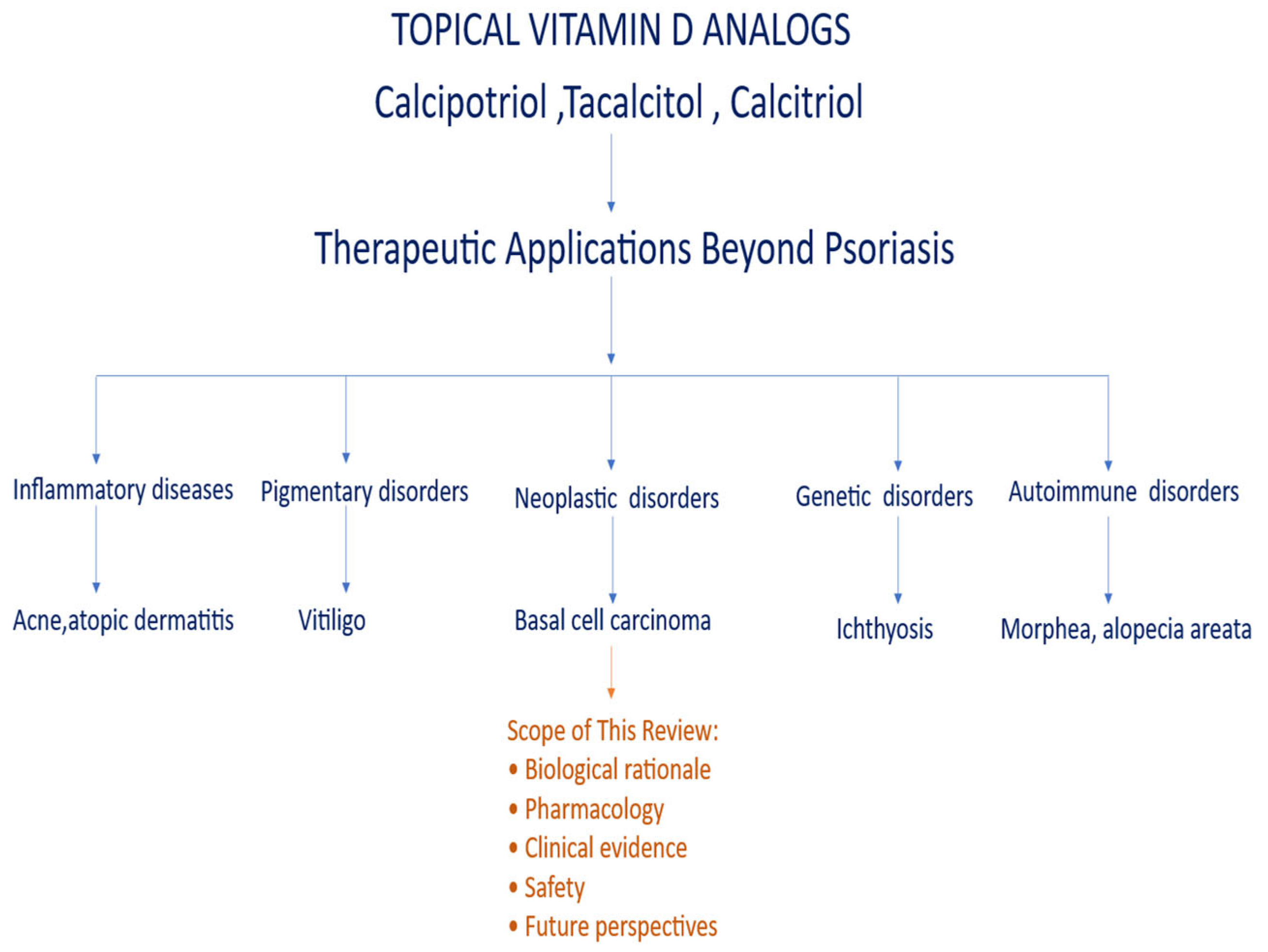
Figure 4.
Future directions for the clinical development of topical vitamin D analogs beyond psoriasis.
Figure 4.
Future directions for the clinical development of topical vitamin D analogs beyond psoriasis.
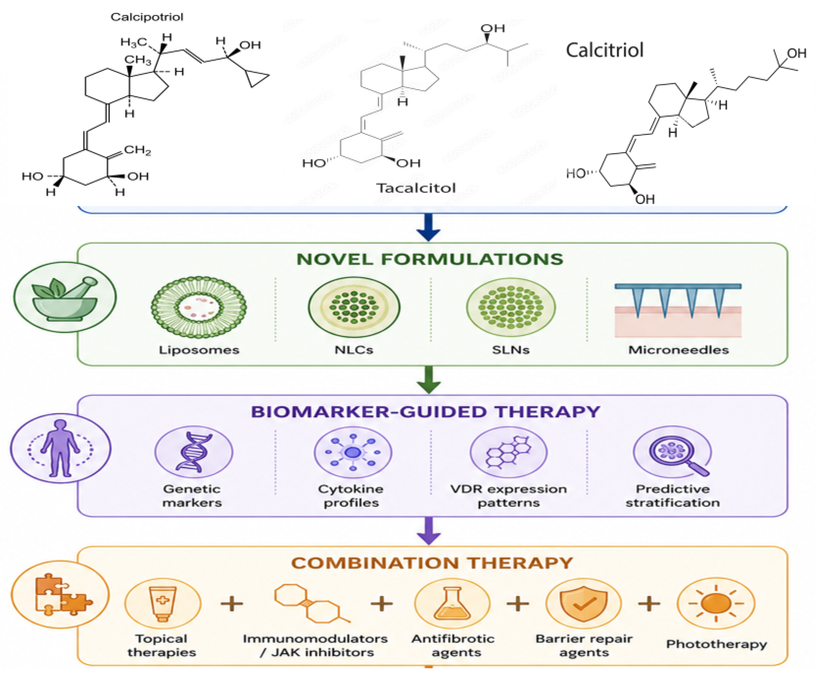
Disclaimer/Publisher’s Note: The statements, opinions and data contained in all publications are solely those of the individual author(s) and contributor(s) and not of MDPI and/or the editor(s). MDPI and/or the editor(s) disclaim responsibility for any injury to people or property resulting from any ideas, methods, instructions or products referred to in the content. |
© 2026 by the authors. Licensee MDPI, Basel, Switzerland. This article is an open access article distributed under the terms and conditions of the Creative Commons Attribution (CC BY) license (http://creativecommons.org/licenses/by/4.0/).
Copyright: This open access article is published under a Creative Commons CC BY 4.0 license, which permit the free download, distribution, and reuse, provided that the author and preprint are cited in any reuse.