
Submitted:
04 September 2026
Posted:
07 September 2026
You are already at the latest version
Abstract
Background: Laminins, a family of heterotrimeric glycoproteins, are fundamental components of the basement membrane and play critical roles in tissue architecture, cell adhesion, migration, differentiation, and hemostasis. Methods: This comprehensive integrative review synthesizes the current state of knowledge on the expression, administration, and modulation of laminin-derived bioactive peptides and isoforms. Results: Laminin peptides and isoforms exhibit diverse, context-dependent effects across physiological and pathological processes, including cancer progression, neurodegeneration, inflammation, tissue regeneration, and exercise-induced adaptations. In cancer, laminins modulate tumor growth, metastasis, and angiogenesis through complex signaling pathways involving PI3K/Akt, MAPK/ERK, and STAT3. In the nervous system, neuronal survival, axonal guidance, and regeneration, offering therapeutic potential for the treatment of neurodegenerative diseases and nerve injury. In inflammation and tissue repair, laminins regulate immune cell trafficking, cytokine production, and wound healing. Furthermore, laminins are integral to exercise-induced muscle adaptation, influencing satellite cell activation, myogenesis, and angiogenesis. Additionally, polylaminin, a biomimetic polymer of laminin assembled under acidic conditions, has emerged as a powerful tool for nerve regeneration, spinal cord repair, and stem cell expansion. Conclusions: The multifaceted nature of laminins underscores their significance as promising targets for novel therapeutic strategies across a broad spectrum of human diseases.
Keywords:
YIGSR
; IKVAV
; C16
; AG73
; laminin-111
; laminin-332
; laminin-511
; laminin γ2
; polylaminin
1. Introduction
Laminins are large, heterotrimeric glycoproteins that are major components of the basement membrane, a specialized extracellular matrix that underlies epithelial and endothelial cells and surrounds most tissues [1,2]. These complex molecules are composed of α, β, and γ chains, and their assembly into a polymer network is crucial for maintaining tissue integrity and regulating cell behavior [3,4]. The laminin family is highly diverse, with at least 16 known isoforms, each exhibiting tissue-specific expression and distinct biological functions [5,6]. The discovery of bioactive peptides derived from laminin, such as YIGSR from the β1 chain and IKVAV from the α1 chain, has opened new avenues for therapeutic intervention in a wide range of diseases [7,8]. These peptides mimic the biological activities of the parent laminin molecule, influencing cell adhesion, migration, differentiation, and survival [9,10].
Figure 1.
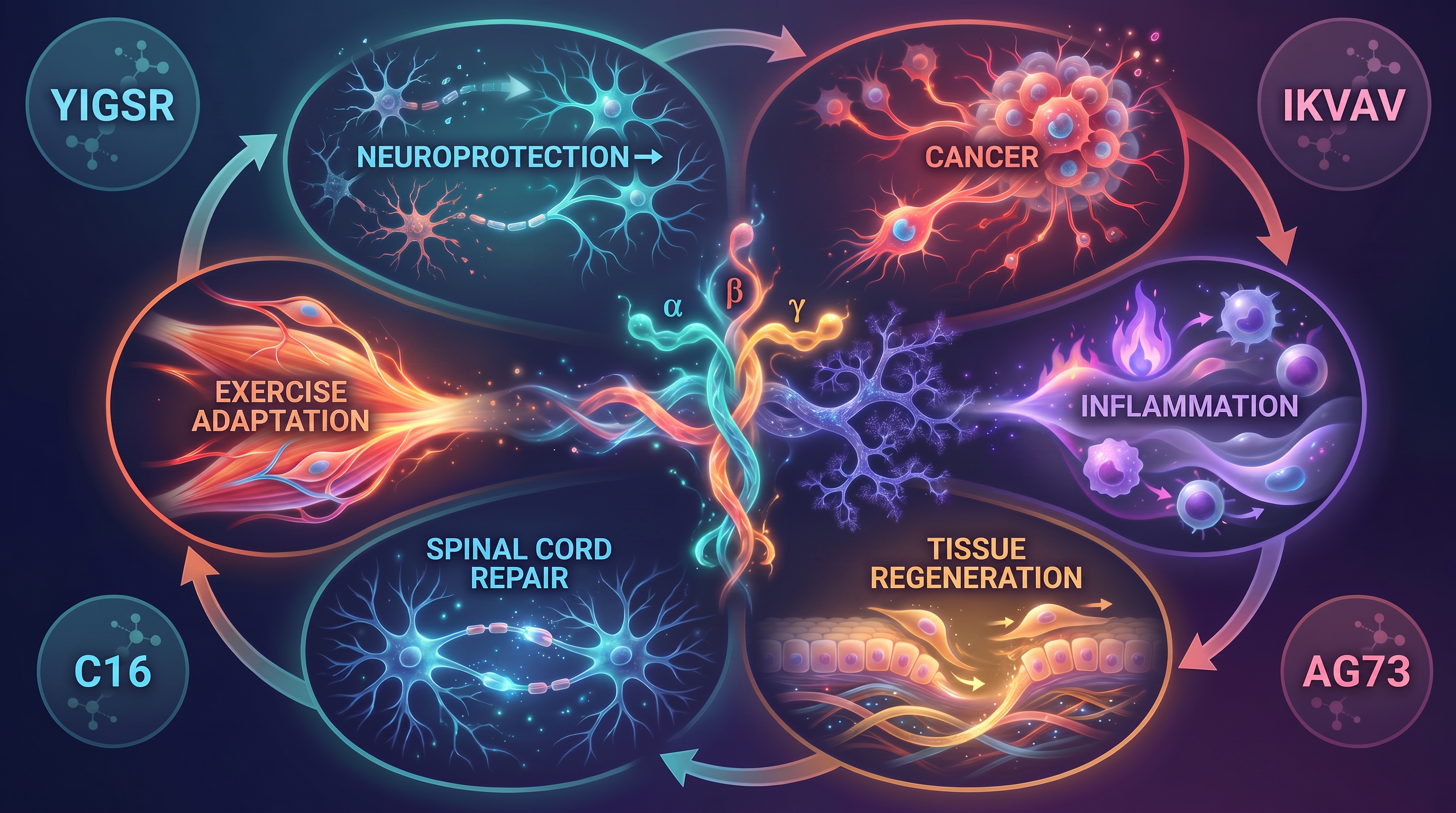
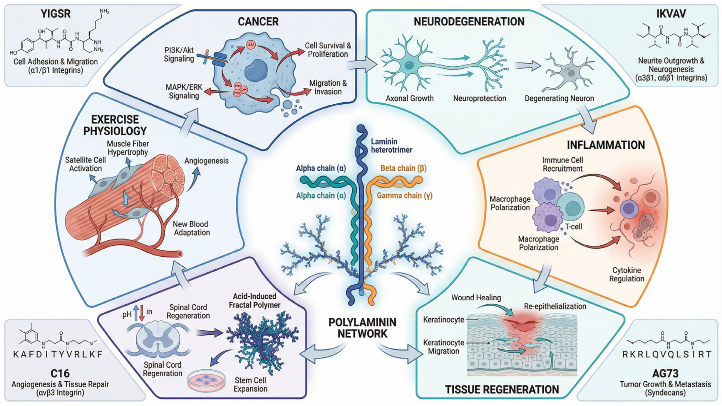
Schematic overview of the multifaceted roles of laminins, laminin-derived bioactive peptides (YIGSR, IKVAV, C16, AG73), and polylaminin in cancer, neurodegeneration, inflammation, tissue regeneration, exercise physiology, and spinal cord repair. The central element depicts the laminin heterotrimer (α, β, γ chains) assembling into a polylaminin fractal network under acidic conditions. Key signaling pathways and cellular interactions are highlighted for each thematic domain.
Figure 1.
Schematic overview of the multifaceted roles of laminins, laminin-derived bioactive peptides (YIGSR, IKVAV, C16, AG73), and polylaminin in cancer, neurodegeneration, inflammation, tissue regeneration, exercise physiology, and spinal cord repair. The central element depicts the laminin heterotrimer (α, β, γ chains) assembling into a polylaminin fractal network under acidic conditions. Key signaling pathways and cellular interactions are highlighted for each thematic domain.
The basement membrane serves as a critical interface between epithelial and mesenchymal tissues, providing structural support and regulating cell behavior through interactions with cell surface receptors, particularly integrins and dystroglycan [11,12]. Laminins interact with these receptors through specific binding domains, initiating intracellular signaling cascades that regulate gene expression, cell proliferation, and apoptosis [13,14]. The α3β1 integrin is particularly important for laminin recognition, and its engagement with laminin-332 has been shown to promote cell migration and invasion in both physiological and pathological contexts [15,16]. Beyond integrin signaling, laminins also interact with other cell surface receptors, including syndecan and perlecan, which are heparan sulfate proteoglycans that modulate laminin-mediated cell behavior [17,18].
This integrative review aims to provide a comprehensive overview of the current literature on the expression, administration, and modulation of laminin-derived peptides and laminin isoforms. We will explore their roles in cancer, neurodegeneration, inflammation, tissue regeneration, and exercise-induced adaptations, highlighting the therapeutic and diagnostic potential of these molecules. By synthesizing the findings from a broad range of studies, we aim to identify key mechanisms of action, discuss the challenges and opportunities in the field, and propose future directions for research and clinical translation.
2. Laminin Overview
2.1. Structural Organization and Biological Functions of Laminins
Laminins are composed of three polypeptide chains (α, β, and γ) that form a cross-shaped molecule with a molecular weight of approximately 900 kDa [19,20]. The α chain contains multiple functional domains, including the N-terminal domain, four tandem laminin-type epidermal growth factor (LE) domains, and a C-terminal globular domain (LG domain) [21,22]. The LG domain is particularly important for cell binding and has been identified as the site of several bioactive peptides, including YIGSR and IKVAV [23,24]. The β and γ chains also contain multiple functional domains and contribute to the overall stability and biological activity of the laminin molecule [25,26]. Specific interactions mediate the assembly of laminin trimers into higher-order structures between the N-terminal domains of the three chains, and this process is essential for the formation of a functional basement membrane [27,28].
The basement membrane, composed primarily of laminins, type IV collagen, nidogen, and perlecan, serves multiple critical functions in tissue homeostasis and pathophysiology [29,30]. In addition to providing structural support, the basement membrane acts as a selective barrier that regulates the passage of molecules and cells between different tissue compartments [31,32]. Laminins also serve as a reservoir for growth factors and cytokines, which can be released and activated by proteolytic cleavage [33,34]. Furthermore, laminins interact with cell surface receptors to initiate signaling cascades that regulate cell proliferation, differentiation, migration, and apoptosis [35,36]. These diverse functions of laminins underscore their importance in maintaining tissue homeostasis and their potential as therapeutic targets in various diseases.
2.2. Laminins in Cancer: Expression, Signaling, and Therapeutic Implications
The expression of laminin isoforms is frequently altered in cancer, and these changes can have profound effects on tumor progression, metastasis, and prognosis [37,38]. Laminin-332 (formerly laminin-5) is often overexpressed in various cancers, including breast, colon, pancreatic, and squamous cell carcinomas, and its expression is associated with increased invasion, epithelial-mesenchymal transition (EMT), and poor clinical outcomes [39,40]. The upregulation of laminin-332 in cancer cells promotes the formation of hemidesmosomes and focal adhesions, which facilitate cell migration and invasion [41,42]. Mechanistically, laminin-332 engagement with α3β1 integrin activates phosphatidylinositol 3-kinase (PI3K) and protein kinase B (Akt) signaling, leading to increased cell survival and proliferation [43,44]. Additionally, laminin-332 can activate mitogen-activated protein kinase (MAPK) signaling pathways, including extracellular signal-regulated kinase (ERK) and p38 MAPK, which promote cell migration and invasion [45,46].
Laminin-511 has also been implicated in cancer progression, particularly in the context of cancer stem cells and tumor-initiating cells [47,48]. Studies have demonstrated that laminin-511 promotes the self-renewal of cancer stem cells by activating signal transducer and activator of transcription 3 (STAT3) signaling, thereby enhancing tumor growth and therapeutic resistance [49,50]. The laminin γ2 chain is a particularly interesting target, as its expression is upregulated in many aggressive cancers and it has been shown to promote tumor cell migration and invasion through interactions with α3β1 integrin and other cell surface receptors [51,52]. Furthermore, the laminin γ2 chain can be proteolytically cleaved to generate a truncated form that exhibits enhanced pro-tumoral activity [53,54]. The phosphorylation of the laminin γ1 chain has also been identified as a critical regulator of cancer progression, with specific phosphorylation sites modulating integrin binding and downstream signaling [55,56].
Laminin-derived peptides have shown considerable promise as anti-cancer agents through multiple mechanisms of action. The YIGSR peptide, derived from the β1 chain of laminin, has been extensively studied for its anti-metastatic properties [57,58]. YIGSR inhibits melanoma cell migration and invasion by competing with the parent laminin molecule for binding to cell-surface receptors, thereby disrupting signaling pathways that promote tumor progression [59,60]. The molecular mechanism of YIGSR-mediated inhibition involves suppression of integrin-mediated signaling, leading to decreased expression of matrix metalloproteinases (MMPs) and reduced cell motility [61,62]. Additionally, YIGSR has been shown to induce apoptosis in cancer cells by activating caspase-dependent pathways and upregulating pro-apoptotic proteins such as Bax [63,64].
The C16 peptide, derived from the laminin α1 chain, represents another promising anti-cancer agent with multiple biological activities [65]. C16 has been shown to inhibit tumor growth and promote apoptosis in human melanoma cells by inducing reactive oxygen species (ROS) and activating the intrinsic mitochondrial apoptotic pathway [66]. Furthermore, C16 can regulate the expression of invadopodia-associated proteins, such as Tks5, thereby reducing the invasive capacity of cancer cells [67]. The C16 peptide also exhibits anti-angiogenic properties, inhibiting endothelial cell proliferation and tube formation, which contributes to its anti-tumor effects [68].
The IKVAV peptide, derived from the α1 chain of laminin, has been shown to inhibit tumor growth and metastasis in a variety of cancer models through multiple mechanisms [69]. IKVAV competes with the parent laminin molecule for binding to cell surface receptors, disrupting integrin-mediated signaling and reducing cell migration and invasion [70]. Additionally, IKVAV has been shown to promote neuronal differentiation and survival, which may contribute to its anti-tumor effects in the context of neural tumors [71]. Recent studies have demonstrated that IKVAV can activate autophagy in cancer cells, leading to cell death through autophagic mechanisms [72].
2.3. Laminins in Neurodegeneration, Neuroprotection, and Neuroregeneration
Laminins play a crucial role in the development and maintenance of the nervous system, and their dysregulation has been implicated in various neurodegenerative diseases, including Alzheimer's disease, Parkinson's disease, and amyotrophic lateral sclerosis (ALS) [73]. Laminin-111 is a major component of the basement membrane in the nervous system and has been shown to promote neuronal survival and axonal growth by activating integrin-mediated signaling pathways [74]. The interaction of laminin-111 with α6β1 integrin activates PI3K/Akt signaling, leading to increased expression of anti-apoptotic proteins and enhanced neuronal survival [75]. Additionally, laminin-111 promotes axonal outgrowth through activation of focal adhesion kinase (FAK) and Src family kinases, which regulate the dynamics of the actin cytoskeleton [76].
The IKVAV peptide, derived from the laminin α1 chain, has been extensively studied for its neurotrophic effects and is among the best-characterized laminin-derived peptide [77]. IKVAV promotes neuronal differentiation and survival by activating integrin-mediated signaling, leading to increased expression of neurotrophic factors such as brain-derived neurotrophic factor (BDNF) and nerve growth factor (NGF) [78]. Furthermore, IKVAV has been shown to promote axonal regeneration and functional recovery following spinal cord injury in animal models [79]. The mechanism of IKVAV-mediated neuroprotection involves activation of the PI3K/Akt and MAPK/ERK signaling pathways, which converge on the transcription factor cAMP response element binding protein (CREB) to promote the expression of genes involved in neuronal survival and plasticity [80].
In the context of neurodegenerative diseases, laminins and their derived peptides have shown significant therapeutic potential. Laminin-111 has been shown to attenuate neuronal death in models of Alzheimer's disease by promoting the clearance of amyloid-beta (Aβ) and reducing tau hyperphosphorylation through activation of integrin-mediated signaling [81]. The YIGSR peptide has also been shown to promote neurite outgrowth and neuronal survival, suggesting its potential for promoting nerve regeneration [82]. These findings suggest that laminin-based therapies could be a promising strategy for treating a variety of neurological disorders and promoting recovery following nerve injury.
2.4. Laminins in Inflammation and Immune Regulation
Laminins are key regulators of inflammation and immune cell function, influencing immune cell trafficking, cytokine production, and the resolution of inflammatory responses [83]. The basement membrane, composed primarily of laminins, serves as a critical barrier that regulates the extravasation of immune cells from the vasculature into inflamed tissues [84]. Laminin-511 is particularly important in this context, as it is highly expressed in endothelial basement membranes and plays a critical role in regulating leukocyte transendothelial migration [85]. The interaction of leukocyte integrins with laminin-511 modulates the efficiency and specificity of immune cell recruitment to inflamed tissues [86].
Laminin-derived peptides have been shown to modulate inflammatory responses through multiple mechanisms. The YIGSR peptide can inhibit the production of pro-inflammatory cytokines, such as tumor necrosis factor-alpha (TNF-α) and interleukin-6 (IL-6), by lipopolysaccharide (LPS)-stimulated macrophages [87]. The molecular mechanism of YIGSR-mediated anti-inflammatory activity involves the suppression of nuclear factor-kappa B (NF-κB) signaling, a critical transcription factor that regulates the expression of pro-inflammatory genes [88]. Additionally, YIGSR can promote macrophage differentiation toward an anti-inflammatory M2 phenotype, which is associated with tissue repair and the resolution of inflammation [89].
The AG73 peptide, derived from the laminin α1 chain, has been shown to promote angiogenesis and accelerate wound healing through multiple mechanisms [90]. AG73 promotes endothelial cell proliferation and tube formation, thereby increasing vascular density in healing tissues [91]. Furthermore, AG73 can promote the recruitment and differentiation of immune cells involved in tissue repair, including macrophages and regulatory T cells [92]. The pro-angiogenic and pro-regenerative effects of AG73 are mediated by the activation of integrin-dependent signaling pathways and the upregulation of vascular endothelial growth factor (VEGF) [93].
Laminin-332 plays a critical role in regulating immune cell function at epithelial and endothelial barriers. The interaction of immune cells with laminin-332 at these barriers modulates their activation state and effector functions [94]. Furthermore, laminin-332 has been shown to regulate the differentiation of regulatory T cells, which are critical for maintaining immune homeostasis and preventing excessive inflammation [95]. These findings suggest that laminins and their derived peptides represent promising therapeutic targets for regulating inflammation and promoting tissue repair.
2.5. Laminins in Tissue Regeneration and Wound Healing
Tissue regeneration and wound healing are complex biological processes that require coordinated interactions between multiple cell types and signaling pathways, and laminins play a critical role in orchestrating these processes [96]. Laminin-332 is essential for proper basement membrane formation in the skin and plays a critical role in wound healing through multiple mechanisms [97]. During the early stages of wound healing, laminin-332 is upregulated at the wound margin and promotes keratinocyte migration through the activation of integrin-mediated signaling [98]. The interaction of α3β1 integrin with laminin-332 activates FAK and Src family kinases, which regulate focal adhesion dynamics and promote cell migration [99].
Laminin-511 has also been shown to play important roles in tissue regeneration and wound healing. The expression of laminin-511 is upregulated during wound healing, and this isoform has been shown to promote keratinocyte migration and re-epithelialization [100]. Laminin-511 interacts with α6β1 integrin on keratinocytes, promoting their migration and differentiation [101]. Furthermore, laminin-511 has been shown to promote the differentiation of bone marrow-derived mesenchymal stem cells into osteoblasts, suggesting its role in bone regeneration [102].
Laminin-111 has been shown to enhance wound healing in animal models, particularly in the context of impaired wound healing associated with diabetes [103]. Laminin-111 promotes keratinocyte migration and proliferation by activating integrin-mediated signaling pathways [104]. Additionally, laminin-111 has been shown to promote angiogenesis and accelerate the formation of new blood vessels during tissue healing [105]. These pro-angiogenic effects of laminin-111 are mediated by the activation of endothelial cell integrins and the upregulation of VEGF signaling [106].
Laminin-derived peptides have also been shown to promote tissue regeneration and wound healing. The AG73 peptide has been shown to promote angiogenesis and accelerate wound healing by promoting endothelial cell proliferation and tube formation [107]. AG73 also promotes the recruitment of immune cells involved in tissue repair and the production of growth factors that support tissue regeneration [108]. The YIGSR peptide has been shown to promote wound healing by promoting keratinocyte migration and differentiation [109]. Additionally, YIGSR can promote angiogenesis by promoting endothelial cell proliferation and tube formation [110].
2.6. Laminins and Exercise Physiology: Molecular Adaptations and Therapeutic Applications
Exercise is a powerful stimulus for tissue remodeling and adaptation, and laminins play a critical role in mediating the exercise-induced changes in muscle structure and function [111]. During exercise, muscle fibers undergo mechanical stress and metabolic challenges, which trigger a cascade of molecular events that lead to muscle protein synthesis, mitochondrial biogenesis, and vascular adaptation [112]. Laminins, as critical components of the basement membrane surrounding muscle fibers, are essential for maintaining muscle structure and facilitating muscle tissue adaptation to exercise [113].
Laminin-111 is a major component of the basement membrane in skeletal muscle and plays a critical role in muscle fiber development, maintenance, and adaptation to exercise [114]. The interaction of muscle satellite cells with laminin-111 promotes their proliferation and differentiation into myoblasts, which fuse to form new muscle fibers or repair damaged muscle tissue [115]. Additionally, laminin-111 interacts with α7β1 integrin on muscle fibers, promoting muscle fiber stability and protecting against exercise-induced muscle damage [116]. Studies have shown that laminin-111 expression is upregulated by exercise, particularly during resistance training [117].
Laminin-511 has also been shown to play important roles in muscle adaptation to exercise. Laminin-511 expression is upregulated in skeletal muscle following endurance exercise, and this isoform has been shown to promote angiogenesis and vascular adaptation in muscle tissue [118]. The pro-angiogenic effects of laminin-511 are mediated by promoting endothelial cell proliferation and tube formation, thereby increasing capillary density in exercised muscles [119]. Furthermore, laminin-511 has been shown to promote the differentiation of muscle satellite cells and enhance muscle fiber regeneration following exercise-induced muscle damage [120].
The molecular mechanisms underlying the laminin-mediated adaptations to exercise involve the activation of integrin-dependent signaling pathways and the upregulation of growth factor signaling [121]. Exercise-induced muscle damage triggers the release of damage-associated molecular patterns (DAMPs) and growth factors, which promote the activation of muscle satellite cells and the initiation of muscle protein synthesis [122]. Laminins, through their interactions with integrins, amplify these signals and promote muscle regeneration and adaptation [123]. Furthermore, laminins interact with other extracellular matrix components, such as perlecan and nidogen, to create a microenvironment that supports muscle regeneration [124].
Laminin-derived peptides have shown potential to enhance exercise-induced adaptations in muscle and promote recovery from exercise-induced muscle damage. The IKVAV peptide has been shown to promote myoblast differentiation and fusion, suggesting its potential for enhancing muscle regeneration following exercise-induced damage [125]. Additionally, IKVAV can promote the differentiation of bone marrow-derived mesenchymal stem cells into myogenic lineages, suggesting its potential for cell-based regenerative therapies in the context of muscle injury [126]. The YIGSR peptide has been shown to promote angiogenesis, which could enhance the vascular adaptation to exercise and improve oxygen delivery to exercised muscles [127].
Recent studies have investigated the potential of laminin-based therapies to enhance exercise performance and promote recovery in athletes. Laminin-derived peptides have been shown to promote muscle protein synthesis and enhance the anabolic response to resistance training [128]. Furthermore, laminin-based therapies have been shown to accelerate recovery from exercise-induced muscle damage and reduce the incidence of exercise-related injuries [129]. These findings suggest that laminin-based interventions may represent a novel approach for enhancing exercise performance and promoting recovery in both healthy individuals and those with muscle-related pathologies.
2.7. Polylaminins: Structure, Functions, and Emerging Therapeutic Applications
In addition to naturally occurring laminin isoforms and derived peptides, recent advancements in biomaterials and tissue engineering have highlighted the significant therapeutic potential of polymerized laminins, commonly referred to as polylaminins. Polylaminin (polyLM) is a biomimetic, non-covalent polymer of the laminin protein that self-assembles into nano- and micro-structured networks upon pH acidification [130]. This structurally distinct form of laminin mimics the complex architecture of the extracellular matrix (ECM) more effectively than ordinary, non-polymerized laminin. The structural characterization of polyLM reveals an intrinsic fractal nature, forming a loose flocculated meshwork that is fundamentally different from the bulky aggregates formed by laminin under neutral pH conditions [131]. This fractal-like organization is hypothesized to be the structural basis for the architecture of basement membranes in neurogenic niches, particularly within the central nervous system (CNS) [132]. The structural properties of polylaminin also influence the organization of other ECM components; for instance, type IV collagen has been shown to immediately conform to the shape of acid-induced polymerized laminin, suggesting a highly coordinated interaction between these critical basement membrane proteins [133].
The unique structural characteristics of polylaminins translate into enhanced biological activities, particularly in the context of neurogenesis and nerve regeneration. Polylaminin has been demonstrated to stimulate neuritogenesis to a significantly greater extent than ordinary laminin [134]. In vitro studies utilizing retinal cells have shown that polyLM induces robust cellular spreading and the outgrowth of neurites, influencing both the proliferation and differentiation of these neural cells through specific signaling pathways, including those involving protein kinase A (PKA) and protein kinase C (PKC) [135]. Furthermore, the biological activity of polylaminin is maintained even when adsorbed onto synthetic scaffolds. For example, poly-ε-caprolactone (PCL) filaments coated with polylaminin have been shown to enhance functional nerve regeneration and tissue replacement in models of sciatic nerve injury, promoting significant motor function recovery and an increased number of motor plates in the target musculature [136].
The therapeutic applications of polylaminins are particularly promising for the treatment of severe neurological injuries, where endogenous regenerative capacity is limited. In experimental models of spinal cord injury (SCI), acute local injection of polylaminin, but not non-polymerized laminin, significantly improved motor function following thoracic compression or complete transection [137]. The mechanism underlying this recovery involves not only the promotion of axonal regeneration across the lesion site but also an unsuspected anti-inflammatory effect that contributes to early functional improvements [137]. This dual action—stimulating neurite outgrowth while modulating the inflammatory microenvironment—positions polylaminin as a multifaceted therapeutic agent. Recent studies have expanded on these findings, demonstrating the efficacy of laminin polymer treatments in accelerating the repair of crushed peripheral nerves [138] and showing promising results in longitudinal trials for the treatment of chronic spinal cord injuries in large animal models, such as dogs [139].
Beyond nerve regeneration, the biocompatibility and structural stability of laminin biopolymers have opened new avenues in stem cell research and broader tissue engineering applications [140]. Polylaminin substrates have been shown to modulate cellular signaling differently than ordinary laminin in various cell types, including follicular thyroid cells [141]. Moreover, polymerized laminins, specifically LN-521, have emerged as highly effective and feasible substrates for the large-scale expansion of induced pluripotent stem cells (iPSCs), maintaining their pluripotency and genetic stability even at low protein concentrations [142]. The ability to generate specific fractal microstructures of laminin-111 to precisely signal to cells further underscores the potential of polylaminins as designable ECM components for advanced regenerative medicine [143]. The ongoing elucidation of the roles of polymerizing laminins in development, health, and disease continues to drive the development of novel, biomimetic therapies across multiple medical disciplines [144].
2.8. Practical Applications
Limitations and Future Directions: While the molecular mechanisms are well-characterized in vitro and in animal models, a primary limitation is the scarcity of human clinical trials translating these findings into applied sports science. Future research must prioritize randomized controlled trials to evaluate the efficacy, safety, and optimal delivery methods of laminin-derived peptides in athletic populations. Bridging this translational gap will be essential to fully realize the potential of laminin-targeted interventions in optimizing human movement and athletic performance.
For Sports Medicine and Rehabilitation: In clinical and rehabilitative settings, the application of laminin-based biomaterials or peptide therapies holds immense promise for treating severe muscle strains, tendinopathies, and denervation injuries. The neurotrophic effects of the IKVAV peptide, combined with the angiogenic properties of the AG73 and YIGSR peptides, offer a multifaceted approach to sports injury rehabilitation—simultaneously promoting revascularization, muscle fiber regeneration, and the re-establishment of neuromuscular junctions. Practitioners should monitor the development of these targeted therapies, as they may soon become integral components of advanced rehabilitation protocols for elite athletes.
For Researchers in Sport Physiology: This review highlights a critical paradigm shift: the ECM is not merely a structural scaffold but a highly dynamic signaling hub. Researchers should focus on quantifying how different exercise modalities (e.g., high-intensity interval training vs. traditional hypertrophy training) specifically modulate laminin isoform expression in human skeletal muscle. There is a pressing need for longitudinal in vivo studies to determine whether an athlete's baseline laminin profile predicts their adaptive response to training or their susceptibility to muscle injury. Additionally, the cross-talk between laminin-mediated integrin signaling (such as α7β1) and systemic inflammatory markers post-exercise represents a fertile ground for discovering novel biomarkers of overtraining and recovery.
For Coaches and Athletes: The understanding that laminin-111 and laminin-511 are upregulated by mechanical stress and endurance exercise provides a molecular rationale for periodized training programs. Coaches can leverage this knowledge by designing resistance and endurance protocols that optimally stimulate extracellular matrix (ECM) remodeling, thereby enhancing muscle satellite cell activation and subsequent hypertrophy. Furthermore, the evidence that laminin-derived peptides (such as IKVAV and YIGSR) promote myoblast differentiation and angiogenesis suggests that nutritional or pharmacological interventions targeting these pathways could accelerate recovery between intense training sessions. Athletes recovering from exercise-induced muscle damage (EIMD) or sports-related injuries may benefit from future therapies that mimic these bioactive peptides to reduce downtime and prevent the chronification of inflammatory responses.
The findings synthesized in this integrative review provide compelling evidence for the critical role of laminins and their derived peptides in exercise physiology, muscle adaptation, and tissue regeneration, offering significant practical applications for sports scientists, coaches, athletes, and clinical practitioners.
3. Materials and Methods
A technical search of the PubMed database was conducted to identify relevant studies for this integrative review. The search strategy included a combination of keywords and MeSH terms related to "laminin," "laminin peptides," "YIGSR," "IKVAV," "C16," "AG73," "laminin-111," "laminin-332," "laminin-511," "laminin γ2," "polylaminin," "cancer," "neurodegeneration," "inflammation," "tissue regeneration," "wound healing," "exercise physiology," "physical activity," and "muscle adaptation." The search was limited to English-language articles with no date restrictions. The reference lists of retrieved articles were also manually screened for additional relevant studies. Studies were included if they investigated the expression, administration, or modulation of laminin-derived peptides or laminin isoforms in the context of cancer, neurodegeneration, inflammation, tissue regeneration, or exercise-induced adaptations. Both in vitro and in vivo studies were considered. Data from the selected studies were extracted and synthesized to provide a comprehensive overview of the current state of knowledge. Quality assessment was performed using established criteria to evaluate the methodological rigor of the included studies.
4. Conclusions
This integrative review highlights the diverse and critical roles of laminin-derived peptides and laminin isoforms in a wide range of physiological and pathological processes. The findings from numerous studies demonstrate their significant therapeutic and diagnostic potential in cancer, neurodegeneration, inflammation, tissue regeneration, and exercise-induced adaptations. The molecular mechanisms underlying the biological effects of laminins and laminin-derived peptides involve the activation of integrin-dependent signaling pathways and the upregulation of growth factor signaling, which converge on critical transcription factors to regulate gene expression and cell behavior.
The emergence of polylaminin as a biomimetic, fractal-structured polymer of laminin represents a significant advance in the field, offering enhanced neuritogenic, regenerative, and anti-inflammatory properties compared to native laminin. The demonstrated efficacy of polylaminin in preclinical models of spinal cord injury and peripheral nerve repair, combined with its biocompatibility and potential for stem cell expansion, positions it as a highly promising candidate for clinical translation.
However, several challenges remain to be addressed before these molecules can be translated into clinical practice. Future research should focus on elucidating the precise mechanisms of action of different laminin peptides and isoforms in specific disease contexts, identifying the most effective delivery strategies, and conducting well-designed clinical trials to evaluate their safety and efficacy in humans. Furthermore, the development of novel laminin-based biomaterials and therapeutic delivery systems will be essential for optimizing the biological activity and bioavailability of these molecules in vivo. A deeper understanding of the complex biology of laminins and their interactions with cellular and tissue processes will undoubtedly pave the way for the development of novel and effective therapies for a variety of diseases and conditions.
Author Contributions
Conceptualization, E.S., G.K., and L.G.; E.S., G.K., and L.G.; formal analysis, all authors; investigation, all authors; resources, all authors; data curation, all authors; writing—original draft preparation, all authors; writing—review and editing, all authors; visualization, all authors; supervision, L.G.; project administration, L.G.. All authors have read and agreed to the published version of the manuscript.
Funding
This research received no external funding.
Institutional Review Board Statement
Not applicable.
Informed Consent Statement
Not applicable.
Data Availability Statement
No new data were created or analyzed in this study. Data sharing is not applicable to this article.
Conflicts of Interest
The authors declare no conflicts of interest.
Abbreviations
The following abbreviations are used in this manuscript:
| ALS | Amyotrophic lateral sclerosis |
| Aβ | Amyloid-beta |
| Akt | Protein kinase B |
| BDNF | Brain-derived neurotrophic factor |
| CREB | cAMP response element binding protein |
| EMT | Epithelial-mesenchymal transition |
| ERK | Extracellular signal-regulated kinase |
| FAK | Focal adhesion kinase |
| IL-6 | Interleukin-6 |
| IKVAV | Isoleucine-lysine-valine-alanine-valine peptide |
| LE | Laminin-type epidermal growth factor |
| LG | Laminin globular domain |
| LPS | Lipopolysaccharide |
| MAPK | Mitogen-activated protein kinase |
| MMPs | Matrix metalloproteinases |
| NF-κB | Nuclear factor-kappa B |
| NGF | Nerve growth factor |
| PI3K | Phosphatidylinositol 3-kinase |
| ROS | Reactive oxygen species |
| STAT3 | Signal transducer and activator of transcription 3 |
| TNF-α | Tumor necrosis factor-alpha |
| VEGF | Vascular endothelial growth factor |
| YIGSR | Tyrosine-isoleucine-glycine-serine-arginine peptide |
| C16 | Laminin α1 chain-derived peptide |
| AG73 | Laminin α1 chain-derived peptide |
| Laminin-111 | Laminin isoform composed of α1, β1, and γ1 chains |
| Laminin-332 | Laminin isoform composed of α3, β3, and γ2 chains |
| Laminin-511 | Laminin isoform composed of α5, β1, and γ1 chains |
| Laminin γ2 | Laminin gamma-2 chain |
| Polylaminin | Biomimetic polymer of laminin assembled under acidic conditions |
References
- Aumailley, M.; Bruckner-Tuderman, L.; Carter, W.G.; Deutzmann, R.; Edgar, D.; Ekblom, P.; Engel, J.; Engvall, E.; Hohenester, E.; Jones, J.C.; et al. A simplified laminin nomenclature. Matrix Biol. 2005, 24, 326–332. [Google Scholar] [CrossRef] [PubMed]
- Colognato, H.; Yurchenco, P.D. Form and function: The laminin family of heterotrimers. Dev. Dyn. 2000, 218, 213–234. [Google Scholar]
- Yurchenco, P.D.; Schittny, J.C. Molecular architecture of the basement membrane. FASEB J. 1990, 4, 1577–1590. [Google Scholar] [CrossRef] [PubMed]
- Pöschl, E.; Schlötzer-Schrehardt, U.; Brachvogel, B.; Saito, K.; Ninomiya, Y.; Mayer, U. Collagen IV is essential for basement membrane stability but dispensable for its initial assembly during early development. Development 2004, 131, 1619–1628. [Google Scholar] [CrossRef] [PubMed]
- Durbeej, M. Laminins. Cell Tissue Res. 2010, 339, 259–268. [Google Scholar] [CrossRef] [PubMed]
- Miner, J.H.; Yurchenco, P.D. Laminin functions in tissue morphogenesis. Annu. Rev. Cell Dev. Biol. 2004, 20, 255–284. [Google Scholar] [CrossRef] [PubMed]
- Tashiro, K.; Sephel, G.C.; Weeks, B.; Sasaki, M.; Martin, G.R.; Kleinman, H.K.; Yamada, Y. A synthetic peptide containing the IKVAV sequence from the A chain of laminin mediates cell attachment, migration, and neurite outgrowth. J. Biol. Chem. 1989, 264, 7044–7050. [Google Scholar] [CrossRef]
- Yamamura, K.; Kibbey, M.C.; Jun, S.H.; Kleinman, H.K. Effect of Matrigel and laminin peptide YIGSR on tumor growth and metastasis. Semin Cancer Biol. 1993, 4, 259–65. [Google Scholar] [PubMed]
- Kleinman, H.K.; Philp, D.; Hoffman, M.P. Role of the extracellular matrix in morphogenesis. Curr. Opin. Biotechnol. 2003, 14, 526–532. [Google Scholar] [CrossRef] [PubMed]
- Giancotti, F.G.; Ruoslahti, E. Integrin signaling. Science 1999, 285, 1028–1032. [Google Scholar] [CrossRef] [PubMed]
- Hynes, R.O. Integrins: Versatility, modulation, and signaling in cell adhesion. Cell 1992, 69, 11–25. [Google Scholar] [CrossRef] [PubMed]
- Plantman, S.; Patarroyo, M.; Fried, K.; Domogatskaya, A.; Tryggvason, K.; Hammarberg, H.; Cullheim, S. Integrin-laminin interactions controlling neurite outgrowth from adult DRG neurons in vitro. Mol. Cell Neurosci. 2008, 39, 50–62. [Google Scholar] [CrossRef] [PubMed]
- Simon-Assmann, P.; Orend, G.; Mammadova-Bach, E.; Spenlé, C.; Lefebvre, O. Role of laminins in physiological and pathological angiogenesis. Int. J. Dev. Biol. 2011, 55, 455–65. [Google Scholar] [CrossRef] [PubMed]
- Halfter, W.; Dong, S.; Yip, Y.P.; Hayes, A.; Ott, S. A critical function of the pial basement membrane in cortical histogenesis. J. Neurosci. 2002, 22, 6029–6040. [Google Scholar] [CrossRef] [PubMed]
- Caplan, I.F.; Hernandez-Morato, I.; Pitman, M.J. Temporal expression of Laminin-111 in the developing rat larynx. Neurosci. Lett. 2022, 781, 136658. [Google Scholar] [CrossRef] [PubMed]
- Ford-Holevinski, T.S.; Hopkins, J.M.; McCoy, J.P.; Agranoff, B.W. Laminin supports neurite outgrowth from explants of axotomized adult rat retinal neurons. Brain Res. 1986, 393, 121–6. [Google Scholar] [CrossRef] [PubMed]
- Perera, T.H.; Lu, X.; Smith Callahan, L.A. Effect of Laminin Derived Peptides IKVAV and LRE Tethered to Hyaluronic Acid on hiPSC Derived Neural Stem Cell Morphology, Attachment and Neurite Extension. J. Funct. Biomater. 2020, 6, 15. [Google Scholar] [CrossRef] [PubMed]
- Jiang, Z.; Xi, Y.; Lai, K.; Wang, Y.; Wang, H.; Yang, G. Laminin-521 Promotes Rat Bone Marrow Mesenchymal Stem Cell Sheet Formation on Light-Induced Cell Sheet Technology. BioMed Res. Int. 2017, 2017, 9474573. [Google Scholar] [CrossRef] [PubMed]
- Zachman, A.L.; Crowder, S.W.; Ortiz, O.; Zienkiewicz, K.J.; Bronikowski, C.M.; Yu, S.S.; Giorgio, T.D.; Guelcher, S.A.; Kohn, J.; Sung, H. J. Pro-angiogenic and anti-inflammatory regulation by functional peptides loaded in polymeric implants for soft tissue regeneration. Tissue Eng. Part A 2013, 19, 437–47. [Google Scholar] [CrossRef] [PubMed]
- Negishi, Y.; Nomizu, M. Laminin-derived peptides: Applications in drug delivery systems for targeting. Pharmacol. Ther. 2019, 202, 91–97. [Google Scholar] [CrossRef] [PubMed]
- Parzymies, P. So Much More Than Just Structural Support: Laminin-332 and Its Multifaceted Functions in Skin Homeostasis and Disease. Exp. Dermatol. 2026, 35, e70293. [Google Scholar] [CrossRef] [PubMed]
- Sugawara, K.; Tsuruta, D.; Ishii, M.; Jones, J.C.; Kobayashi, H. Laminin-332 and -511 in skin. Exp. Dermatol. 2008, 17, 473–480. [Google Scholar] [CrossRef] [PubMed]
- Pouliot, N.; Kusuma, N. Laminin-511: a multi-functional adhesion protein regulating cell migration, tumor invasion and metastasis. Cell Adh Migr. 2013, 7, 142–9. [Google Scholar] [CrossRef] [PubMed]
- Negishi, Y.; Omata, D.; Iijima, H.; Hamano, N.; Endo-Takahashi, Y.; Nomizu, M.; Aramaki, Y. Preparation and characterization of laminin-derived peptide AG73-coated liposomes as a selective gene delivery tool. Biol. Pharm. Bull. 2010, 33, 1766–9. [Google Scholar] [CrossRef] [PubMed]
- Van Ry, P.M.; Minogue, P.; Hodges, B.L.; Burkin, D.J. Laminin-111 improves muscle repair in a mouse model of merosin-deficient congenital muscular dystrophy. Hum. Mol. Genet 2014, 23, 383–96. [Google Scholar] [CrossRef] [PubMed]
- Simon, T.; Bromberg, J.S. Regulation of the Immune System by Laminins. Trends Immunol. 2017, 38, 858–871. [Google Scholar] [CrossRef] [PubMed]
- Sakamoto, N.; Iwahana, M.; Tanaka, N.G.; Osada, Y. Inhibition of angiogenesis and tumor growth by a synthetic laminin peptide, CDPGYIGSR-NH2. Cancer Res. 1991, 51, 903–6. [Google Scholar] [PubMed]
- Ekblom, P.; Lonai, P.; Talts, J.F. Expression and biological role of laminin-1. Matrix Biol. 2003, 22, 35–47. [Google Scholar] [CrossRef] [PubMed]
- De Arcangelis, A.; Neuville, P.; Boukamel, R.; Lefebvre, O.; Kedinger, M.; Simon-Assmann, P. Inhibition of laminin alpha 1-chain expression leads to alteration of basement membrane assembly and cell differentiation. J. Cell Biol. 1996, 133, 417–30. [Google Scholar] [CrossRef] [PubMed]
- Hohenester, E.; Engel, J. Domain structure and organisation in extracellular matrix proteins. Matrix Biol. 2002, 21, 115–128. [Google Scholar] [CrossRef] [PubMed]
- Timpl, R.; Tisi, D.; Talts, J.F.; Andac, Z.; Sasaki, T.; Hohenester, E. Structure and function of laminin LG modules. Matrix Biol. 2000, 19, 309–317. [Google Scholar] [CrossRef] [PubMed]
- Akter, L.; Flechsig, H.; Marchesi, A.; Franz, C.M. Observing Dynamic Conformational Changes within the Coiled-Coil Domain of Different Laminin Isoforms Using High-Speed Atomic Force Microscopy. Int. J. Mol. Sci. 2024, 25, 1951. [Google Scholar] [CrossRef] [PubMed]
- Aumailley, M.; Gayraud, B. Structure and biological activity of the extracellular matrix. J. Mol. Med. 1998, 76, 253–265. [Google Scholar] [CrossRef] [PubMed]
- Ocampo, J.S.; de Brito, J.M.; Corrêa-de-Santana, E.; Borojevic, R.; Villa-Verde, D.M.; Savino, W. Laminin-211 controls thymocyte--thymic epithelial cell interactions. Cell Immunol. 2008, 254, 1–9. [Google Scholar] [CrossRef] [PubMed]
- Lugassy, C.; Kleinman, H.K.; Vernon, S.E.; Welch, D.R.; Barnhill, R.L. C16 laminin peptide increases angiotropic extravascular migration of human melanoma cells in a shell-less chick chorioallantoic membrane assay. Br. J. Dermatol. 2007, 157, 780–2. [Google Scholar] [CrossRef] [PubMed]
- Qin, Y.; Rodin, S.; Simonson, O.E.; Hollande, F. Laminins and cancer stem cells: Partners in crime? Semin Cancer Biol. 2017, 45, 3–12. [Google Scholar] [CrossRef] [PubMed]
- Iwamuro, M.; Shiraha, H.; Oyama, A.; Uchida, D.; Horiguchi, S.; Okada, H. Laminin-411 and -511 modulate the proliferation, adhesion, and morphology of gastric cancer cells. Cell Biochem. Biophys. 2021, 79, 407–418. [Google Scholar] [CrossRef] [PubMed]
- Katayama, H. Mechanism of anchorage-independency and tumor formation of cancer cells: possible involvement of cell membrane-bound laminin-332. Cell Tissue Res. 2020, 379, 255–259. [Google Scholar] [CrossRef] [PubMed]
- Kinoshita, T.; Hanazawa, T.; Nohata, N.; Kikkawa, N.; Enokida, H.; Yoshino, H.; Yamasaki, T.; Hidaka, H.; Nakagawa, M.; Okamoto, Y.; Seki, N. Tumor suppressive microRNA-218 inhibits cancer cell migration and invasion through targeting laminin-332 in head and neck squamous cell carcinoma. Oncotarget 2012, 3, 1386–400. [Google Scholar] [CrossRef] [PubMed]
- Qin, Y.; Shembrey, C.; Smith, J.; Paquet-Fifield, S.; Behrenbruch, C.; Beyit, L.M.; Thomson, B.N.J.; Heriot, A.G.; Cao, Y.; Hollande, F. Laminin 521 enhances self-renewal via STAT3 activation and promotes tumor progression in colorectal cancer. Cancer Lett. 2020, 476, 161–169. [Google Scholar] [CrossRef] [PubMed]
- Rodin, S.; Domogatskaya, A.; Ström, S.; Typeaeus, L.; Xia, J.; Yokota, C.; Sasaki, T.; Sorokin, L.; Tryggvason, K.; Fässler, R.; et al. Long-term self-renewal of human pluripotent stem cells on human recombinant laminin-511. Nat. Biotechnol. 2010, 28, 611–615. [Google Scholar] [CrossRef] [PubMed]
- Galliou, P.A.; Verrou, K.M.; Papanikolaou, N.A.; Koliakos, G. Phosphorylation mapping of laminin gamma1-chain: Kinases, functional interaction sequences, and phosphorylation-interfering cancer mutations. J. Biosci. 2024, 49, 85. [Google Scholar] [CrossRef]
- Diao, B.; Yang, P. Comprehensive analysis of the expression and prognosis for laminin genes in ovarian cancer. Pathol. Oncol. Res. 2021, 27, 1609855. [Google Scholar] [CrossRef] [PubMed]
- Yoshida, N.; Ishii, E.; Nomizu, M.; Yamada, Y.; Mohri, S.; Kinukawa, N.; Matsuzaki, A.; Oshima, K.; Hara, T.; Miyazaki, S. The laminin-derived peptide YIGSR (Tyr-Ile-Gly-Ser-Arg) inhibits human pre-B leukaemic cell growth and dissemination to organs in SCID mice. Br. J. Cancer 1999, 80, 1898–904. [Google Scholar] [CrossRef] [PubMed]
- Nomizu, M.; Kim, W.H.; Yamamura, K.; Utani, A.; Song, S.Y.; Otaka, A.; Roller, P.P.; Kleinman, H.K.; Yamada, Y. Identification of cell binding sites in the laminin alpha 1 chain carboxyl-terminal G domain. J. Biol. Chem. 1998, 273, 32491–32499. [Google Scholar] [CrossRef] [PubMed]
- Nomizu, M.; Weeks, B.S.; Weston, C.A.; Kim, W.H.; Kleinman, H.K.; Yamada, Y. Structure-activity study of a laminin alpha 1 chain aspartic acid-rich and cysteine-rich domain peptide, LAMININA. Biochemistry 1995, 34, 12995–13003. [Google Scholar] [CrossRef] [PubMed]
- Malinda, K.M.; Nomizu, M.; Chung, M.; Delgado, M.; Kuratomi, Y.; Yamada, Y.; Kleinman, H.K.; Ponce, M.L. Identification of laminin alpha 1 and beta 1 chain peptides active for endothelial cell adhesion, tube formation, and aortic sprouting. FASEB J. 1999, 13, 53–62. [Google Scholar] [PubMed]
- Miner, J.H.; Cunningham, J.; Sanes, J.R. Roles for laminin in embryogenesis: Exencephaly and ocular abnormalities in mice lacking the laminin alpha5 chain. J. Cell Biol. 1998, 143, 415–425. [Google Scholar] [CrossRef] [PubMed]
- Miner, J.H.; Li, C.; Mudd, J.L.; Go, G.; Sutherland, A.E. Compositional and structural requirements for laminin and basement membranes during mouse embryo implantation and gastrulation. Development 2004, 131, 2247–2256. [Google Scholar] [CrossRef] [PubMed]
- Li, X.; Liu, X.; Josey, B.; Chou, C.J.; Tan, Y.; Zhang, N.; Wen, X. Short laminin peptide for improved neural stem cell growth. Stem Cells Transl. Med. 2014, 3, 662–70. [Google Scholar] [CrossRef] [PubMed]
- Caires-dos-Santos, L.; da Silva, S.V.; Smuczek, B.; de Siqueira, A.S.; Cruz, K.S.P.; Barbuto, J.A.M.; Augusto, T.M.; Freitas, V.M.; Carvalho, H.F.; Jaeger, R.G. Laminin-derived peptide C16 regulates Tks expression and reactive oxygen species generation in human prostate cancer cells. J. Cell. Physiol. 2020, 235, 587–598. [Google Scholar] [CrossRef] [PubMed]
- Christensen, L. The distribution of fibronectin, laminin and tetranectin in human breast cancer with special attention to the extracellular matrix. APMIS Suppl. 1992, 26, 1–39. [Google Scholar] [PubMed]
- Qiu, X.; Tan, H.; Fu, D.; Zhu, Y.; Zhang, J. Laminin is over expressed in breast cancer and facilitate cancer cell metastasis. J. Cancer Res. Ther. 2018, 14, S1170–S1172. [Google Scholar] [CrossRef] [PubMed]
- Menezes, K.; de Menezes, J.R.; Nascimento, M.A.; Santos, R. de S.; Coelho-Sampaio, T. Polylaminin, a polymeric form of laminin, promotes regeneration after spinal cord injury. FASEB J. 2010, 24, 4513–4522. [Google Scholar] [CrossRef] [PubMed]
- Hochman-Mendez, C.; Cantini, M.; Moratal, D.; Salmeron-Sanchez, M.; Coelho-Sampaio, T. A fractal nature for polymerized laminin. PLoS ONE 2014, 9, e109388. [Google Scholar] [CrossRef] [PubMed]
- Coelho-Sampaio, T.; Tenchov, B.; Nascimento, M.A.; Hochman-Mendez, C.; Morandi, V.; Caarls, M.B.; Altankov, G. Type IV collagen conforms to the organization of polylaminin adsorbed on planar substrata. Acta Biomater. 2020, 111, 242–253. [Google Scholar] [CrossRef] [PubMed]
- Hochman-Mendez, C.; Lacerda de Menezes, J.R.; Sholl-Franco, A.; Coelho-Sampaio, T. Polylaminin recognition by retinal cells. J. Neurosci. Res. 2014, 92, 24–34. [Google Scholar] [CrossRef] [PubMed]
- de Siqueira-Santos, R.; Sardella-Silva, G.; Nascimento, M.A.; Teixeira de Oliveira, L.; Coelho-Sampaio, T.; Ribeiro-Resende, V.T. Biological activity of laminin/polylaminin-coated poly-ε-caprolactone filaments on the regeneration and tissue replacement of the rat sciatic nerve. Mater. Today Bio 2019, 3, 100026. [Google Scholar] [CrossRef] [PubMed]
- Haggerty, A.E.; Bening, M.R.; Pherribo, G.; Dauer, E.A.; Oudega, M. Laminin polymer treatment accelerates repair of the crushed peripheral nerve in adult rats. Acta Biomater. 2019, 86, 185–193. [Google Scholar] [CrossRef] [PubMed]
- Chize, C.M.; Vivas, D.G.; Menezes, K.; Freire, M.N.; Jiddu, R.F.P.; Graça-Souza, A.V.; de Souza-Leite, E.; Louzada, P.R.; Coelho-Sampaio, T. A laminin-based therapy for dogs with chronic spinal cord injury: Promising results of a longitudinal trial. Front. Vet. Sci. 2025, 12, 1592687. [Google Scholar] [CrossRef] [PubMed]
- Freire, E.; Barroso, M.M.; Klier, R.N.; Coelho-Sampaio, T. Biocompatibility and structural stability of a laminin biopolymer. Macromol. Biosci. 2012, 12, 67–74. [Google Scholar] [CrossRef] [PubMed]
- Palmero, C.Y.; M,iranda-Alves, L.; Sant'Ana Barroso, M.M.; Souza, E.C.; Machado, D.E.; Palumbo-Junior, A.; Santos, C.A.; Portilho, D.M.; Mermelstein, C.S.; Takiya, C.M.; Carvalho, D.P.; Hochman-Mendez, C.; Coelho-Sampaio, T.; Nasciutti, L.E. The follicular thyroid cell line PCCL3 responds differently to laminin and to polylaminin, a polymer of laminin assembled in acidic pH. Mol. Cell. Endocrinol. 2013, 376, 12–22. [Google Scholar] [CrossRef] [PubMed]
- Mesquita, F.C.P.; Leite, E.S.; Morrissey, J.; Freitas, C.; Coelho-Sampaio, T.; Hochman-Mendez, C. Polymerized laminin-521: A feasible substrate for expanding induced pluripotent stem cells at a low protein concentration. Cells 2022, 11, 3955. [Google Scholar] [CrossRef] [PubMed]
- Hochman-Mendez, C.; Coelho-Sampaio, T.; Kent, A.J.; Inman, J.L.; Bissell, M.J.; Robertson, C. Generating a fractal microstructure of laminin-111 to signal to cells. J. Vis. Exp. 2020, 163, e61134. [Google Scholar] [CrossRef] [PubMed]
- Kuratomi, Y.; Nomizu, M.; Tanaka, K.; Ponce, M.L.; Komiyama, S.; Kleinman, H.K.; Yamada, Y. Laminin gamma 1 chain peptide, C-16 (KAFDITYVRLKF), promotes migration, MMP-9 secretion, and pulmonary metastasis of B16-F10 mouse melanoma cells. Br. J. Cancer 2002, 86, 1169–73. [Google Scholar] [CrossRef] [PubMed]
- Catalani, E.; Giovarelli, M.; Zecchini, S.; Perrotta, C.; Cervia, D. Oxidative Stress and Autophagy as Key Targets in Melanoma Cell Fate. Cancers 2021, 13, 5791. [Google Scholar] [CrossRef] [PubMed]
- Siqueira, A.S.; Pinto, M.P.; Cruz, M.C.; Smuczek, B.; Cruz, K.S.; Barbuto, J.A.; Hoshino, D.; Weaver, A.M.; Freitas, V.M.; Jaeger, R.G. Laminin-111 peptide C16 regulates invadopodia activity of malignant cells through β1 integrin, Src and ERK 1/2. Oncotarget 2016, 7, 47904–47917. [Google Scholar] [CrossRef] [PubMed]
- 68.; Qu, H.; Fu, X.X.; Han, S. C16 peptide and angiopoietin-1 alleviate the side effects of glucocorticoids in a rat multiple sclerosis model. Life Sci. 2025, 363, 123402. [Google Scholar] [CrossRef] [PubMed]
- Kikkawa, Y.; Hozumi, K.; Katagiri, F.; Nomizu, M.; Kleinman, H.K.; Koblinski, J.E. Laminin-111-derived peptides and cancer. Cell Adh Migr. 2013, 7, 150–256. [Google Scholar] [CrossRef] [PubMed]
- Bresalier, R.S.; Schwartz, B.; Kim, Y.S.; Duh, Q.Y.; Kleinman, H.K.; Sullam, P.M. The laminin alpha 1 chain Ile-Lys-Val-Ala-Val (IKVAV)-containing peptide promotes liver colonization by human colon cancer cells. Cancer Res. 1995, 55, 2476–80. [Google Scholar] [PubMed]
- Xie, C.; Chen, Y.; Wang, L.; Liao, K.; Xue, B.; Han, Y.; Li, L.; Jiang, Q. Recent research of peptide-based hydrogel in nervous regeneration. Bioact. Mater. 2024, 40, 503–523. [Google Scholar] [CrossRef] [PubMed]
- Linder, B.; Kögel, D. Autophagy in Cancer Cell Death. Biology 2019, 8, 82. [Google Scholar] [CrossRef] [PubMed]
- Nguyen, B.; Bix, G.; Yao, Y. Basal lamina changes in neurodegenerative disorders. Mol. Neurodegener. 2021, 16, 81. [Google Scholar] [CrossRef] [PubMed]
- Nirwane, A.; Yao, Y. Cell-specific expression and function of laminin at the neurovascular unit. J. Cereb. Blood Flow Metab. 2022, 42, 1979–1999. [Google Scholar] [CrossRef] [PubMed]
- Nakashima, Y.; Tsukahara, M. Laminin-511 Activates the Human Induced Pluripotent Stem Cell Survival via α6β1 Integrin-Fyn-RhoA-ROCK Signaling. Stem Cells Dev. 2022, 31, 706–719. [Google Scholar] [CrossRef] [PubMed]
- Hermann, H.J.; Wuebbles, R.D.; Dagda, M.; Muñoz, A.; Parker, L.L.; Guzman, P.C.; Byrne, L.T.; Moore, S.A.; Burkin, D.J. Spatial proteomics reveals recombinant human laminin-111 restores adhesion signaling to laminin-α2-deficient muscle. JCI Insight 2025, 10, 194581. [Google Scholar] [CrossRef] [PubMed]
- Yamada, M.; Kadoya, Y.; Kasai, S.; Kato, K.; Mochizuki, M.; Nishi, N.; Watanabe, N.; Kleinman, H.K.; Yamada, Y.; Nomizu, M. Ile-Lys-Val-Ala-Val (IKVAV)-containing laminin alpha1 chain peptides form amyloid-like fibrils. FEBS Lett. 2002, 530, 48–52. [Google Scholar] [CrossRef] [PubMed]
- Farrukh, A.; Ortega, F.; Fan, W.; Marichal, N.; Paez, J.I.; Berninger, B.; Campo, A.D.; Salierno, M.J. Bifunctional Hydrogels Containing the Laminin Motif IKVAV Promote Neurogenesis. Stem Cell Rep. 2017, 9, 1432–1440. [Google Scholar] [CrossRef] [PubMed]
- Kazemi, S.; Baltzer, W.; Schilke, K.; Mansouri, H.; Mata, J.E. IKVAV-linked cell membrane-spanning peptide treatment induces neuronal reactivation following spinal cord injury. Future Sci. OA 2015, 1, FSO81. [Google Scholar] [CrossRef] [PubMed]
- Li, B.; Qiu, T.; Zhang, P.; Wang, X.; Yin, Y.; Li, S. IKVAV regulates ERK1/2 and Akt signalling pathways in BMMSC population growth and proliferation. Cell Prolif. 2014, 47, 133–45. [Google Scholar] [CrossRef] [PubMed]
- Rodin, S.; Kozin, S.A.; Kechko, O.I.; Mitkevich, V.A.; Makarov, A.A. Aberrant interactions between amyloid-beta and alpha5 laminins as possible driver of neuronal disfunction in Alzheimer's disease. Biochimie 2020, 174, 44–48. [Google Scholar] [CrossRef] [PubMed]
- Wang, Z.; Jia, S.; Xu, H.; Wang, X.; Lu, B.; Wu, W.; Huang, D.; Kong, L.; Kang, X.; Tian, F.; et al. A functionalized self-assembling peptide containing E7 and YIGSR sequences enhances neuronal differentiation of spermatogonial stem cells on aligned PCL fibers for spinal cord injury repair. Theranostics 2022, 12, 7567–7585. [Google Scholar] [CrossRef] [PubMed]
- Silva-Oliveira, G.; Linhares-Lacerda, L.; Mattos, T.R.F.; Sanches, C.; Coelho-Sampaio, T.; Riederer, I.; Saraiva, E.M. Laminin Triggers Neutrophil Extracellular Traps (NETs) and Modulates NET Release Induced by Leishmania amazonensis. Biomedicines 2022, 10, 521. [Google Scholar] [CrossRef] [PubMed]
- Wright, S.A.; Lennon, R.; Greenhalgh, A.D. Basement membranes' role in immune cell recruitment to the central nervous system. J. Inflamm. (Lond) 2024, 21, 53. [Google Scholar] [CrossRef] [PubMed]
- Di Russo, J.; Luik, A.L.; Yousif, L.; Budny, S.; Oberleithner, H.; Hofschröer, V.; Klingauf, J.; van Bavel, E.; Bakker, E.N.; Hellstrand, P.; et al. Endothelial basement membrane laminin 511 is essential for shear stress response. EMBO J. 2017, 36, 183–201. [Google Scholar] [CrossRef] [PubMed]
- Lowell, C.A. INTEGRIN FUNCTION IN LEUKOCYTE-MEDIATED INFLAMMATION-ACTINOPATHIES IN IMMUNE DISEASES. Trans. Am. Clin. Climatol. Assoc. 2025, 135, 74–86. [Google Scholar] [PubMed]
- Dekkers, B.G.; Bos, I.S.; Halayko, A.J.; Zaagsma, J.; Meurs, H. The laminin β1-competing peptide YIGSR induces a hypercontractile, hypoproliferative airway smooth muscle phenotype in an animal model of allergic asthma. Respir. Res. 2010, 11, 170. [Google Scholar] [CrossRef] [PubMed]
- Liu, T.; Zhang, L.; Joo, D.; Sun, S. C. NF-κB signaling in inflammation. Signal Transduct. Target Ther. 2017, 2, 17023. [Google Scholar] [CrossRef] [PubMed]
- Yan, L.; Wang, J.; Cai, X.; Liou, Y.C.; Shen, H.M.; Hao, J.; Huang, C.; Luo, G.; He, W. Macrophage plasticity: signaling pathways, tissue repair, and regeneration. MedComm (2020) 2024, 5, e658. [Google Scholar] [CrossRef] [PubMed]
- Puchalapalli, M.; Mu, L.; Edwards, C.; Kaplan-Singer, B.; Eni, P.; Belani, K.; Finkelstein, D.; Patel, A.; Sayyad, M.; Koblinski, J.E. The Laminin-α1 Chain-Derived Peptide, AG73, Binds to Syndecans on MDA-231 Breast Cancer Cells and Alters Filopodium Formation. Anal. Cell Pathol. (Amst) 2019, 2019, 9192516. [Google Scholar] [CrossRef] [PubMed]
- Liang, T.; Liu, J.; Liu, F.; Su, X.; Li, X.; Zeng, J.; Chen, F.; Wen, H.; Chen, Y.; Tao, J.; et al. Application of Pro-angiogenic Biomaterials in Myocardial Infarction. ACS Omega 2024, 9, 37505–37529. [Google Scholar] [CrossRef] [PubMed]
- Li, J.; Tan, J.; Martino, M.M.; Lui, K.O. Regulatory T-Cells: Potential Regulator of Tissue Repair and Regeneration. Front Immunol. 2018, 9, 585. [Google Scholar] [CrossRef] [PubMed]
- Shaw, P.; Dwivedi, S.K.D.; Bhattacharya, R.; Mukherjee, P.; Rao, G. VEGF signaling: Role in angiogenesis and beyond. Biochim Biophys. Acta Rev. Cancer 2024, 1879, 189079. [Google Scholar] [CrossRef] [PubMed]
- Tsuruta, D.; Kobayashi, H.; Imanishi, H.; Sugawara, K.; Ishii, M.; Jones, J.C. Laminin-332-integrin interaction: a target for cancer therapy? Curr. Med. Chem. 2008, 15, 1968–75. [Google Scholar] [CrossRef] [PubMed]
- Zhang, W.; Xiang, Y.; Lu, C.; Wang, F.; Ren, H.; Wu, H.; Yan, M. The extracellular matrix in inflammation and cancer. Mol. BioMed 2026, 7, 21. [Google Scholar] [CrossRef] [PubMed]
- Nayak, M.; Banerjee, D.; Venugopal, V.; Nethi, S.K.; Barui, A.K.; Mukherjee, S. Cell-engineered technologies for wound healing and tissue regeneration. npj BioMed Innov. 2025, 2, 38. [Google Scholar] [CrossRef] [PubMed]
- Chung, H.; Suh, E.K.; Han, I.O.; Oh, E.S. Keratinocyte-derived laminin-332 promotes adhesion and migration in melanocytes and melanoma. J. Biol. Chem. 2011, 286, 13438–47. [Google Scholar] [CrossRef] [PubMed]
- Chang, Y.C.; Sabourin, C.L.; Lu, S.E.; Sasaki, T.; Svoboda, K.K.; Gordon, M.K.; Riley, D.J.; Casillas, R.P.; Gerecke, D.R. Upregulation of gamma-2 laminin-332 in the mouse ear vesicant wound model. J. Biochem Mol. Toxicol. 2009, 23, 172–84. [Google Scholar] [CrossRef] [PubMed]
- Cavaco, A.C.M.; Rezaei, M.; Caliandro, M.F.; Lima, A.M.; Stehling, M.; Dhayat, S.A.; Haier, J.; Brakebusch, C.; Eble, J.A. The Interaction between Laminin-332 and α3β1 Integrin Determines Differentiation and Maintenance of CAFs, and Supports Invasion of Pancreatic Duct Adenocarcinoma Cells. Cancers 2018, 11, 14. [Google Scholar] [CrossRef] [PubMed]
- Iorio, V.; Troughton, L.D.; Hamill, K.J. Laminins: Roles and Utility in Wound Repair. Adv. Wound Care (New Rochelle) 2015, 4, 250–263. [Google Scholar] [CrossRef] [PubMed]
- Gao, J.; DeRouen, M.C.; Chen, C.H.; Nguyen, M.; Nguyen, N.T.; Ido, H.; Harada, K.; Sekiguchi, K.; Morgan, B.A.; Miner, J.H.; et al. Laminin-511 is an epithelial message promoting dermal papilla development and function during early hair morphogenesis. Genes Dev. 2008, 22, 2111–24. [Google Scholar] [CrossRef] [PubMed]
- Sun, Y.; Wang, T.L.; Toh, W.S.; Pei, M. The role of laminins in cartilaginous tissues: from development to regeneration. Eur. Cell Mater. 2017, 34, 40–54. [Google Scholar] [CrossRef] [PubMed]
- Malinda, K.M.; Wysocki, A.B.; Koblinski, J.E.; Kleinman, H.K.; Ponce, M.L. Angiogenic laminin-derived peptides stimulate wound healing. Int. J. Biochem Cell Biol. 2008, 40, 2771–80. [Google Scholar] [CrossRef] [PubMed]
- Horejs, C.M.; Serio, A.; Purvis, A.; Gormley, A.J.; Bertazzo, S.; Poliniewicz, A.; Wang, A.J.; DiMaggio, P.; Hohenester, E.; Stevens, M.M. Biologically-active laminin-111 fragment that modulates the epithelial-to-mesenchymal transition in embryonic stem cells. Proc. Natl. Acad. Sci. U S A 2014, 111, 5908–13. [Google Scholar] [CrossRef] [PubMed]
- Ishihara, J.; Ishihara, A.; Fukunaga, K.; et al. Laminin heparin-binding peptides bind to several growth factors and enhance diabetic wound healing. Nat. Commun. 2018, 9, 2163. [Google Scholar] [CrossRef] [PubMed]
- Stamati, K.; Priestley, J.V.; Mudera, V.; Cheema, U. Laminin promotes vascular network formation in 3D in vitro collagen scaffolds by regulating VEGF uptake. Exp. Cell Res. 2014, 327, 68–77. [Google Scholar] [CrossRef] [PubMed]
- Saklani, M.; Jha, C.B.; Baidya, A.T.K.; Singh, S.; Kumar, R.; Mathur, R.; Tiwari, A.K.; Varshney, R. Laminin mimetic angiogenic and collagen peptide hydrogel for enhance dermal wound healing. Biomater. Adv. 2024, 158, 213761. [Google Scholar] [CrossRef] [PubMed]
- Lee, J.H.; Shin, S.J.; Lee, J.H.; Knowles, J.C.; Lee, H.H.; Kim, H.W. Adaptive immunity of materials: Implications for tissue healing and regeneration. Bioact. Mater. 2024, 41, 499–522. [Google Scholar] [CrossRef] [PubMed]
- Kim, Y.Y.; Li, H.; Song, Y.S.; Jeong, H.S.; Yun, H.Y.; Baek, K.J.; Kwon, N.S.; Shin, Y.K.; Park, K.C.; Kim, D.S. Laminin peptide YIGSR enhances epidermal development of skin equivalents. J. Tissue Viability 2018, 27, 117–121. [Google Scholar] [CrossRef] [PubMed]
- Ngo, M.T.; Harley, B.A.C. Angiogenic biomaterials to promote therapeutic regeneration and investigate disease progression. Biomaterials 2020, 255, 120207. [Google Scholar] [CrossRef] [PubMed]
- Ferraro, E.; Giammarioli, A.M.; Chiandotto, S.; Spoletini, I.; Rosano, G. Exercise-induced skeletal muscle remodeling and metabolic adaptation: redox signaling and role of autophagy. Antioxid. Redox Signal 2014, 21, 154–76. [Google Scholar] [CrossRef] [PubMed]
- Smith, J.A.B.; Murach, K.A.; Dyar, K.A.; Zierath, J.R. Exercise metabolism and adaptation in skeletal muscle. Nat. Rev. Mol. Cell Biol. 2023, 24, 607–632. [Google Scholar] [CrossRef] [PubMed]
- Aumailley, M.; Smyth, N. The role of laminins in basement membrane function. J. Anat. 1998, 193, 1–21. [Google Scholar] [CrossRef] [PubMed]
- Zou, K.; De Lisio, M.; Huntsman, H.D.; Pincu, Y.; Mahmassani, Z.; Miller, M.; Olatunbosun, D.; Jensen, T.; Boppart, M.D. Laminin-111 improves skeletal muscle stem cell quantity and function following eccentric exercise. Stem Cells Transl. Med. 2014, 3, 1013–22. [Google Scholar] [CrossRef] [PubMed]
- Penton, C.M.; Badarinarayana, V.; Prisco, J.; Powers, E.; Pincus, M.; Allen, R.E.; August, P.R. Laminin 521 maintains differentiation potential of mouse and human satellite cell-derived myoblasts during long-term culture expansion. Skelet. Muscle 2016, 6, 44. [Google Scholar] [CrossRef] [PubMed]
- Rooney, J.E.; Gurpur, P.B.; Yablonka-Reuveni, Z.; Burkin, D.J. Laminin-111 restores regenerative capacity in a mouse model for alpha7 integrin congenital myopathy. Am. J. Pathol. 2009, 174, 256–64. [Google Scholar] [CrossRef] [PubMed]
- Garg, K.; Mahmassani, Z.S.; Dvoretskiy, S.; Valero, M.C.; Huntsman, H.D.; Lapp, S.; Wu, Y.F.; Hauschka, S.D.; Burkin, D.J.; Boppart, M.D. Laminin-111 Improves the Anabolic Response to Mechanical Load in Aged Skeletal Muscle. J. Gerontol. A Biol. Sci. Med. Sci. 2021, 76, 586–590. [Google Scholar] [CrossRef] [PubMed]
- Okumura, N.; Kakutani, K.; Numata, R.; Nakahara, M.; Schlötzer-Schrehardt, U.; Kruse, F.; Kinoshita, S.; Koizumi, N. Laminin-511 and -521 enable efficient in vitro expansion of human corneal endothelial cells. Invest Ophthalmol. Vis. Sci. 2015, 56, 2933–42. [Google Scholar] [CrossRef] [PubMed]
- Xu, H.; Pumiglia, K.; LaFlamme, S.E. Laminin-511 and α6 integrins regulate the expression of CXCR4 to promote endothelial morphogenesis. J. Cell Sci. 2020, 133, jcs246595. [Google Scholar] [CrossRef] [PubMed]
- Riederer, I.; Bonomo, A.C.; Mouly, V.; Savino, W. Laminin therapy for the promotion of muscle regeneration. FEBS Lett. 2015, 589, 3449–53. [Google Scholar] [CrossRef] [PubMed]
- Speer, J.; Barcellona, M.; Jing, L.; Liu, B.; Lu, M.; Kelly, M.; Buchowski, J.; Zebala, L.; Luhmann, S.; Gupta, M.; Setton, L. Integrin-mediated interactions with a laminin-presenting substrate modulate biosynthesis and phenotypic expression for cells of the human nucleus pulposus. Eur. Cell Mater. 2021, 41, 793–810. [Google Scholar] [CrossRef] [PubMed]
- Stožer, A.; Vodopivc, P.; Križančić Bombek, L. Pathophysiology of exercise-induced muscle damage and its structural, functional, metabolic, and clinical consequences. Physiol. Res. 2020, 69, 565–598. [Google Scholar] [CrossRef] [PubMed]
- Boppart, M.D.; Mahmassani, Z.S. Integrin signaling: linking mechanical stimulation to skeletal muscle hypertrophy. Am. J. Physiol. Cell Physiol. 2019, 317, C629–C641. [Google Scholar] [CrossRef] [PubMed]
- Walker, C.; Mojares, E.; Del Río Hernández, A. Role of Extracellular Matrix in Development and Cancer Progression. Int. J. Mol. Sci. 2018, 19, 3028. [Google Scholar] [CrossRef] [PubMed]
- Bischof, K.; Stafilidis, S.; Bundschuh, L.; Oesser, S.; Baca, A.; König, D. Reduction in systemic muscle stress markers after exercise-induced muscle damage following concurrent training and supplementation with specific collagen peptides - a randomized controlled trial. Front Nutr. 2024, 11, 1384112. [Google Scholar] [CrossRef] [PubMed]
- Namjoo, A.R.; Abrbekoh, F.N.; Saghati, S.; Amini, H.; Saadatlou, M.A.E.; Rahbarghazi, R. Tissue engineering modalities in skeletal muscles: focus on angiogenesis and immunomodulation properties. Stem Cell Res. Ther. 2023, 14, 90. [Google Scholar] [CrossRef] [PubMed]
- Jha, A.; Moore, E. YIGSR, A Laminin-Derived Peptide, Dictates a Concentration-Dependent Impact on Macrophage Phenotype Response. Cell Mol. Bioeng. 2024, 17, 423–440. [Google Scholar] [CrossRef] [PubMed]
- Casarella, S.; Ferla, F.; Di Francesco, D.; Pagani, V.; Di Varsavia, C.; Regano, I.; Boccafoschi, F. Laminin-derived peptide drives the cardiomyogenic potential and cardiac cells functionality. Front Bioeng. Biotechnol. 2025, 13, 1629412. [Google Scholar] [CrossRef] [PubMed]
- Marcinczyk, M.; Dunn, A.; Haas, G.; Madsen, J.; Scheidt, R.; Patel, K.; Talovic, M.; Garg, K. The Effect of Laminin-111 Hydrogels on Muscle Regeneration in a Murine Model of Injury. Tissue Eng. Part A 2019, 25, 1001–1012. [Google Scholar] [CrossRef] [PubMed]
- Speer, J.E.; Barcellona, M.N.; Lu, M.Y.; Zha, Z.; Jing, L.; Gupta, M.C.; Buchowski, J.M.; Kelly, M.P.; Setton, L.A. Development of a library of laminin-mimetic peptide hydrogels for control of nucleus pulposus cell behaviors. J. Tissue Eng. 2021, 12, 20417314211021220. [Google Scholar] [CrossRef] [PubMed]
- Hohenester, E. Structural biology of laminins. Essays Biochem 2019, 63, 285–295. [Google Scholar] [CrossRef] [PubMed]
- Hamill, K.J.; Kligys, K.; Hopkinson, S.B.; Jones, J.C. Laminin deposition in the extracellular matrix: a complex picture emerges. J. Cell Sci. 2009, 122, 4409–17. [Google Scholar] [CrossRef] [PubMed]
- Charonis, A.S.; Tsilibary, E.C.; Yurchenco, P.D.; Furthmayr, H. Binding of laminin to type IV collagen: a morphological study. J. Cell Biol. 1985, 100, 1848–53. [Google Scholar] [CrossRef] [PubMed]
- Hochman-Mendez, C.; Cantini, M.; Moratal, D.; Salmeron-Sanchez, M.; Coelho-Sampaio, T. A fractal nature for polymerized laminin. PLoS ONE 2014, 9, e109388. [Google Scholar] [CrossRef] [PubMed]
- Guo, Y.M.; Jiang, X.; Min, J.; Huang, J.; Huang, X.F.; Ye, L. Advances in the study of Müller glia reprogramming in mammals. Front Cell Neurosci. 2023, 17, 1305896. [Google Scholar] [CrossRef] [PubMed]
- Gadalla, D.; Kennedy, M.; Ganem, J.; Suppah, M.; Schmitt, A.; Lott, D.G. Polycaprolactone Fiber and Laminin and Collagen IV Protein Incorporation in Implants Enhances Wound Healing in a Novel Mouse Skin Splint Model. J. Tissue Eng. Regen. Med. 2024, 2024, 2515383. [Google Scholar] [CrossRef] [PubMed]
- Chize, C.M.; Vivas, D.G.; Menezes, K.; Freire, M.N.; Jiddu, R.F.P.; Graça-Souza, A.V.; de Souza-Leite, E.; Louzada, P.R.; Coelho-Sampaio, T. A laminin-based therapy for dogs with chronic spinal cord injury: promising results of a longitudinal trial. Front Vet. Sci. 2025, 12, 1592687. [Google Scholar] [CrossRef] [PubMed]
- Chang, W.; Shah, M.B.; Zhou, G.; Walsh, K.; Rudraiah, S.; Kumbar, S.G.; Yu, X. Polymeric nanofibrous nerve conduits coupled with laminin for peripheral nerve regeneration. BioMed Mater. 2020, 15, 035003. [Google Scholar] [CrossRef] [PubMed]
- Haggerty, A.E.; Bening, M.R.; Pherribo, G.; Dauer, E.A.; Oudega, M. Laminin polymer treatment accelerates repair of the crushed peripheral nerve in adult rats. Acta Biomater. 2019, 86, 185–193. [Google Scholar] [CrossRef] [PubMed]
- Boni, R.; Ali, A.; Shavandi, A.; Clarkson, A.N. Current and novel polymeric biomaterials for neural tissue engineering. J. BioMed Sci. 2018, 25, 90. [Google Scholar] [CrossRef] [PubMed]
- Miyazaki, K. Laminin-5 (laminin-332): Unique biological activity and role in tumor growth and invasion. Cancer Sci. 2006, 97, 91–8. [Google Scholar] [CrossRef] [PubMed]
- Paccola Mesquita, F.C.; Hochman-Mendez, C.; Morrissey, J.; Sampaio, L.C.; Taylor, D.A. Laminin as a Potent Substrate for Large-Scale Expansion of Human Induced Pluripotent Stem Cells in a Closed Cell Expansion System. Stem Cells Int. 2019, 2019, 9704945. [Google Scholar] [CrossRef] [PubMed]
- Hochman-Mendez, C.; Coelho-Sampaio, T.; Kent, A.J.; Inman, J.L.; Bissell, M.J.; Robertson, C. Generating a Fractal Microstructure of Laminin-111 to Signal to Cells. J. Vis. Exp. 2020, 28, 10.3791/61134. [Google Scholar] [CrossRef] [PubMed]
- Horita, S.I.M.; Bensalah, M.; Bigot, A.; Mamchaoui, K.; Butler Browne, G.S.; Beghini, D.G.; Savino, W.; Trollet, C.; Mouly, V.; Negroni, E. Human Laminin-111-Derived AG73 Increases Proliferation, Migration, and Differentiation of Human Myoblasts: A Promising Candidate in Regenerative Medicine. ACS Omega 2025, 10, 58480–58489. [Google Scholar] [CrossRef] [PubMed]
Disclaimer/Publisher’s Note: The statements, opinions and data contained in all publications are solely those of the individual author(s) and contributor(s) and not of MDPI and/or the editor(s). MDPI and/or the editor(s) disclaim responsibility for any injury to people or property resulting from any ideas, methods, instructions or products referred to in the content. |
© 2026 by the authors. Licensee MDPI, Basel, Switzerland. This article is an open access article distributed under the terms and conditions of the Creative Commons Attribution (CC BY) license (http://creativecommons.org/licenses/by/4.0/).
Copyright: This open access article is published under a Creative Commons CC BY 4.0 license, which permit the free download, distribution, and reuse, provided that the author and preprint are cited in any reuse.