

Submitted:
03 September 2026
Posted:
04 September 2026
You are already at the latest version
Abstract
Background/Objectives: Cutaneous melanoma is predominantly sporadic, although a subset of patients presents clinical features suggestive of inherited susceptibility. We introduced the pragmatic concept of genetically enriched (GE) melanoma, defined by the presence of early-onset melanoma, familial melanoma, or multiple primary melanomas. Methods: Its clinicopathological characteristics and prognosis were compared with those of sporadic melanoma. Demographic, clinicopathological, and survival data were analyzed using descriptive statistics, survival analysis, and Cox regression models. Results: In this retrospective single-center study, 1,874 patients were classified as having GE (n = 796, 42.5%) or sporadic melanoma (n = 1,078, 57.5%). Patients with GE melanoma were significantly younger at diagnosis (median age, 37 years [IQR 32–43] vs 57 years [IQR 50–66], p < 0.001) and were more frequently females than those with sporadic melanoma (56.8% vs. 51.4%, p = 0.024). The GE cohort also showed a distinct clinicopathological profile, with a higher proportion of melanoma in situ (20.2% vs. 15.2%), lower proportions of acral lentiginous (3.5% vs. 1.1%) and lentigo maligna (5.2% vs. 1.4%) melanoma, and a lower prevalence of previous malignancies. After a median follow-up of 8 years, disease-free survival did not differ significantly between the two cohorts (log-rank p = 0.23). Within the GE cohort, older age and male sex were associated with a higher risk of recurrence. Conclusions: Overall, patients fulfilling clinical criteria suggestive of inherited melanoma susceptibility appear to represent a distinct clinicopathological subgroup without evidence of poorer prognosis. The concept of GE melanoma may offer a pragmatic framework for identifying patients who may benefit from genetic counseling and tailored surveillance strategies.
Keywords:
cutaneous melanoma
; hereditary melanoma
; genetic susceptibility
; disease-free survival
; genetic counseling
1. Introduction
Cutaneous melanoma represents the most aggressive form of skin cancer, with a rising incidence globally despite notable advances in prevention and early diagnosis [1]. Despite the fact that ultraviolet (UV) radiation is established as the main risk factor, melanoma development is considered as the result of complex interactions between environmental exposures and genetic susceptibility [2,3,4]. While most melanomas arise sporadically, approximately a 7%–15% of them occur in individuals with clinical features suggestive of genetic predisposition [5,6,7]. Early identification of these patients is important, as they may benefit from genetic counseling, germline testing, tailored surveillance programs, and individualized management [8,9].
Over the past two decades, our knowledge on melanoma genetics has broadened our understanding of hereditary melanoma. Germline pathogenic variants in high- and intermediate-penetrance genes, including CDKN2A, CDK4, BAP1, MITF, POT1, and TERT, have been associated with an increased risk of melanoma development [5,10,11,12]. Patients with melanoma harboring these germline variants, usually present at a younger age, develop multiple primary melanomas, and report a positive family history of melanoma. However, currently recognized susceptibility genes account for only a proportion of melanoma cases with positive personal or family history, suggestive of genetic susceptibility. Indeed, hereditary melanoma, linked to high penetrance susceptibility genes, accounts for 2% of melanoma cases, while familial melanoma defined as melanoma diagnosis in two first-degree relatives or three or more relatives of any degree from the same side of the family with a melanoma diagnosis, accounts for 5% - 12% of melanoma cases [7]. This observation indicates that additional genetic variations, biological–environment interactions, and yet unidentified factors contribute to melanoma predisposition [13,14,15,16].
Several clinical characteristics have been associated with an increased likelihood of inherited genetic susceptibility to melanoma, most notably early-onset melanoma, familial melanoma, and multiple primary melanomas. To our knowledge, these patient groups have predominantly been investigated as distinct clinical entities [5,17,18,19,20,21]. Despite their phenotypic heterogeneity, they share a common biological and clinical rationale: each identifies patients with a substantially higher probability of harboring inherited genetic susceptibility than unselected melanoma populations. Although not all individuals meeting these clinical criteria carry identifiable pathogenic germline variants, they remain enriched for genetic predisposition and therefore constitute a clinically relevant population for genetic investigation. Current international guidelines for genetic counseling rely largely on combinations of clinical characteristics rather than on molecular testing alone [22,23,24]. However, little is known about clinicopathological characteristics and prognosis of patients belonging to these groups compared with patients with sporadic melanoma. Most published studies have focused on certain hereditary melanoma syndromes, specific germline gene mutations, or limited high-risk populations, providing only a fragmented understanding of the broader phenotype associated with increased genetic susceptibility [10,18,25,26,27]. A broader evaluation of this population may improve patient selection for genetic assessment and contribute to more personalized surveillance and management strategies.
In the present study, we use the pragmatic term of genetically enriched (GE) melanoma, defined as melanoma occurring in patients presenting with at least one of the following characteristics: early-onset melanoma, familial melanoma, or multiple primary melanomas. We highlight that this concept does not represent a distinct biological subtype but rather describes a clinically defined cohort consisting of subjects with an increased probability of inherited genetic susceptibility. By combining these overlapping high-risk clinical phenotypes into a single study population, we aimed to investigate whether they represent a subgroup with unique phenotypic and prognostic characteristics.
Therefore, we aimed to compare patients with sporadic melanoma and GE melanoma from a demographic, clinicopathological, and prognostic perspective. Specifically, we evaluated differences in tumor features, recurrence patterns, and survival outcomes between the two cohorts. We aimed to investigate whether patients fulfilling established clinical criteria suggestive of genetically predisposed melanoma represent a distinct subgroup characterized by unique clinicopathological features and different disease behavior compared with sporadic melanoma, reflecting the contribution of inherited genetic susceptibility to melanoma development and progression.
2. Materials and Methods
Patients with histologically confirmed primary cutaneous melanoma diagnosed and followed at ‘Andreas Sygros’ Hospital Melanoma Center between January 1980 and December 2024 were eligible for inclusion. Individuals with incomplete information regarding age at diagnosis, family history, or number of primary melanomas were excluded. Subjects were divided into two distinct cohorts as follows —GE melanomas (Cohort A) and sporadic melanomas (Cohort B). Patients were included in the GE cohort if they fulfilled at least one of the following criteria: a. Early onset (EO) melanoma, defined as first melanoma diagnosis at < 40 years; b. familial melanoma (FM), defined as melanoma in patients with at least one first-degree relative diagnosed with melanoma, or with at least two relatives of any degree from the same side of the family diagnosed with melanoma; or c. Multiple primary melanoma (MPM) defined as diagnosis of two or more primary melanomas in the same patient, either synchronous or metachronous [7,28,29]. Patients fulfilling more than one criterion remained classified within the GE cohort; therefore, the EO, FM, and MPM categories were not mutually exclusive. Cases with none of the aforementioned criteria were categorized as sporadic.
Demographic and histopathological data, including age, sex, history of malignancy, Breslow thickness, ulceration, histological subtype, and lymph node involvement was retrospectively collected from patients’ records. Lymph node involvement was defined as the presence of regional lymph node metastasis, either detected clinically (palpable and/or radiologically suspicious lymph nodes) or confirmed pathologically following sentinel lymph node biopsy or lymph node dissection. Subjects were routinely followed according to institutional protocols, including regular clinical examination and imaging when clinically indicated. Disease recurrence was defined as the first documented local, regional, or distant melanoma recurrence occurring after primary melanoma diagnosis. Disease-free survival (DFS) was calculated from the date of primary melanoma diagnosis to the date of first recurrence.
Variables with missing values were analyzed using available case analysis. The number of patients included in each analysis is reported where appropriate.
Continuous variables were summarized as median and interquartile range (IQR), whereas categorical variables were presented as frequencies and percentages. Comparisons between Cohort A and Cohort B were performed using the chi-square test or Fisher’s exact test, as appropriate, for categorical variables, with odds ratios (ORs) and corresponding 95% confidence intervals (CIs) calculated where applicable. Continuous variables were compared using the Mann–Whitney U test. Comparisons of age at diagnosis among the four melanoma subgroups (EO, FM, MPM and sporadic melanoma) were performed using the Kruskal–Wallis test, followed by Dunn’s post hoc test with Holm correction for multiple comparisons. To determine whether the association between melanoma cohort and previous malignancy was independent of age, a multivariable binary logistic regression model was constructed, including cohorts and age at melanoma diagnosis as independent variables. Adjusted odds ratios (aORs) with 95% confidence intervals (CIs) were calculated. Disease-free survival (DFS) was estimated using the Kaplan–Meier method and compared using the log-rank test. Univariate Cox proportional hazards regression models were used to estimate hazard ratios (HRs) and 95% confidence intervals (CIs) for variables associated with recurrence. Statistical significance was defined as a two-sided p-value < 0.05. Statistical analyses were performed using R version 4.5.1.
The study was approved by the Institutional Ethics Committee of ‘Andreas Sygros’ University Hospital. Owing to the retrospective nature of the study, the requirement for informed consent was waived.
3. Results
A total of 1,874 patients with cutaneous melanoma were included in the study. Of these, 796 (42.5%) fulfilled at least one criterion for GE melanoma and were assigned to Cohort A, whereas 1,078 (57.5%) were classified as sporadic melanoma (Cohort B). Within the GE cohort, 506 patients (27.0%) had EO melanoma, 184 (9.8%) had FM, and 106 (5.7%) had MPM.
The clinicopathological characteristics of the study cohorts are summarized in Table 1. Age at diagnosis differed significantly across the four melanoma subgroups (Kruskal–Wallis test, p < 0.001). Median age at diagnosis was 35 years (IQR, 31–38) in patients with EO melanoma, 47.5 years (IQR, 38–60.25) in those with FM, 54.5 years (IQR, 43–62) in patients with MPM, and 57 years (IQR, 50–66) in patients with sporadic melanoma (Table 2a). Patients with GE melanoma were diagnosed at a significantly younger age than those with sporadic melanoma (median age, 37 years [IQR 32–43] vs 57 years [IQR 50–66], Mann–Whitney U test, p < 0.001). Post hoc Dunn analyses with Holm correction showed significant differences in age at diagnosis across all pairwise subgroup comparisons (Table 2b). Female patients were more frequently represented in Cohort A than in Cohort B (56.8% vs. 51.4%, p = 0.024).
A history of previous malignancy was reported in 56/774 (7.2%) patients in Cohort A compared with 144/1,057 (13.6%) patients in Cohort B. This difference was statistically significant in the unadjusted analysis (χ2 = 18.6, p < 0.001), with GE status associated with lower odds of a previous malignancy (OR, 0.49; 95% CI, 0.36–0.68). However, after adjustment for age using multivariable logistic regression, cohort status was no longer associated with previous malignancy (adjusted OR 0.93, 95% CI 0.64–1.34; p = 0.687). In contrast, increasing age remained an independent predictor of previous malignancy (adjusted OR 1.039 per year, 95% CI 1.027–1.051; p < 0.001). The distribution of previous malignancies according to primary tumor site is presented in Table 3.
Among patients with available data, superficial spreading melanoma was the predominant histological subtype in both cohorts, accounting for 63.6% and 64.4% of Cohort A and Cohort B respectively. However, the distribution of histological subtype differed significantly between the two cohorts (p<0.001). Compared with GE melanomas, sporadic melanomas demonstrated a higher proportion of acral lentiginous melanoma (3.5% vs. 1.1%) and lentigo maligna melanoma (5.2% vs. 1.4%). Melanoma in situ was more frequently observed in the GE cohort (20.2% vs. 15.2%). The majority of tumors in both cohorts had a Breslow thickness <1 mm (52.5% and 51.8%, respectively). Median Breslow thickness was 0.90 mm (IQR, 0.55–1.98) in the GE melanoma cohort and 0.96 mm (IQR, 0.53–1.81) in the sporadic melanoma cohort. There was no statistically significant difference between the two cohorts (Mann–Whitney U test, p = 0.826). Ulceration was present in 143/584 (24.5%) patients in the GE cohort and 214/860 (24.9%) patients in the sporadic cohort, with no significant difference (χ2 = 0.03, p = 0.864; OR, 1.02; 95% CI, 0.80–1.30). Similarly, lymph node involvement was observed in 94/796 (11.8%) patients in the GE cohort and 120/1,078 (11.1%) patients in the sporadic cohort, with no significant difference (χ2 = 0.20, p = 0.65; OR, 1.07; 95% CI, 0.80–1.42).
Follow-up information was available for 1,680 patients, including 706 (88.7%) in the GE cohort and 974 (90.4%) in the sporadic cohort. The median follow-up duration was 8 years (0.5 – 41).
During follow-up, melanoma recurrence occurred in 183 patients, including 77 (10.9%) in the GE cohort and 106 (10.9%) in the sporadic cohort. The median disease-free survival (DFS) was not reached in either cohort during the study period. Kaplan–Meier analysis demonstrated no statistically significant difference in DFS between patients in the two cohorts (log-rank p=0.23) (Figure 1).
The estimated 5-year DFS rate was 90.0% (95% CI, 87.5–92.5) in the GE cohort and 94.5% (95% CI, 93.0–95.9) in the sporadic cohort. Likewise, the 10-year and 15-year DFS rates were comparable between the two groups (Table 4).
Univariate Cox regression analyses were performed separately for each study cohort (Table 5 and Table 6).
Within the GE cohort, both age and sex were significantly associated with DFS. Increasing age was associated with a higher risk of melanoma recurrence, with each additional year of age increasing recurrence risk by approximately 2% (p=0.003, HR 1.02, 95% CI, 1.01–1.04). Male patients had approximately two-fold higher risk of recurrence compared with female patients (p=0.003, HR 2.01, 95% CI, 1.27–3.16) (Table 5, Figure 2).
Similarly, in the sporadic melanoma cohort, increasing age was significantly associated with recurrence risk, with each additional year increasing the risk of recurrence by approximately 3% (p=0.004, HR 1.03, 95% CI, 1.01–1.05) (Table 6). No other evaluated variables demonstrated a statistically significant association with disease-free survival in the univariate analyses.
4. Discussion
The present study adopts the pragmatic concept of GE melanoma, a clinically defined population comprising patients with EO melanoma, FM, or MPM [5,30,31]. Rather than representing a distinct biological subtype, this cohort encompasses patients with an increased likelihood of genetic susceptibility based on clinical criteria, traditionally used for referral to genetic counseling. By evaluating this broader high-risk population as a single entity, our study provides more comprehensive insights of the clinicopathological characteristics and prognosis associated with suspected hereditary melanoma in routine clinical practice.
The main finding of our study is that, despite clear demographic and clinicopathological differences, GE melanoma was not associated with inferior DFS compared with sporadic melanoma. Although patients in the GE cohort were diagnosed at a substantially younger age, recurrence rates and long-term DFS were comparable between the two groups. These findings suggest that clinical features associated with inherited melanoma susceptibility primarily influence melanoma development rather than subsequent tumor aggressiveness and disease progression.
Our findings are broadly consistent with previous observations indicating that hereditary melanoma often presents earlier in life and is characterized by the occurrence of multiple primary tumors rather than an intrinsically more aggressive biological behavior [27,28,32,33]. Germline pathogenic variants in melanoma susceptibility genes, particularly CDKN2A, have been consistently associated with increased melanoma risk; however, evidence regarding their impact on survival has remained inconsistent. Several studies have suggested no significant differences in melanoma-specific survival between carriers and non-carriers after adjustment for established prognostic factors, whereas others have reported conflicting results [34,35,36,37]. Importantly, our study extends prognostic observations beyond genetically confirmed carriers by examining a clinically defined population enriched for genetic predisposition, thereby reflecting real-world clinical decision-making.
An interesting finding of our study was the clear gradient in age at melanoma diagnosis across the four clinical subgroups. Patients with EO melanoma were diagnosed at a markedly younger age than all other groups, which was anticipated. Patients with FM were diagnosed significantly earlier than those with MPM and sporadic melanoma. Furthermore, individuals with MPM were also diagnosed at a younger age than patients with sporadic melanomas. These findings support a biological rationale underlying the concept of GE melanoma, as each of its subgroups demonstrates an earlier age of onset than sporadic melanoma to a different extent. The observed age gradient likely reflects varying contributions of inherited genetic susceptibility, environmental exposure, and cumulative lifetime risk. The remarkably young age of patients with EO melanoma may indicate an underlying genetic susceptibility, even in the absence of a positive family history or multiple primary tumors. Collectively, these results suggest that age at diagnosis is not a simple demographic characteristic but may serve as a clinical marker for identifying patients who may benefit from genetic counseling and intensified surveillance.
An additional noteworthy finding was the higher proportion of female patients within the GE cohort. Similar sex differences have occasionally been reported in some genetically predisposed melanoma cohorts [38,39], although the underlying mechanisms remain uncertain. Increased healthcare utilization, greater participation in skin cancer surveillance programs or differences in sun-exposure behavior may all contribute to this observation [38,40]. Likewise, the higher proportion of melanoma in situ and the lower frequency of acral lentiginous and lentigo maligna melanoma likely reflect differences in melanoma pathogenesis and surveillance practices. Individuals with familial risk or previous melanoma diagnoses frequently undergo intensive dermatological follow-up, facilitating earlier detection of thinner and in situ lesions before progression to invasive disease [32,41].
Interestingly, patients in the GE cohort had a significantly lower prevalence of previous malignancies compared with those with sporadic melanoma. This difference could be explained, at least in part, by the substantially younger age of the GE population, which provides less cumulative time for the development of additional primary cancers. In fact, this association was no longer observed after adjustment for age. This finding indicates that the lower prevalence of previous malignancies among GE patients is largely attributable to their substantially younger age at melanoma diagnosis rather than to GE melanoma status itself. Given that cancer incidence increases with age, this result is biologically plausible and highlights the importance of accounting for age as a major confounder when comparing cancer histories between melanoma populations. These findings further emphasize that crude differences between cohorts should be interpreted cautiously and reinforce the value of multivariable analyses in characterizing the clinical phenotype of GE melanoma. The distribution of sites of previous malignancies, as shown in Table 3, are in line with literature [42].
The most clinically relevant finding is the absence of a worse oncological outcome among GE patients despite their increased inherited susceptibility. This observation supports the concept that inherited predisposition primarily increases the probability of developing melanoma rather than conferring an aggressive tumor phenotype [20,43]. Consequently, the rationale for intensified surveillance in these patients may lie principally in facilitating earlier diagnosis of subsequent primary melanomas rather than anticipating more aggressive disease behavior following melanoma diagnosis.
Within the genetically enriched cohort, increasing age and male sex emerged as significant predictors of disease recurrence, findings that mirror those observed in the general melanoma population [44]. Male sex has consistently been associated with poorer melanoma outcomes across numerous studies, potentially reflecting biological differences, delayed diagnosis, or behavioral factors [45,46]. Similarly, increasing age remained independently associated with recurrence risk, reinforcing the notion that conventional prognostic determinants continue to influence outcomes even among patients with increased inherited susceptibility [47,48].
The present study possesses several important strengths. It represents one of the largest single-center analyses evaluating patients selected according to clinical criteria suggestive of genetic predisposition. Especially, in the context of global disparities in genetic testing availability. Furthermore, by combining EO melanoma, FM, and MPM into a unified clinically defined cohort, our study more closely reflects the way patients are currently identified for genetic counseling in everyday practice. The long follow-up period further enhances the robustness of survival analyses.
Several limitations should also be acknowledged. First, the retrospective single-center design introduces the possibility of selection and information bias. Second, GE melanoma represents a clinically defined rather than molecularly confirmed population. Although this approach reflects current clinical practice, some patients classified as GE may not harbor identifiable pathogenic germline variants. In contrast, a proportion of patients categorized as sporadic may possess unrecognized hereditary susceptibility. AJCC stage was not included in the analysis because the study covered a period during which several revisions of the AJCC staging system were implemented, potentially introducing stage migration and limiting the comparability of staging data across the cohort. Instead, individual pathological characteristics were analyzed.
5. Conclusions
In conclusion, our findings suggest that patients fulfilling established clinical criteria suggestive of genetically predisposed melanoma constitute a distinct demographic and clinicopathological subgroup with no inferior DFS compared with patients with sporadic melanoma. The concept of GE melanoma provides a pragmatic clinical foundation for investigating inherited melanoma susceptibility beyond molecularly confirmed hereditary syndromes and may provide a practical framework for identifying patients who could benefit from genetic counseling, intensified surveillance, and future translational research.
Author Contributions
Conceptualization, T.Z. and I.S.; Methodology, T.Z., K.K. and I.S.; Formal Analysis, T.Z. and I.S.; Investigation, T.Z., K.K., M.P., A.B. K.L., V.N., G.C., M.S., E.N. and I.S.; Resources, M.P., A.B., K.L., V.N., G.C., M.S., E.N. and I.S.; Data Curation, T.Z. and K.K.; Writing – Original Draft Preparation, T.Z., A.S. and I.S.; Writing – Review & Editing, K.K., V.N., E.N., V.C., A.S. and I.S.; Visualization, T.Z., A.S. and I.S.; Supervision, V.N., E.N., V.C., A.S. and I.S.; Project Administration, T.Z., E.N., V.C., A.S. and I.S. All authors have read and agreed to the published version of the manuscript.
Funding
This research received no external funding.
Institutional Review Board Statement
The study was conducted in accordance with the Declaration of Helsinki and approved by the Institutional Ethics Committee of ‘Andreas Sygros’ University Hospital.
Informed Consent Statement
Patient informed consent was waived due to the retrospective nature of the study.
Data Availability Statement
Deidentified data underlying the analyses are available from the corresponding author upon reasonable request.
Acknowledgments
During the preparation of this manuscript, the authors used ChatGPT (OpenAI) for the purposes of language editing and improving clarity and structure. All generated content was critically reviewed, verified, and substantially revised by the authors. The authors take full responsibility for the final content of the manuscript.
Conflicts of Interest
The authors declare no conflict of interest.
References
- Caraviello, C.; Nazzaro, G.; et al. Melanoma Skin Cancer: A Comprehensive Review of Current Knowledge. Cancers 2025, 17(17), 2920. [Google Scholar] [CrossRef] [PubMed]
- Arnold, M.; Singh, D.; et al. Global Burden of Cutaneous Melanoma in 2020 and Projections to 2040. JAMA Dermatol. 2022, 158(5), 495–503. [Google Scholar] [CrossRef] [PubMed]
- Gilchrest, B.A.; Eller, M.S.; et al. The pathogenesis of melanoma induced by ultraviolet radiation. N Engl. J. Med. 1999, 340(17), 1341–8. [Google Scholar] [CrossRef] [PubMed]
- Stefanaki, I.; D’Ecclesiis, O.; et al. Association of MC1R variants with melanoma risk and interaction with sun exposure: An M-SKIP project. J. Eur. Acad. Dermatol. Venereol. 2025, 39(7), e568–e571. [Google Scholar] [CrossRef] [PubMed]
- Scarano, C.; Veneruso, I.; et al. Genetic Landscape of Familial Melanoma. Genes 2025, 16(8), 857. [Google Scholar] [CrossRef] [PubMed]
- Fiasconaro, C.A.; Carbone, A.; et al. Germline Non-CDKN2A Variants in Melanoma and Associated Hereditary Cancer Syndromes. Diseases 2025, 13(6), 180. [Google Scholar] [CrossRef] [PubMed]
- López Riquelme, I.; Martínez García, S.; et al. Germline mutations predisposing to melanoma and associated malignancies and syndromes: a narrative review. Int. J. Dermatol. 2025, 64(6), 1027–1041. [Google Scholar] [CrossRef] [PubMed]
- Leachman, S.A.; Carucci, J.; et al. Selection criteria for genetic assessment of patients with familial melanoma. J. Am. Acad. Dermatol. 2009, 61(4), 677.e1–14. [Google Scholar] [CrossRef] [PubMed]
- Soura, E.; Eliades, P.J.; et al. Hereditary melanoma: Update on syndromes and management: Genetics of familial atypical multiple mole melanoma syndrome. J. Am. Acad. Dermatol. 2016, 74(3), 395-407; quiz 408-10. [Google Scholar] [CrossRef] [PubMed]
- Pellegrini, C.; Cardelli, L.; et al. High- and intermediate-risk susceptibility variants in melanoma families from the Mediterranean area: A multicentre cohort from the MelaNostrum Consortium. J. Eur. Acad. Dermatol. Venereol. 2023, 37(12), 2498–2508. [Google Scholar] [CrossRef] [PubMed]
- Ferrara, G.; Paiella, S.; et al. Prevalence of CDKN2A, CDK4, POT1, BAP1, MITF, ATM, and TERT Pathogenic Variants in a Single-Center Retrospective Series of Patients with Melanoma and Personal or Family History Suggestive of Genetic Predisposition. Dermatol. Pract. Concept 2024, 14(3), e2024120. [Google Scholar] [CrossRef] [PubMed]
- Papakostas, D.; Stefanaki, I.; et al. Genetic epidemiology of malignant melanoma susceptibility. Melanoma Manag 2015, 2(2), 165–169. [Google Scholar] [CrossRef] [PubMed]
- Johansson, P.A.; Palmer, J.M.; et al. Genetic architecture of multiple primary cutaneous melanoma in the absence of high-penetrance susceptibility genes. J. Invest Dermatol. 2026, S0022-202X (26)00134-X. [Google Scholar] [CrossRef] [PubMed]
- Potrony, M.; Badenas, C.; et al. Update in genetic susceptibility in melanoma. Ann. Transl. Med. 2015, 3(15), 210. [Google Scholar] [CrossRef] [PubMed]
- Truderung, O.A.; Sagi, J.C.; et al. Melanoma susceptibility: an update on genetic and epigenetic findings. Int. J. Mol. Epidemiol. Genet. 2021, 12(5), 71–89. [Google Scholar] [PubMed]
- Landi, M.T.; Bishop, D.T.; et al. Genome-wide association meta-analyses combining multiple risk phenotypes provide insights into the genetic architecture of cutaneous melanoma susceptibility. Nat. Genet. 2020, 52(5), 494–504. [Google Scholar] [CrossRef] [PubMed]
- Zocchi, L.; Lontano, A.; et al. Familial Melanoma and Susceptibility Genes: A Review of the Most Common Clinical and Dermoscopic Phenotypic Aspect, Associated Malignancies and Practical Tips for Management. J. Clin. Med. 2021, 10(16), 3760. [Google Scholar] [CrossRef] [PubMed]
- Chatziioannou, E.; Kypreou, K.; et al. Genetic susceptibility in early-onset melanoma: An observational Greek cohort study. J. Eur. Acad. Dermatol. Venereol. 2026, 40(6), e431–e434. [Google Scholar] [CrossRef] [PubMed]
- Antúnez-Lay, A.; Podlipnik, S.; et al. Synchronous primary cutaneous melanomas: a descriptive study of their clinical features, histology, genetic background of the patients and clinical outcomes. J. Eur. Acad. Dermatol. Venereol. 2022, 36(12), 2364–2372. [Google Scholar] [CrossRef] [PubMed]
- Adler, N.R.; Kelly, J.W.; et al. Clinicopathological characteristics and prognosis of patients with multiple primary melanomas. Br. J. Dermatol. 2018, 178(1), e44–e45. [Google Scholar] [CrossRef] [PubMed]
- Cust, A.E.; Goumas, C.; et al. MC1R genotype as a predictor of early-onset melanoma, compared with self-reported and physician-measured traditional risk factors: an Australian case-control-family study. BMC Cancer 2013, 13, 406. [Google Scholar] [CrossRef] [PubMed]
- Badenas, C.; Aguilera, P.; et al. Genetic counseling in melanoma. Dermatol. Ther. 2012, 25(5), 397–402. [Google Scholar] [CrossRef]
- Rossi, M.; Pellegrini, C.; et al. Familial Melanoma: Diagnostic and Management Implications. Dermatol. Pract. Concept 2019, 9(1), 10–16. [Google Scholar] [CrossRef] [PubMed]
- Goldstein, A.M.; Kim, J.; et al. Prevalence of Familial Melanoma Genes and Cancer Risk Among Genomically Ascertained Individuals. JAMA Dermatol. 2026, 162(7), 692–700. [Google Scholar] [CrossRef] [PubMed] [PubMed Central]
- Begg, C.B.; Orlow, I.; et al. Genes Environment and Melanoma Study Group. Lifetime risk of melanoma in CDKN2A mutation carriers in a population-based sample. J. Natl. Cancer Inst. 2005, 97(20), 1507–15. [Google Scholar] [CrossRef] [PubMed]
- van der Rhee, J.I.; Krijnen, P.; et al. Clinical and histologic characteristics of malignant melanoma in families with a germline mutation in CDKN2A. J. Am. Acad. Dermatol. 2011, 65(2), 281–288. [Google Scholar] [CrossRef] [PubMed]
- Karagianni, F.; Njauw, C.N.; et al. CDKN2A/CDK4 Status in Greek Patients with Familial Melanoma and Association with Clinico-epidemiological Parameters. Acta Derm. Venereol. 2018, 98(9), 862–866. [Google Scholar] [CrossRef] [PubMed]
- Florell, S.R.; Boucher, K.M.; et al. Population-based analysis of prognostic factors and survival in familial melanoma. J. Clin. Oncol. 2005, 23(28), 7168–77. [Google Scholar] [CrossRef] [PubMed]
- Cust, A.E.; Schmid, H.; et al. Population-based, case-control-family design to investigate genetic and environmental influences on melanoma risk: Australian Melanoma Family Study. Am. J. Epidemiol. 2009, 170(12), 1541–54. [Google Scholar] [CrossRef] [PubMed]
- Zheng, C.; Sarin, K.Y. Unveiling the genetic landscape of hereditary melanoma: From susceptibility to surveillance. Cancer Treat. Res. Commun. 2024, 40, 100837. [Google Scholar] [CrossRef] [PubMed]
- Chatziioannou, E.; Kypreou, K.; et al. Germline predisposition in early-onset melanoma. Molecular Analysis for Precision Oncology Congress (MAP), Paris, France, September 2025; p. 158eP. [Google Scholar]
- Nagore, E.; Botella-Estrada, R.; et al. Comparison between familial and sporadic cutaneous melanoma in Valencia, Spain. J. Eur. Acad. Dermatol. Venereol. 2008, 22(8), 931–6. [Google Scholar] [CrossRef] [PubMed]
- Aguilera, P.; Malvehy, J.; et al. Clinical and Histopathological Characteristics between Familial and Sporadic Melanoma in Barcelona, Spain. J. Clin. Exp. Dermatol. Res. 2014, 5(5), 231. [Google Scholar] [CrossRef] [PubMed]
- Helgadottir, H.; Höiom, V.; et al. Germline CDKN2A Mutation Status and Survival in Familial Melanoma Cases. J. Natl. Cancer Inst. 2016, 108(11). [Google Scholar] [CrossRef] [PubMed]
- Ipenburg, N.A.; El Sharouni, M.A.; et al. Lack of association between CDKN2A germline mutations and survival in patients with melanoma: A retrospective cohort study. J. Am. Acad. Dermatol. 2022, 87(2), 479–482. [Google Scholar] [CrossRef] [PubMed]
- Helgadottir, H.; Tuominen, R.; et al. Cancer risks and survival in patients with multiple primary melanomas: Association with family history of melanoma and germline CDKN2A mutation status. J. Am. Acad. Dermatol. 2017, 77(5), 893–901. [Google Scholar] [CrossRef] [PubMed]
- Dalmasso, B.; Pastorino, L.; et al. CDKN2A germline mutations are not associated with poor survival in an Italian cohort of melanoma patients. J. Am. Acad. Dermatol. 2019, 80(5), 1263–1271. [Google Scholar] [CrossRef] [PubMed]
- D’Ecclesiis, O.; Caini, S.; et al. Gender-Dependent Specificities in Cutaneous Melanoma Predisposition, Risk Factors, Somatic Mutations, Prognostic and Predictive Factors: A Systematic Review. Int. J. Environ. Res. Public Health 2021, 18(15), 7945. [Google Scholar] [CrossRef] [PubMed]
- Olsen, C.M.; Carroll, H.J.; et al. Familial melanoma: a meta-analysis and estimates of attributable fraction. Cancer Epidemiol. Biomark. Prev. 2010, 19(1), 65–73. [Google Scholar] [CrossRef] [PubMed]
- Olsen, C.M.; Thompson, J.F.; et al. Evaluation of Sex-Specific Incidence of Melanoma. JAMA Dermatol. Erratum in: JAMA Dermatol. 2020 May 1;156(5):604. doi: 10.1001/jamadermatol.2020.1353. 2020, 156(5), 553–560. [Google Scholar] [CrossRef] [PubMed]
- Vecchiato, A.; Pasquali, S.; et al. Histopathological characteristics of subsequent melanomas in patients with multiple primary melanomas. J. Eur. Acad. Dermatol. Venereol. 2014, 28(1), 58–64. [Google Scholar] [CrossRef] [PubMed]
- Yang, G.B.; Barnholtz-Sloan, J.S.; et al. Risk and survival of cutaneous melanoma diagnosed subsequent to a previous cancer. Arch. Dermatol. 2011, 147, 1395–1402. [Google Scholar] [CrossRef] [PubMed]
- Leeman, A.; Hillary, T. Family history in melanoma risk: Revisiting criteria for surveillance. J. Eur. Acad. Dermatol. Venereol. 2025, 39(11), 1864–1865. [Google Scholar] [CrossRef] [PubMed]
- Lasithiotakis, K.; Leiter, U.; et al. Age and gender are significant independent predictors of survival in primary cutaneous melanoma. Cancer 2008, 112(8), 1795–804. [Google Scholar] [CrossRef] [PubMed]
- Lyngstrand, J.E.; Røssell, E.L.; et al. Sex disparities in melanoma stage, recurrence and mortality: A Danish cohort study, 2004 to 2024. J. Eur. Acad. Dermatol. Venereol. 2025. [Google Scholar] [CrossRef] [PubMed]
- Morgese, F.; Sampaolesi, C.; et al. Gender Differences and Outcomes in Melanoma Patients. Oncol. Ther. 2020, 8(1), 103–114. [Google Scholar] [CrossRef] [PubMed]
- Balch, C.M.; Soong, S.J.; et al. Age as a prognostic factor in patients with localized melanoma and regional metastases. Ann. Surg. Oncol. 2013, 20(12), 3961–8. [Google Scholar] [CrossRef] [PubMed]
- Ribero, S.; Stucci, L.S.; et al. Effect of Age on Melanoma Risk, Prognosis and Treatment Response. Acta Derm. Venereol. 2018, 98(7), 624–629. [Google Scholar] [CrossRef] [PubMed]
Figure 1.
Kaplan-Meier curves for DFS by study cohort.
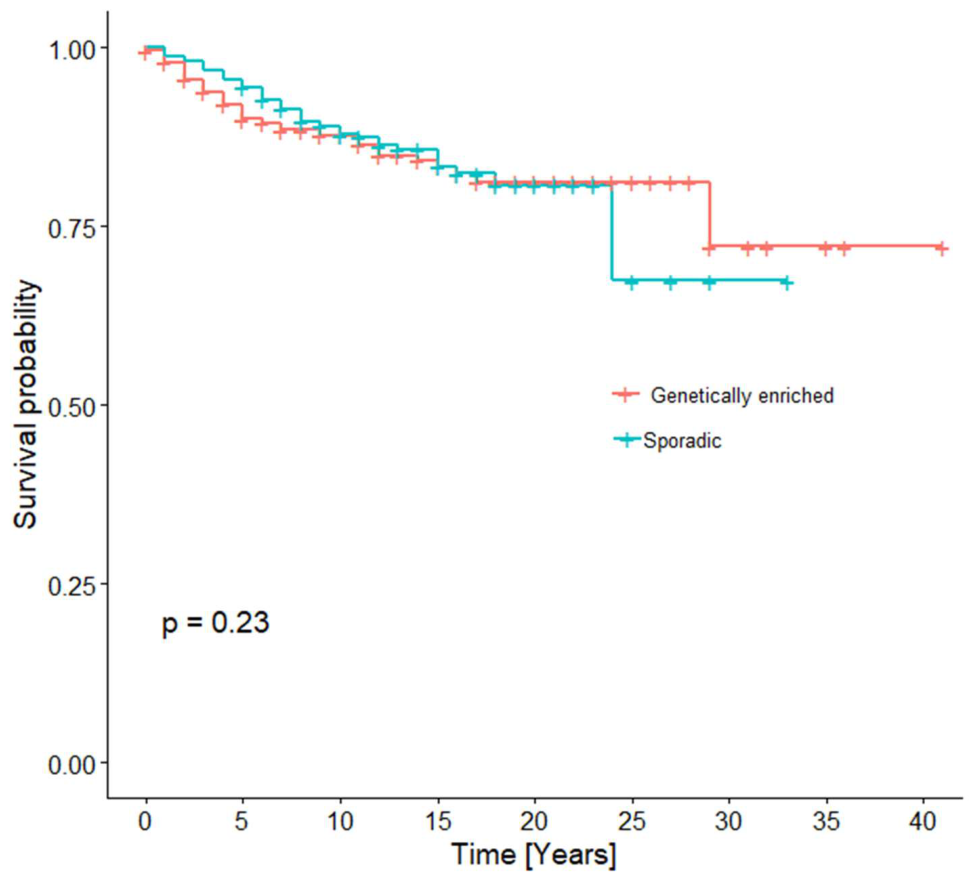
Figure 2.
Kaplan-Meier curves for DFS by sex in the genetically enriched cohort.
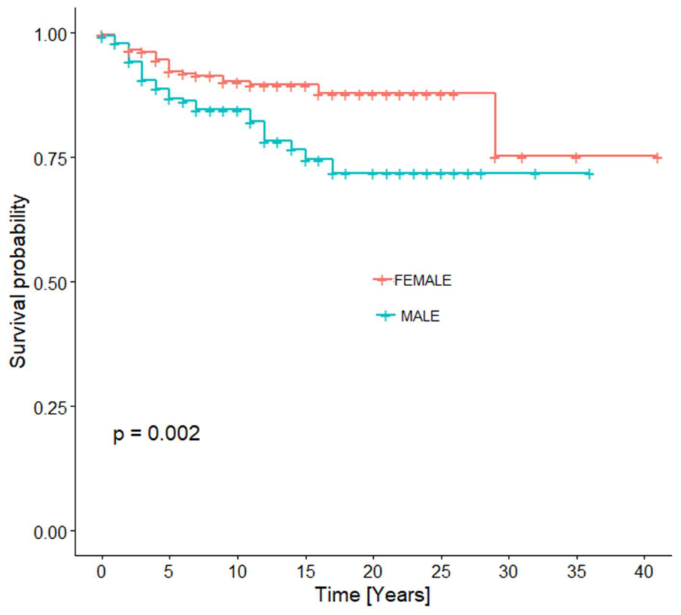

Table 1.
Comparison of clinicopathological characteristics between the two study cohorts.
| Parameter | Genetically enriched (N=796) | Sporadic (N=1078) | p-value*** |
| N (%) * | N (%) * | ||
| Histological type (N=1811) | <0.001 | ||
| ALM | 8(1.1) | 37(3.5) | |
| IN SITU | 153(20.2) | 160(15.2) | |
| LMM | 11(1.4) | 55(5.2) | |
| NM | 84(11.1) | 100(9.5) | |
| OTHER | 20(2.6) | 23(2.2) | |
| SSM | 483(63.6) | 677(64.4) | |
| Sex (N=1874) | 0.024 | ||
| Female | 452(56.8) | 554(51.4) | |
| Male | 344(43.2) | 524(48.6) | |
| History of malignancy (N=1831) | 56(7.2) | 144(13.6) | <0.001 |
| Breslow (N=1484) | 0.41 | ||
| <1 mm | 316(52.5) | 457(51.8) | |
| 1-2 mm | 146(24.3) | 244(27.7) | |
| 2-4 mm | 98(16.3) | 125(14.2) | |
| >4 mm | 42(7.0) | 56(6.3) | |
| Breslow median (mm) (IQR) | 0.90 (0.55-1.98) | 0.96(0.53-1.81) | 0.826 |
| Ulceration(N=1444) | 143(24.5) | 214(24.9) | 0.864 |
| Positive Lymph nodes (N=1874) | 94(11.8) | 120(11.1) | 0.65 |
| Locoregional recurrence (N=183) | 0.30 | ||
| Yes | 26(33.8) | 45(42.5) | |
| No | 51(66.2) | 61(57.5) | |
| Distant recurrence (N=183) | 0.051 | ||
| Yes | 56(72.7) | 61(57.5) | |
| No | 21(27.3) | 45(42.5) |
ALM: Acral lentiginous melanoma, LMM: Lentigo maligna melanoma, NM: Nodular melanoma, SSM: Superficial spreading melanoma. *Data are presented as N (column percentage). **Percentages were calculated out of the total number of informative cases per group. ***Statistically significant p-values are shown in bold.
Table 2.
a. Age at diagnosis across the melanoma study subgroups.
| Pairwise comparison | N | Median age, years (IQR) |
| Early-onset melanoma | 506 | 35 (31–38) |
| Familial melanoma | 184 | 47.5 (38–60.25) |
| Multiple primary melanoma | 106 | 54.5 (43–62) |
| Sporadic melanoma | 1078 | 57 (50–66) |
Table 2.
b. Pairwise comparisons of age at diagnosis among the four melanoma subgroups using Dunn’s test with Holm correction.
Table 2.
b. Pairwise comparisons of age at diagnosis among the four melanoma subgroups using Dunn’s test with Holm correction.
| Pairwise comparison | Holm-adjusted p-value |
| Early-onset vs Familial | <0.001 |
| Early-onset vs Multiple primary | <0.001 |
| Early-onset vs Sporadic | <0.001 |
| Familial vs Multiple primary | 0.014 |
| Familial vs Sporadic | <0.001 |
| Multiple primary vs Sporadic | 0.003 |
Table 3.
Distribution of previous cancer primary sites in patients with reported previous malignancy.
Table 3.
Distribution of previous cancer primary sites in patients with reported previous malignancy.
| Previous cancer site | Genetically enriched (N=56) | Sporadic (N=144) |
| N (%) | N (%) | |
| Non – melanoma skin cancer | 28(50) | 55(38.19) |
| Breast cancer | 4(7.14) | 16(11.12) |
| Gynecological cancer | 4(7.14) | 4(2.78) |
| Gastrointestinal cancer | 3(5.36) | 9(6.25) |
| Prostate cancer | 3(5.36) | 17(11.8) |
| Thyroid cancer | 6(10.72) | 14(9.73) |
| Other | 8(14.28) | 29(20.13) |
Table 4.
Five-, ten- and fifteen-year DFS rates by cohort.
| Genetically enriched | Sporadic | |
| 5-year DFS rate (95% CI) | 90% (87.5-92.5) | 94.5% (93-95.9) |
| 10-year DFS rate | 87.8% (84.5-90.7) | 87.8 (85.4-90.3) |
| 15-year DFS rate | 83.4% (79.4-87.6) | 83.4% (79.5-87.4) |
DFS: Disease-free Survival, CI: confidence interval.
Table 5.
Results of univariate Cox regression models for DFS in the genetically enriched cohort.
| Parameter | HR | 95% CI | p-value* |
| Age | 1.02 | 1.01-1.04 | 0.003 |
| Sex | |||
| Male | 2.01 | 1.27-3.16 | 0.003 |
| Female | [Reference] | ||
HR: Hazard Ratio, CI: confidence interval; *Statistically significant p-values are shown in bold, italics.
Table 6.
Results of univariate Cox regression models for DFS in the sporadic cohort.
| Parameter | HR | 95% CI | p-value* |
| Age | 1.03 | 1.01-1.05 | 0.004 |
| Sex | |||
| Male | 1.16 | 0.79-1.70 | 0.40 |
| Female | [Reference] | ||
HR: Hazard Ratio, CI: confidence interval; *Statistically significant p-values are shown in bold, italics.
Disclaimer/Publisher’s Note: The statements, opinions and data contained in all publications are solely those of the individual author(s) and contributor(s) and not of MDPI and/or the editor(s). MDPI and/or the editor(s) disclaim responsibility for any injury to people or property resulting from any ideas, methods, instructions or products referred to in the content. |
© 2026 by the authors. Licensee MDPI, Basel, Switzerland. This article is an open access article distributed under the terms and conditions of the Creative Commons Attribution (CC BY) license (http://creativecommons.org/licenses/by/4.0/).
Copyright: This open access article is published under a Creative Commons CC BY 4.0 license, which permit the free download, distribution, and reuse, provided that the author and preprint are cited in any reuse.