

Submitted:
04 September 2026
Posted:
04 September 2026
You are already at the latest version
Abstract
Pigmented lesions arising at sites of remote trauma can create diagnostic uncertainty when their clinical appearance overlaps with melanocytic neoplasms. A 64-year-old man presented with an asymptomatic 6-mm blue dermal nodule on the left fifth digit at the site of a penetrating pencil injury sustained during childhood. After remaining unchanged for decades, the lesion gradually enlarged over three years and produced mild nail plate deformity without periungual or subungual pigmentation. Cellular blue nevus, nodular melanoma, and graphite foreign-body granuloma were considered. Shave biopsy revealed a well-demarcated intradermal lesion containing abundant dark amorphous pigment and sclerosis. Higher-power examination demonstrated coarse granular black pigment with multinucleated giant cells and surrounding fibrosis, without melanocytic proliferation. These findings established the diagnosis of graphite foreign-body granuloma. The prolonged latency and recent enlargement illustrate how retained graphite may become clinically concerning many years after implantation. Because clinical features may overlap with melanoma and other pigmented tumors, remote trauma history can refine the differential diagnosis but cannot replace tissue examination. Histopathologic evaluation remains essential for definitive distinction of exogenous pigment from melanocytic proliferation.
Keywords:
graphite foreign-body granuloma
; graphite granuloma
; pencil injury
; foreign-body reaction
; pigmented lesion
; melanoma mimic
; dermatopathology
Introduction
Foreign material introduced into the skin can produce clinical findings long after the inciting event has been forgotten. The cutaneous response depends on the material’s size, composition, depth, anatomic site, and persistence, as well as the host inflammatory response. Pencil punctures are common minor injuries, particularly in childhood, and are often managed without medical evaluation. Nevertheless, retained pencil-core fragments may leave a permanent blue-gray mark or, less commonly, produce a delayed nodular foreign-body reaction. Published cases document latency periods measured in decades, indicating that apparent clinical quiescence does not necessarily imply that implanted material has been eliminated [1,2,3,4,5,6,7].
The term “pencil lead” is a historical misnomer. Contemporary pencil cores are composed principally of graphite blended with clay and binders rather than elemental lead [2,7]. Graphite is generally regarded as relatively inert, which helps explain why a puncture tattoo may remain unchanged for many years. However, mechanical fragmentation, gradual dispersion of particulate material, local tissue remodeling, or changes in immune recognition may eventually be accompanied by histiocytic inflammation, multinucleated giant cells, fibrosis, and clinically apparent enlargement [1,5,6,7,8]. The precise trigger for delayed growth is not established, and the rarity of reported cases limits conclusions regarding incidence and natural history.
A deeply situated dark pigment may appear blue because shorter wavelengths of visible light are preferentially scattered as light passes through the skin, a clinicopathologic phenomenon often described as the Tyndall effect. This optical appearance is not specific to graphite. It can occur with dermal melanin in blue nevi, melanoma, and metastatic melanoma, and with other exogenous pigments. Consequently, a homogeneous blue-black nodule cannot be classified reliably on color alone [8,9,10]. Dermoscopy improves assessment of pigmented lesions by revealing structures that are not visible to the unaided eye, but individual dermoscopic features overlap between benign and malignant diagnoses. In a meta-analysis, blue-white veil and irregular pigmentation were associated with melanoma, yet no single structure provided perfect sensitivity and specificity [9].
The diagnostic stakes are heightened on a digit and near the nail apparatus. Pigmented lesions in this location encompass melanocytic nevi, melanoma, hemorrhage, infection, vascular and adnexal tumors, squamous neoplasia, and exogenous pigment. Nail-unit melanoma is uncommon but is frequently challenging to recognize and sample. Contemporary reviews emphasize integration of clinical examination, dermoscopy, procedural information, and histopathology rather than reliance on any single feature [11,12,13,14,15]. Although the present lesion was adjacent to the nail plate rather than presenting as classic longitudinal melanonychia, its interval growth and associated nail deformation appropriately raised concern for a neoplasm.
Graphite granuloma can therefore create a two-sided diagnostic problem. Overconfidence in a history of pencil trauma may delay biopsy of melanoma, whereas failure to elicit remote trauma may lead to unnecessary alarm or extensive investigation of a benign foreign-body reaction. The safest use of history is as a means of refining the differential diagnosis. A changing pigmented lesion should still undergo tissue evaluation when malignancy cannot be excluded clinically.
We report a 64-year-old man with a 6-mm blue dermal nodule of the left fifth digit that enlarged decades after childhood pencil trauma. The case is notable for its prolonged latency, periungual location, mild nail plate deformity, and close clinical resemblance to a melanocytic tumor. We expand the clinicopathologic analysis to clarify the diagnostic workflow, histologic differential diagnosis, practical applications of the findings, and priorities for future study. The central lesson is that remote penetrating trauma is diagnostically valuable, but definitive distinction between graphite and melanocytic proliferation rests on representative histopathologic examination.
Materials and Methods
This single-patient case report was prepared from the clinical history, physical examination, dermoscopic assessment, procedural findings, and histopathologic material available during routine care. The report is descriptive and does not test an intervention or estimate disease frequency. Patient age, lesion location, size, symptoms, temporal evolution, relevant cancer history, and remembered trauma were recorded because these variables directly influence the differential diagnosis of an acral or periungual pigmented nodule. The clinical and dermoscopic images were selected to demonstrate the lesion’s morphology and relationship to the nail plate.
The lesion was evaluated by visual examination and dermoscopy. The assessment focused on symmetry, border definition, color distribution, surface characteristics, nail involvement, and the presence or absence of periungual or subungual pigmentation. Because interval enlargement could not be reconciled confidently with a benign diagnosis, tissue sampling was undertaken. A shave biopsy was performed for diagnostic evaluation. The procedural observation that the nodule was circumscribed and separated readily from adjacent tissue and the underlying nail matrix was documented as supportive context, but it was not treated as a substitute for microscopic evaluation.
Histopathologic interpretation was based on hematoxylin and eosin-stained sections examined at progressively higher magnification. Review addressed lesion circumscription, epidermal and dermal architecture, pigment quality and distribution, sclerosis, inflammatory response, multinucleated giant cells, and evidence of a melanocytic proliferation. The diagnosis required concordance among the remote pencil injury, the blue-black clinical appearance, and the microscopic identification of coarse exogenous pigment with a foreign-body giant-cell reaction and fibrosis [8,10,16]. Absence of a melanocytic proliferation in the examined specimen was essential to excluding the leading melanocytic mimics.
The literature discussion was organized around four clinically relevant domains: reported graphite and pencil-core granulomas, dermoscopic interpretation of blue lesions, differential diagnosis of acral and nail-adjacent pigmentation, and biopsy/histopathologic evaluation of suspected nail-unit melanoma. Recent reviews and diagnostic studies were included to place this rare case within current practice, while older case reports were retained because they provide the most direct evidence concerning long latency after pencil injury. This combination is necessary in a rare condition for which contemporary comparative studies are not available.
Case Presentation
A 64-year-old man presented for evaluation of an asymptomatic blue lesion on the left fifth digit. He recalled a penetrating graphite-pencil injury at the same site during childhood. A visible lesion remained stable and asymptomatic for several decades. During the three years preceding presentation, however, it gradually enlarged and subsequently stabilized. He denied pain, bleeding, drainage, or recent trauma. His medical history was negative for melanoma and nonmelanoma skin cancer.
Physical examination demonstrated a 6-mm, well-circumscribed blue dermal nodule adjacent to the nail plate, with mild associated nail plate deformity. There was no periungual extension of pigment and no subungual pigmentation. Dermoscopy showed a homogeneous, sharply circumscribed blue-black lesion (Figure 1). The remembered injury supported implanted graphite, but the lesion’s recent growth and periungual location prevented a purely clinical diagnosis. The principal differential diagnoses were cellular blue nevus, nodular melanoma, and graphite foreign-body granuloma; other considerations for a dark digital lesion include hemorrhage, vascular lesions, and nonmelanocytic tumors depending on the broader clinical context.
A shave biopsy was performed because the lesion had changed and melanoma could not be excluded. During sampling, the lesion appeared as a circumscribed dermal nodule that separated readily from the surrounding tissue and underlying nail matrix. This finding helped localize the process but did not determine its lineage. The specimen was submitted for routine histopathologic examination.
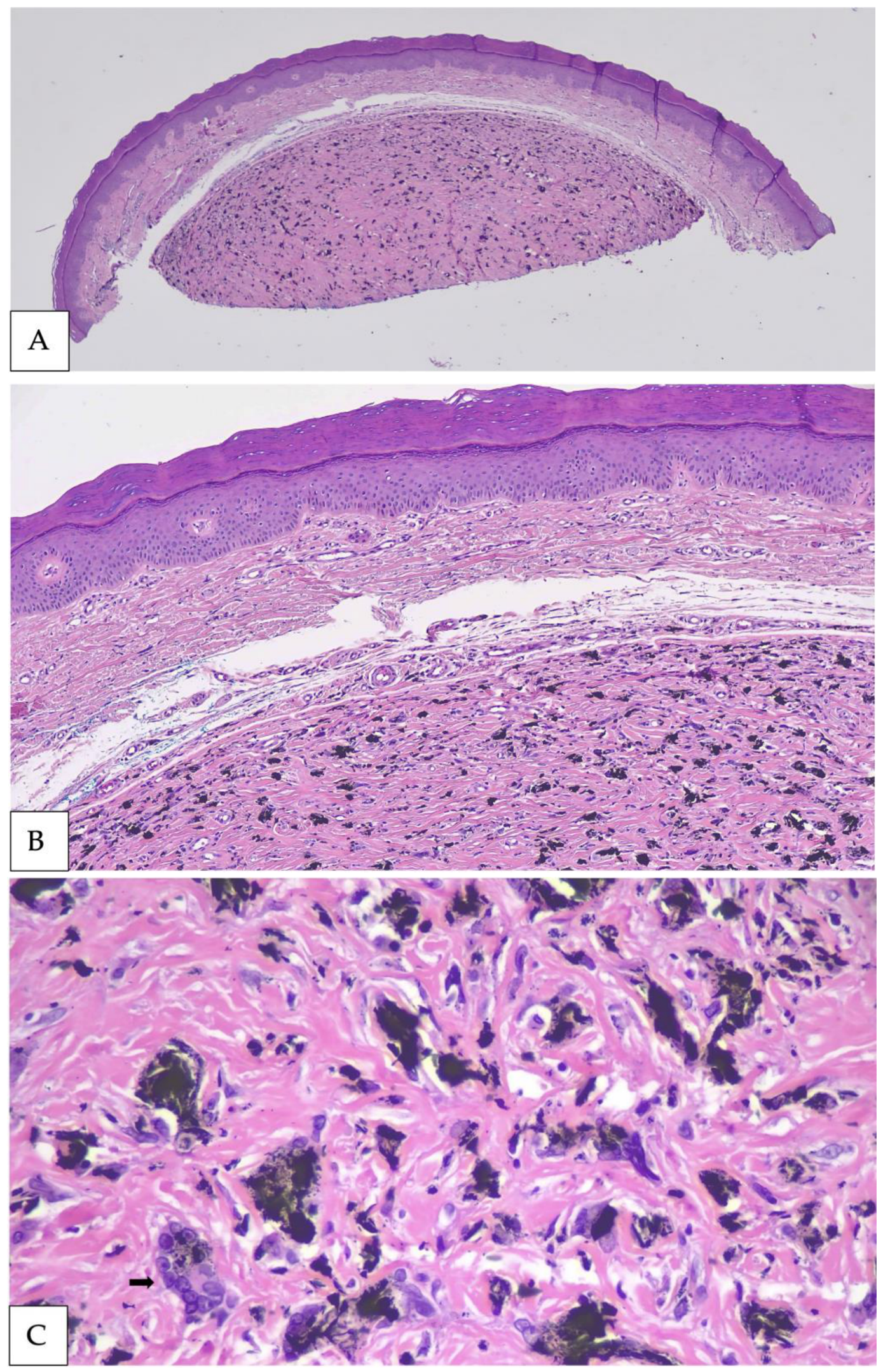
Histopathologic examination demonstrated a well-demarcated intradermal lesion with prominent sclerosis and abundant dark amorphous pigment (Figure 2A). At intermediate magnification, coarse black particulate material was dispersed throughout the fibrotic dermis (Figure 2B). High-power examination showed pigment associated with multinucleated foreign-body giant cells and surrounding fibrosis (Figure 2C). No melanocytic proliferation was identified in the examined sections.
The morphology was concordant with the history of remote pencil trauma and established the diagnosis of graphite foreign-body granuloma. In particular, the combination of coarse exogenous pigment, a granulomatous giant-cell response, and fibrosis explained both the blue clinical color and the firm dermal nodule. The absence of a melanocytic proliferation distinguished the lesion from blue nevus and melanoma.
Discussion
This case illustrates the diagnostic challenge posed by traumatic pencil-core implantation. Although the published literature consists largely of individual case reports, a consistent clinicopathologic pattern emerges: a remote puncture injury is followed by persistent blue-gray pigmentation, a prolonged clinically silent interval, and the later development or enlargement of a papule, nodule, or mass [1,2,3,4,5,6,7]. One recent report described a pencil-core granuloma that developed on a finger decades after injury [3], while another lesion was initially considered localized argyria because the history of trauma was unavailable [2]. Similar lesions have been reported on the forehead, palm, thigh, eyelid, and other sites [1,4,5,6,7], with latency periods exceeding [4,6]. Graphite granuloma should therefore be considered when evaluating a changing pigmented lesion at a site of possible prior pencil injury.
The mechanism underlying delayed clinical change remains incompletely understood. The relative chemical inertness of graphite permits prolonged tissue retention with little acute inflammation. Over time, a retained pencil core may fragment, allowing smaller particles to disperse among collagen bundles and within macrophages. Repetitive motion or minor trauma, particularly at mechanically active sites such as the digits, may contribute to fragmentation or tissue remodeling, although this mechanism has not been established experimentally. Persistent, nondegradable particles may induce macrophage aggregation and multinucleated giant-cell formation, followed by fibroblast activation and collagen deposition. This process produces the chronic granulomatous and sclerotic architecture observed histologically. Degradation, calcification, necrosis, and granulation tissue have also been reported [1,5,6]. In the present specimen, abundant pigment, multinucleated giant cells, and dense fibrosis were consistent with a chronic foreign-body reaction, without evidence of a melanocytic neoplasm.
The clinical color is an important but nonspecific clue. Blue coloration reflects the optical effect of pigment or other material located within the dermis and does not establish whether that material represents melanin, graphite, tattoo ink, hemosiderin, or another substance. The differential diagnosis of a blue lesion therefore includes common, cellular, and atypical blue nevi; melanoma; pigmented basal cell carcinoma; dermatofibroma; vascular lesions; and exogenous pigment [8]. Cutaneous melanoma metastases may also appear as structureless blue lesions on dermatoscopy and mimic blue nevi [16]. Thus, a homogeneous and well-circumscribed blue lesion may appear reassuring in the appropriate context, but these features cannot independently establish benignity.
Dermatoscopy remains useful for characterizing morphology, selecting an appropriate biopsy site, and documenting lesions that are observed over time. In this case, dermatoscopy demonstrated a homogeneous, well-circumscribed blue-black lesion but did not reveal a feature capable of excluding melanoma. This limitation is consistent with a systematic review and meta-analysis showing that melanoma-associated findings including blue-white veil, irregular pigmentation, shiny white structures, and peppering which have useful but incomplete diagnostic performance [9]. Nodular melanoma is a particular concern because it may be symmetric and relatively feature-poor despite rapid growth. For these reasons, the inclusion of nodular melanoma in the original clinical differential diagnosis was appropriate [10].
The lesion’s proximity to the nail apparatus added diagnostic complexity. It caused mild nail-plate deformation but lacked longitudinal melanonychia or periungual and subungual pigmentation. Although these findings reduced its resemblance to classic pigmented nail-unit melanoma, their absence did not exclude malignancy. Nail melanoma may present with nail dystrophy, splitting, bleeding, a mass, or amelanotic change rather than a classic pigmented band. Accordingly, interval enlargement or distortion of the nail apparatus warrants careful evaluation even when longitudinal melanonychia and Hutchinson sign are absent [11,12,13].
Biopsy technique should be selected according to the morphology and anatomic origin of the lesion. Suspected nail-matrix lesions producing longitudinal melanonychia require sampling of the pigment source while preserving sufficient architecture and minimizing permanent nail dystrophy. Incomplete or poorly oriented specimens may compromise interpretation, and longitudinal or tangential approaches may be preferable to small matrix punches in selected cases [12,13,14,15]. In contrast, the lesion in this case was a discrete dermal nodule adjacent to the nail rather than a pigmented band arising within the matrix. Shave biopsy provided the diagnostically relevant dermal tissue, and the nodule separated from the underlying matrix. The successful use of this technique should therefore not be generalized to lesions suspected of representing nail-matrix melanoma.
Histopathologic examination provides the decisive distinction between exogenous pigment and melanocytic disease. Graphite generally appears as coarse black particulate material that may be non-refractile or variably refractile and may lie freely in the dermis or within histiocytes. The associated foreign-body reaction can include epithelioid histiocytes, multinucleated giant cells, chronic inflammation, fibrosis, and, less commonly, necrosis or calcification [1,2,3,4,5,6,7]. In contrast, a blue nevus contains a dermal proliferation of dendritic or spindle melanocytes, melanophages, and variable fibrosis. Melanoma is characterized by an atypical melanocytic proliferation accompanied by architectural disorder, cytologic atypia, mitotic activity, or other site-appropriate malignant features. Interpretation of nail-unit specimens may be particularly challenging because trauma, inflammation, poor specimen orientation, and subtle melanocytic proliferations can obscure the diagnosis [14].
Routine hematoxylin and eosin examination, interpreted in conjunction with the clinical history, is generally sufficient when coarse pigment and a characteristic foreign-body reaction are present. If cellular lineage remains uncertain, melanocytic immunohistochemical markers may help demonstrate or exclude a melanocytic proliferation, although pigmented macrophages and technical background require careful interpretation. Polarization can identify some foreign materials, but a negative result does not exclude graphite. Elemental or spectroscopic methods, including energy-dispersive X-ray analysis, Raman spectroscopy, and micro–Fourier transform infrared spectroscopy, may help characterize carbonaceous material in analytically difficult cases. Such testing is not routinely necessary when morphology and clinicopathologic correlation provide a secure diagnosis.
This case has several practical clinical implications. First, evaluation of a blue or black nodule should include questions about remote puncture wounds, occupational exposures, tattoos, injections, and previous procedures. Patients may not volunteer a childhood pencil injury unless specifically asked, and some may not recall the event at all [2,7]. Second, a history of trauma should not provide false reassurance. It does not reliably distinguish a foreign-body granuloma from melanoma or exclude a coincidental malignancy. Interval growth or another concerning clinical change should therefore prompt appropriate evaluation despite a plausible traumatic explanation. Third, the pathology requisition should document the lesion’s color, location, duration, evolution, dermatoscopic appearance, and relevant trauma history. This information allows the dermatopathologist to consider an exogenous source of pigment while deliberately evaluating for a melanocytic proliferation.
The diagnosis also has implications for patient counseling. Once examination of a representative biopsy specimen confirms graphite granuloma and excludes malignancy, the patient can be reassured that the lesion represents retained foreign material rather than melanoma or lead poisoning. The colloquial term “pencil lead” may cause unnecessary anxiety; explaining that modern pencil cores contain graphite and clay clarifies the nature of the lesion. Subsequent management can be individualized according to symptoms, residual lesion, functional or nail impairment, cosmetic concern, and diagnostic certainty. Observation may be reasonable when the biopsy is adequate and the clinicopathologic findings are concordant, whereas complete removal may be considered for persistent symptoms, recurrent enlargement, nail impairment, or residual diagnostic uncertainty.
This report is limited by its description of a single patient and therefore cannot establish the frequency of pencil-core granulomas, identify predictors of delayed enlargement, or compare biopsy techniques. The chronology also depends partly on the patient’s recollection of a childhood injury and subsequent clinical change. Although the clinical and histopathologic findings were concordant, serial dermatoscopic imaging, chemical characterization of the pigment, and standardized long-term follow-up were unavailable. These limitations restrict conclusions regarding the lesion’s natural history, optimal biopsy approach, and risk of recurrence.
Additional systematic investigation could help address these uncertainties. A multicenter registry incorporating injury-to-presentation interval, anatomic site, symptoms, growth pattern, dermatoscopic findings, biopsy method, histologic reaction, treatment, and recurrence could determine whether digits and other mechanically active sites are disproportionately affected. It could also clarify whether delayed enlargement correlates with fragmentation, secondary trauma, or a particular inflammatory response. Correlation of routine histology with analytical methods such as Raman spectroscopy or energy-dispersive X-ray analysis could define the uncommon circumstances in which ancillary material identification adds diagnostic value. Noninvasive imaging techniques, including reflectance confocal microscopy and high-frequency ultrasound, could also be studied for their ability to identify a retained fragment or a characteristic pigment-fibrosis pattern before biopsy. Nevertheless, any diagnostic algorithm must maintain a low threshold for tissue sampling when melanoma remains clinically plausible.
Ultimately, this case demonstrates the importance of clinicopathologic correlation in the evaluation of a changing blue-black lesion. A remote pencil injury may suggest graphite implantation, but neither trauma history nor dermatoscopic appearance can exclude melanoma. Conversely, recognition of coarse exogenous pigment accompanied by a foreign-body giant-cell reaction can prevent misclassification as a melanocytic lesion. Specifically asking about remote puncture injuries and communicating that history to the dermatopathologist are therefore essential. Once an adequate biopsy confirms graphite granuloma and excludes malignancy, management can be tailored to the patient’s symptoms, nail impairment, residual lesion, and preferences.
Conclusions
Graphite foreign-body granuloma should be considered when a blue-black digital or periungual nodule occurs at the site of a remote pencil puncture, even when decades separate injury from enlargement. The prolonged latency in this case is consistent with the relative inertness of graphite and a delayed chronic foreign-body response. Clinically, however, the lesion’s recent growth and nail plate deformation appropriately raised concern for melanoma and other pigmented neoplasms.
The diagnosis rests on clinicopathologic concordance: a compatible traumatic history, a dermal pigmented nodule, and histology showing coarse black exogenous material with multinucleated giant cells and fibrosis but no melanocytic proliferation. Remote trauma should refine the differential diagnosis, not replace biopsy of a changing lesion. Careful history taking, representative sampling, communication with the dermatopathologist, and deliberate exclusion of melanocytic proliferation allow clinicians to recognize this benign melanoma mimic without compromising oncologic vigilance.
Author Contributions
Conceptualization, C.N. and T.B.; investigation, B.G., T.B. and W.C.; writing—original draft preparation, C.N.; writing—review and editing, C.N., B.G., T.B. and W.C.; visualization, C.N. and B.G.; supervision, T.B. All authors have read and agreed to the published version of the manuscript.
Funding
This research received no external funding.
Institutional Review Board Statement
Ethical review and approval were waived for this single-patient case report in accordance with institutional policy.
Informed Consent Statement
Written informed consent has been obtained from the patient to publish this paper and the accompanying clinical images.
Data Availability Statement
No new datasets were created or analyzed in this case report. Data supporting the findings are contained within the article, subject to patient privacy restrictions.
Conflicts of Interest
The authors declare no conflicts of interest.
Figure 1.
Figure Clinical and dermatoscopic presentation of graphite foreign-body granuloma. (A) Blue dermal nodule involving the left fifth digit with mild associated nail plate deformity. (B) Dermatoscopic view demonstrating a well-circumscribed blue-black lesion adjacent to the nail plate.
Figure 1.
Figure Clinical and dermatoscopic presentation of graphite foreign-body granuloma. (A) Blue dermal nodule involving the left fifth digit with mild associated nail plate deformity. (B) Dermatoscopic view demonstrating a well-circumscribed blue-black lesion adjacent to the nail plate.
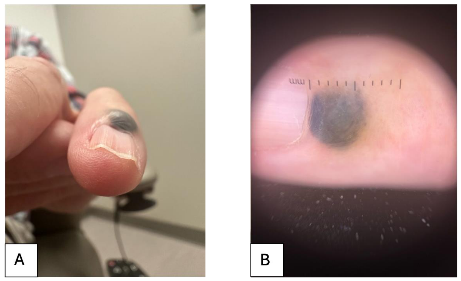
Figure 2.
Figure Histopathologic features of graphite foreign-body granuloma. (A) Low-power hematoxylin and eosin (H&E)-stained section demonstrating a well-circumscribed dermal lesion with prominent sclerosis and abundant dark pigment (×20). (B) Intermediate-power H&E-stained section demonstrating coarse black exogenous pigment dispersed throughout the fibrotic dermis (×100). (C) High-power H&E-stained section demonstrating coarse black pigment associated with a multinucleated foreign-body giant-cell reaction (arrow) (×400).
Figure 2.
Figure Histopathologic features of graphite foreign-body granuloma. (A) Low-power hematoxylin and eosin (H&E)-stained section demonstrating a well-circumscribed dermal lesion with prominent sclerosis and abundant dark pigment (×20). (B) Intermediate-power H&E-stained section demonstrating coarse black exogenous pigment dispersed throughout the fibrotic dermis (×100). (C) High-power H&E-stained section demonstrating coarse black pigment associated with a multinucleated foreign-body giant-cell reaction (arrow) (×400).
References
- Terasawa, N.; Kishimoto, S.; Kibe, Y.; Takenaka, H.; Yasuno, H. Graphite foreign body granuloma. Br. J. Dermatol. 1999, 141, 774–776. [Google Scholar] [CrossRef] [PubMed]
- Sharma, A. Graphite foreign body misdiagnosed as a blue naevus-like localised argyria. Clin. Cosmet. Investig. Dermatol. 2021, 14, 1253–1256. [Google Scholar] [CrossRef] [PubMed]
- Matsuoka, K.; Tanaka, R.; Nomura, T. Pencil-core granuloma. CMAJ 2022, 194, E14. [Google Scholar] [CrossRef] [PubMed]
- Bittencourt, M.J.S.; Dos Santos, J.E.B.; Barros Junior, J.N.S.; Xerfan, E.M.S. Pencil-core granuloma. An. Bras. Dermatol. 2017, 92, 578–579. [Google Scholar] [CrossRef] [PubMed]
- Shibuya, R.; Endo, Y.; Fujisawa, A.; Tanioka, M.; Miyachi, Y. Granuloma caused by carbon deposition in the dermis. Case Rep. Dermatol. Med. 2014, 2014, 686489. [Google Scholar] [CrossRef] [PubMed]
- Aswani, V.H.; Kim, S.L. Fifty-three years after a pencil puncture wound. Case Rep. Dermatol. 2015, 7, 303–305. [Google Scholar] [CrossRef] [PubMed]
- Phelps, P.O.; Carlson, J.A.; Meyer, D.R. Enlarging pigmented eyelid mass associated with remote pencil trauma. Ocul. Oncol. Pathol. 2017, 3, 310–312. [Google Scholar] [CrossRef] [PubMed]
- Longo, C.; Scope, A.; Lallas, A.; Zalaudek, I.; Moscarella, E.; Gardini, S.; Argenziano, G.; Pellacani, G. Blue lesions. Dermatol. Clin. 2013, 31, 637–647. [Google Scholar] [CrossRef] [PubMed]
- Williams, N.M.; Rojas, K.D.; Reynolds, J.M.; Kwon, D.; Shum-Tien, J.; Jaimes, N. Assessment of diagnostic accuracy of dermoscopic structures and patterns used in melanoma detection: A systematic review and meta-analysis. JAMA Dermatol. 2021, 157, 1078–1088. [Google Scholar] [CrossRef] [PubMed]
- Menzies, S.W.; Moloney, F.J.; Byth, K.; Avramidis, M.; Argenziano, G.; Zalaudek, I.; et al. Dermoscopic evaluation of nodular melanoma. JAMA Dermatol. 2013, 149, 699–709. [Google Scholar] [CrossRef] [PubMed]
- Darmawan, C.C.; Ohn, J.; Mun, J.H.; Kim, S.; Lim, Y.; Jo, S.J.; et al. Diagnosis and treatment of nail melanoma: A review of the clinicopathologic, dermoscopic, and genetic characteristics. J. Eur. Acad. Dermatol. Venereol. 2022, 36, 651–660. [Google Scholar] [CrossRef] [PubMed]
- Das De, S.; Tan, K.B.; Ho, S.A.J.E.; Athanasian, E.A.; Puhaindran, M.E. Pigmented nail lesions: When to observe, when to biopsy, when to widely excise, and when to amputate? J. Hand Surg. Am. 2022, 47, 988–997. [Google Scholar] [CrossRef] [PubMed]
- Conway, J.; Bellet, J.S.; Rubin, A.I.; Lipner, S.R. Adult and pediatric nail unit melanoma: Epidemiology, diagnosis, and treatment. Cells 2023, 12, 964. [Google Scholar] [CrossRef] [PubMed]
- Jiang, A.J.; Abbott, J.J.; Higgins, H.W.; Elenitsas, R.; Rubin, A.I. Clinicohistopathologic challenges and traps in the diagnosis of nail unit melanoma. J. Cutan. Pathol. 2023, 50, 580–590. [Google Scholar] [CrossRef] [PubMed]
- Dika, E.; Starace, M.; Alessandrini, A.; Patrizi, A.; Baraldi, C.; Misciali, C.; et al. The histopathologic evaluation of diagnostic procedures in nail melanoma. Dermatol. Pract. Concept. 2023, 13, e2023092. [Google Scholar] [CrossRef] [PubMed]
- Tiodorovic, D.; Stojkovic-Filipovic, J.; Marghoob, A.; Argenziano, G.; Puig, S.; Malvehy, J.; et al. Dermatoscopic patterns of cutaneous metastases: A multicentre cross-sectional study of the International Dermoscopy Society. J. Eur. Acad. Dermatol. Venereol. 2024, 38, 1432–1438. [Google Scholar] [CrossRef] [PubMed]
Disclaimer/Publisher’s Note: The statements, opinions and data contained in all publications are solely those of the individual author(s) and contributor(s) and not of MDPI and/or the editor(s). MDPI and/or the editor(s) disclaim responsibility for any injury to people or property resulting from any ideas, methods, instructions or products referred to in the content. |
© 2026 by the authors. Licensee MDPI, Basel, Switzerland. This article is an open access article distributed under the terms and conditions of the Creative Commons Attribution (CC BY) license (http://creativecommons.org/licenses/by/4.0/).
Copyright: This open access article is published under a Creative Commons CC BY 4.0 license, which permit the free download, distribution, and reuse, provided that the author and preprint are cited in any reuse.