

Submitted:
26 August 2026
Posted:
02 September 2026
You are already at the latest version
Abstract
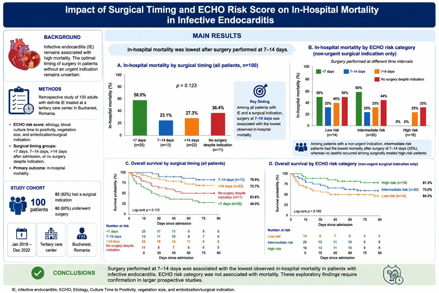
Introduction: Infective endocarditis (IE) remains associated with high mortality, and the optimal timing of surgery in patients without an urgent indication remains uncertain. We evaluated the association between surgical timing, in-hospital mortality, and a study-specific ECHO risk score. Methods: We retrospectively studied 100 adults with definite IE treated at a tertiary care center in Bucharest, Romania. The ECHO score incorporated etiology, blood culture time to positivity, vegetation size, and embolization or surgical indication. Surgical timing was categorized as < 7 days, 7–14 days, or >14 days after hospital admission. The primary outcome was in-hospital mortality. Results: Eighty-two patients had a surgical indication, and 60 underwent surgery. Mortality was lowest after surgery at 7–14 days (23.1%), compared with >14 days (27.3%), no surgery despite indication (36.4%), and < 7 days (56.0%) (p = 0.123). Among patients with a non-urgent surgical indication, intermediate-risk patients had the lowest mortality after surgery at 7–14 days (25%), whereas no deaths occurred among surgically treated high-risk patients. ECHO risk category was not associated with mortality (p = 0.165). Conclusions: Surgery at 7–14 days was associated with the lowest observed mortality. These exploratory findings require confirmation in larger prospective studies.
Keywords:
infective endocarditis
; surgery
; surgical timing
; risk assessment
; mortality
; survival
1. Background
Despite advances in diagnostic and therapeutic strategies, infective endocarditis (IE) remains a serious and potentially life-threatening condition characterized by high morbidity and mortality [1]. Contemporary data report in-hospital mortality rates of 20% to 30%, reflecting the disease’s complexity and frequent complications, such as heart failure, systemic embolization, and uncontrolled infection [2,3].
Early identification of high-risk patients and timely intervention are essential for improving outcomes [4]. Large vegetations and Staphylococcus aureus infections have been associated with an increased risk of embolism and worse outcomes [5]. Meanwhile, prosthetic valve- and device-related endocarditis present additional therapeutic challenges [6,7].
Current strategies combine antimicrobial therapy and surgery, with surgery indicated in 50–70% of cases. However, determining the optimal timing of surgery remains challenging [8]. While urgent surgery is recommended in cases of hemodynamic instability, uncontrolled infection, or high embolic risk, the management of non-urgent indications is less clear.
Current ESC 2023 guidelines emphasize that surgical timing in infective endocarditis should be individualized and classified as emergent, urgent, or elective, although the optimal timing in patients with non-urgent indication remains uncertain [9,10].
Previous studies have shown that early surgical intervention in infective endocarditis can reduce embolic events and improve clinical outcomes, particularly in patients with large vegetations or high-risk features, although its impact on overall mortality remains variable [11,12,13].
Risk stratification tools have been proposed to guide clinical decision-making in IE. These models incorporate parameters associated with adverse outcomes [14,15,16].
We developed the ECHO score, a study-specific composite risk stratification tool integrating etiology, blood culture time to positivity, vegetation size, and embolization or surgical indication. While such tools aim to identify patients at increased risk, their ability to predict outcomes and guide therapeutic strategies in real-world settings remains uncertain.
This study evaluated the association between surgical timing and in-hospital mortality and survival in patients with infective endocarditis. It also assessed the prognostic value of the ECHO score. We hypothesized that surgical timing may be more informative than baseline risk in patients without an urgent surgical indication.
2. Methods
2.1. Study Design and Setting
We conducted a retrospective observational cohort study at a university-affiliated tertiary care hospital in Bucharest, Romania. We evaluated IE etiology and management, including surgical indication and timing, together with in-hospital and one-year outcomes.
The study adhered to the Strengthening the Reporting of Observational Studies in Epidemiology (STROBE) guidelines.
2.2. Study Population
The study included 100 patients hospitalized with IE in a Bucharest Tertiary Care hospital. Diagnosis was established according to the modified Duke criteria.
2.3. Data Collection
Data were collected retrospectively from hospital records, including etiology, blood culture time to positivity, vegetation size, embolization, surgical indication and timing, and clinical outcomes. The ECHO score is a study-specific composite risk stratification tool in which one point was assigned for each of the following: (E) aggressive etiology (Staphylococcus spp. or fungal infection), (C) blood culture positivity within 24 hours, (H) vegetation >10 mm, and (O) embolization or a surgical indication. The total score ranged from 0 to 4, with higher scores indicating greater clinical risk. Patients were classified as low risk (0–1), intermediate risk (2), or high risk (3–4).
Analyses of surgical timing were restricted to patients with a documented surgical indication. Timing was calculated from hospital admission to surgery and categorized as <7 days, 7–14 days, or >14 days. Patients with an indication who did not undergo surgery were classified as “no surgery despite indication.” Exploratory risk-stratified analyses were restricted to patients with a non-urgent surgical indication.
The primary outcome of the study was in-hospital mortality, analyzed in relation to surgical timing.
2.4. Statistical Analysis
Statistical analysis was performed using standard descriptive and comparative methods. Continuous variables are presented as mean ± standard deviation, and categorical variables as frequencies and percentages.
Comparisons between categorical groups were performed using the chi-square test. Because mortality was binary, the association between the continuous ECHO score and mortality was summarized using a point-biserial correlation coefficient. Survival curves were estimated using the Kaplan–Meier method.
Kaplan–Meier analyses used time from hospital admission to in-hospital death, with discharge treated as censoring. Comparisons between groups were evaluated descriptively because of the small subgroup sizes. A two-sided p-value <0.05 was considered statistically significant for formal group comparisons.
Statistical analyses were performed using SPSS Statistics version 26.0 (IBM Corp., Armonk, NY, USA). Figures were generated using Python version 3.10, utilizing the Matplotlib and Seaborn libraries for data visualization.
2.5. Ethics
The study was approved by the Clinical Emergency Hospital Prof. Dr. Agrippa Ionescu Ethics Committee. Informed consent was waived due to the study’s retrospective nature. All data were anonymized and analyzed according to the Declaration of Helsinki.
3. Results
3.1. Baseline Characteristics
A total of 100 patients with definite infective endocarditis were included in the study. The mean ECHO score was 2.22 ± 0.88. According to the predefined risk stratification, 22 patients (22.0%) were classified as low risk (ECHO score 0–1), 42 (42.0%) as intermediate risk (score 2), and 36 (36.0%) as high risk (score 3–4).
Staphylococcus spp.were identified in 24.0% of patients, whereas Candida spp. accounted for 4.0%; all remaining cases were caused by other microorganisms. Blood cultures became positive within 24 hours in 51.0% of patients, vegetations larger than 10 mm were present in 49.0%, and embolic events occurred in 57.0%.
A surgical indication was established in 82 patients (82.0%), including 51 (51.0%) with a non-urgent indication and 31 (31.0%) requiring urgent surgery. Among patients with a surgical indication, 60 (73.2%) underwent surgery: 25 (30.5%) at <7 days, 13 (15.9%) at 7–14 days, and 22 (26.8%) at >14 days after admission. Twenty-two patients (26.8%) did not undergo surgery despite indication. Overall in-hospital mortality was 37.0%. At 1 year, 62 patients (62.0% of the full cohort) were alive. Mean length of stay was 39.1 ± 22.7 days (Table 1).
3.2. Surgical Timing and Clinical Outcomes
Among patients with a surgical indication, mortality varied according to surgical timing. The lowest observed mortality was in patients undergoing surgery at 7–14 days after admission (23.1%), followed by surgery at >14 days (27.3%), no surgery despite indication (36.4%), and surgery at <7 days (56.0%) (Table 2).
The overall difference between timing groups was not statistically significant (p = 0.123); therefore, the observed pattern should be interpreted as exploratory (Figure 1D).
3.3. Risk Stratification
Mortality was 31.8% in the low-risk group, 47.6% in the intermediate-risk group, and 27.8% in the high-risk group (Table 2; Figure 1C). The association between ECHO risk category and mortality was not statistically significant (p = 0.165). The continuous ECHO score showed a negligible correlation with mortality (r = −0.05).
Mortality was also similar across native valve, prosthetic valve, and device-related infective endocarditis, with no significant differences observed between valve types (p = 0.89).
3.4. Exploratory Analyses by Surgical Timing and ECHO Risk
Exploratory analyses combining surgical timing and ECHO risk category suggested distinct outcome patterns (Figure 1).
Among high-risk patients (ECHO score 3–4) with a non-urgent surgical indication, no deaths occurred among patients who underwent surgery, irrespective of timing, whereas mortality was 22% among patients managed without surgery (Figure 1A).
Among intermediate-risk patients (ECHO score = 2) with a non-urgent surgical indication, mortality was lowest after surgery at 7–14 days (25%); higher mortality was observed after surgery at <7 days or >14 days and with no surgery despite indication (Figure 1B).
Overall mortality according to the three predefined ECHO risk categories is shown in Figure 1C, and mortality among all patients with a surgical indication according to treatment timing is shown in Figure 1D.
Four-panel figure showing in-hospital mortality according to surgical timing and ECHO risk. (A) High-risk patients (ECHO score 3–4) with a non-urgent surgical indication. (B) Intermediate-risk patients (ECHO score = 2) with a non-urgent surgical indication. (C) Mortality in the overall cohort according to the predefined ECHO categories: low (0–1), intermediate (2), and high (3–4). (D) Mortality among patients with a documented surgical indication according to timing: <7 days, 7–14 days, >14 days, or no surgery despite indication. Percentages are descriptive; the overall timing comparison was not statistically significant (p = 0.123).
3.5. Survival Analysis in Patients with Non-Urgent Surgical Indications
Kaplan–Meier analysis was performed in patients with a non-urgent surgical indication (Figure 2). Survival curves were examined separately for intermediate- and high-risk patients and according to surgical timing.
Among intermediate-risk patients, surgery at 7–14 days was associated with the highest estimated in-hospital survival probability, whereas surgery at <7 days or >14 days and no surgery despite indication showed less favorable curves.
Among high-risk patients, surgically treated patients showed higher estimated in-hospital survival than patients managed without surgery.
Within this subgroup, the association between the continuous ECHO score and survival was weak (r = 0.16). Because subgroup sizes were small and comparisons were descriptive, these findings should be interpreted cautiously.
Kaplan–Meier curves of time from hospital admission to in-hospital death, with discharge treated as censoring, stratified by surgical timing: <7 days (green), 7–14 days (blue), >14 days (orange), and no surgery despite indication (red). The left panel shows high-risk patients (ECHO score 3–4), and the right panel shows intermediate-risk patients (ECHO score = 2). Curves are descriptive because of the limited subgroup sizes.
3.6. Overall In-Hospital Survival According to Surgical Timing
Kaplan–Meier analysis among the 82 patients with a documented surgical indication showed separation of the in-hospital survival curves according to surgical timing (Figure 3). The 7–14-day group had the most favorable estimated survival, whereas the <7-day group had the least favorable curve. Patients who did not undergo surgery despite indication also had lower estimated survival than those treated at 7–14 days.
Because the timing-group comparison was not statistically significant and the analysis was vulnerable to confounding by indication, these findings are exploratory and do not establish a causal benefit or an optimal surgical window.
Kaplan–Meier curves of time from hospital admission to in-hospital death among patients with a documented surgical indication, stratified as surgery at <7 days, 7–14 days, or >14 days, and no surgery despite indication. The 7–14-day group had the most favorable estimated survival curve. Comparisons are descriptive and should be interpreted in light of small group sizes and potential confounding by indication.
4. Discussion
This study evaluated the association of surgical timing and ECHO risk stratification with outcomes in patients with infective endocarditis. The principal observation was that, among patients with a surgical indication, mortality was lowest in those treated at 7–14 days; however, the difference between timing groups was not statistically significant.
Mortality was lowest among patients who underwent surgery at 7–14 days after hospital admission. Higher mortality in the <7-day group likely reflects, at least in part, the greater severity of illness among patients selected for earlier intervention.
The observed pattern suggests a possible treatment window for selected patients with non-urgent indications, but it does not establish an optimal interval. Initial stabilization and targeted antimicrobial therapy may be beneficial in some patients, while excessive delay may allow disease progression or complications [2,8,9,17].
Notably, early surgery was associated with the highest mortality rate. This likely reflects confounding by indication, as patients requiring urgent intervention may have had more severe disease at presentation. Similar observations have been reported in large registries, where early surgery is often performed in the sickest patients, making direct comparisons challenging [18,19,20]. Therefore, the observed worse outcomes in this group should not be interpreted as a detrimental effect of early surgery per se, but rather as a marker of disease severity.
The ECHO score showed a negligible correlation with mortality in the overall cohort (r = −0.05) and a weak association with survival in patients with a non-urgent surgical indication (r = 0.16). Although the score incorporates clinically relevant variables, these findings do not support its use as a stand-alone prognostic tool in this cohort.
These findings suggest that, while the ECHO score incorporates clinically relevant variables—such as etiology, blood culture kinetics, vegetation size, and embolization—it may not adequately capture the complexity of disease evolution or the dynamic factors influencing outcomes in IE.
Exploratory subgroup analyses suggested that the association between surgical timing and mortality may differ according to ECHO risk category. Among intermediate-risk patients, the lowest observed mortality was in the 7–14-day group.
Among high-risk patients with a non-urgent indication, no deaths occurred in surgically treated patients, whereas mortality was observed in patients managed without surgery. Because the subgroup was small and treatment allocation was non-random, this finding should not be interpreted as proof of a treatment effect.
Kaplan–Meier curves showed the most favorable estimated in-hospital survival in the 7–14-day group. The poorer curve in patients treated at <7 days is likely influenced by confounding by indication and greater baseline severity.
Differences between timing groups appeared more clearly in the exploratory non-urgent subgroup, but these descriptive findings require confirmation in larger cohorts.
Patients with prosthetic valve and device-related endocarditis tended to have more complex clinical courses compared to those with native valve involvement. Although detailed statistical comparisons were limited, this observation is consistent with existing literature, which describes higher complication rates and therapeutic challenges in these subgroups [6,7,21,22]. Despite this, the absence of an effect of valve type on outcome suggests that prognosis is primarily driven by clinical status and treatment strategy rather than anatomical substrate.
No multivariable model was performed in the present analysis; therefore, independent predictors of mortality could not be established.
The findings have several potential clinical implications, but they should be interpreted as hypothesis-generating.
In patients with non-urgent indications, surgical timing should be individualized by the multidisciplinary Endocarditis Team.
The 7–14-day interval was associated with the lowest observed mortality, but an optimal timing window cannot be established from these data.
Risk scores alone may be insufficient to guide management decisions.
Greater emphasis should be placed on individualized decision-making and dynamic clinical assessment.
Overall, the findings suggest that treatment timing and dynamic clinical assessment should be considered alongside baseline risk stratification. The ECHO score alone was insufficient to discriminate mortality risk in this cohort.
4.1. Study Limitations
This study has several limitations. First, its retrospective design introduces potential selection and information bias. Second, the sample size may have limited the study’s statistical power. Third, the observed association between early surgery and higher mortality is likely influenced by confounding by indication. Additionally, the ECHO score is a study-specific tool that has not been externally validated, limiting its generalizability.
4.2. Future Directions
Additional prospective, multicenter studies are necessary to validate these findings and better define optimal timing strategies for infective endocarditis. Combining dynamic clinical variables and advanced risk models may improve patient stratification and guide therapeutic decisions
5. Conclusion
Surgery performed 7–14 days after admission was associated with the lowest observed mortality among patients with a surgical indication, but differences between timing groups were not statistically significant. These exploratory findings do not establish an optimal surgical window and should be validated in prospective, multicenter studies. Clinical decisions should integrate surgical urgency, dynamic disease severity, and multidisciplinary assessment rather than rely on a baseline risk score alone.
Funding
Publication of this paper was supported by the University of Medicine and Pharmacy Carol Davila, through the institutional program Publish not Perish.
Institutional Review Board Statement
The study was conducted in accordance with the Declaration of Helsinki, and approved by the Institutional Ethics Committee of Clinical Emergency Hospital Prof. Dr. Agrippa Ionescu (750262/ 21 Nov 2023).
Informed Consent Statement
The requirement for informed patient consent was waived by the Ethics Committee due to the retrospective observational design of the study, which involved the analysis of anonymized routinely collected clinical data without any intervention or direct patient contact.
Data Availability Statement
The data presented in this study are not publicly available due to patient privacy and ethical restrictions. De-identified data may be made available from the corresponding author upon reasonable request and with permission from the Institutional Ethics Committee, in accordance with institutional policies and applicable data protection regulation.
Transparency declaration
The authors declare no conflicts of interest. During the preparation of this manuscript, the authors used AI-assisted language tools to enhance the clarity and readability of the text. These tools were used solely for editorial purposes and did not contribute to the design of the study, the collection or analysis of the data, the interpretation of the findings, or the drawing of scientific conclusions. The authors reviewed and edited all AI-generated suggestions, and take full responsibility for the content of the manuscript.
References
- Li M, Kim JB, Sastry BKS, Chen M. Infective endocarditis. The Lancet [Internet]. Elsevier; 2024 [cited 2026 Mar 31];404:377–92. [CrossRef]
- Patel SK, Hassan SMA, Côté M, Leis B, Yanagawa B. Current trends and challenges in infective endocarditis. Curr Opin Cardiol. Lippincott Williams and Wilkins; 2025;40:75–84. [CrossRef]
- Menon T. Uncommon pathogens causing infective endocarditis. Indian J Thorac Cardiovasc Surg [Internet]. Springer; 2024 [cited 2026 Mar 30];40:8–15. [CrossRef]
- Tzoumas A, Sagris M, Xenos D, Ntoumaziou A, Kyriakoulis I, Kakargias F, et al. Epidemiological Profile and Mortality of Infective Endocarditis Over the Past Decade: A Systematic Review and Meta-Analysis of 133 Studies. American Journal of Cardiology [Internet]. Elsevier Inc.; 2025 [cited 2026 Mar 30];244:67–88. [CrossRef]
- Santos-Patarroyo SD, Quintero-Martinez JA, Lahr BD, Chesdachai S, Abu Saleh O, Michelena HI, et al. Correlation Between Blood Culture Time to Positivity and Vegetation Size in Staphylococcus aureus Infective Endocarditis. Antibiotics 2025, Vol 14, Page 456 [Internet]. Multidisciplinary Digital Publishing Institute; 2025 [cited 2026 Mar 30];14:456. [CrossRef]
- Hutt E, Canosa FJM, Unai S, Jaber WA. Manifestations of Prosthetic Valve Endocarditis: Lessons From Multimodality Imaging and Pathological Correlation. Circ Cardiovasc Imaging [Internet]. Lippincott Williams & WilkinsHagerstown, MD; 2024 [cited 2026 Mar 30];17:E016435. [CrossRef]
- Baddour LM, Esquer Garrigos Z, Rizwan Sohail M, Havers-Borgersen E, Krahn AD, Chu VH, et al. Update on Cardiovascular Implantable Electronic Device Infections and Their Prevention, Diagnosis, and Management: A Scientific Statement From the American Heart Association. Circulation [Internet]. Lippincott Williams & WilkinsHagerstown, MD; 2024 [cited 2026 Mar 30];149:E201–16. [CrossRef]
- Bishev D, DiCaro M V., Chakraborty S, Stanger GT, Ho C, Tak T. Infective Endocarditis and Complications; Surgical Indications and Management: An Integrative Review. Journal of Personalized Medicine 2026, Vol 16, Page 103 [Internet]. Multidisciplinary Digital Publishing Institute; 2026 [cited 2026 Mar 30];16:103. [CrossRef]
- Delgado V, Ajmone Marsan N, De Waha S, Bonaros N, Brida M, Burri H, et al. 2023 ESC Guidelines for the management of endocarditis. Eur Heart J [Internet]. Eur Heart J; 2023 [cited 2026 Mar 31];44:3948–4042. [CrossRef]
- Fowler VG, Durack DT, Selton-Suty C, Athan E, Bayer AS, Chamis AL, et al. The 2023 Duke-International Society for Cardiovascular Infectious Diseases Criteria for Infective Endocarditis: Updating the Modified Duke Criteria. [cited 2026 Mar 31]. [CrossRef]
- Sohail MR, Martin KR, Wilson WR, Baddour LM, Harmsen WS, Steckelberg JM. Medical versus surgical management of Staphylococcus aureus prosthetic valve endocarditis. American Journal of Medicine. Elsevier Inc.; 2006;119:147–54. [CrossRef]
- D’Alonzo M, Di Bacco L, Fiore A, Baudo M, Zanin F, Baldelli C, et al. The Dilemma of Surgical Timing in Acute Aortic Valve Endocarditis: Does Early Surgery Improve Risks or Prognosis? J Cardiovasc Dev Dis [Internet]. J Cardiovasc Dev Dis; 2025 [cited 2026 Mar 30];12. [CrossRef]
- Arregle F, Martel H, Philip M, Gouriet F, Casalta JP, Riberi A, et al. Infective endocarditis with neurological complications: Delaying cardiac surgery is associated with worse outcome. Arch Cardiovasc Dis [Internet]. Elsevier Masson; 2021 [cited 2026 Mar 22];114:527–36. [CrossRef]
- Üstünışık ÇT, Duman ZM, Timur B, Aksu T, İyigün T, Göde S, et al. Early Mortality Predictors in İnfective Endocarditis Patients: A Single-Center Surgical Experience. Braz J Cardiovasc Surg [Internet]. Sociedade Brasileira de Cirurgia Cardiovascular; 2022 [cited 2026 Apr 4];37:829. [CrossRef]
- Suwa Y, Miyasaka Y, Taniguchi N, Harada S, Nakai E, Shiojima I. Predictors of in-hospital mortality in patients with infective endocarditis. Acta Cardiol [Internet]. Acta Cardiol; 2021 [cited 2026 Apr 4];76:642–9. [CrossRef]
- Palraj BR, Baddour LM, Hess EP, Steckelberg JM, Wilson WR, Lahr BD, et al. Predicting Risk of Endocarditis Using a Clinical Tool (PREDICT): Scoring System to Guide Use of Echocardiography in the Management of Staphylococcus aureus Bacteremia. Clin Infect Dis [Internet]. Oxford University Press; 2015 [cited 2026 Apr 4];61:18. [CrossRef]
- Lau L, Baddour L, Fernández Hidalgo N, Brothers TD, Kong WKF, Borger MA, et al. Infective endocarditis: it takes a team. Eur Heart J [Internet]. Eur Heart J; 2025 [cited 2026 Mar 30];46:2275–88. [CrossRef]
- Benedetto U, Spadaccio C, Gentile F, Moon MR, Nappi F. A narrative review of early surgery versus conventional treatment for infective endocarditis: do we have an answer? Ann Transl Med [Internet]. AME Publishing Company; 2020 [cited 2026 Apr 4];8:1626–1626. [CrossRef]
- Musleh R, Schlattmann P, Caldonazo T, Kirov H, Witte OW, Doenst T, et al. Surgical Timing in Patients With Infective Endocarditis and With Intracranial Hemorrhage: A Systematic Review and Meta-Analysis. J Am Heart Assoc [Internet]. American Heart Association Inc.; 2022 [cited 2026 Apr 4];11:24401. [CrossRef]
- Narayanan MA, Haddad TM, Kalil AC, Kanmanthareddy A, Suri RM, Mansour G, et al. Early versus late surgical intervention or medical management for infective endocarditis: A systematic review and meta-analysis. Heart. BMJ Publishing Group; 2016;102:950–7. [CrossRef]
- Raad II, Hanna HA. Intravascular Catheter-Related Infections: New Horizons and Recent Advances. Arch Intern Med [Internet]. American Medical Association; 2002 [cited 2026 Mar 30];162:871–8. [CrossRef]
- Hayley B, Leung Chan K. Infectious Complications in Infective Endocarditis. Infective Endocarditis: Epidemiology, Diagnosis, Imaging, Therapy, and Prevention [Internet]. Springer, Cham; 2016 [cited 2026 Mar 30];123–36. [CrossRef]
Figure 1.
Association of ECHO risk category and surgical timing with in-hospital mortality.
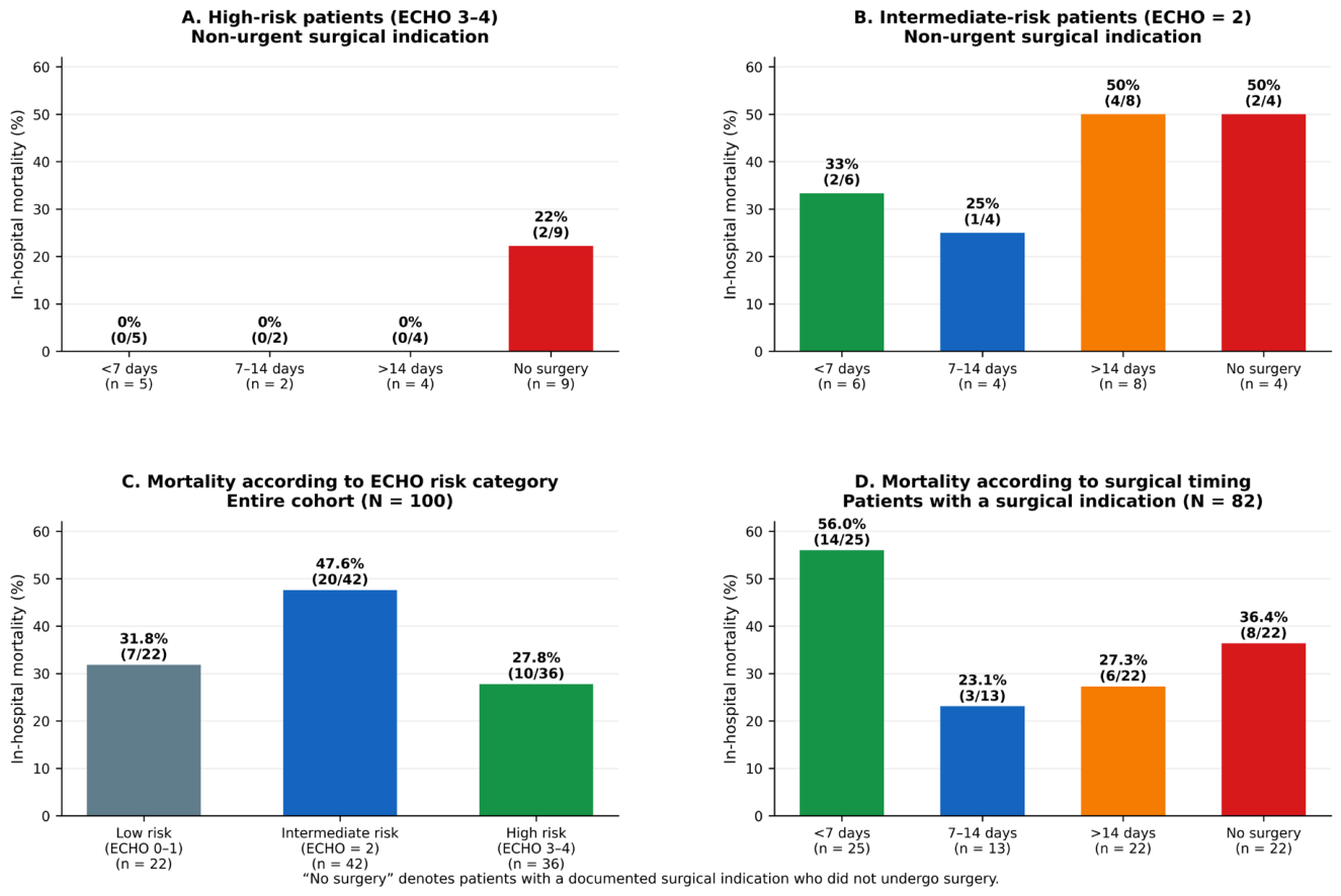
Figure 2.
Kaplan–Meier curves according to surgical timing in high- and intermediate-risk infective endocarditis with a non-urgent surgical indication.
Figure 2.
Kaplan–Meier curves according to surgical timing in high- and intermediate-risk infective endocarditis with a non-urgent surgical indication.
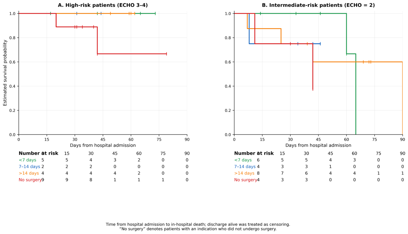
Figure 3.
Kaplan–Meier in-hospital survival according to surgical timing among patients with a surgical indication.
Figure 3.
Kaplan–Meier in-hospital survival according to surgical timing among patients with a surgical indication.
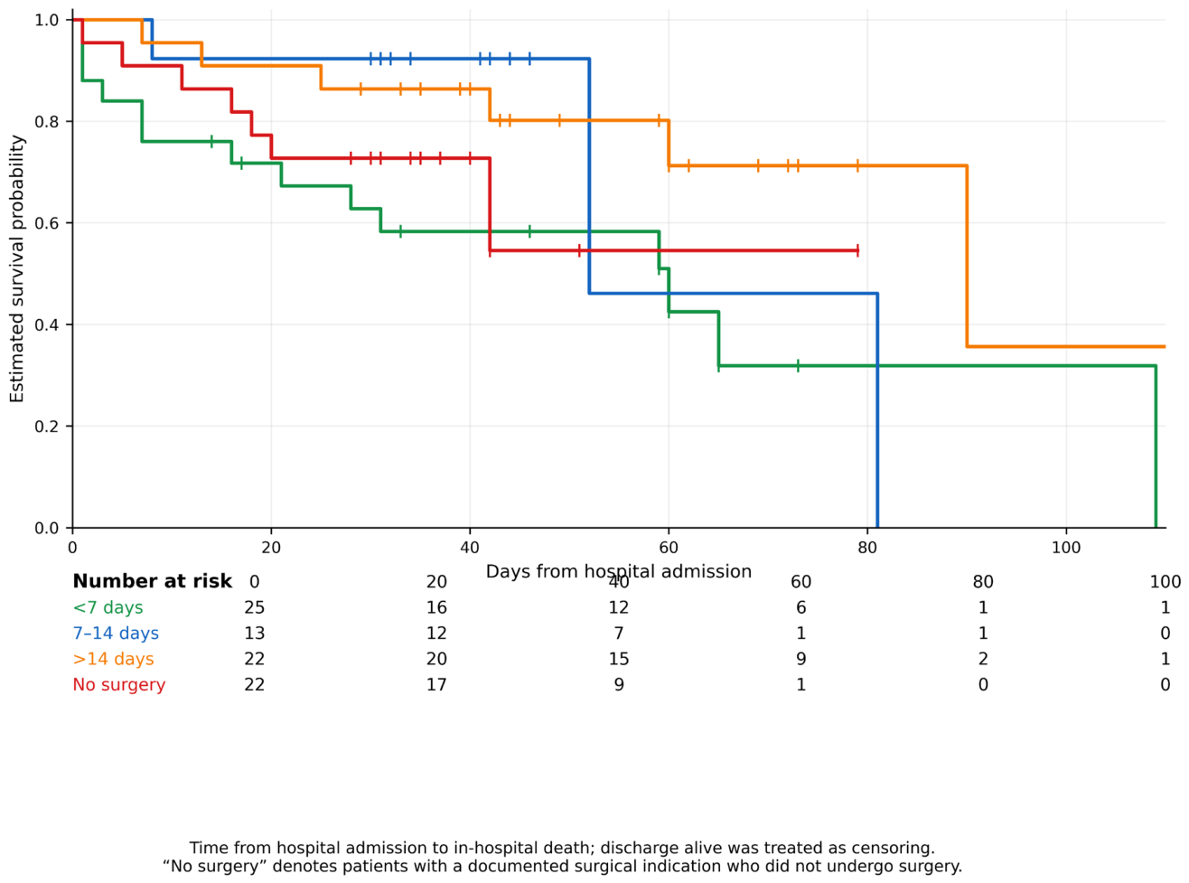

Table 1.
Baseline Clinical and Echocardiographic Characteristics.
| Characteristic | Value |
|---|---|
| Number of patients | 100 |
| ECHO score (mean ± SD) | 2.22 ± 0.88 |
| Low risk (0–1) | 22 (22.0%) |
| Intermediate (2) | 42 (42.0%) |
| High (3–4) | 36 (36.0%) |
| Staphylococcus spp. | 24 (24.0%) |
| Candida spp. | 4 (4.0%) |
| Other etiology | 72 (72.0%) |
| Blood culture ≤24 h | 51 (51.0%) |
| Vegetation >10 mm | 49 (49.0%) |
| Embolization | 57 (57.0%) |
| Any surgical indication | 82 (82.0%) |
| Non-urgent indication | 51 (51.0%) |
| Urgent indication | 31 (31.0%) |
| No indication | 18 (18.0%) |
| Surgery performed (among indicated) | 60 (73.2%) |
| <7 days (among indicated) | 25 (30.5%) |
| 7–14 days (among indicated) | 13 (15.9%) |
| >14 days (among indicated) | 22 (26.8%) |
| No surgery despite indication | 22 (26.8%) |
| Alive | 63 (63.0%) |
| Death | 37 (37.0%) |
| Relapse | 4 (4.0%) |
| 1-year survival | 62 (62.0%) |
| Length of stay (days) | 39.1 ± 22.7 |
Values are n (%) or mean ± SD. Surgical timing is measured from hospital admission. “No surgery despite indication” refers only to patients with a documented surgical indication.
Table 2.
In-hospital outcomes according to surgical timing among patients with an indication and according to ECHO risk category in the overall cohort (p timing = 0.123; p risk = 0.165).
Table 2.
In-hospital outcomes according to surgical timing among patients with an indication and according to ECHO risk category in the overall cohort (p timing = 0.123; p risk = 0.165).
| Timing / Risk Group | Alive | Death | Mortality % |
| <7 days | 11 | 14 | 56.0% |
| 7–14 days | 10 | 3 | 23.1% |
| >14 days | 16 | 6 | 27.3% |
| No surgery despite indication | 14 | 8 | 36.4% |
| Risk group (overall cohort) | Alive | Death | Mortality % |
| Low | 15 | 7 | 31.8% |
| Intermediate | 22 | 20 | 47.6% |
| High | 26 | 10 | 27.8% |
Mortality percentages are calculated within each timing or ECHO risk category.
Disclaimer/Publisher’s Note: The statements, opinions and data contained in all publications are solely those of the individual author(s) and contributor(s) and not of MDPI and/or the editor(s). MDPI and/or the editor(s) disclaim responsibility for any injury to people or property resulting from any ideas, methods, instructions or products referred to in the content. |
© 2026 by the authors. Licensee MDPI, Basel, Switzerland. This article is an open access article distributed under the terms and conditions of the Creative Commons Attribution (CC BY) license (http://creativecommons.org/licenses/by/4.0/).
Copyright: This open access article is published under a Creative Commons CC BY 4.0 license, which permit the free download, distribution, and reuse, provided that the author and preprint are cited in any reuse.