

Submitted:
31 August 2026
Posted:
01 September 2026
You are already at the latest version
Abstract
Functional human humoral immunity remains one of the greatest challenges in humanized mouse research. Although numerous humanized mouse models have been developed, efficient antigen-specific antibody responses remain difficult to reproduce. This review proposes a conceptual framework for evaluating humanized mouse models based on the reconstruction of functional humoral immune modules rather than the presence of individual immune-cell populations. Functional humoral immunity depends on the coordinated reconstruction of functional modules established through productive cognate interactions among immune cells within spatially organized lymphoid microenvironments. Comparative analyses of representative humanized mouse models demonstrate that these functional modules are only partially reconstructed and differ substantially among currently available models. Moreover, the duration of the humoral immunity window—the period during which the functional modules required for humoral immunity remain sufficiently coordinated to support productive cognate interactions and effective antigen-specific antibody responses—is limited in current humanized mouse models. We therefore propose a spatiotemporal framework for the design, evaluation, and fit-for-purpose application of next-generation humanized mouse models.
Keywords:
humanized mouse
; humoral immunity
; cognate interaction
; functional modules
; humoral immunity window
1. Introduction
The generation of antigen-specific human antibodies has long been recognized as one of the major challenges in the development of humanized mouse models. Reconstituting human immune responses in an experimentally tractable animal model has therefore remained an important goal in immunological research. To address this challenge, numerous humanized mouse models have been established by transplanting human immune cells into severely immunodeficient mice. However, the mere engraftment of human T and B lymphocytes is insufficient to reproduce functional antigen-specific antibody responses.
Rather than reviewing the history of humanized mouse development, which has been comprehensively covered elsewhere [1,2,3,4,5,6], this review focuses on the design principles required for reconstructing functional human humoral immunity. By comparing representative humanized mouse models, we discuss how the functional modules required for antibody production are reconstructed and how their spatial and temporal coordination determines the quality of human antibody responses.
2.1. Functional Modules Required for Human Humoral Immunity
Human humoral immunity is established through the coordinated function of multiple immunological modules rather than by individual immune cell populations alone. These modules include self/non-self-discrimination, antigen presentation, T-cell activation, B-cell activation, and germinal center reactions. Functional antibody production emerges only when these modules are appropriately organized in both space and time (Figure 1).
Compared with cellular immune responses, functional humoral immunity requires additional functional modules involving antigen-specific B cells, follicular dendritic cells (FDCs), and germinal center (GC) reactions, which are organized hierarchically and spatially. The spatial organization of these functional modules becomes increasingly complex as immune responses progress. Comparison of representative humanized mouse models indicates that efficient reconstruction of humoral immunity depends on the coordinated reconstruction of these functional modules rather than on the presence of individual immune-cell populations alone.
A central mechanism underlying this process is cognate interaction, in which T-cell receptors recognize peptide–major histocompatibility complex (MHC) molecules presented by antigen-presenting cells and B cells. Although the basic principle of cognate interaction is conserved between mice and humans, many of the molecules involved differ substantially in amino acid sequence, expression pattern, or molecular compatibility between the two species [7]. Consequently, the murine immune environment cannot fully support human immune cell interactions, and human immune responses reconstructed in mice inevitably differ from those occurring in humans. Throughout this review, we therefore evaluate humanized mouse models not simply by the presence of individual immune cell populations, but by how effectively these functional modules are reconstructed and maintained.
2.2. Humanized Mouse Models for Antibody Production
2.2.1. T-Cell Differentiation and Cellular Requirements for Humoral Immunity
Successful reconstruction of the human immune system in mice depends not only on the engraftment of human cells but also on the ability to reproduce the developmental environments in which these cells differentiate and interact. Designing a humanized mouse model therefore requires careful consideration of two fundamental questions: how human immune cells can be successfully engrafted into the murine host, and how xenogeneic immune responses can be sufficiently controlled to permit antigen-specific immune responses against the intended target.
Because productive cognate interactions mediated by helper T cells are central to functional humoral immunity, the cellular environments required for T-cell differentiation and maturation provide the logical starting point for understanding the reconstruction of human humoral immunity.
T-lymphocyte development proceeds through multiple stages, each requiring distinct cellular niches and supporting cell populations [8]. Accordingly, the major cellular environments supporting immune-cell development and function in representative humanized mouse models are summarized in Table 1.
The thymus provides the essential microenvironment for T-cell development. In BLT-humanized mice, transplantation of human fetal liver, which supplies HSCs, together with human thymic tissue establishes a human hematopoietic system and a human thymic microenvironment, providing one of the most physiologically relevant environments currently available for human T-cell development and selection [9,10,11,12]. In PBMC-humanized mice, mature peripheral lymphocytes are directly transferred and therefore bypass thymic development [3,4]. In contrast, HSC-humanized mice rely on the murine thymus, where developing human T cells undergo selection on mouse thymic epithelial cells [13].
Secondary lymphoid organs constitute the major sites of adaptive immune responses. In BLT mice, lymphoid tissues consist of a murine microenvironment populated by human lymphocytes and myeloid cells derived from the transplanted human tissues [12,14]. PBMC-humanized mice do not have human thymic environment, and retain murine secondary lymphoid organs. However, they contain abundant mature human lymphocytes and antigen-presenting cells, thereby creating a hybrid human–mouse immune environment. In contrast, T cells developing in HSC-humanized mice are positively selected by murine MHC molecules, whereas B cells and antigen-presenting cells express human HLA molecules. This species mismatch results in inefficient cognate interactions between T cells and human antigen-presenting cells or B cells.
B-cell development depends on both the primary and secondary lymphoid microenvironments. Whereas early B-cell differentiation is initiated in the primary lymphoid organs, subsequent maturation, survival, and antigen-dependent differentiation require the specialized cellular environments of secondary lymphoid tissues [15]. Following antigen encounter, B cells receive helper signals from activated helper T cells through cognate interactions and subsequently differentiate into antibody-producing plasma cells and memory B cells [16]. Therefore, successful reconstruction of secondary lymphoid tissues is essential for functional humoral immunity.
2.2.2. Hierarchical Organization of Functional Humoral Immunity
Unlike cellular immunity, which can be initiated through interactions between antigen-presenting cells (APCs) and T cells, effective humoral immunity requires an additional layer of cellular cooperation (Figure 1). Following antigen presentation by APCs, naïve helper T cells are activated through cognate interactions. These activated helper T cells subsequently engage antigen-specific B cells, providing the signals necessary for B-cell activation, class-switch recombination, and differentiation into antibody-producing plasma cells.
Generation of high-affinity, class-switched antibodies further requires the formation of germinal centers, highly organized microenvironments established by follicular dendritic cells (FDCs) [17]. Within germinal centers, B cells repeatedly migrate between the FDC-rich light zone, where they acquire antigen, and the T follicular helper (Tfh) cell-rich zone, where they receive survival and differentiation signals [18]. This spatial organization is essential for affinity maturation, somatic hypermutation, and the selection of high-affinity memory B cells and plasma cells [19]. The germinal center therefore represents one of the most prominent examples in which the spatial organization of immune cells directly determines immunological function. Reconstructing such a highly coordinated immunological network within severely immunodeficient mice represents one of the greatest challenges in humanized mouse development. This complexity explains why functional humoral immunity has lagged behind cellular immunity in conventional humanized mouse models.
2.3. Conventional Humanized Mouse Models
Based on the conceptual framework described above, the following sections compare three representative humanized mouse models by addressing a common question: to what extent can each model reconstruct the functional modules required for productive cognate interactions and functional humoral immunity, and what biological limitations remain?
2.3.1. HSC-Humanized Mice
Transplantation of human hematopoietic stem cells (HSCs) into severely immunodeficient mice offers, in principle, the opportunity to generate virtually all human hematopoietic lineages in vivo [20]. This concept has made HSC-humanized mice one of the most widely used platforms for investigating human hematopoiesis and immune development [1,21,22,23]. Early studies by Manz and colleagues demonstrated that intrahepatic transplantation of HSCs into neonatal recipients markedly improved engraftment efficiency and supported the formation of lymphoid tissue-like structures, raising expectations that antigen-specific human IgG responses might eventually be reproduced in vivo [2].
However, subsequent studies revealed fundamental biological limitations. Human and murine immune systems differ substantially in both their cellular interactions and the molecular signals that regulate immune development [7]. Consequently, several developmental and intercellular processes required for optimal human immune function are only partially supported within the murine microenvironment.
One of the most important limitations arises during thymic selection. Human T cells developing in HSC-humanized mice undergo positive selection on murine thymic epithelial cells expressing mouse MHC molecules [13,24]. In contrast, peripheral human B cells and antigen-presenting cells express human HLA molecules. This species mismatch compromises cognate interactions between T cells and human antigen-presenting cells or B cells, thereby limiting effective T-cell help for humoral immune responses [24,25].
Additional limitations arise during peripheral immune maturation. As illustrated in Figure 1, antigen-presenting cells can develop to some extent and are capable of initiating T-cell activation [26]. However, both the T cell–DC module and the T cell–B cell module remain functionally incomplete. Moreover, follicular dendritic cells (FDCs), which are essential for germinal center formation, are not generated because they are not derived from hematopoietic stem cells [27,28]. Consequently, germinal center reactions cannot be fully established, preventing efficient affinity maturation and sustained antigen-specific class-switched antibody responses.
Although improvements in HSC transplantation protocols and the generation of cytokine-expressing next-generation humanized mice have enhanced the development of several immune cell populations [11,29,30,31] these advances have not fundamentally overcome the inability to reconstruct functional humoral immunity.
Taken together, HSC-humanized mice provide an excellent platform for studying human hematopoietic stem cell biology, hematopoietic differentiation, and early immune development. However, they incompletely reconstruct the functional modules required for humoral immunity, particularly those involving cognate T cell–B cell interactions and FDC-dependent germinal center reactions (Figure 1).
2.3.2. PBMC-Humanized Mice
Unlike HSC-humanized mice, PBMC-humanized mice are established by transferring mature human peripheral blood mononuclear cells, including T cells, B cells, monocytes, and NK cells [32,33,34]. Because these immune cells originate from the same donor, cognate interactions mediated by T-cell receptors and HLA molecules are largely preserved. Consequently, PBMC-humanized mice initially preserve the cellular interactions required for cognate immune responses.
However, the major limitation of PBMC-humanized mice is not the reconstruction of immune modules but their maintenance over time. Although most of the cellular components required for humoral immunity are initially present, these cells cannot be maintained for extended periods within the murine host [35]. Conventional NOG mice support only limited survival of dendritic cells, the most potent antigen-presenting cells for initiating adaptive immune responses [36]. Consequently, these models have frequently been used to evaluate dendritic cell-based immunotherapies rather than endogenous antigen presentation [37].
Another major obstacle is xenogeneic graft-versus-host disease (xeno-GVHD). Because the peripheral immune environment is fundamentally xenogeneic, human T cells recognize murine MHC molecules as foreign antigens, leading to progressive T-cell expansion and the establishment of a T cell-dominant immune environment [32]. As GVHD develops, B cells progressively disappear, resulting in the collapse of the B-cell activation module and severely limiting antibody production [38]. Humanized mouse strains lacking murine MHC molecules have been developed to suppress xenogeneic GVHD [36,39]. These approaches successfully reduced T-cell-mediated GVHD, although B-cell maintenance and efficient antigen-specific antibody production remain suboptimal. These observations indicate that preventing xenogeneic T-cell activation is insufficient to reconstruct functional humoral immunity.
This finding suggests that the limitation of PBMC-humanized mice extends beyond MHC incompatibility. Appropriate cytokine environments, balanced immune cell composition, and sustained intercellular communication are also required to maintain functional humoral immune modules. Therefore, successful reconstruction of humoral immunity depends not only on preserving cognate interactions themselves but also on maintaining the functional modules that support, regulate, and sustain these interactions over time.
Taken together, PBMC-humanized mice preserve cognate interactions but fail to maintain the cellular environment required for sustained humoral immunity because of progressive immune imbalance and B-cell loss.
2.4. Next Generation Humanized Mice: Improving Functional Modules and Their Maintenance
2.4.1. HLA-Tg Humanized Mice: Improving HLA-Restricted Cognate Interaction
To overcome the problem of cognate interaction, several HLA-transgenic humanized mouse strains have been developed by introducing human HLA class I or class II molecules into immunodeficient recipient strains [39,40,41]. HLA class I-transgenic models support the development and activation of HLA-restricted human CD8+ T cells, whereas HLA class II-transgenic models promote HLA-restricted CD4+ T-cell responses and can improve cognate interactions between helper T cells and HLA-matched human antigen-presenting cells and B cells. Some HLA-transgenic models have shown enhanced antigen-specific T-cell responses and human IgG production following immunization. However, these responses depend on compatibility between the HLA transgene and the HLA alleles of the donor cells. Moreover, HLA expression does not fully restore mature B-cell differentiation, the human secondary lymphoid microenvironment, germinal center formation, affinity maturation, or robust and reproducible antigen-specific IgG production [42]. HLA-transgenic models therefore improve selected HLA-restricted immune interactions but do not by themselves reconstruct complete human humoral immunity.
Collectively, these findings indicate that HLA compatibility facilitates productive cognate interactions in humanized mice but remains insufficient for robust and reproducible production of antigen-specific human IgG antibodies.
2.4.2. IL-4-Tg Humanized Mice: Extending the Humoral Immunity Window
The limitations of conventional PBMC-humanized mice suggest that reconstructing functional humoral immunity requires more than preserving cognate interactions alone. Although mature human immune cells initially coexist within the recipient, progressive activation of human T cells against xenogeneic murine antigens gradually disrupts immune homeostasis, leading to the loss of dendritic cells and B cells and ultimately impairing the functional modules required for sustained antibody production. These observations indicate that the persistence of coordinated immune-cell interactions, rather than their initial establishment alone, is essential for functional humoral immunity.
To address this limitation, we developed a human IL-4 transgenic NOG mouse in which human IL-4 is constitutively and systemically expressed [38,43,44]. Among the cytokines involved in adaptive immunity, IL-4 was selected because of its pleiotropic functions, including the promotion of B-cell survival, proliferation, class-switch recombination, memory B-cell formation, and helper T-cell function [45]. These pleiotropic properties suggested that sustained systemic expression of IL-4 might improve the maintenance of several cellular components associated with humoral immune responses rather than acting on a single immune-cell population.
Indeed, transplantation of human PBMCs into IL-4 transgenic NOG mice resulted in prolonged maintenance of human B cells and dendritic cells, while preserving a diverse B-cell receptor repertoire and supporting antigen-specific human IgG production [44]. Histological analyses further demonstrated the formation of follicle-like structures, suggesting partial reconstruction of the spatial organization required for humoral immune responses. Although authentic germinal centers containing functional follicular dendritic cells were suboptimal, these observations indicate that several functional modules supporting cognate interactions were simultaneously stabilized.
Collectively, these findings suggest that IL-4 does not simply preserve individual immune-cell populations but rather stabilizes the functional modules that support, regulate, and sustain cognate interactions. By maintaining dendritic cells, T cells, and B cells over an extended period, IL-4 prolongs the interval during which functional humoral immunity can be elicited.
2.4.3. Spatiotemporal Coordination of Functional Humoral Immunity
The comparative analyses presented above reveal a common design principle underlying the successful reconstruction of functional humoral immunity. Rather than depending simply on the presence of human immune cells, functional antibody responses require both the appropriate spatial organization of cellular environments supporting productive cognate interactions and their temporal maintenance throughout the immune response.
BLT-humanized mice provide a major spatial advantage at the level of thymic T-cell development by introducing a human thymic microenvironment; however, this advantage does not extend to the entire architecture required for functional humoral immunity. Consequently, BLT-humanized mice provide a physiologically relevant developmental environment for human T-cell education and selection. However, despite this advantage, long-term maintenance of functional humoral immunity remains incomplete because progressive xenogeneic immune responses eventually disrupt immune homeostasis.
Successful humoral immunity requires the coordinated coexistence of functional helper T cells and B cells within peripheral immune tissues, particularly secondary lymphoid organs, where productive cognate interactions can occur. Although all representative humanized mouse models support the coexistence of both cell populations, each model differs in the duration over which this coordinated immune environment is maintained. Progressive immune imbalance, including B-cell loss and/or xenogeneic immune activation, eventually disrupts the functional modules required for sustained antibody production. We refer to this coordinated period as the humoral immunity window, during which the functional modules required for humoral immunity remain sufficiently coordinated to support productive cognate interactions and antigen-specific antibody responses. Thy, Thymus; BM, Bone marrow; SPL, spleen.
In contrast, HSC-humanized mice establish continuous human hematopoiesis but depend on a murine thymic microenvironment for T-cell selection and predominantly murine secondary lymphoid architecture. Consequently, although human immune cells differentiate continuously, the cellular environments required for efficient productive cognate interactions are only partially reconstructed, resulting in relatively inefficient antigen-specific humoral immune responses.
PBMC-humanized mice represent a different situation. Because mature human immune cells originating from the same donor are transferred simultaneously, the functional modules supporting productive cognate interactions are initially preserved under physiological human leukocyte antigen restriction. However, progressive activation of human T cells against murine tissues gradually disrupts immune homeostasis, leading to the loss of B cells and dendritic cells and ultimately destabilizing multiple functional modules required for sustained humoral immunity.
To overcome this temporal limitation, we developed human IL-4 transgenic PBMC-humanized mice. Constitutive systemic expression of the pleiotropic cytokine IL-4 improved the maintenance of T cells, B cells, and DCs, thereby contributing to the persistence of multiple functional modules and extending the period during which coordinated humoral immune responses can occur.
We refer to this interval as the humoral immunity window—the period during which the functional modules required for humoral immunity remain sufficiently coordinated to support productive cognate interactions and effective antigen-specific antibody responses (Figure 2).
Taken together, these observations indicate that currently available humanized mouse models reconstruct different aspects of functional humoral immunity rather than reproducing the entire immune system equally well. BLT-humanized mice provide a physiologically relevant human thymic microenvironment for T-cell education and selection. HSC-humanized mice provide continuous hematopoiesis, while HSC-humanized mice expressing human HLA class II address one critical bottleneck by improving HLA-restricted cognate interactions. PBMC-humanized mice provide donor-matched mature immune modules, and IL-4 transgenic PBMC-humanized mice primarily improve the temporal persistence of multiple functional modules. No single model reconstructs all spatial and temporal components required for functional humoral immunity. Rather, each model reconstructs distinct functional modules or extends their persistence to different degrees.
Importantly, this design principle should not be interpreted as implying that complete reconstruction of human humoral immunity is currently achievable. Rather, it provides a conceptual framework for defining which functional modules have been successfully reconstructed and, consequently, which biological questions can—and cannot—be addressed using each humanized mouse model. Careful interpretation of experimental findings therefore requires an understanding of both the capabilities and the biological scope of each model.
3. Conclusions and Future Perspectives
The goal of next-generation humanized mouse models is therefore not simply to reconstruct individual human immune-cell populations, but to establish and sustain the humoral immunity window through the coordinated spatiotemporal reconstruction of multiple functional modules supporting productive cognate interactions. This conceptual framework provides a rational strategy for designing future humanized mouse models capable of reproducing functional humoral immunity with greater physiological fidelity.
Humanized mouse models should therefore be regarded as hypothesis-generating experimental platforms rather than complete replicas of human immunity. Their greatest strength lies in enabling mechanistic investigation within well-defined biological boundaries, allowing researchers to formulate and test hypotheses while avoiding conclusions that extend beyond the functions actually reconstructed in each model. Such an approach will maximize both the scientific value and the translational relevance of humanized mouse research.
Author Contributions
Conceptualization, X.X. and Y.Y.; writing—original draft preparation, Y.K., R.I., Y.O.; writing—review and editing, Y.K., R.I., Y.O.; visualization, Y.K., R.I; supervision, T.S.; project administration, Y.K.; funding acquisition, Y.K. All authors have read and agreed to the published version of the manuscript.”.
Funding
This study was funded by Tokai University Institute of Advanced Biosciences (Tokyo, Japan).
Institutional Review Board Statement
Not applicable.
Informed Consent Statement
Not applicable.
Data Availability Statement
No new data were created or analyzed in this study. Data sharing is not applicable to this article.
Acknowledgments
During the preparation of this manuscript, the authors used ChatGPT (OpenAI) to assist with conceptual organization, manuscript structuring, logical analysis, and English-language editing. The authors critically reviewed and revised all AI-assisted content and take full responsibility for the scientific interpretation and content of the publication.
Conflicts of Interest
The authors declare no conflicts of interest. The funders had no role in the design of the study; in the collection, analyses, or interpretation of data; in the writing of the manuscript; or in the decision to publish the results”.
Abbreviations
The following abbreviations are used in this manuscript:
| APC | Antigen-presenting cell |
| BLT | Bone marrow–liver–thymus |
| BM | Bone marrow |
| DC | Dendritic cell |
| FDC | Follicular dendritic cell |
| GVHD | Graft-versus-host disease |
| HLA | Human leukocyte antigen |
| HSC | Hematopoietic stem cell |
| IgG | Immunoglobulin G |
| IL-4 | Interleukin-4 |
| MHC | Major histocompatibility complex |
| NK | Natural killer |
| NOG | NOD/Shi-scid/IL-2Rγnull |
| PBMC | Peripheral blood mononuclear cell |
| SPL | Spleen |
| Tfh | T follicular helper cell |
| Tg | Transgenic |
References
- Shultz, L.D.; Ishikawa, F.; Greiner, D.L. Humanized mice in translational biomedical research. Nat. Rev. Immunol. 2007, 7, 118–130. [Google Scholar] [CrossRef] [PubMed]
- Manz, M.G. Human-hemato-lymphoid-system mice: opportunities and challenges. Immunity 2007, 26, 537–541. [Google Scholar] [CrossRef] [PubMed]
- Walsh, N.C.; Kenney, L.L.; Jangalwe, S.; Aryee, K.E.; Greiner, D.L.; Brehm, M.A.; Shultz, L.D. Humanized Mouse Models of Clinical Disease. Annu Rev. Pathol. 2017, 12, 187–215. [Google Scholar] [CrossRef] [PubMed]
- Allen, T.M.; Brehm, M.A.; Bridges, S.; Ferguson, S.; Kumar, P.; Mirochnitchenko, O.; Palucka, K.; Pelanda, R.; Sanders-Beer, B.; Shultz, L.D.; et al. Humanized immune system mouse models: progress, challenges and opportunities. Nat. Immunol. 2019, 20, 770–774. [Google Scholar] [CrossRef] [PubMed]
- Yin, L.; Wang, X.J.; Chen, D.X.; Liu, X.N.; Wang, X.J. Humanized mouse model: a review on preclinical applications for cancer immunotherapy. Am. J. Cancer Res. 2020, 10, 4568–4584. [Google Scholar] [PubMed]
- Ye, W.; Chen, Q. Potential Applications and Perspectives of Humanized Mouse Models. Annu Rev. Anim. Biosci. 2022, 10, 395–417. [Google Scholar] [CrossRef] [PubMed]
- Mestas, J.; Hughes, C. Of mice and not men: differences between mouse and human immunology. J. Immunol. 2004, 172, 2731–2738. [Google Scholar] [CrossRef] [PubMed]
- Harris, K.M.; Clements, M.A.; Kwilasz, A.J.; Watkins, L.R. T cell transgressions: Tales of T cell form and function in diverse disease states. Int. Rev. Immunol. 2022, 41, 475–516. [Google Scholar] [CrossRef] [PubMed]
- McCune, J.M.; Namikawa, R.; Kaneshima, H.; Shultz, L.D.; Lieberman, M.; Weissman, I.L. The SCID-hu mouse: murine model for the analysis of human hematolymphoid differentiation and function. Science 1988, 241, 1632–1639. [Google Scholar] [CrossRef] [PubMed]
- Wege, A.K.; Melkus, M.W.; Denton, P.W.; Estes, J.D.; Garcia, J.V. Functional and phenotypic characterization of the humanized BLT mouse model. Curr. Top. Microbiol. Immunol. 2008, 324, 149–165. [Google Scholar] [CrossRef] [PubMed]
- Covassin, L.; Jangalwe, S.; Jouvet, N.; Laning, J.; Burzenski, L.; Shultz, L.D.; Brehm, M.A. Human immune system development and survival of non-obese diabetic (NOD)-scid IL2rgamma(null) (NSG) mice engrafted with human thymus and autologous haematopoietic stem cells. Clin. Exp. Immunol. 2013, 174, 372–388. [Google Scholar] [CrossRef] [PubMed]
- Chung, Y.; Son, J.; Choi, B.; Joo, S.; Lee, Y.; Park, J.; Moon, H.; Kim, T.; Kim, S.; Hong, S.; et al. Co-transplantation of human fetal thymus, bone and CD34(+) cells into young adult immunodeficient NOD/SCID IL2Rgamma(null) mice optimizes humanized mice that mount adaptive antibody responses. Clin. Immunol. 2015, 157, 156–165. [Google Scholar] [CrossRef] [PubMed]
- Saito, Y.; Kametani, Y.; Hozumi, K.; Mochida, N.; Ando, K.; Ito, M.; Nomura, T.; Tokuda, Y.; Makuuchi, H.; Tajima, T.; et al. The in vivo development of human T cells from CD34(+) cells in the murine thymic environment. Int. Immunol. 2002, 14, 1113–1124. [Google Scholar] [CrossRef] [PubMed]
- Zhang, T.; Liu, W.; Yang, Y.G. B cell development and antibody responses in human immune system mice: current status and future perspective. Sci. China Life Sci. 2024, 67, 645–652. [Google Scholar] [CrossRef] [PubMed]
- Hobeika, E.; Dautzenberg, M.; Levit-Zerdoun, E.; Pelanda, R.; Reth, M. Conditional Selection of B Cells in Mice With an Inducible B Cell Development. Front Immunol. 2018, 9, 1806. [Google Scholar] [CrossRef] [PubMed]
- Gómez-Manríquez, J.; Hernández-Bello, J.; Muñoz-Valle, J.; Sifuentes-Franco, S.; Graciano-Machuca, O.; Morales-Núñez, J. B cell development: transcriptional regulation and immunological mechanisms in homeostasis. Front Immunol. 2025, 16, 1593338. [Google Scholar] [CrossRef] [PubMed]
- Ochsenbein, A.F.; Pinschewer, D.D.; Sierro, S.; Horvath, E.; Hengartner, H.; Zinkernagel, R.M. Protective long-term antibody memory by antigen-driven and T help-dependent differentiation of long-lived memory B cells to short-lived plasma cells independent of secondary lymphoid organs. Proc. Natl. Acad. Sci. U S A 2000, 97, 13263–13268. [Google Scholar] [CrossRef] [PubMed]
- Victora, G.D.; Nussenzweig, M.C. Germinal centers. Annu Rev. Immunol. 2012, 30, 429–457. [Google Scholar] [CrossRef] [PubMed]
- Corcoran, L.M.; Tarlinton, D.M. Regulation of germinal center responses, memory B cells and plasma cell formation-an update. Curr. Opin. Immunol. 2016, 39, 59–67. [Google Scholar] [CrossRef] [PubMed]
- Doulatov, S.; Notta, F.; Laurenti, E.; Dick, J.E. Hematopoiesis: a human perspective. Cell Stem Cell 2012, 10, 120–136. [Google Scholar] [CrossRef] [PubMed]
- Ito, R.; Takahashi, T.; Katano, I.; Ito, M. Current advances in humanized mouse models. Cell Mol. Immunol. 2012, 9, 208–214. [Google Scholar] [CrossRef] [PubMed]
- Shultz, L.D.; Lyons, B.L.; Burzenski, L.M.; Gott, B.; Chen, X.; Chaleff, S.; Kotb, M.; Gillies, S.D.; King, M.; Mangada, J.; et al. Human lymphoid and myeloid cell development in NOD/LtSz-scid IL2R gamma null mice engrafted with mobilized human hemopoietic stem cells. J. Immunol. 2005, 174, 6477–6489. [Google Scholar] [CrossRef] [PubMed]
- Ito, M.; Hiramatsu, H.; Kobayashi, K.; Suzue, K.; Kawahata, M.; Hioki, K.; Ueyama, Y.; Koyanagi, Y.; Sugamura, K.; Tsuji, K.; et al. NOD/SCID/gamma(c)(null) mouse: an excellent recipient mouse model for engraftment of human cells. Blood 2002, 100, 3175–3182. [Google Scholar] [CrossRef] [PubMed]
- Matsumura, T.; Kametani, Y.; Ando, K.; Hirano, Y.; Katano, I.; Ito, R.; Shiina, M.; Tsukamoto, H.; Saito, Y.; Tokuda, Y.; et al. Functional CD5+ B cells develop predominantly in the spleen of NOD/SCID/gammac(null) (NOG) mice transplanted either with human umbilical cord blood, bone marrow, or mobilized peripheral blood CD34+ cells. Exp. Hematol. 2003, 31, 789–797. [Google Scholar] [CrossRef] [PubMed]
- Kametani, Y.; Shiina, M.; Katano, I.; Ito, R.; Ando, K.; Toyama, K.; Tsukamoto, H.; Matsumura, T.; Saito, Y.; Ishikawa, D.; et al. Development of human-human hybridoma from anti-Her-2 peptide-producing B cells in immunized NOG mouse. Exp. Hematol. 2006, 34, 1240–1248. [Google Scholar] [CrossRef] [PubMed]
- Akkina, R. Humanized Mice for Studying Human Immune Responses and Generating Human Monoclonal Antibodies. Microbiol. Spectr. 2014, 2. [Google Scholar] [CrossRef] [PubMed]
- Abd El-Aleem, S.A.; Saber, E.A.; Aziz, N.M.; El-Sherif, H.; Abdelraof, A.M.; Djouhri, L. Follicular dendritic cells. J. Cell Physiol. 2022, 237, 2019–2033. [Google Scholar] [CrossRef] [PubMed]
- Heesters, B.A.; Myers, R.C.; Carroll, M.C. Follicular dendritic cells: dynamic antigen libraries. Nat. Rev. Immunol. 2014, 14, 495–504. [Google Scholar] [CrossRef] [PubMed]
- Willis, E.; Verrelle, J.; Banerjee, E.; Assenmacher, C.A.; Tarrant, J.C.; Skuli, N.; Jacobson, M.L.; O’Rouke, D.M.; Binder, Z.A.; Radaelli, E. Humanization with CD34-positive hematopoietic stem cells in NOG-EXL mice results in improved long-term survival and less severe myeloid cell hyperactivation phenotype relative to NSG-SGM3 mice. Vet. Pathol. 2024, 61, 664–674. [Google Scholar] [CrossRef] [PubMed]
- Katano, I.; Takahashi, T.; Ito, R.; Kamisako, T.; Mizusawa, T.; Ka, Y.; Ogura, T.; Suemizu, H.; Kawakami, Y.; Ito, M. Predominant development of mature and functional human NK cells in a novel human IL-2-producing transgenic NOG mouse. J. Immunol. 2015, 194, 3513–3525. [Google Scholar] [CrossRef] [PubMed]
- Mu, Y.; Ohno, Y.; Mochizuki, M.; Kawai, K.; Goto, M.; Ogura, T.; Takahashi, R.; Ito, M.; Ito, R. Human dendritic cell differentiation in hematopoietic stem cell-transplanted NOG hFLT3L Tg/mFlt3 KO humanized mice. Immunol. Lett. 2024, 270, 106943. [Google Scholar] [CrossRef] [PubMed]
- Ali, N.; Flutter, B.; Sanchez Rodriguez, R.; Sharif-Paghaleh, E.; Barber, L.D.; Lombardi, G.; Nestle, F.O. Xenogeneic graft-versus-host-disease in NOD-scid IL-2Rgammanull mice display a T-effector memory phenotype. PLoS ONE 2012, 7, e44219. [Google Scholar] [CrossRef] [PubMed]
- Ito, R.; Katano, I.; Kawai, K.; Hirata, H.; Ogura, T.; Kamisako, T.; Eto, T.; Ito, M. Highly sensitive model for xenogenic GVHD using severe immunodeficient NOG mice. Transplantation 2009, 87, 1654–1658. [Google Scholar] [CrossRef] [PubMed]
- van Rijn, R.; Simonetti, E.; Hagenbeek, A.; Hogenes, M.; Weger, R.; Canninga-van Dijk, M.; Weijer, K.; Spits, H.; Storm, G.; van Bloois, L.; et al. A new xenograft model for graft-versus-host disease by intravenous transfer of human peripheral blood mononuclear cells in RAG2-/- gammac-/- double-mutant mice. Blood 2003, 102, 2522–2531. [Google Scholar] [CrossRef] [PubMed]
- Ehx, G.; Ritacco, C.; Baron, F. Pathophysiology and preclinical relevance of experimental graft-versus-host disease in humanized mice. Biomark. Res. 2024, 12, 139. [Google Scholar] [CrossRef] [PubMed]
- Yaguchi, T.; Kobayashi, A.; Inozume, T.; Morii, K.; Nagumo, H.; Nishio, H.; Iwata, T.; Ka, Y.; Katano, I.; Ito, R.; et al. Human PBMC-transferred murine MHC class I/II-deficient NOG mice enable long-term evaluation of human immune responses. Cell Mol. Immunol. 2018, 15, 953–962. [Google Scholar] [CrossRef] [PubMed]
- Spranger, S.; Frankenberger, B.; Schendel, D.J. NOD/scid IL-2Rg(null) mice: a preclinical model system to evaluate human dendritic cell-based vaccine strategies in vivo. J. Transl. Med. 2012, 10, 30. [Google Scholar] [CrossRef] [PubMed]
- Kametani, Y.; Katano, I.; Miyamoto, A.; Kikuchi, Y.; Ito, R.; Muguruma, Y.; Tsuda, B.; Habu, S.; Tokuda, Y.; Ando, K.; et al. NOG-hIL-4-Tg, a new humanized mouse model for producing tumor antigen-specific IgG antibody by peptide vaccination. PLoS ONE 2017, 12, e0179239. [Google Scholar] [CrossRef] [PubMed]
- Ka, Y.; Katano, I.; Nishinaka, E.; Welcker, J.; Mochizuki, M.; Kawai, K.; Goto, M.; Tomiyama, K.; Ogura, T.; Yamamoto, T.; et al. Improved engraftment of human peripheral blood mononuclear cells in NOG MHC double knockout mice generated using CRISPR/Cas9. Immunol. Lett. 2021, 229, 55–61. [Google Scholar] [CrossRef] [PubMed]
- Kollet, O.; Peled, A.; Byk, T.; Ben-Hur, H.; Greiner, D.; Shultz, L.; Lapidot, T. beta2 microglobulin-deficient (B2m(null)) NOD/SCID mice are excellent recipients for studying human stem cell function. Blood 2000, 95, 3102–3105. [Google Scholar] [CrossRef]
- Shultz, L.D.; Saito, Y.; Najima, Y.; Tanaka, S.; Ochi, T.; Tomizawa, M.; Doi, T.; Sone, A.; Suzuki, N.; Fujiwara, H.; et al. Generation of functional human T-cell subsets with HLA-restricted immune responses in HLA class I expressing NOD/SCID/IL2r gamma(null) humanized mice. Proc. Natl. Acad. Sci. U S A 2010, 107, 13022–13027. [Google Scholar] [CrossRef] [PubMed]
- Suzuki, M.; Takahashi, T.; Katano, I.; Ito, R.; Ito, M.; Harigae, H.; Ishii, N.; Sugamura, K. Induction of human humoral immune responses in a novel HLA-DR-expressing transgenic NOD/Shi-scid/gammacnull mouse. Int. Immunol. 2012, 24, 243–252. [Google Scholar] [CrossRef] [PubMed]
- Kametani, Y.; Ito, R.; Ohshima, S.; Manabe, Y.; Ohno, Y.; Shimizu, T.; Yamada, S.; Katano, N.; Kirigaya, D.; Ito, K.; et al. Construction of the systemic anticancer immune environment in tumour-bearing humanized mouse by using liposome-encapsulated anti-programmed death ligand 1 antibody-conjugated progesterone. Front Immunol. 2023, 14, 1173728. [Google Scholar] [CrossRef] [PubMed]
- Kametani, Y.; Ohshima, S.; Ito, R.; Ohno, Y.; Yamada, S.; Hoshino, Y.; Miyamoto, A.; Suzuki-Ohno, M.; Katano, N.; Tsuda, B.; et al. Human interleukin-4-dependent facilitation of human IgG production in PBL-NOG-hIL-4-Tg mice. Front Immunol. 2025, 16, 1670682. [Google Scholar] [CrossRef] [PubMed]
- Chakma, C.R.; Good-Jacobson, K.L. Requirements of IL-4 during the Generation of B Cell Memory. J. Immunol. 2023, 210, 1853–1860. [Google Scholar] [CrossRef] [PubMed]
Figure 1.
Spatial organization of the functional modules required for human humoral immunity.
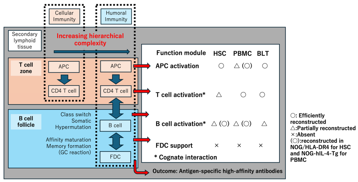
Figure 2.
Temporal dynamics of human immune cell maintenance in humanized mouse models.
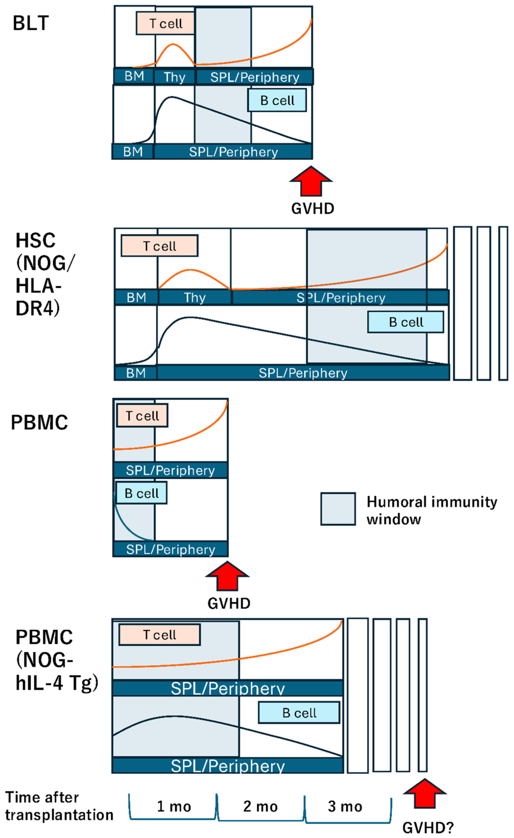

Table 1.
Immune-cell environments in representative humanized mouse models.
| Model | Transplanted human cells | Primary lymphoid organ | Peripheral Tissues | Secondary lymphoid organs |
| BLT | HSC Liver tissues Thymic tissues | Human BM and thymus | Mouse tissues with human immune cells | Mouse tissues with human immune cells |
| HSC | HSC | Mouse thymus | Mouse tissues with human immune cells | Mouse tissues with human immune cells |
| PBMC | PBMC | NA | Mouse tissues with human immune cells | Mouse tissues with human immune cells |
Disclaimer/Publisher’s Note: The statements, opinions and data contained in all publications are solely those of the individual author(s) and contributor(s) and not of MDPI and/or the editor(s). MDPI and/or the editor(s) disclaim responsibility for any injury to people or property resulting from any ideas, methods, instructions or products referred to in the content. |
© 2026 by the authors. Licensee MDPI, Basel, Switzerland. This article is an open access article distributed under the terms and conditions of the Creative Commons Attribution (CC BY) license (http://creativecommons.org/licenses/by/4.0/).
Copyright: This open access article is published under a Creative Commons CC BY 4.0 license, which permit the free download, distribution, and reuse, provided that the author and preprint are cited in any reuse.