

Submitted:
28 August 2026
Posted:
31 August 2026
You are already at the latest version
Abstract
Introduction: Active scars can be defined as ‘pain generators’: the pain associated with them may have an important impact on the patient’s quality of life. During the healing process, several local and systemic neurochemical changes occur, leading to alterations in both the tissue and systemic levels, with changes in the central nervous system (CNS) implicated in the development of chronic pain. Clinically, the features that define a scar as a pathological one include increased firmness, increased tissue thickness, and increased reactivity of the skin after palpation. The therapeutic approach, therefore, includes both local treatments which interferes with structural changes in the scar tissue and systemic treatments. Materials and Methods: A critical review of the literature regarding the role of local anaesthetics in the clinical management of painful scars has been conducted. The paper also reports a series of cases involving patients affected by painful scars who were successfully treated with local lidocaine injections. A series of five cases of adult patients with active painful scars of post-traumatic and post-surgical aetiology, have been treated with intra-lesional infiltration of 0.5% lidocaine. When appropriate, we associated other techniques. Pain intensity has been assessed using the NRS scale before the start of treatment, at the end of treatment, and at 3 months’ follow-up. Results and Discussion: Lidocaine infiltration has been demonstrated effective in the treatment of painful scars, as it reduces the expression of inflammatory mediators. It also has a neuromodulatory effect by reducing the pain signal transduction. Eventually, a key role is the inhibition of NGF binding to its TyrkA receptor. In all patients, we observed a long-lasting reduction in pain symptoms, suggesting that changes had occurred in the tissue, in the peripheral nervous system and a modulation of central nervous system connectivity had been detected. No complications occurred. Conclusion: local treatment with intralesional infiltration with 0.5% lidocaine has demonstrated to be safe and effective in the treatment of painful scars, maintaining the benefit on pain over time, with no complications.
Keywords:
painful scar
; pain generator
; lidocaine infiltration
1. Introduction
1.1. The Scar: “A Pain Generator”
Skin or mucosal scars resulting from trauma or surgery can cause pain of varying intensity, which may also involve the autonomic nervous system and the internal organs. The term ‘pain generator’ refers to the various anatomical or functional structures responsible for clinical pain.
Scars can therefore be regarded as ‘pain generators’, and the pain associated with them can have an important impact on the patient’s quality of life.
The pain resulting from active scars can have several components. Firstly, there is the nociceptive pain component, arising from tissue damage where nociceptors are activated; there may also be a neuropathic pain component, when the somatosensory system is injured; and eventually, a nociplastic pain component may play a pivotal role: it arises from altered nociception involving the activation of peripheral nociceptors without evidence of active tissue damage causing the pain [1]. The estimated prevalence of painful scars is 2% of all scars; in the subgroup of burn scars, it is estimated around 30–68% [2].
1.2. Physical Examination
The wound healing process can deviate from the physiological pathway, leadong to hypertrophic scars, keloids and atrophic scars. A hypertrophic scar is characterised by increased secretion of extracellular matrix, though this is contained into the area of the initial wound. In a keloid, the scar tissue proliferates beyond the edges of the original wound. These two conditions are regarded as two different stages of the same pathology. An atrophic scar, instead, appears as a depression in the skin characterised by a deficit of extracellular matrix. [3].
Scars can involve all components of the soft tissues: the skin, the subcutaneous tissue, the superficial and deep fasciae, muscles and visceral structures [4]. When the dermis and fascia are altered and involved by scar tissue, their function and ability to interact with the microenvironment are impaired [3]. On physical examination, the findings of a pathological scar – defined as an ‘active scar’ – include increased firmness, increased tissue thickness and increased reactivity of the skin surface following palpation [4]. Spontaneous pain or pain induced by palpation can also occur. To carry out an effective treatment of a painful scar, understanding its extent within the layers beneath the skin and the condition of these tissues is essential [5]. If the scar tissue extends beneath the skin and through the underlying layers, it may cause injury to or entrapment of nerve structures, as well as alterations at the fascial level, profoundly altering the function of entire muscle groups. In such cases, a condition known as a neuroma may develop, in which neurons become trapped within the scar tissue, leading to mechanical compression of the Aδ and C fibres, which causes pain [6]. Both intra-neural and extra-neural structures may be involved in the development of a painful scar; for this reason, a systematic approach to diagnosis is required in order to plan the appropriate treatment. Ultrasound is an important tool for assessing the depth and extent of the scar in different tissues and can also provide information on the involvement of nerve structures and the condition of the connective tissue [7]. It can be routinely used in clinical practice to complete the physical examination [5] (Figure1).
1.3. Pathophysiology of “Active Scars”
Fibrosis and the scar-forming process can alter biomechanics and biochemical signalling, leading to painful symptoms [8]. However, neurochemical changes do not occur only in the site affected by the injury, but also at a systemic level: the neurotransmitters released can alter central connectivity and plasticity, a systemic response implicated in the aetiology of chronic pain [9]. In particular, after the injury, both free nerve endings and keratinocytes are involved in nociception. Both secrete pro-inflammatory neuropeptides, establishing an inflammatory state that can become chronic, on the basis of the pathogenesis of neuropathic pain and central sensitisation [10]. Wound healing is a process triggered by inflammatory mediators. Afterwards, mechanical forces guide the remodelling and maturation of the scar by activating mechanoreceptors, which regulate the gene expression of proteins underlying cell proliferation, angiogenesis and epithelialisation. Sometimes, however, the remodelling process is disrupted due to persistent inflammation within the scar. Another factor playing a key regulatory role in scar formation, whether normal or pathological, is the skin’s peripheral sensory and autonomic nervous system. Other neuropeptides, such as VIP, Substance P and CGRP, secreted mainly by the autonomic nervous system (ANS), play a role in modulating metalloproteinases, thereby regulating the granulation tissue and the collagen structure. Inflammatory cytokines, such as TNF-α, IL-1 and IL-6, are secreted by both immune cells and nerve endings [11]. In this context, mast cells also secrete histamine, which plays both a vasoactive role and a role in stimulating fibrosis [12]. Hypertrophic scar tissue exhibits increased expression of Toll-like receptors (epidermal TLR-6 and TLR-7, and dermal TLR-8), the persistent activation of which exacerbates the inflammatory cascade responsible for fibrosis. Finally, during the healing process, calcium plays a key role in intracellular signalling; indeed, the in vitro administration of a calcium antagonist (verapamil) can reduce inflammation and the production of extracellular matrix [12]. In conclusion, pathological fibrosis results from the persistence of the inflammatory response in the tissue, which promotes the proliferation of fibroblasts and the deposition of extracellular matrix by these cells. In this context, dysregulation of TGF-β/Smad signalling is one of the main pathogenic factors. It is a ubiquitous cytokine in the wound-healing process, fundamental in the deposition of the extracellular matrix by regulating scar formation and can be synthesised by damaged tissues and M2-phenotype macrophages [13]. TGF-β1 also induces angiogenesis via the stimulation of VEGF and PDGF [12]. Furthermore, other essential mediators in the pathogenesis of painful scars are neuropeptides, expressed by autonomic and sensory nerve endings, including calcitonin gene-related peptide (CGRP), substance P (SP), neurokinin A (NKA) and vasoactive intestinal peptide (VIP) [14]; in particular, changes in substance P levels are observed in hypertrophic scars [11]. Neuropeptides modulate wound healing by inducing vasodilation and inflammation, and by stimulating the proliferation of epithelial, vascular and connective tissues. Moreover, nerve fibres regenerate spontaneously within scars, but their density, morphology and activity are different from those of healthy tissue; they have a greater density in pathological scars, which consequently exhibit higher concentrations of NPY, VIP, SP and CGRP compared with healthy tissue, a sign of increased neurogenic inflammation (12, 14): the cause of the symptoms of the active scar: pain, itching, allodynia and altered sensitivity to thermal stimuli [11]. A systemc increase in neuropeptides, particularly CGRP, is also observed, the process underlying chronic pain [9]. Local neuro-inflammatory overstimulation is probably the result of a spinal reflex arc: tissue damage stimulates C and Aδ fibres, which triggers a central neuro-inflammatory signal leading to increased neuropeptide secretion [3]. In painful scars, in fact, there is an increase in free nerve endings secreting neuropeptides [6].
1.3.1. The Role of Nerve Grow Factor
A mediator which plays a pivotal role in wound healing and is therefore implicated in the pathophysiology of painful scars when dysregulated is NGF (Nerve Growth Factor) [6]. NGF promotes the proliferation of fibroblasts and keratinocytes, the deposition of the extracellular matrix (ECM), angiogenesis, the differentiation of fibroblasts into myofibroblasts, and wound contraction and ECM remodelling [15] following skin tissue damage. Exposed nerve endings release Substance P and NKA, which induce the synthesis and release of NGF in keratinocytes; this, in turn, exacerbates the release of vasoactive peptides by the exposed nerve endings, triggering and sustaining neurogenic inflammation [6]. Overexpression of NGF leads to fibrotic tissue abnormalities, whilst downregulation results in delayed wound healing. NGF binds primarily to two receptors, TyrkA and p75, which mediate proliferation, survival and apoptosis through various complex mechanisms during the three phases of wound healing: the inflammatory, proliferative and remodelling phases. In the first phase, the immune-activating effect on immune cells predominates, promoting phagocytosis and degranulation of mast cells. In the following stages, NGF induces re-epithelialisation, angiogenesis and neurogenesis, with an anti-apoptotic effect on keratinocytes and neurons, stimulating axonal growth and promoting fibroblast migration. During the remodelling phase, it induces the differentiation of fibroblasts into myofibroblasts and wound contraction. The production of NGF by keratinocytes also increases the expression of α1-adrenergic receptors on free nerve endings, causing pain mediated by sympathetic nervous system activation [16].
1.4. Therapeutic Options
In this context, an effective treatment for painful scars is acupuncture, which plays a role in suppressing local and systemic inflammation, stimulating re-epithelialisation, and improving microcirculatory function – all of which have a trophic effect by enhancing oxygenation and normalising pH [17]. Furthermore, micro-needling has been shown to have regenerative effects, inducing the release of trophic and neoangiogenic mediators, normalising the pattern of collagen organisation, and reducing the local inflammatory response [11]. By remodelling the extracellular matrix with other collagen proteins, it also activates the central anti-nociceptive system [18].
Furthermore, acupuncture modulates the peripheral and central nervous systems through the release of endogenous opioids, which have an inhibitory effect on the dorsal horns (by activating inhibitory interneurons), and of oxytocin, which inhibits the activity of C-fibres [10].
In cases of chronic pain, the therapeutic approach must include both local treatment that interferes with structural changes within the scar tissue, and systemic treatment, such as the use of drugs with systemic action, including gabapentinoids, sodium channel modulators, ketamine, NSAIDs, opioids and SNRIs [19]. One systemic approach is auricular acupuncture, which works through the release of β-endorphins, the regulation of pro-inflammatory and anti-inflammatory cytokines, and the activation of the descending anti-nociceptive system [6].
One of local techniques is the intralesional injection of corticosteroids, which is effective in reducing pain and itching, and acts by inhibiting fibroblast proliferation and promoting collagen degradation [7]. However, it is associated with complications such as atrophy, hyperpigmentation, telangiectasia, ulcers, dermal atrophy, necrosis, and pain during treatment [20].
Other techniques employ electrical neuromodulation: TENS (Transcutaneous Electrical Nerve Stimulation), EMS (Electrical Muscle Stimulation) and IFC (Interferential Current) [19]. Finally, a widely used local approach is the infiltration of the lesion with a local anaesthetic, an effective technique with long-lasting effects [7]. This treatment is the therapy of choice precisely because of the effect these drugs can have on local changes and on the central processes involved in the development of chronic conditions.
1.5. The Role of Local Anaesthetics
Thanks to these effects, local anaesthetics form the cornerstone of the neural therapy, which developed in Europe – primarily in Germany – during the first half of the 20th century; the term ‘neural therapy’ was coined to distinguish the use of these drugs from that of surgical anaesthesia. The theory underpinning neural therapy is based on the identification of the so-called ‘disturbance field’, a region of the body characterised by altered local homeostasis which amplifies to become a systemic pathology. Both the somatosensory system and the autonomic nervous system – in its sympathetic and parasympathetic components – are therefore involved. Local anaesthetics form the basis of the therapy, which utilises their regulatory effect on nerve endings to restore tissue homeostasis. One of the main targets of neural therapy is scar tissue: according to the theory of neural therapy, every scar has the potential to interfere with the sympathetic nervous system, to the extent that it becomes a ‘disturbing field’. In this context, the infiltration of scar tissue has two effects: the interruption of the afferent stimulus from the disturbance field and a systemic effect on the modulation of sympathetic activity [21].
2. Materials and Methods
This paper presents a critical review of the literature concerning the role of local anaesthetics in the clinical management of painful scars. Furthermore, a case series of patients with painful scars treated with local lidocaine infiltration is presented, exploiting its role in modulating scar tissue remodelling and the expression of pro- and anti-inflammatory mediators, as well as its ability to modify the neuronal activity underlying the chronicity of pain. A significant improvement in pain symptoms was observed, indicating that lidocaine was effective in silencing the pain generator.
We report five cases of adult patients suffering from painful active scars of post-traumatic and post-surgical aetiology, treated with 0.5% lidocaine infiltration; the total dose of lidocaine administered never exceeded 100 mg. Patients underwent personalised treatment, involving a variable number of sessions, a variable volume of anaesthetic administered, and, where appropriate, combination with techniques having a systemic effect. Pain intensity was assessed using the NRS (Numeric Rating Scale). The NRS has been detected prior to the start of treatment, at the end of treatment, and at the 3-month follow-up. No complications arose because of the lidocaine infiltration treatment.
3. Cases Presentation
3.1. Case 1: Pain Like Scapulohumeral Periarthritis of the Right Shoulder
Female, 68 years old.
Past medical history:
- -
- previous excision of dorsal lipoma;
- -
- arterial hypertension;
- -
- Dyslipidemia.
Medication history: enalapril, atorvastatin.
Diagnostic investigations already performed:
- -
- MRI of the right shoulder: negative.
PAIN HISTORY: For 24 months, pain in the shoulder with functional impairment in abduction and external rotation. NRS at rest 4, NRS during activity 7.
PHYSICAL EXAMINATION: Whitish, thickened scar, tenaciously adherent to underlying planes, tender on palpation (Figure 2a).
TREATMENT: Five sessions of scar infiltration with 0.5% lidocaine performed on a weekly basis (Figure 2b).
OUTCOME: At the end of the treatment, complete regression of painful symptoms, NRS 0, NRS at three months 0.
3.2. Case 2: Pain at the Site of Previous Hip Prosthesis Wound, Complicated by Infection
79-year-old male.
Past medical history:
- -
- Lumbar disc herniation;
- -
- Previous right hip replacement. Followed by six more revision surgeries at the same site. Complicated by multidrug-resistant Acinetobacter baumannii infection.
Medication history: NSAIDs as needed.
PAIN HISTORY: For about 36 months, he has reported pain only at the wound site; present with movement, reduced at rest, NRS 8. Walks with an aid (cane).
PHYSICAL EXAMINATION: Irregular scar with hypertrophic and atrophic areas, wide, and firmly adhered to the underlying tissues (Figure 3).
Diagnostic tests already performed:
- -
- Lumbar MRI: negative.
- -
- Scintigraphy: negative for infectious foci.
TREATMENT: Scar infiltrations with 0.5% lidocaine were performed for a total of eight weekly sessions.
OUTCOME: Clinical improvement was already evident after the first session. After six sessions, pain was absent while sitting. After eight sessions, pain was absent both standing and lying down. NRS 3 with movement, NRS 0 at rest. Walks without aid. NRS at three months 4 with movement, 1 at rest.
3.3. Case 3: Pain in the Right Leg with Claudication, Site of Previous Trauma
75-year-old male.
Past medical history: negative.
Medication history: negative.
Diagnostic tests already performed: none.
PAIN HISTORY: For the past 5 months, pain in the back of the right leg with claudication at 100 m, site of a previous wound from an accidental fall (from a ladder). NRS 7.
PHYSICAL EXAM: Red, raised scar, tender to touch, adherent to deeper layers (Figure 4). TREATMENT: Five sessions of scar infiltration with 0.5% lidocaine were performed weekly. In the first two sessions, only the superficial scar was infiltrated: no improvement noted. In the following sessions, the deep muscle wound was also infiltrated, resolving the symptoms.
OUTCOME: At the end of treatment, NRS 0, normal mobility restored, NRS at three months 0.
3.4. Case 4: Pain in the Right Hypochondrium with Dysesthesia in the Lower Limbs
30-year-old female.
Past medical history: surgery for dorsal hernia via right thoracotomy.
Medication history: negative.
Diagnostic investigations already performed:
- Spinal cord MRI: mild cord softening at T7 and T8.
PAIN HISTORY: For 7 months, pain in the right hypochondrium with dysesthesia in the lower limbs, NRS 8.
PHYSICAL EXAMINATION: The scar is red, thickened, raised, spontaneously painful, hyperalgesic on palpation (Figure 5a).
TREATMENT: Six sessions of scar infiltration with 0.5% lidocaine were performed on a weekly basis (Figure 5b and Figure 5c).
OUTCOME: At the end of treatment, NRS 0, normal mobility recovered, NRS at three months 0.
3.5. Case 5: Pain in the Tongue
65-year-old woman.
Past medical history: previous removal of a lingual angioma.
Medication history: none.
Diagnostic investigations already carried out: none.
PAIN HISTORY: Persistent pain at the tip of the tongue for 72 months. NRS score of 8 during activity, 5 at rest.
PHYSICAL EXAMINATION: A thickened, whitish scar with increased firmness (Figure 6a).
TREATMENT: Eight sessions of infiltration of the scar with 0.5% lidocaine were carried out at weekly intervals, combined with a block of the sphenopalatine ganglion and of the left lingual nerve.
OUTCOME: There is complete remission of pain lasting for several days, with a mean NRS of 1 at rest, 3 during activity (Figure 7b). Mean NRS at three months 1.This section may be divided by subheadings. It should provide a concise and precise description of the experimental results, their interpretation, and the experimental conclusions that can be drawn.
4. Results
5. Discussion
Lidocaine infiltration has proved to be effective in the treatment of painful scars because it acts directly on the pathophysiological mechanisms underlying the aetiology of the condition. More specifically, lidocaine has been demonstrated to reduce the expression of inflammatory mediators. It is hypothesised that it regulates the expression of vasoactive neuropeptides, thereby alleviating neuropathic pain and neurogenic inflammation by inhibiting the transcription of receptors for chemokines and inflammatory cytokines. Secondly, lidocaine exerts a neuromodulatory effect by reducing pain signal transduction in C-fibres and inhibiting aberrant discharges from damaged neurons, as well as inhibiting sodium channels in spinal cord neurons [22].
Furthermore, at the neuronal level, lidocaine inhibits Gq-coupled receptors by binding to the α subunit, resulting in a reversible and transient downstream effect on neuronal signal transduction, which inhibits cAMP activity and produces an anti-inflammatory effect. [23,24].
Eventually, a key regulatory role described for lidocaine is its effect on the binding of NGF to the TyrkA receptor: in vitro, a lidocaine concentration of 40–50 mmol inhibits the growth of dendrites in NGF-stimulated neurons.
It has in fact been demonstrated that lidocaine is a competitive ligand for ATP at the TyrkA receptor binding site, as the two binding sites of the two molecules have a similar steric structure: the protein sequence of the cytoplasmic site where lidocaine binds the sodium channel is very similar to the one for ATP of the tyrosine kinase receptor activator. Therefore, lidocaine at a concentration greater than 40 mmol, inhibits NGF receptor activity, acting as a competitive antagonist. In vivo, a local concentration of 0.5% lidocaine is considered sufficient to suppress TyrkA receptor activation, thereby blocking the pathways responsible for generating chronic pain [25] and inducince the persistent inflammatory state.
Despite differences in the number of sessions and whether or not further systemic treatments were combined, local lidocaine infiltration in all patients resulted in a long-lasting reduction in pain symptoms, indicating that a change had occurred at the tissue level and in the activity of the peripheral nervous system at the site, as well as a modulation of central nervous system connectivity.
6. Conclusions
In conclusion, local treatment consisting in intralesional infiltration of 0.5% lidocaine has been shown to be safe and effective in the treatment of painful scars, providing sustained relief from pain symptoms over time, with no complications. From a biochemical perspective, lidocaine plays a key role in disrupting the intercellular cross-talk which sustains local inflammation and in inhibiting the neuronal signalling underlying the development of chronic pain. To confirm these findings, further studies are needed involving a larger sample size, as well as studies comparing these results with systemic and other local techniques.
Author Contributions
Conceptualization, G.G. and V.G.; methodology, V.G.; software, S.A.; validation, G.G., A.L. and F.C.; formal analysis, V.G.; investigation, G.G.; resources, G.G.; data curation, V.G.; writing—original draft preparation, V.G.; writing—review and editing, G.G.; visualization, S.A.; supervision, G.G.; project administration, G.G.; funding acquisition, F.C. All authors have read and agreed to the published version of the manuscript.
Funding
This research received no external funding.
Informed Consent Statement
Informed consent was obtained from all subjects involved in the study.
Conflicts of Interest
The authors declare no conflicts of interest.
Abbreviations
The following abbreviations are used in this manuscript:
| NGF: | nerve growth factor |
| TyrKA: | tyrosine-kinase A |
| VIP: | vasoactive Intestinal Peptide |
| CGRP: | Calcitonin Gene-Related Peptide |
| TNF | tumor necrosis factor |
| IL-1: | interleukin 1 |
| IL-6: | interleukin 6 |
| TLR: | toll-like receptor |
| TGF: | Transforming Growth Factor |
| VEGF: | Vascular Endothelial Growth Factor |
| PDGF: | Platelet-Derived Growth Factor |
| NKA: | neurokinin A |
| NPY: | neuropeptide Y |
| ECM: | extracellular matrix |
| NSAIDs: | non-steroidal anti-inflammatory drugs |
| SNRI: | Serotonin-Norepinephrine Reuptake Inhibitors |
| TENS: | Transcutaneous Electrical Nerve Stimulation |
| EMS | Electrical Muscle Stimulation |
| IFC | Interferential Current |
| NRS | numerical rating scale |
References
- Bonezzi, C.; Fornasari, D.; Cricelli, C.; Magni, A.; Ventriglia, G. Not All Pain is Created Equal: Basic Definitions and Diagnostic Work-Up. Pain Ther. 2020, 9 (Suppl 1), 1–15. [Google Scholar] [CrossRef] [PubMed] [PubMed Central]
- Bijlard, E.; Uiterwaal, L.; Kouwenberg, C.A.; Mureau, M.A.; Hovius, S.E.; Huygen, F.J. A Systematic Review on the Prevalence, Etiology, and Pathophysiology of Intrinsic Pain in Dermal Scar Tissue. Pain Physician 2017, 20(2), 1–13. [Google Scholar] [CrossRef] [PubMed]
- Bordoni, B.; Zanier, E. Skin, fascias, and scars: symptoms and systemic connections. J. Multidiscip. Healthc. 2013, 7, 11–24. [Google Scholar] [CrossRef] [PubMed] [PubMed Central]
- Lewit, K.; Olsanska, S. Clinical importance of active scars: abnormal scars as a cause of myofascial pain. J. Manip. Physiol. Ther. 2004, 27(6), 399–402. [Google Scholar] [CrossRef] [PubMed]
- Pirri, C.; Stecco, A.; Fede, C.; De Caro, R.; Stecco, C.; Özçakar, L. Ultrasound imaging of a scar on the knee: Sonopalpation for fascia and subcutaneous tissues. Eur. J. Transl. Myol. 2020, 30(1), 8909. [Google Scholar] [CrossRef] [PubMed] [PubMed Central]
- Bintoro, D.A.; Helianthi, D.R. Combination of Battlefield Acupuncture and Local Point Acupuncture for Post Laparotomy Scar Pain. Med. Acupunct. 2022, 34(1), 66–70. [Google Scholar] [CrossRef] [PubMed] [PubMed Central]
- Abd-Elsayed, A.; Pope, J.; Mundey, D.A.; Slavin, K.V.; Falowski, S.; Chitneni, A.; Popielarski, S.R.; John, J.; Grodofsky, S.; Vanetesse, T.; Fishman, M.A.; Kim, P. Diagnosis, Treatment, and Management of Painful Scar: A Narrative Review. J. Pain Res. 2022, 15, 925–937. [Google Scholar] [CrossRef] [PubMed] [PubMed Central]
- Lee, S.W.; Van Dien, C.; Won, S.J. Adipose Tissue as Pain Generator in the Lower Back and Lower Extremity: Application in Musculoskeletal Medicine. HCA Healthc. J. Med. 2020, 1(5), 257–268. [Google Scholar] [CrossRef] [PubMed] [PubMed Central]
- Hamed, K.; Giles, N.; Anderson, J.; Phillips, J.K.; Dawson, L.F.; Drummond, P.; Wallace, H.; Wood, F.M.; Rea, S.M.; Fear, M.W. Changes in cutaneous innervation in patients with chronic pain after burns. Burns 2011, 37(4), 631–7. [Google Scholar] [CrossRef] [PubMed]
- Lauder, G.; West, N. Clinical Insights into the Importance of Scars and Scar Release in Paediatric Chronic Myofascial Pain. Pain Management - Practices, Novel Therapies and Bioactives. IntechOpen 2021. [Google Scholar] [CrossRef]
- Téot, L.; Mustoe, T.A.; Middelkoop, E.; Gauglitz, G.G. (Eds.) Textbook on Scar Management: State of the Art Management and Emerging Technologies [Internet]; Springer: Cham (CH), 2020. [Google Scholar] [PubMed]
- Lu, Y.Y.; Qin, H.; Lu, C.C.; Wu, M.K.; Zhang, C.L.; Wu, C.H. Migraines and keloids: a 15-year Taiwan claim database analysis. Postgrad. Med. J. 2022, 98(1160), 450–455. [Google Scholar] [CrossRef] [PubMed] [PubMed Central]
- Zhang, T.; Wang, X.F.; Wang, Z.C.; Lou, D.; Fang, Q.Q.; Hu, Y.Y.; Zhao, W.Y.; Zhang, L.Y.; Wu, L.H.; Tan, W.Q. Current potential therapeutic strategies targeting the TGF-β/Smad signaling pathway to attenuate keloid and hypertrophic scar formation. BioMed Pharmacother. 2020, 129, 110287. [Google Scholar] [CrossRef] [PubMed]
- Altun, V.; Hakvoort, T.E.; van Zuijlen, P.P.; van der Kwast, T.H.; Prens, E.P. Nerve outgrowth and neuropeptide expression during the remodeling of human burn wound scars. A 7-month follow-up study of 22 patients. Burns 2001, 27(7), 717–22. [Google Scholar] [CrossRef] [PubMed]
- Hirose, M.; Kuroda, Y.; Murata, E. NGF/TrkA Signaling as a Therapeutic Target for Pain. Pain Pract. 2016, 16(2), 175–82. [Google Scholar] [CrossRef] [PubMed]
- G El Baassiri, M.; Dosh, L.; Haidar, H.; Gerges, A.; Baassiri, S.; Leone, A.; Rappa, F.; Jurjus, A. Nerve growth factor and burn wound healing: Update of molecular interactions with skin cells. Burns 2023, 49(5), 989–1002. [Google Scholar] [CrossRef] [PubMed]
- Allam, H. Acupuncture and trigger point injection in the management of poststernotomy pain syndrome: A case series study. Explore (NY) 2020, 16(3), 161–164. [Google Scholar] [CrossRef] [PubMed]
- Rozenfeld, E.; Sapoznikov Sebakhutu, E.; Krieger, Y.; Kalichman, L. Dry needling for scar treatment. Acupunct. Med. 2020, 38(6), 435–439. [Google Scholar] [CrossRef] [PubMed]
- Abd-Elsayed, A.; Pope, J.; Mundey, D.A.; Slavin, K.V.; Falowski, S.; Chitneni, A.; Popielarski, S.R.; John, J.; Grodofsky, S.; Vanetesse, T.; Fishman, M.A.; Kim, P. Diagnosis, Treatment, and Management of Painful Scar: A Narrative Review. J. Pain Res. 2022, 15, 925–937. [Google Scholar] [CrossRef] [PubMed] [PubMed Central]
- Artzi, O.; Koren, A.; Niv, R.; Mehrabi, J.N.; Friedman, O. The Scar Bane, Without the Pain: A New Approach in the Treatment of Elevated Scars: Thermomechanical Delivery of Topical Triamcinolone Acetonide and 5-Fluorouracil. Dermatol. Ther. (Heidelb) 2019, 9(2), 321–326. [Google Scholar] [CrossRef] [PubMed] [PubMed Central]
- Barop, Hans. Manuale e Atlante di Terapia Neurale; Edi-Ermes, 2003. [Google Scholar]
- Li, M.; Jiang, H.; Gu, K.; Sun, X.; Gu, J.; Li, C.; Wang, G. Lidocaine Alleviates Neuropathic Pain and Neuroinflammation by Inhibiting HMGB1 Expression to Mediate MIP-1α/CCR1 Pathway. J. Neuroimmune Pharmacol. 2021, 16(2), 318–333. [Google Scholar] [CrossRef] [PubMed]
- Chen, Y.; Granger, A.J.; Tran, T.; Saulnier, J.L.; Kirkwood, A.; Sabatini, B.L. Endogenous Gαq-Coupled Neuromodulator Receptors Activate Protein Kinase A. Neuron 2017, 96(5), 1070–1083.e5. [Google Scholar] [CrossRef] [PubMed] [PubMed Central]
- Hollmann, M.W.; Herroeder, S.; Kurz, K.S.; Hoenemann, C.W.; Struemper, D.; Hahnenkamp, K.; Durieux, M.E. Time-dependent inhibition of G protein-coupled receptor signaling by local anesthetics. Anesthesiology 2004, 100(4), 852–60. [Google Scholar] [CrossRef] [PubMed]
- Liu, Z.; Wu, H.; Huang, S. Role of NGF and its receptors in wound healing (Review). Exp. Ther. Med. 2021, 21(6), 599. [Google Scholar] [CrossRef] [PubMed] [PubMed Central]
Figure 1.
Example of physical and ultrasound examination of the scar. (a) Skin scar, wide and adherent, not hypertrophic. (b) Ultrasound view of the scar, deep, involving the muscular fascia.
Figure 1.
Example of physical and ultrasound examination of the scar. (a) Skin scar, wide and adherent, not hypertrophic. (b) Ultrasound view of the scar, deep, involving the muscular fascia.
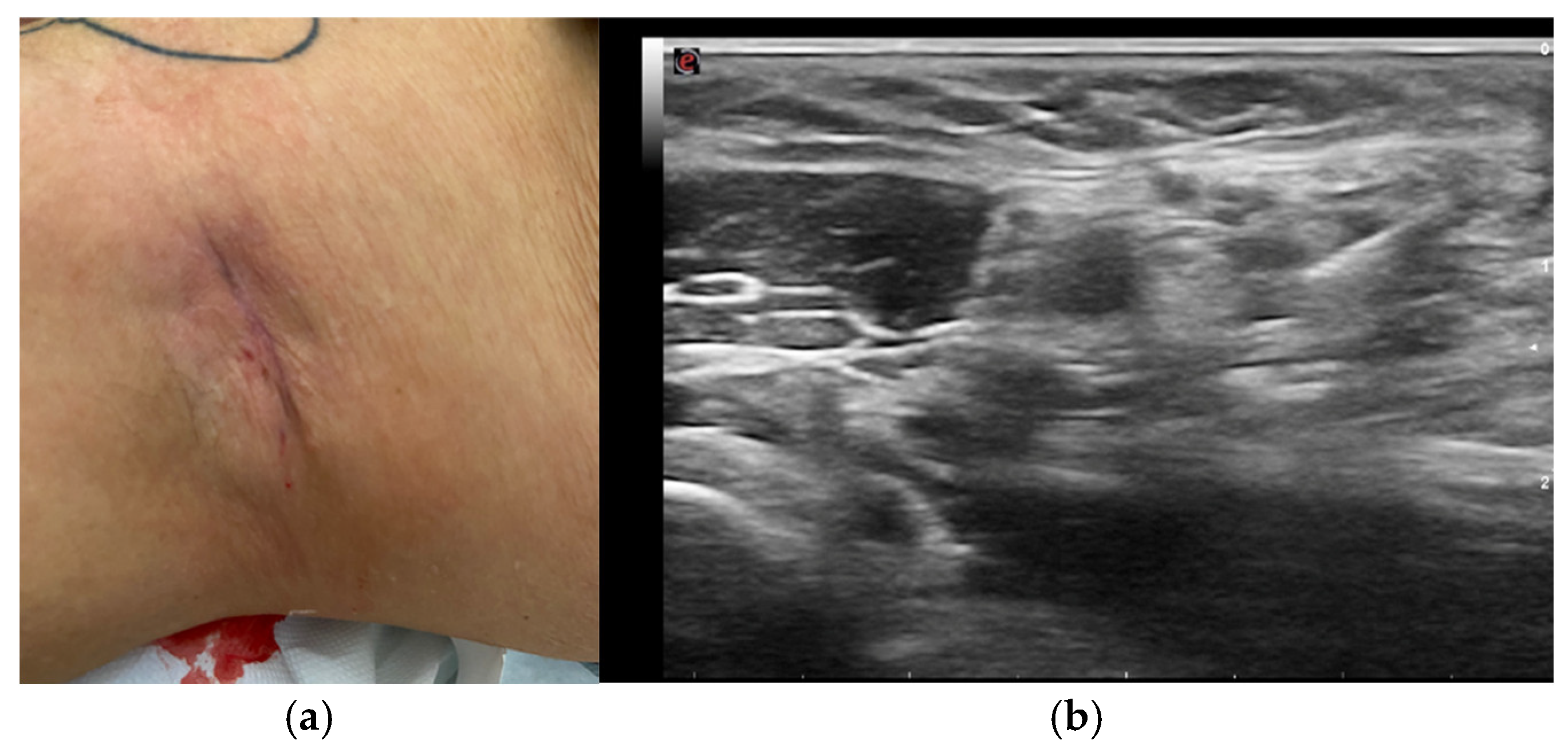
Figure 2.
(a) scar at the site of previous lipoma removal. (b) after infiltrative treatment.
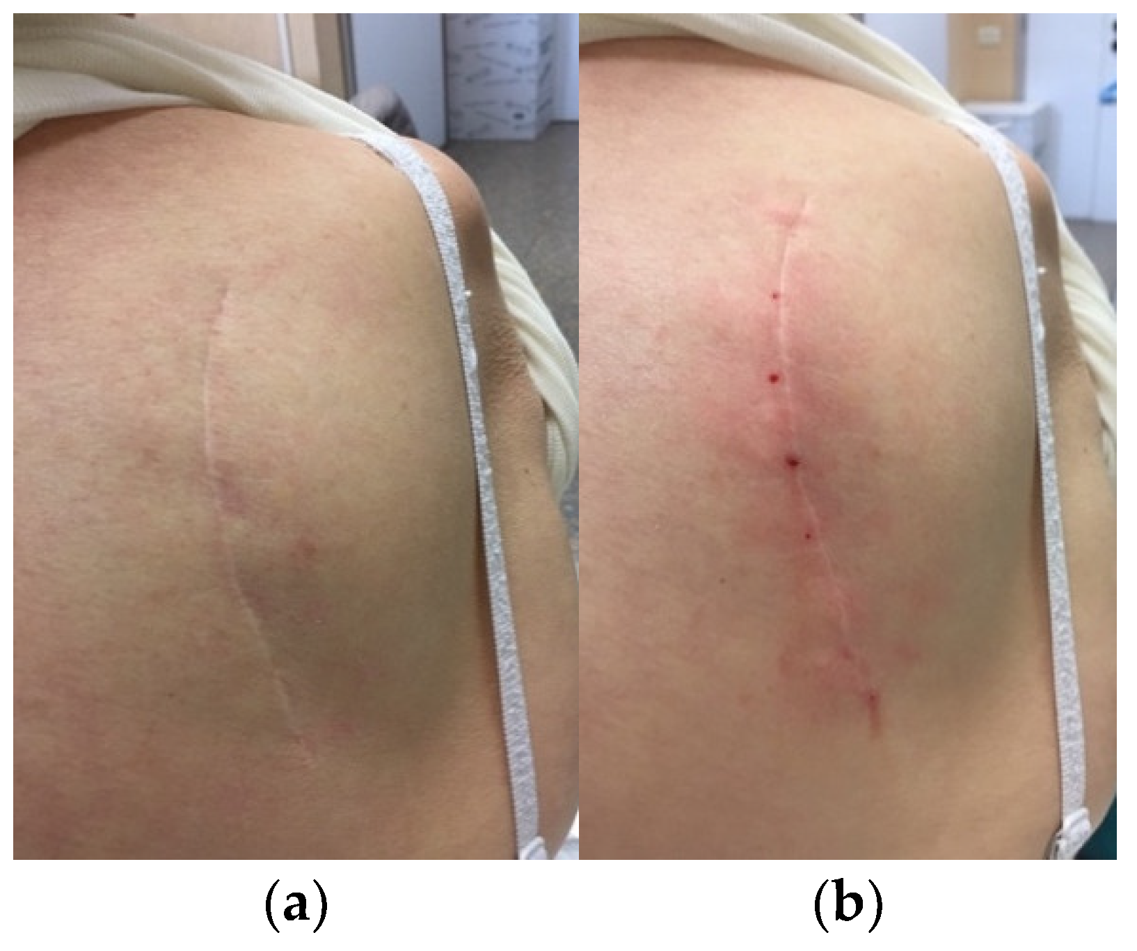
Figure 3.
wide, atrophic scar, site of previous surgery complicated by infection.
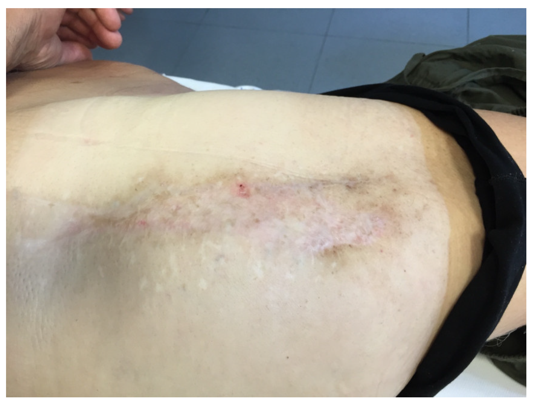
Figure 4.
scar after a fall injury.
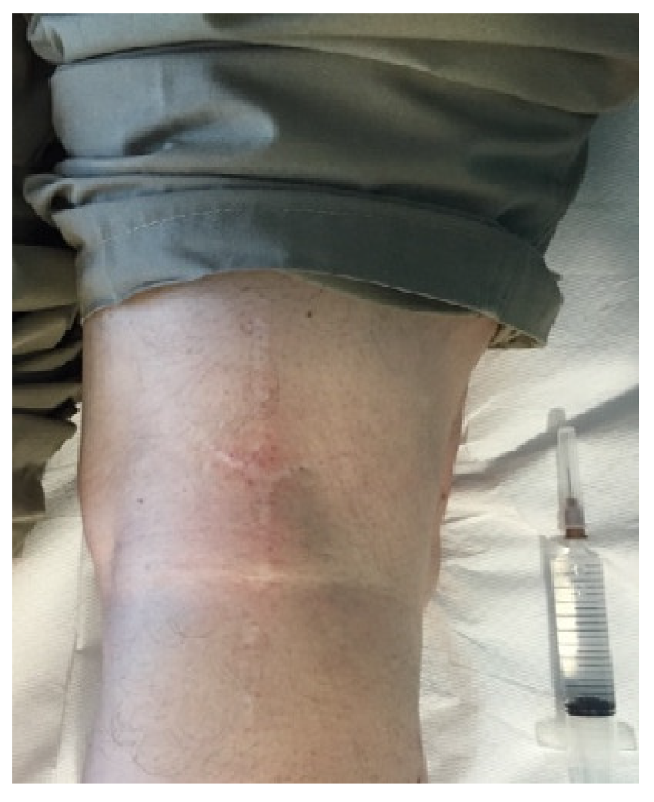
Figure 5.
(a) active post-surgical scars. (b) partial intralesional infiltration. (c) complete intralesional infiltration.
Figure 5.
(a) active post-surgical scars. (b) partial intralesional infiltration. (c) complete intralesional infiltration.
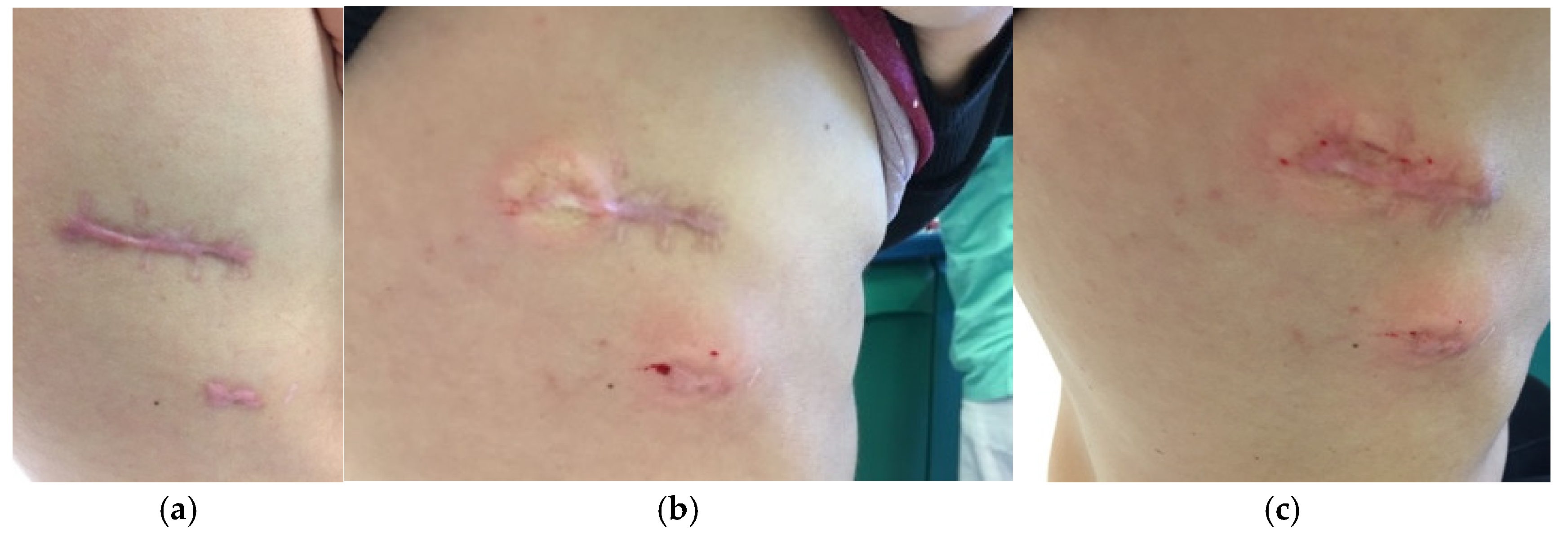
Figure 6.
(a) mucosal scar prior to treatment. (b) scar after 5 sessions.
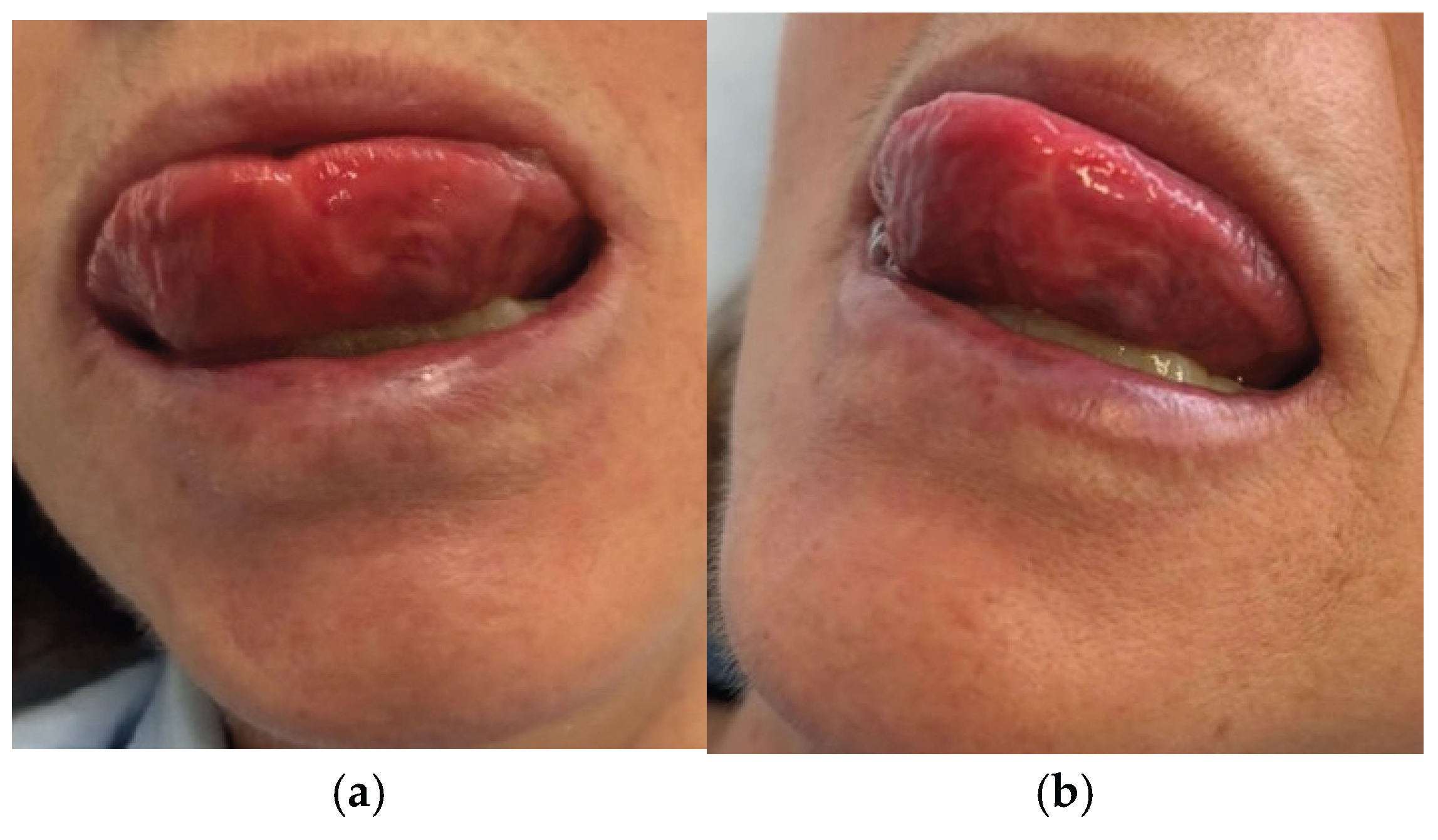
Figure 7.
Graph reporting the NRS scores before the treatment, after the treatment and at the three-month follow-up.
Figure 7.
Graph reporting the NRS scores before the treatment, after the treatment and at the three-month follow-up.
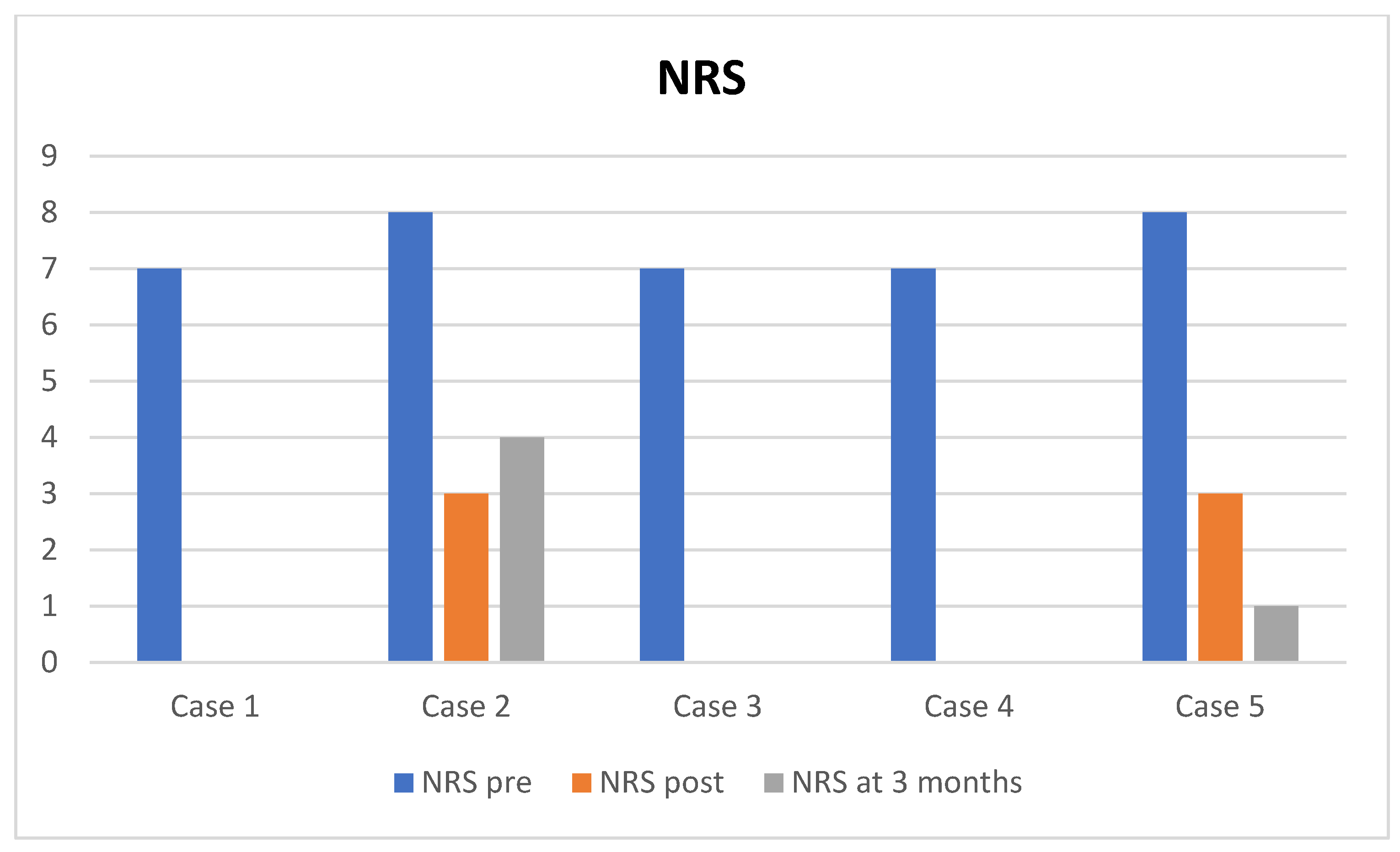

Table 1.
Cases features: age, sex, duration of pain, any associated systemic treatment, number of sessions, pre-treatment NRS, post-treatment NRS, NRS at 3 months, complications.
Table 1.
Cases features: age, sex, duration of pain, any associated systemic treatment, number of sessions, pre-treatment NRS, post-treatment NRS, NRS at 3 months, complications.
| Reference | Age | Sex | Duration of pain (months) | Systemic treatment associated | Number of therapy sessions | NRS pre | NRS post | NRS at 3 months | Complications |
| Case 1 | 68 | F | 24 | none | 5 | 7 | 0 | 0 | none |
| Case 2 | 79 | M | 36 | FANS | 8 | 8 | 3 | 4 | none |
| Case 3 | 75 | M | 5 | none | 5 | 7 | 0 | 0 | none |
| Case 4 | 27 | F | 9 | none | 8 | 7 | 0 | 0 | none |
| Case 5 | 30 | F | 7 | block of the sphenopalatine ganglion and of the left lingual nerve | 8 | 8 | 3 | 1 | none |
Disclaimer/Publisher’s Note: The statements, opinions and data contained in all publications are solely those of the individual author(s) and contributor(s) and not of MDPI and/or the editor(s). MDPI and/or the editor(s) disclaim responsibility for any injury to people or property resulting from any ideas, methods, instructions or products referred to in the content. |
© 2026 by the authors. Licensee MDPI, Basel, Switzerland. This article is an open access article distributed under the terms and conditions of the Creative Commons Attribution (CC BY) license (http://creativecommons.org/licenses/by/4.0/).
Copyright: This open access article is published under a Creative Commons CC BY 4.0 license, which permit the free download, distribution, and reuse, provided that the author and preprint are cited in any reuse.