

Submitted:
27 August 2026
Posted:
28 August 2026
You are already at the latest version
Abstract
Reliable molecular detection of foot-and-mouth disease virus (FMDV) depends on RT-qPCR performance and pre-analytical factors, including sample matrix, RNA extraction method, carrier RNA supplementation, and storage conditions. This study compared silica membrane- and magnetic bead-based RNA extraction for detection of FMDV serotypes O, A, and SAT2 in serum, swab suspensions, and tissue homogenates at three virus dilutions, with and without carrier RNA. FMDV RNA stability in serum stored at room temperature or −20 °C for up to 60 days and following five freeze–thaw cycles was also evaluated using serotype O. Extraction performance was matrix-dependent. In serum, silica-based extraction yielded significantly lower Ct values than magnetic bead-based extraction. In tissue homogenates, magnetic bead extraction performed better with carrier RNA, whereas carrier RNA impaired silica-based extraction. No significant differences were observed for swabs. FMDV RNA detectability remained stable at −20 °C for 60 days, whereas room-temperature storage caused progressive signal loss, which was reduced by carrier RNA. Five freeze–thaw cycles did not progressively affect RNA detectability. These findings demonstrate that FMDV RNA detectability depends on interactions between extraction method, sample matrix, and carrier RNA, highlighting the importance of matrix-specific extraction optimization and appropriate frozen storage for reliable RT-qPCR diagnosis.
Keywords:
foot-and-mouth disease virus
; RNA extraction
; RT-qPCR
; carrier RNA
; RNA stability
; sample matrix
1. Introduction
Foot-and-mouth disease (FMD) is a highly contagious transboundary disease affecting domestic and wild cloven-hoofed animals [1]. It is caused by foot-and-mouth disease virus (FMDV), a small, non-enveloped, positive-sense, single-stranded RNA virus belonging to the genus Aphthovirus within the family Picornaviridae [2]. The virus is classified into seven immunologically distinct serotypes, O, A, C, Asia 1, SAT1, SAT2, and SAT3, between which there is little or no cross-protection [3]. Foot-and-mouth disease remains endemic in large parts of Africa, the Middle East, and Asia, as well as in limited areas of South America, whereas Europe, North and Central America, Australia, New Zealand, and several other regions are free from the disease [4]. Nevertheless, FMD-free countries remain vulnerable to incursions through the movement of infected animals, contaminated animal products, fomites, and other transmission pathways. This continuing threat was demonstrated by the re-emergence of FMD in Germany, Hungary, and Slovakia in 2025, following decades of freedom from the disease in these countries [5]. These outbreaks highlighted the importance of maintaining preparedness and effective diagnostic capacity even in historically FMD-free regions. Reliable laboratory diagnosis depends not only on the analytical performance of the detection assay but also on the quality, type, and quantity of the submitted sample, as well as its collection, transport, storage, and processing. Field conditions may compromise sample integrity through delayed transport, exposure to unsuitable temperatures, or repeated freezing and thawing. Moreover, samples may need to be stored for extended periods before testing or subsequently used for retrospective analyses. As FMDV has an RNA genome, degradation during sample handling and storage may reduce the amount of detectable target RNA and consequently affect diagnostic sensitivity. Real-time reverse transcription polymerase chain reaction (RT-qPCR) is widely used for rapid FMDV detection because of its high sensitivity, specificity, and suitability for high-throughput testing [6,7]. However, efficient and reproducible RNA extraction is a prerequisite for reliable molecular detection. Extraction efficiency may be influenced by viral RNA concentration, sample composition, the presence of inhibitors, and the physicochemical principles of the extraction method. Silica membrane- and magnetic bead-based procedures are among the most frequently used nucleic acid extraction approaches in diagnostic laboratories. Although both rely on the selective binding of nucleic acids under appropriate chemical conditions, they differ in their workflows, automation options, throughput, and susceptibility to matrix-related effects. Carrier RNA is frequently used to improve the recovery of small amounts of nucleic acid by enhancing binding and reducing target loss during extraction [8]. Despite widespread use of silica- and magnetic bead-based extraction in FMD diagnostic laboratories, there is limited comparative evidence regarding how their performance interacts with clinically relevant matrices and carrier RNA supplementation.
Therefore, this study aimed to compare silica membrane- and magnetic bead-based RNA extraction methods for RT-qPCR detection of FMDV serotypes O, A, and SAT2 in serum, swab suspensions, and tissue homogenates, and to evaluate the effect of carrier RNA supplementation. In addition, FMDV RNA stability during prolonged storage at room temperature and −20 °C and following repeated freeze–thaw cycles were assessed in serum.
2. Materials and Methods
Viruses and Sample Matrices
Inactivated foot-and-mouth disease virus (FMDV) serotypes O (O1 Manisa), A (A22 IRQ 24/64), and SAT2 (SAT2 Eritrea), obtained from The Pirbright Institute, United Kingdom, were used to prepare experimentally spiked samples in three matrices: bovine spleen homogenate, bovine serum, and swab suspension. All biological matrices had previously tested negative for FMDV.
For preparation of the spleen matrix, approximately 3 g of previously stored FMDV-negative bovine spleen tissue was homogenized in 27 mL of phosphate-buffered saline (PBS) at a ratio of 1:10 (w/v) using pestle and mortar. The homogenates were clarified by centrifugation at 4,000 rpm for 10 min, and the resulting supernatants were used as the matrix for virus spiking. Aliquots of 22.5 mL of spleen homogenate were mixed with 2.5 mL of each FMDV preparation to obtain a virus-to-matrix ratio of 1:10 (v/v).
For preparation of the swab suspension matrix, 50 swabs were individually immersed in 1 mL of PBS and vortexed for 2 min. Aliquots of 22.5 mL of the resulting swab suspensions were subsequently spiked with 2.5 mL of each virus preparation at a virus-to-matrix ratio of 1:10 (v/v). Thus, the experimental setup was designed to assess FMDV RNA recovery from a swab suspension matrix rather than to reproduce the complete field swab collection procedure.
For the serum matrix, FMDV-negative bovine serum samples were used without additional pretreatment. Aliquots of 40.5 ml of serum were directly spiked with 4.5 mL of each FMDV preparation at a virus-to-matrix ratio of 1:10 (v/v).
Following preparation of the three spiked matrices, serial 10-fold dilutions from 10−1 to 10−3, were prepared, and the samples were subsequently subjected to nucleic acid extraction and RT-qPCR analysis as described below.
RNA Extraction Methods and Carrier RNA Supplementation
RNA extraction was performed with and without carrier RNA using two extraction methods: a magnetic bead-based method employing the IndiMag Pathogen Kit (INDICAL BIOSCIENCE GmbH, Germany) and a silica membrane-based method employing the IndiSpin Pathogen Kit (INDICAL BIOSCIENCE GmbH, Germany). In this experiment, carrier RNA (INDICAL BIOSCIENCE GmbH, Germany) was added as part of the extraction procedure to evaluate its effect on FMDV RNA extraction performance and subsequent RT-qPCR detectability. All extractions were performed according to the manufacturers’ instructions. Magnetic bead-based extraction was automated using the IndiMag 48 instrument (INDICAL BIOSCIENCE GmbH, Germany), whereas silica membrane-based extraction was performed using the QIAcube Connect instrument (QIAGEN, Germany). For both extraction methods, 200 µL of sample was used as the starting volume, with 100 µL of lysis buffer and 1 µL per sample of carrier RNA added in the carrier RNA-supplemented conditions. The final RNA elution volume was 100 µL for both extraction methods. All experimental conditions were tested in triplicate (Supplementary table S1).
Stability of FMDV RNA Under Different Storage Conditions
The stability of FMDV RNA under different storage conditions was evaluated using bovine serum spiked with FMDV O/Manisa at a ratio of 1:10. The spiked serum was divided into two groups: samples supplemented with carrier RNA at a ratio of 1:100 and samples without carrier RNA. In contrast to the extraction experiment, carrier RNA was intentionally added directly to the serum before storage and was not added during subsequent RNA extraction. This design was used to evaluate whether the presence of carrier RNA during storage influenced the preservation and subsequent RT-qPCR detectability of FMDV RNA, independently of its conventional use during the extraction procedure. Aliquots from both groups were stored either at room temperature (22 ± 3 °C) or at −20 ± 2 °C and analysed on days 0, 3, 5, 30, and 60. At each time point, RNA was extracted using the IndiSpin Pathogen Kit (INDICAL BIOSCIENCE GmbH, Germany) and the QIAcube Connect instrument (QIAGEN, Germany). No additional carrier RNA was added during extraction. All experimental conditions were tested in triplicate (Supplementary table S1).
Effect of Repeated Freeze–Thaw Cycles
The effect of repeated freeze–thaw cycles on FMDV RNA detection was evaluated using bovine serum spiked with FMDV O/Manisa. Before freeze–thaw treatment, the spiked serum was divided into two groups: samples supplemented with carrier RNA at a ratio of 1:100 and samples without carrier RNA. Carrier RNA was added before the first freeze–thaw cycle to assess whether its presence in the sample influenced FMDV RT-qPCR detectability following repeated freezing and thawing. No carrier RNA was added during subsequent RNA extraction, thereby avoiding any additional effect of carrier RNA on extraction efficiency. Samples were subjected to five consecutive freeze–thaw cycles. For each cycle, the same sample tubes were frozen at −20 ± 2 °C for 1 h and subsequently thawed completely at room temperature (22 ± 3 °C). Following thawing, samples were thoroughly mixed before being returned to −20 °C for the subsequent cycle. Following each cycle, RNA was extracted using the IndiSpin Pathogen Kit (INDICAL BIOSCIENCE GmbH, Germany) and the QIAcube Connect instrument (QIAGEN, Germany), without the addition of carrier RNA during extraction. All experimental conditions were tested in triplicate (Supplementary table S1).
FMDV RT-qPCR
RNA extracted in all three experiments was analysed by RT-qPCR using the Luna® Universal Probe One-Step RT-qPCR Kit (New England Biolabs, USA). The assay targeted the conserved 3D region of the FMDV genome and was performed according to the previously published protocol for the molecular detection of FMDV described by Moniwa et al. [7]. The total reaction volume was 12.5 µL and comprised 1.88 µL of nuclease-free water, 6.25 µL of Luna Universal One-Step Reaction Mix (2×), 0.63 µL of Luna WarmStart® RT Enzyme Mix (20×), 0.50 µL of each primer (10 µM), 0.25 µL of probe (10 µM), and 2.50 µL of RNA template. The thermal cycling conditions consisted of reverse transcription at 55 °C for 10 min, initial denaturation at 95 °C for 1 min, followed by 45 cycles of denaturation at 95 °C for 10 s and annealing/extension at 60 °C for 30 s. Amplification was performed using a QuantStudio™ 3 Real-Time PCR System (Applied Biosystems, Thermo Fisher Scientific, USA). Appropriate positive controls, negative extraction controls, and no-template controls were included in each run. The VetMAX™ Xeno™ Internal Positive Control (IPC) RNA (Thermo Fisher Scientific, USA) was included as an internal extraction and amplification control to monitor the efficiency of nucleic acid extraction and to identify potential RT-qPCR inhibition. Cycle threshold (Ct) values obtained under the different experimental conditions were used for comparative analysis.
Statistical Analysis
Ct values obtained from three technical replicates were summarized as mean ± standard deviation (SD). For inferential statistical analyses, the mean Ct value of the three technical replicates was calculated for each experimental condition and used as a single observation. Technical repeatability was additionally assessed using the range between the minimum and maximum Ct values within each triplicate. The effects of the extraction method (magnetic bead-based versus silica membrane-based extraction) and carrier RNA supplementation were evaluated separately for serum, swab, and tissue matrices. Comparisons were performed using the paired two-sided Wilcoxon signed-rank test, with virus serotype and dilution combinations treated as matched experimental units. Differences were expressed as ΔCt, calculated as the mean Ct under the first condition minus the mean Ct under the reference condition. To account for multiple testing across matrices and experimental contrasts, raw p-values were adjusted using the Holm method. An adjusted p-value < 0.05 was considered statistically significant. The stability and freeze–thaw experiments were analysed descriptively because they were based on technical replicates of the same experimental material. For each storage condition, time point and carrier RNA treatment, results were expressed as mean Ct ± SD. Changes during storage were reported as ΔCt relative to day 0, whereas changes following repeated freeze–thaw cycles were calculated relative to the first cycle. Statistical analyses were performed using Python 3.13. with the SciPy package 1.17.0.
3. Results
Influence of Extraction Method and Carrier RNA on FMDV RNA Detection
The performance of magnetic-bead and silica membrane-based extraction was evaluated in serum, swab and tissue matrices spiked with FMDV serotypes O, A and SAT2 at three dilutions. Most technical triplicates showed good repeatability, with a median within-triplicate SD of 0.20 Ct. Nine reactions yielded no detectable amplification. All non-detectable reactions were obtained for serotype O at the highest dilution in the swab matrix, except when silica membrane-based extraction was performed with carrier RNA. The effect of the extraction method varied according to the sample matrix and carrier RNA supplementation. In serum samples extracted without carrier RNA, silica membrane-based extraction system yielded consistently lower Ct values than magnetic bead-based extraction, with a mean difference of −6.63 Ct (median ΔCt: −7.67; Holm-adjusted p = 0.047). Silica extraction also performed significantly better in serum samples extracted with carrier RNA, producing Ct values that were, on average, 3.15 cycles lower than those obtained using magnetic bead-based extraction (median ΔCt: −3.19; Holm-adjusted p = 0.047). For magnetic bead-based extraction of serum samples, the addition of carrier RNA reduced the mean Ct by 3.51 cycles compared with extraction without carrier RNA. However, this difference was not statistically significant after adjustment for multiple comparisons (median ΔCt: −4.25; Holm-adjusted p = 0.094). Carrier RNA had virtually no overall effect on silica membrane-based extraction of serum samples (mean ΔCt: −0.03; median ΔCt: −0.05; Holm-adjusted p = 1.000). No significant differences between extraction methods or carrier RNA conditions were detected in the swab matrix. Without carrier RNA, silica extraction produced Ct values that were, on average, 1.32 cycles lower than those obtained using magnetic extraction (Holm-adjusted p = 1.000). In the presence of carrier RNA, the corresponding mean difference was −0.87 Ct (Holm-adjusted p = 1.000). Carrier RNA did not significantly affect magnetic bead-based extraction (mean ΔCt: −0.37; Holm-adjusted p = 1.000) or silica membrane-based extraction (mean ΔCt: +0.08; Holm-adjusted p = 1.000). Interpretation of the swab results was limited by the non-detectable reactions for 10−3 diluted serotype O and by the variable triplicate identified for the 10−2 serotype O dilution. In tissue samples extracted with carrier RNA, magnetic bead-based extraction performed significantly better than silica membrane-based extraction system. The mean Ct obtained using silica extraction was 3.71 cycles higher than that obtained using magnetic extraction (median ΔCt: 3.44; Holm-adjusted p = 0.047). In the absence of carrier RNA, no consistent difference between the two extraction methods was detected (mean ΔCt: −0.89; median ΔCt: −0.06; Holm-adjusted p = 1.000). The effect of carrier RNA in tissue samples also depended on the extraction method. With magnetic bead-based extraction, carrier RNA supplementation led to a reduction of the Ct by 2.44 cycles on average; however, the difference was not significant after adjustment for multiple comparisons (median ΔCt: −1.29; Holm-adjusted p = 0.191). In contrast, the addition of carrier RNA during silica membrane-based extraction significantly increased the mean Ct by 2.16 cycles (median ΔCt: 2.23; Holm-adjusted p = 0.047) (Supplementary table S2).
Stability of FMDV RNA During Storage
Storage temperature had a pronounced effect on the stability of FMDV RNA in serum. At −20 °C, the RT-qPCR signal remained stable throughout the 60-day observation period. After 60 days, the mean Ct changed by only +0.47 cycles in samples supplemented with carrier RNA and by −0.60 cycles in samples without carrier RNA relative to day 0. The fluctuations observed at intermediate time points were not progressive and remained within approximately 1.3 Ct of the baseline values. At room temperature, Ct values increased with prolonged storage, particularly between days 30 and 60. After 30 days, the mean Ct had increased by 1.30 cycles in samples containing carrier RNA and by 2.37 cycles in samples without carrier RNA. After 60 days, the increases reached 4.33 and 6.57 Ct, respectively. After 60 days at room temperature, the increase in Ct was smaller in samples stored with carrier RNA than in those stored without carrier RNA (4.33 versus 6.57 Ct), corresponding to a difference of approximately 2.23 Ct. This finding indicates better preservation of RT-qPCR detectability in the presence of carrier RNA under the tested conditions (Figure 1).
Effect of Repeated Freeze–Thaw Cycles
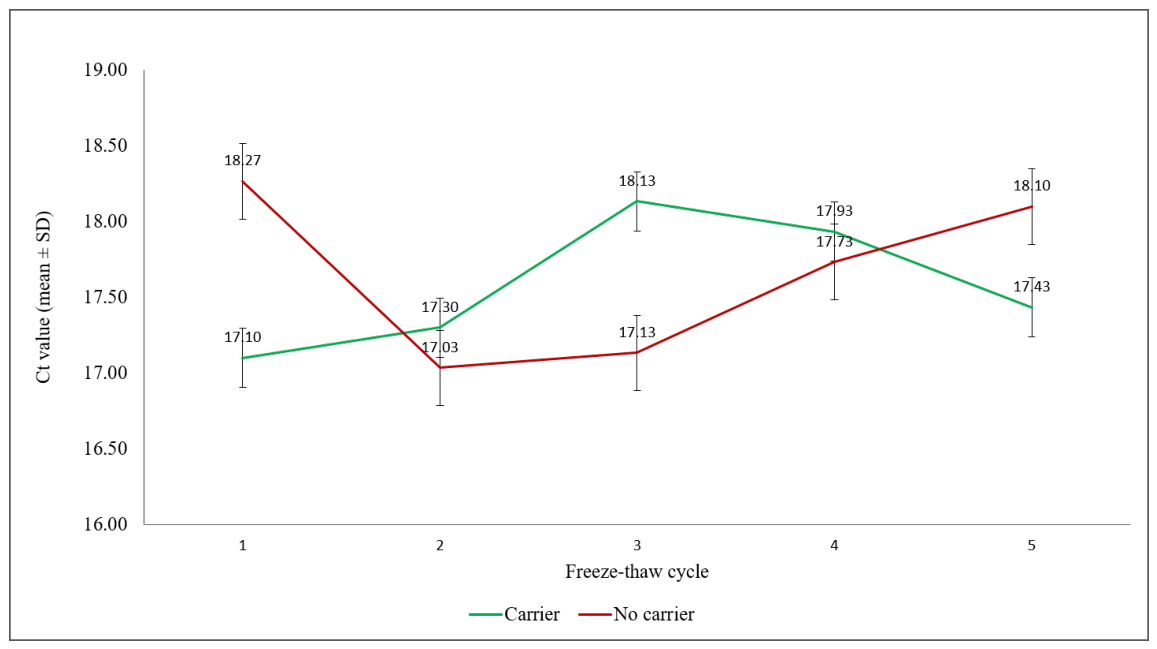
Repeated freezing and thawing did not produce a progressive increase in Ct values over five cycles. In samples containing carrier RNA, the mean Ct increased from 17.10 after the first cycle to 17.43 after the fifth cycle, corresponding to a net change of +0.33 Ct. In samples without carrier RNA, the mean Ct changed from 18.27 after the first cycle to 18.10 after the fifth cycle, representing a net change of −0.17 Ct. Although fluctuations of approximately 1 Ct were observed at some intermediate cycles, there was no evidence of cumulative loss of detectable FMDV RNA under the tested conditions (Figure 2).
4. Discussion
The present study demonstrates that FMDV RT-qPCR detectability is strongly influenced by the interaction between sample matrix, RNA extraction method, and carrier RNA supplementation. No single extraction approach performed optimally across all matrices, emphasizing the need for matrix-specific validation of pre-analytical procedures.
Silica membrane-based extraction system consistently produced significantly lower Ct values in serum samples, irrespective of carrier RNA supplementation, whereas magnetic bead-based extraction performed significantly better for tissue homogenates when carrier RNA was included. No consistent difference between extraction methods was observed for swab samples. One possible explanation for these matrix-dependent differences is the interaction between the physicochemical characteristics of the sample matrix and the different nucleic acid capture mechanisms of the two extraction systems.
The observed improvement associated with carrier RNA during magnetic extraction of tissue samples may similarly reflect enhanced nucleic acid capture or reduced target loss during purification. However, because RNA yield, purity, residual PCR inhibition, and extraction efficiency were not independently quantified, the mechanisms underlying these matrix-dependent differences cannot be determined from the present study. These observations agree with previous studies demonstrating that extraction efficiency depends on the interaction between extraction chemistry and sample matrix rather than on the extraction method alone [9,10,11]. The effect of carrier RNA was likewise dependent on both the extraction method and the sample matrix. Although carrier RNA tended to improve RNA RT-qPCR detectability from serum and tissue samples extracted using the magnetic bead-based method, these improvements were not statistically significant after correction for multiple comparisons. In contrast, carrier RNA provided no measurable benefit during silica extraction of serum and significantly increased Ct values during silica extraction of tissue samples. These findings indicate that carrier RNA should not be regarded as universally beneficial but rather as a parameter requiring validation for each extraction protocol and sample type. Previous investigations have similarly reported that the effect of carrier RNA depends on the extraction chemistry and the characteristics of the sample [12,13].
For FMDV molecular diagnostics, neither silica membrane- nor magnetic bead-based extraction is universally superior, and method selection should consider both sample matrix and laboratory workflow requirements. Automated magnetic bead-based systems may offer operational advantages in high-throughput laboratories despite comparable amplification performance [14].
An additional practical finding of the present study was that pre-analytical storage conditions significantly influenced FMDV RNA detectability. However, it should be noted that carrier RNA served different experimental purposes in the extraction and stability experiments. In the extraction experiment, carrier RNA was included during the extraction procedure to assess its effect on extraction performance, whereas in the stability experiment it was added directly to serum before storage and omitted during subsequent extraction. The latter experiment therefore evaluated whether the presence of carrier RNA during storage was associated with preservation of subsequent FMDV RT-qPCR detectability rather than its conventional role in facilitating nucleic acid extraction. Serum samples stored at −20 °C remained suitable for RT-qPCR analysis throughout the 60-day observation period, whereas prolonged storage at room temperature resulted in progressively increasing Ct values, particularly after 30 and 60 days. Although carrier RNA supplementation partially mitigated this loss of signal, it did not completely prevent RNA degradation.
In contrast, repeated freeze–thaw cycles had only a limited effect on FMDV RNA detection, with no progressive loss of RT-qPCR sensitivity observed after up to five cycles. RNA degradation is strongly influenced by storage temperature and duration, while low temperatures generally preserve RNA integrity [15,16,17,18]. Although viral RNA stability varies depending on the biological matrix and storage medium, prolonged exposure to higher temperatures generally has a greater impact on RNA integrity than appropriate frozen storage, while the effect of repeated freeze–thaw cycles appear to be context-dependent [17,18,19]. Although unnecessary freeze–thaw cycles should generally be avoided as part of good sample-management practice, the present results indicate that up to five cycles did not materially impair FMDV RT-qPCR detectability under the tested conditions.
The present study has several limitations that should be considered when interpreting the findings. First, the experiments were performed using experimentally spiked matrices rather than naturally infected clinical specimens. In particular, PBS-based swab suspensions represent standardized experimental matrices and may not fully reproduce the biological complexity and potential inhibitory components of swab samples. Similarly, although bovine tissue homogenates provide a more complex biological matrix, spiking after homogenization does not completely reproduce the distribution and association of viral material present in naturally infected tissues. Furthermore, only one commercial extraction system representing each extraction principle was evaluated, and the observed differences should not be generalized to silica membrane- or magnetic bead-based technologies as a whole. RNA yield and purity were not independently quantified; therefore, differences in Ct values reflect overall RT-qPCR performance and cannot be attributed specifically to differences in RNA recovery. Finally, the storage stability and freeze–thaw experiments were restricted to a single FMDV isolate, serum matrix, and starting virus concentration, and further studies using naturally infected specimens, additional matrices, viral concentrations and serotypes, and extraction platforms would be valuable to confirm the broader applicability of these findings.
This study highlights the critical importance of pre-analytical variables for reliable molecular detection of FMDV. Matrix-specific selection of extraction methods, careful validation of carrier RNA supplementation, appropriate frozen storage, and standardized sample handling can all improve RT-qPCR performance. Rather than recommending a single universally optimal extraction strategy, the results support tailoring extraction protocols to the specific diagnostic matrix to maximize analytical sensitivity and robustness in routine veterinary diagnostic laboratories.
Supplementary Materials
The following supporting information can be downloaded at the website of this paper posted on Preprints.org. Table S1: Experimental design and raw RT-qPCR Ct data for the evaluation of FMDV RNA extraction, storage stability, and repeated freeze–thaw cycles; Table S2: Pairwise statistical comparisons of FMDV RNA extraction methods and carrier RNA supplementation across sample matrices.
Author Contributions
Conceptualization, V.M. and Lj.V.; methodology, V.M. and LjV; investigation, V.M., B.M., Z.Z.S., S.S., D.G., Đ.A. and L.V.; formal analysis, V.M.; validation, V.M., B.M. and D.G.; visualization, V.M.; original draft preparation, V.M.; writing review and editing, V.M., B.M., Z.Z.S., S.S., D.G., Đ.A. Lj.V., G.V.; supervision, V.M., G.V.; All authors have read and agreed to the published version of the manuscript.
Funding
This research was funded by the Ministry of Science, Technological Development and Innovation of the Republic of Serbia, Contract No. 451-03-33/2026-03/200030.
Data Availability Statement
The data supporting the findings of this study are available within the article and its supplementary material.
Conflicts of Interest
The authors declare no conflicts of interest.
Abbreviations
The following abbreviations are used in this manuscript:
| FMD | Foot-and-mouth disease |
| FMDV | Foot-and-mouth disease virus |
| RT-qPCR | Real-time reverse transcription quantitative polymerase chain reaction |
| RNA | Ribonucleic acid |
| PBS | Phosphate-buffered saline |
| Ct | Cycle threshold |
| SD | Standard deviation |
| IPC | Internal positive control |
| RT | Room temperature |
References
- Alexandersen, S.; Mowat, N. Foot-and-Mouth Disease: Host Range and Pathogenesis. In Current Topics in Microbiology and Immunology; Mahy, B., Ed.; Springer: Berlin, 2005; Vol. 288, pp. 9–42. [Google Scholar]
- Rodriguez, L.L.; Arzt, J. Picornaviridae. Vet. Microbiol. 2022, 533–542. [Google Scholar] [CrossRef]
- Davies, G. Foot and Mouth Disease. Res. Vet. Sci. 2002, 73, 195–199. [Google Scholar] [CrossRef] [PubMed]
- Humphreys, J.M.; Stenfeldt, C.; King, D.P.; Knight-Jones, T.; Perez, A.M.; VanderWaal, K.; Sanderson, M.W.; Di Nardo, A.; Jemberu, W.T.; Pamornchainavakul, N.; et al. Epidemiology and Economics of Foot-and-Mouth Disease: Current Understanding and Knowledge Gaps. Vet. Res. 2025, 56, 141. [Google Scholar] [CrossRef] [PubMed]
- Girault, G.; Romey, A.; Bernelin-Cottet, C.; Bakkali Kassimi, L.; Zientara, S.; Blaise-Boisseau, S. Re-Emergence of Foot-and-Mouth Disease in Europe in 2025: Impact and Future Prospects. Virologie 2025, Vol. 29, 393–396. [Google Scholar] [CrossRef] [PubMed]
- Hwang, J.H.; Shin, Y.K.; Park, S.Y.; Kim, J.; Kim, S.M.; Kim, B.; Park, J.H.; Lee, J.S.; Lee, K.N. Robust Real-Time Reverse Transcription-PCR for Detection of Foot-and-Mouth Disease Virus Neutralizing Carryover Contamination. J. Clin. Microbiol. 2016, 54, 216–219. [Google Scholar] [CrossRef] [PubMed]
- Moniwa, M.; Clavijo, A.; Li, M.; Collignon, B.; Kitching, R.P. Performance of a Foot-and-Mouth Disease Virus Reverse Transcription-Polymerase Chain Reaction with Amplification Controls between Three Real-Time Instruments. J. Vet. Diagn. Investig. 2007, 19, 9–20. [Google Scholar] [CrossRef] [PubMed]
- Made Artika, I.; Ma’roef, C.N.; Johar, E.; Yudhaputri, F.A.; Myint, K.S.A. Comprehensive Review on Viral RNA Extraction Strategies for Enhanced Molecular Diagnostics. Interdiscip. Perspect. Infect. Dis. 2025. [Google Scholar] [CrossRef] [PubMed]
- Ortega-Pinazo, J.; Pacheco-Rodríguez, M.J.; Serrano-Castro, P.J.; Martínez, B.; Pinto-Medel, M.J.; Gómez-Zumaquero, J.M.; Lago-Sampedro, A.; García-Díaz, B.; Estivill-Torrús, G.; Emilio Ferro Gallego, P. Comparing RNA Extraction Methods to Face the Variations in RNA Quality Using Two Human Biological Matrices. Mol. Biol. Rep. 2023, 50, 9263–9271. [Google Scholar] [CrossRef] [PubMed]
- Girard, M.; Morales-Rayas, R.; Jean, J. Comparison of RNA Extraction Methods for the Detection of a Norovirus Surrogate in Ready-to-Eat Foods. Foodborne Pathog. Dis. 2013, 10, 47–54. [Google Scholar] [CrossRef] [PubMed]
- Husakova, M.; Kralik, P.; Babak, V.; Slana, I. Efficiency of DNA Isolation Methods Based on Silica Columns and Magnetic Separation Tested for the Detection of Mycobacterium Avium Subsp. Paratuberculosis in Milk and Faeces. Materials 2020, 13, 1–11. [Google Scholar] [CrossRef] [PubMed]
- Andreasen, D.; Fog, J.U.; Biggs, W.; Salomon, J.; Dahslveen, I.K.; Baker, A.; Mouritzen, P. Improved MicroRNA Quantification in Total RNA from Clinical Samples. Methods 2010, 50, S6–S9. [Google Scholar] [CrossRef] [PubMed]
- El-Shorbagy, H.M.; El-Liethy, S.S.; Moussa, M.K.; Elghor, A.A. Carrier RNA Is a Key Factor Affecting Fully Integrated Short Tandem Repeats Profiling in Challenging Forensic Samples Models. J. Basic Appl. Zool. 2022, 83. [Google Scholar] [CrossRef]
- Zamuner, F.T.; Ramos-López, A.; García-Negrón, A.; Purcell-Wiltz, A.; Cortés-Ortiz, A.; Cuevas, A.R.; Gosala, K.; Winkler, E.; Sidransky, D.; Guerrero-Preston, R. Evaluation of Silica Spin-Column and Magnetic Bead Formats for Rapid DNA Methylation Analysis in Clinical and Point-of-Care Settings. Biomed. Rep. 2024, 21. [Google Scholar] [CrossRef] [PubMed]
- Kornienko, I. V.; Aramova, O.Y.; Tishchenko, A.A.; Rudoy, D. V.; Chikindas, M.L. RNA Stability: A Review of the Role of Structural Features and Environmental Conditions. Molecules 2024, 29. [Google Scholar] [CrossRef] [PubMed]
- Chheda, U.; Pradeepan, S.; Esposito, E.; Strezsak, S.; Fernandez-Delgado, O.; Kranz, J. Factors Affecting Stability of RNA – Temperature, Length, Concentration, PH, and Buffering Species. J. Pharm. Sci. 2024, 113, 377–385. [Google Scholar] [CrossRef] [PubMed]
- Al Qaisieh, B.A.; Ababneh, M.M.K.; Al-Zghoul, M.B.F.; Alghizzawi, D.A.A.; Aboomer, H.A. Evaluation of Various Filter Paper and Reagent Systems for the Preservation of Newcastle Disease Virus RNA Samples. J. Vet. Res. 2025, 69, 183–190. [Google Scholar] [CrossRef] [PubMed]
- Williams, R.C.; Perry, W.B.; Lambert-Slosarska, K.; Futcher, B.; Pellett, C.; Richardson-O’Neill, I.; Paterson, S.; Grimsley, J.M.S.; Wade, M.J.; Weightman, A.J.; et al. Examining the Stability of Viral RNA and DNA in Wastewater: Effects of Storage Time, Temperature, and Freeze-Thaw Cycles. Water Res. 2024, 259. [Google Scholar] [CrossRef] [PubMed]
- Dzung, A.; Cheng, P.F.; Stoffel, C.; Tastanova, A.; Turko, P.; Levesque, M.P.; Bosshard, P.P. Prolonged Unfrozen Storage and Repeated Freeze-Thawing of SARS-CoV-2 Patient Samples Have Minor Effects on SARS-CoV-2 Detectability by RT-PCRJ. Mol. Diagn. 2021, 23, 691–697. [Google Scholar] [CrossRef] [PubMed]
Figure 1.
Effect of storage conditions on FMDV RNA detection. Mean Ct values obtained for serum samples stored at room temperature (RT) and −20 °C for up to 60 days with and without carrier RNA supplementation. Values represent mean Ct ± SD of three technical replicates.
Figure 1.
Effect of storage conditions on FMDV RNA detection. Mean Ct values obtained for serum samples stored at room temperature (RT) and −20 °C for up to 60 days with and without carrier RNA supplementation. Values represent mean Ct ± SD of three technical replicates.
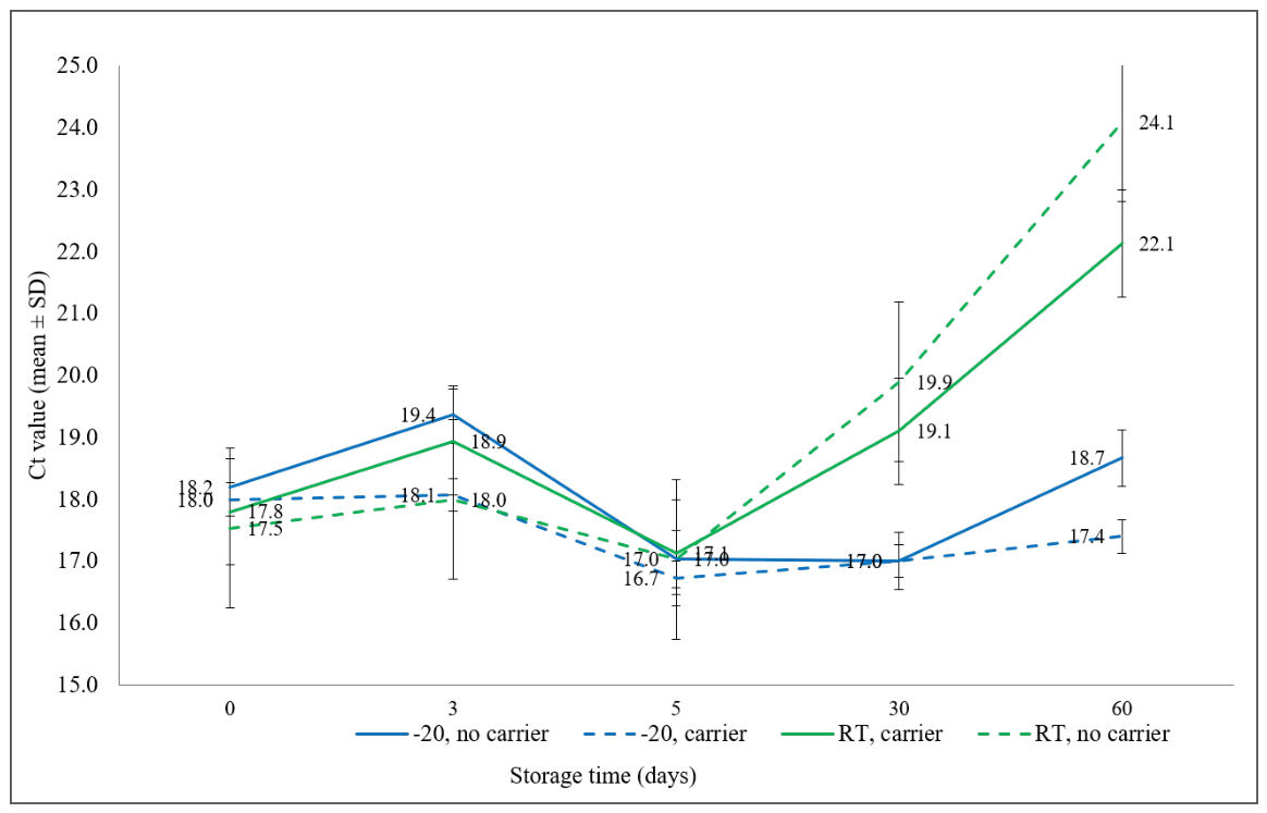
Figure 2.
Effect of repeated freeze–thaw cycles on FMDV RNA detection. Mean Ct values obtained after one to five freeze–thaw cycles for serum samples with and without carrier RNA supplementation. Values represent mean Ct ± SD of three technical replicates.
Figure 2.
Effect of repeated freeze–thaw cycles on FMDV RNA detection. Mean Ct values obtained after one to five freeze–thaw cycles for serum samples with and without carrier RNA supplementation. Values represent mean Ct ± SD of three technical replicates.
Disclaimer/Publisher’s Note: The statements, opinions and data contained in all publications are solely those of the individual author(s) and contributor(s) and not of MDPI and/or the editor(s). MDPI and/or the editor(s) disclaim responsibility for any injury to people or property resulting from any ideas, methods, instructions or products referred to in the content. |
© 2026 by the authors. Licensee MDPI, Basel, Switzerland. This article is an open access article distributed under the terms and conditions of the Creative Commons Attribution (CC BY) license (http://creativecommons.org/licenses/by/4.0/).
Copyright: This open access article is published under a Creative Commons CC BY 4.0 license, which permit the free download, distribution, and reuse, provided that the author and preprint are cited in any reuse.