

Submitted:
27 August 2026
Posted:
28 August 2026
You are already at the latest version
Abstract
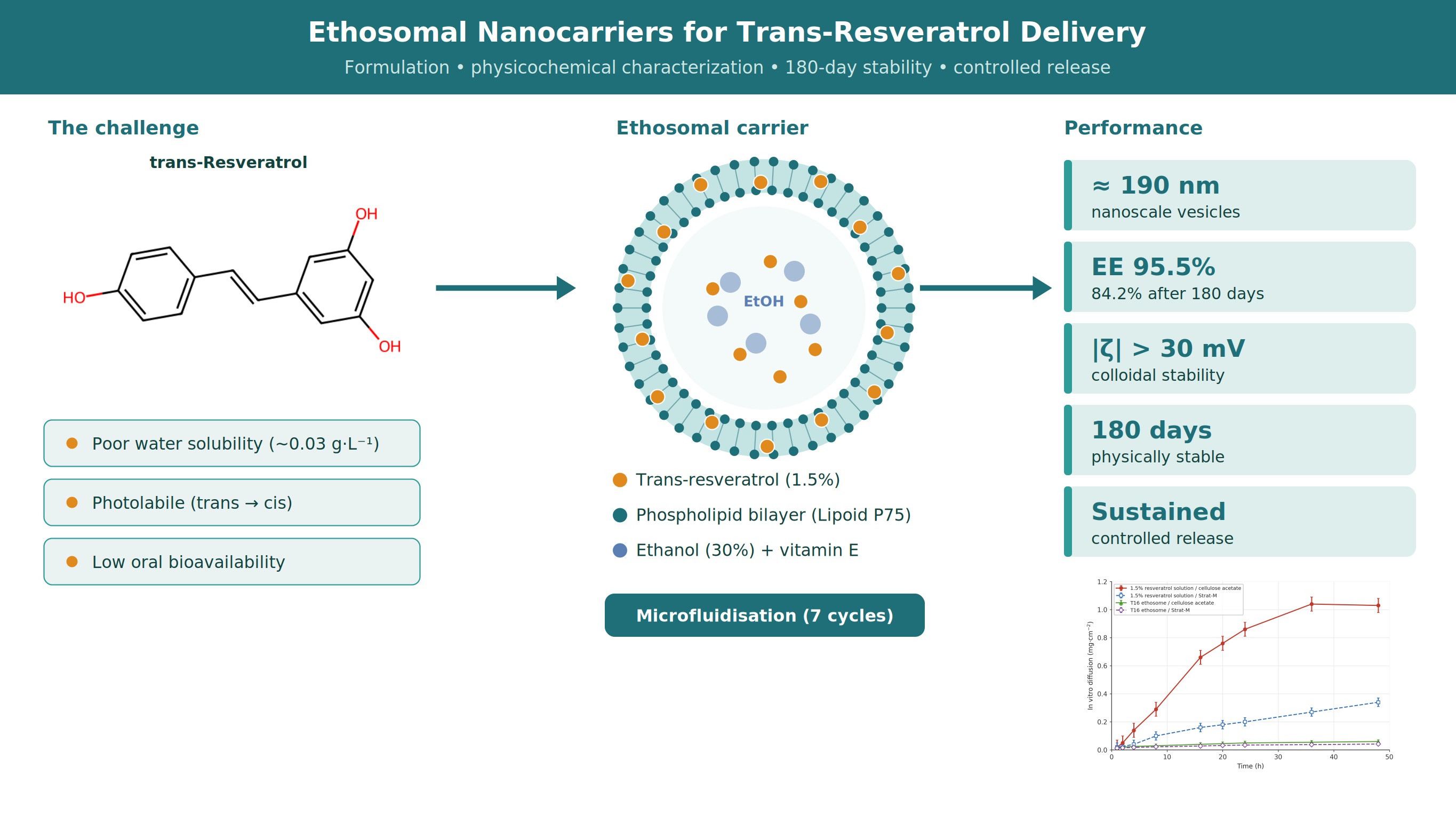
Background: Trans-resveratrol (3,5,4′-trihydroxystilbene) is a natural polyphenolic antioxidant widely used in anti-aging dermocosmetics for its strong radical-scavenging capacity and its activation of cell-protective pathways such as SIRT1. However, its poor aqueous solubility, photochemical lability, and low bioavailability limit its incorporation into topical formulations and its delivery into the skin. Objective: In this study, ethosomal nanocarriers were designed as a phospholipid–ethanol vesicular system to solubilize, stabilize, and control the release of trans-resveratrol for dermocosmetic applications. Microfluidization process is not a commonly used method, however under optimized pressure circulating the formulation through the interaction chamber can produce ethosomes with desirable colloidal stability as an easy method. Methods: Resveratrol-loaded ethosomes were prepared with synthetic phosphatidylcholine (Lipoid P75), ethanol, and vitamin E. Microfluidisation was used as an easy method which was optimized by varying the number of high-pressure homogenization cycles and the pressure applied. Vesicle size, size distribution and distribution uniformity, zeta potential, pH, conductivity, density, and long-term stability were monitored for up to 180 days; morphology was examined by cryo-SEM and molecular compatibility by FTIR. A trans-resveratrol HPLC assay was developed and validated according to ICH Q2guidelines for quantitative analysis. Encapsulation efficiency was determined by HPLC after ultracentrifugation, cytotoxicity was assessed in HaCaT keratinocytes, and in vitro release was evaluated using Franz diffusion cells with two different membranes. Results: All ethosome formulations yielded a nanoscale size distribution (median diameter around 190 nm) and good colloidal stability, with absolute zeta potentials above the 30 mV threshold at early time points and had skin-compatible pH (around 6.5). The optimized formulation (T16) achieved a high encapsulation efficiency (EE) of 95.5% on day 1, 84.2% EE was retained after 180 days, consistent with strong partitioning of the lipophilic active into the ethanol–phospholipid bilayer. FTIR confirmed preservation of the phospholipid bilayer and indicated non-covalent loading, with the resveratrol bands largely masked by the dominant lipid signals. Cryo-SEM confirmed near-spherical vesicles with narrow size distribution. In vitro release showed a sustained, controlled release profile relative to a 1.5% resveratrol solution. Slower diffusion across the skin-mimicking Strat-M membrane was observed compared to cellulose acetate membrane. Conclusions: Using Optimized trans-resveratrol-loaded ethosomes represent a stable, efficient ethosomes enabling formulation stability and controlled topical release. The antioxidant and photoprotective efficacy of the loaded system was not assessed in this study and is identified as a topic for future work.
Keywords:
trans-resveratrol
; ethosomes
; antioxidant
; photoaging
; dermocosmetics
1. Introduction
Human skin ages through two overlapping processes. Intrinsic (chronological) aging is driven by time and genetically programmed changes, whereas extrinsic aging is dominated by environmental insults such as chronic ultraviolet (UV) exposure, a process called photoaging that accounts for most of visible symptoms of aging [1,2]. A central mechanism common to both ageing is oxidative stress: UV radiation and metabolic activity generate reactive oxygen species (ROS) that damage lipids, proteins, and DNA, and that activate the mitogen-activated protein kinase and NF-κB signaling cascades. These cascades up-regulate matrix metalloproteinases (MMPs), which destroy dermal collagen and elastin, while the same time down-regulating new collagen synthesis. These processes result in wrinkling, loss of firmness and elasticity, and dyspigmentation [2,3,4]. All these processes are increased in photoaging. Since ROS are main reason of this cascade, topical antioxidants have become very important for anti-aging dermacosmetics [4].
Resveratrol (3,5,4′-trihydroxystilbene) is a stilbenoid polyphenol produced by many plants as a phytoalexin which is a defensive compound synthesized in response to microbial infection, UV radiation, or physical injury [5]. It can be found in the food mainly in grape skins and red wine, as well as in peanuts and berries. Its popular reputation is tied to the so-called “French paradox”: the epidemiological observation, first mentioned by Renaud and de Lorgeril, that populations in southern France have relatively low rates of coronary heart disease although their diet is rich in saturated fat. This effect is attributed in part to regular moderate level consumption of polyphenol-rich red wine [6]. The molecule exists as two geometric isomers; the trans-isomer is the biologically dominant and more stable form and is the one intended in most therapeutic and cosmetic applications.
Resveratrol has the molecular formula C₁₄H₁₂O₃ and a molar mass of 228.25 g/mol and has three phenolic hydroxyl groups that cause much of its antioxidant, radical-scavenging properties. An important practical limitation is its solubility and stability profile: resveratrol is poorly soluble in water (0.03 g/L) but freely soluble in ethanol, is photolabile (the trans-isomer isomerizes to the cis form), and is sensitive to oxygen, pH, and temperature. Together with extensive first-pass metabolism, this poor aqueous solubility and photochemical instability give resveratrol a low bioavailability and represent a formulation problem [7].
Resveratrol counteracts skin aging through several complementary routes, which act directly onto the photoaging cascade described above. First, its phenolic hydroxyl groups directly scavenge ROS; on top of this stoichiometric effect, resveratrol activates the Nrf2–ARE pathway and thereby up-regulates endogenous antioxidant enzymes such as superoxide dismutase, catalase, and glutathione-related defenses. This provides a catalytic and self-renewing layer of protection against UV-induced oxidative stress [4,14,15]. Secondly, it is a well-characterized activator of sirtuin 1 (SIRT1), an NAD-dependent deacetylase linked to the beneficial effects of caloric restriction, mitochondrial function, and cellular longevity, which is thought to contribute to its protective effects in keratinocytes and fibroblasts [8,9]. Thirdly, it is markedly anti-inflammatory molecule, because it inhibits NF-κB signaling and lowers pro-inflammatory mediators (TNF-α, IL-6, COX-2). By this and related pathways it suppresses the MMP up-regulation that drives collagen breakdown, helping to preserve the dermal matrix [3,10,14]. Trans-resveratrol has been shown to inhibit hydrogen-peroxide accumulation in human keratinocytes and to protect skin cells against UVA-induced damage. It also exhibits anti-melanogenic (tyrosinase-inhibiting) activity relevant to skin brightening [14,15,19]. Its antioxidant potency is frequently cited as being of the order of 50 times that of vitamin E and 30 times that of vitamin C on a comparative basis [20].
These mechanisms are explained many dermatological and clinical literature. In cosmetic science, resveratrol is valued as a topical antioxidant and anti-aging active whose antioxidant, photoprotective, anti-inflammatory, and skin-brightening actions have been reviewed extensively [16]. Clinically, a topical antioxidant combining resveratrol with baicalin and vitamin E produced measurable improvements in fine lines, firmness, and photodamage [17], and a recent randomized, double-blind, placebo-controlled trial reported that oral plus topical trans-resveratrol significantly reduced visible wrinkle scores in women over 40 with good tolerability [18]. At the systemic level the same molecule has shown cardiovascular, metabolic, neuroprotective, and anticancer activity in preclinical and early-phase clinical work [8,11,12,13]. In all of these important topical and systemic uses, the limitation is its delivery and bioavailability rather than any lack of activity [7,11].
To overcome these limitations, lipid-based vesicular carriers such as liposomes have been widely explored to stabilize and deliver dermacosmetic antioxidants, which help protecting unstable actives and providing controlled release [21,22]. Classical liposomes, however, can show limited entrapment of some actives and relatively rapid release, and they are not always very stable. Ethosomes, which are modified liposomal carriers first introduced by Touitou and co-workers (23). Many researchers addresed limitations of liposomes and discussed ethosomes containing a relatively high ethanol content (approximately 20–50%), which acts as a skin-penetration enhancer, reduces vesicle size, increases bilayer fluidity and deformability, and improves the entrapment of lipophilic and amphiphilic actives [23,24,25,26,27,28,29]. These properties make ethosomes particularly suitable for a poorly water-soluble, photolabile antioxidant such as trans-resveratrol, which can partition efficiently into, and be protected within, the ethanol–phospholipid bilayer.
The present work is a companion study to our previously published investigation of ethosomal nanocarriers for hydrolyzed collagen peptides [30], and follows the same formulation platform and characterization framework so that the actives can be compared directly. Although hydrolyzed collagen is a highly hydrophilic peptide, trans-resveratrol is a lipophilic small molecule. Therefore, the present study examines how the same phospholipid–ethanol system accommodates a bilayer-partitioning active. The aims of this study were to (a) develop and validate an HPLC assay for trans-resveratrol, (b) prepare and optimize resveratrol-loaded ethosomes by varying pressure applied and the cycles of microfluidisation process to easily produce and reduce the sizes of ethosomes, (c) characterize vesicle size, colloidal stability, encapsulation efficiency, morphology, cytotoxicity, and (d) investigate in-vitro release profile in order to evaluate ethosomes as a delivery platform for this antioxidant dermacosmetic active.
2. Results and Discussion
2.1. Analytical Method Validation of the Trans-Resveratrol Assay
2.1.1. Identity Confirmation by FT-IR
The identity of trans-resveratrol was confirmed by Fourier-transform infrared (FT-IR) spectroscopy (Figure 1). The spectrum showed the characteristic broad O–H stretching bands at 3215 and 3177 cm⁻¹ arising from the three phenolic hydroxyl groups, together with aromatic and aliphatic C=C stretching and stilbene-backbone bands at 1584, 1502, 1446, 1377, 1322, 1214, 1145, 968, 826, 666, and 609 cm⁻¹. These bands are consistent with reference spectra of trans-resveratrol and confirm the chemical integrity of the active before formulation.
2.1.2. HPLC Method Validation
A reversed phase HPLC method was developed for trans-resveratrol (Inertsil ODS-3 column; acetonitrile / 0.2% formic acid gradient; 1.5 mL/min; 40 °C; detection at 306 nm) and validated according to ICH Q2 (45) for linearity, accuracy, precision, limit of detection (LOD), limit of quantitation (LOQ), robustness, and specificity. Linearity over six concentration levels gave a regression equation y = 1×10⁸x + 430677 and R² = 0.9994, exceeding the R² ≥ 0.998 criterion (Figure S1). Accuracy at 21, 50, and 100 µg/mL gave mean recoveries of 102.8–104.2%, within the accepted 90–110% range. Repeatability at 10, 20, and 110 µg/mL and at 220 µg/mL gave coefficients of variation below 2%. The LOD and LOQ were 4.8 and 14.5 µg/mL, respectively. Robustness testing of a 200 µg·mL⁻¹ sample over 24 h showed only 0.83% variation, and specificity was confirmed by the absence of interfering peaks from unloaded ethosomal formulations at the resveratrol retention time (around 15.2 min; Figure 2). Full validation data is provided in the Supporting Information section.
Solubility studies defined the medium in which resveratrol can actually be dissolved and quantified, and therefore shaped the practical formulation decisions. The active was highly soluble in ethanol (65 mg/L) but only sparingly soluble in phosphate-buffered saline (PBS, pH 7.2; 100 µg/L), increasing to about 500 µg/L in PBS containing 2% polysorbate 20, Resveratrol dissolves readily in ethanol. The ethanol phase of the ethosome is not only carrying the active into the phospholipid bilayer and the core of the ethosome, but also it is a penetration enhancer.
Because resveratrol is almost insoluble in aqueous buffer, a simple PBS receptor medium would not maintain the sink conditions in the release experiments. The solubility data indicated that a surfactant-containing receptor medium would be used for the Franz-cell studies.; the 2% polysorbate-20 PBS medium was therefore selected to keep released resveratrol in solution and allow a valid release measurement.
2.2. Formulation, Particle Size, and Stability of Resveratrol Ethosomes
Resveratrol-loaded ethosomes were prepared following the cold method established earlier [30], with the lipophilic active dissolved in the ethanolic phospholipid phase prior to hydration, and then processed by high-pressure homogenization interaction chamber (M110P) with 1, 3, 5, or 7 cycles. All resveratrol loaded formulations contained 5% lecithin (Lipoid P75), 30% ethanol, 0.3% vitamin E, and 1.5% trans-resveratrol; by using the same method and identical conditions, loaded/unloaded ethosomes were prepared. Compositions are summarized in Table 1.
Particle size and distribution uniformity were monitored by laser diffraction over 180 days (Table 2). The resveratrol-loaded ethosomes formed a homogeneous nanosized population with volume-weighted median diameters (d₀.₅) of approximately 0.19 µm (190 nm) that remained essentially unchanged over the study period, indicating good physical stability with no significant aggregation or phase separation. The larger diameter of the loaded vesicles relative to the unloaded ethosomes (average 90 nm) is consistent with accommodation of the lipophilic active within the phospholipid bilayer. Increasing the number of homogenization cycles helped to improve the size distribution uniformity. Formulation T16 (seven cycles) had a consistent size and low, stable uniformity at all time points tested. Formulation T16, along with the unloaded T24, was used in the later encapsulation, morphology, and release studies. Size and uniformity are shown in separate figures (Figure 3 and Figure 4).
2.3. Physicochemical Properties and Colloidal Stability
The pH, conductivity, density, and zeta potential of the resveratrol-loaded (T13–T16) and unloaded (T21–T24) ethosomes are summarized in Table 3 and Figure 5. The pH of the loaded formulations (6.50–6.54) is close to the physiological pH of skin and therefore suitable for topical application, while conductivity (44.4–44.6 µS/cm) and density (0.949–0.961 g/cm³) were consistent across formulations, indicating uniform, reproducible dispersions. Zeta potential is a one of the key indicators of colloidal stability, with absolute values above 30 mV generally indicating good electrostatic stabilization [33]. Initial absolute zeta potentials of the loaded formulations were 38.7, 36.0, 39.3, and 41.2 mV for T13–T16, and although a gradual decrease was observed over storage, no visible aggregation or phase separation occurred.
Notably, the unloaded ethosomes exhibited consistently higher absolute zeta potentials than the loaded systems. The zeta potential of phospholipid ethosomes is negative, arising from the anionic phosphate groups of the phosphatidylcholine head-groups; incorporation of resveratrol lowers the magnitude of this surface charge.
Most likely some polyphenol molecules move into the outer bilayer, where its hydroxyl groups form hydrogen bonds with and partly shield the phosphate charges on the vesicle surface, which lowers the measured zeta-potential. Similar decreases in zeta potential after drug loading have been reported: Verma and Pathak observed −48.8 to −75.1 mV for econazole-loaded ethanolic vesicles [34], and Al-Ameri and Al-Gawhari reported −36.3 to −51.0 mV for soya-lecithin ethosomes [35], matching the range and the loading-related decrease in our experiments [33,34,35].
2.4. Encapsulation Efficiency
The encapsulation efficiency (EE) of the optimized resveratrol ethosome (T16) was determined by HPLC quantification of free active in the supernatant after ultracentrifugation, following the method described in our earlier study [30] (Table 4). On day 1, T16 exhibited a very high EE of 95.5% (4.5% unencapsulated), markedly higher than the 86% obtained for the hydrophilic collagen peptide ethosomes prepared by the same method [30]. This high entrapment is consistent with the lipophilic character of trans-resveratrol, which partitions efficiently into the ethanol-fluidized phospholipid bilayer rather than residing in the aqueous core. After 180 days of storage, EE decreased moderately to 84.2%, retaining the majority of the encapsulated active; such gradual reductions are commonly reported for phospholipid vesicular systems and attributed to slow bilayer rearrangement and diffusion during prolonged storage. Retention of >84% of the active after six months indicates that the ethosomal bilayer provides an effective and stable reservoir for resveratrol.
2.5. Morphological and Molecular Characterization
2.5.1. Cryo-SEM Analysis
Vesicle morphology was examined by cryo-scanning electron microscopy (cryo-SEM), which preserves the native hydrated state of the vesicles and minimises the structural collapse often seen with conventional SEM. The unloaded (T24) and loaded (T16) ethosomes both exhibited generally spherical to near-spherical morphology, confirming successful vesicle formation (Figure 6 a–d). Quantitative morphometry (Table 5) found a mean aspect ratio of 1.43 for resveratrol-loaded vesicles and 1.37 for the unloaded which is a small deviation from perfect spheres that may come from interactions between the bilayer and resveratrol. Both formulations had very low image-based size dispersion (PDI ≈ 0.038 and 0.026), showing narrow and uniform shapes. This is common with cryo-fixation of phospholipid vesicles, SEM diameters appear larger than median diameters from laser diffraction because of ice-crystal growth and 2D projection during sample preparation that inflate sizes; a similar SEM vs DLS difference has been reported previously [36].
2.5.2. FTIR Spectroscopy of the Formulations
FTIR was used to probe whether resveratrol is chemically altered on encapsulation and whether it perturbs the phospholipid bilayer, by comparing the loaded ethosome with the corresponding empty (unloaded) ethosome (Figure 7). The spectra of the loaded and empty vesicles were essentially superimposable and were dominated by phospholipid-associated bands—a broad O–H/N–H envelope at ~3340 cm⁻¹, CH₂/CH₃ stretching at 2980 cm⁻¹, a carbonyl/amide-region band at ~1645 cm⁻¹, and phosphate- and ester-related vibrations at 1407, 1326, 1277, and 1044 cm⁻¹. Critically, no characteristic lipid peaks were lost, and no new covalent bands appeared on loading resveratrol, and the loaded spectrum did not differ materially from the empty-ethosome spectrum, indicating that resveratrol did not chemically modify the bilayer. The absence of distinct resveratrol bands in the loaded spectrum is expected: in lipid-based systems the strong lipid-matrix signals dominate and mask the weaker absorbance of the entrapped active, because no new resveratrol peaks were observed, encapsulation was confirmed indirectly through the intact lipid structure, combined with the separate encapsulation efficiency and release measurements. This behavior, and the interpretation that entrapment occurs through non-covalent, physicochemical partitioning, are consistent with FTIR reports for other bioactive-loaded liposomal and ethosomal systems [38,39,40] and with the companion collagen study [30].
2.6. In Vitro and Biological Evaluation
2.6.1. Cytotoxicity Assay (HaCaT Keratinocytes)
The cytotoxicity of pure trans-resveratrol, resveratrol-loaded ethosomes (T16), and unloaded ethosomes (T24) was evaluated in HaCaT keratinocytes using the XTT viability assay over a wide concentration range (0.1–25%, v/v; Figure 8). These are high test concentrations relative to the levels at which resveratrol is used in finished topical products. Pure trans-resveratrol reduced keratinocyte viability to approximately 45–50% across the tested range, with only a weak concentration dependence, indicating a moderate intrinsic cytotoxicity of resveratrol at these concentrations. The empty ethosome (T24) was strongly concentration-dependent: viability was essentially complete (around 100%) at 0.1% but fell sharply at higher concentrations (to 12% at 10% and 4% at 25%). The resveratrol-loaded ethosome (T16) showed reduced viability across the whole range (14–25%), combining the intrinsic effect of the active with the carrier effect, and recovered only partially (to around 25%) at the lowest concentration.
The cytotoxicity observed at high test concentrations is driven mainly by the ethosomes, while the loaded system additionally reflects the moderate intrinsic cytotoxicity of resveratrol. As discussed for the companion collagen ethosomes [30], the reduced viability of the vesicles at elevated concentrations is most possibly related to the ethanol content and the surfactant-like membrane action of the phospholipid vesicles, which can disrupt cell membranes at high doses; consistent with this, the empty carrier was well tolerated only at the lowest concentration. A similar dose-dependent pattern—minimal cytotoxicity at low doses and reduced viability at higher doses or longer exposure—has been reported for other ethosomal systems, for example Gossypin-loaded ethosomes on melanoma cells [37]. These findings underscore that appropriately low use concentrations, and further optimization to improve keratinocyte tolerance, are important for topical application. However, the results should be interpreted cautiously because the test concentrations were higher than what would occur naturally in the body. The control experiments performed as expected, which supports the reliability of the measurements.
2.6.2. In Vitro Release Using Franz Diffusion Cells
The in vitro release of trans-resveratrol from the optimized ethosome (T16) was evaluated using Franz diffusion cells fitted with either a cellulose acetate dialysis membrane (12–14 kDa) or a skin-mimicking Strat-M membrane, effective area of the cells was 0.21 cm²); a 2% polysorbate-20 PBS receptor solution was selected as receiving medium on the basis of the solubility data. A 1.5% resveratrol solution served as the free-active comparator; the cumulative amount diffused per unit area was measured over 1–48 h (Figure 9). A consistent rank order was obtained: the free 1.5% solution diffused fastest across cellulose acetate, reaching around 1.03 mg/cm² at 48 h, followed by the solution across Strat-M (0.34 mg/cm²), then the loaded ethosome across cellulose acetate (0.06 mg·cm⁻²), with the loaded ethosome across Strat-M the slowest 0.04 mg/cm²). The encapsulated resveratrol was therefore released in a markedly sustained, controlled manner relative to the free resveratrol solution. The difference was almost an order-of-magnitude reduction in diffused amount confirming that incorporation into the ethosomal bilayer strongly controls release. The slower diffusion across Strat-M relative to cellulose acetate reflects the higher barrier effect of the skin-mimicking membrane against resveratrol [41,42,43]. From a dermocosmetic perspective, this prolonged availability of resveratrol at the skin surface or within the upper skin layers is advantageous and is consistent with the sustained-release behaviour reported for ethosomal systems [26,30].
3. Materials and Methods
The formulation platform, instrumentation, and characterization procedures used here are identical to those established for the earlier study of hydrolyzed-collagen ethosome [30]. The resveratrol-specific materials, the HPLC assay, and the points of difference are described in detail below.
3.1. Materials
Trans-resveratrol was used as received; a certificate of analysis was provided by the supplier (CAS No:, Organic Herb Inc.,Changsha, China). Ethanol (>99%, Merck, Darmstadt, Germany) and soybean lecithin containing phosphatidylcholine (Lipoid P75, Lipoid GmbH, Ludwigshafen am Rhein, Germany; >70% phosphatidylcholine) were used as the phospholipid source, and vitamin E (BASF, Ludwigshafen, Germany) was used as an antioxidant excipient. Disodium phosphate, monopotassium phosphate, sodium chloride, and potassium chloride (Merck/Sigma-Aldrich, Darmstadt, Germany) were used to prepare phosphate-buffered saline (PBS), and polysorbate 20 (Emulgin SML 20, BASF, Ludwigshafen, Germany) was added at 2% to the PBS receptor medium. Acetonitrile and formic acid (HPLC grade) were used for chromatography, and ultrapure water (Merck, Darmstadt, Germany) was used for all analyses. The XTT Cell Proliferation Kit (Roche Diagnostics, Mannheim, Germany) and DMEM (Sigma-Aldrich, Darmstadt, Germany) were used for cell-viability assays.
A high-pressure homogenizer (Microfluidizer M-110P, Microfluidics, Middleborough, MA, USA) was used for ethosome preparation and particle-size reduction. Particle size and distribution were analyzed using a Mastersizer 2000 (Malvern Instruments, Malvern, UK), and zeta potential using an Anton Paar Litesizer 500 (Anton Paar, Graz, Austria). FT-IR spectra were collected on a Thermo Scientific Nicolet iS5 spectrometer with an iD5 ATR accessory (Thermo Fisher Scientific, Waltham, MA, USA). pH was measured with a WTW pH meter (Xylem/WTW, Weilheim, Germany) and density with an Anton Paar DMA 38 digital density meter (Anton Paar, Graz, Austria). Skin-permeation experiments used a LOGAN FDC-6 Franz diffusion cell system (Logan Instruments, Somerset, NJ, USA); Strat-M synthetic membranes (Merck, Darmstadt, Germany) and cellulose acetate membranes with a 12–14 kDa molecular-weight cut-off (Spectrum Laboratories, Rancho Dominguez, CA, USA) were used as diffusion membranes.
3.2. HPLC Assay and Validation of Trans-Resveratrol
Trans-resveratrol was quantified by HPLC (Shimadzu SIL-20AC HT autosampler and pump) on an Inertsil ODS-3 column (250 mm × 4.6 mm, 5 µm). Mobile phase A was acetonitrile and mobile phase B was 0.2% formic acid, delivered as a gradient at 1.5 mL·min⁻¹ with a 40 °C column temperature; 20 µL of sample was injected and detection was at 306 nm. Test and reference solutions were prepared in ethanol (sonicated 10 min, cooled, made to volume) and filtered through 0.45 µm PTFE filters; calibration standards were prepared by dilution over the range 0.0075–0.25 mg/mL. The method was validated according to ICH Q2(R1) [45] for linearity, accuracy, precision (repeatability), LOD, LOQ, range, robustness, and specificity, as detailed in the enclosed Supporting Information section.
Solubility of trans-resveratrol was determined by stirring 1 g of active in 30 mL of medium (ethanol, PBS pH 7.2, or PBS containing 2% polysorbate 20) at 150 rpm and 25 °C for 1 h, filtering through a 0.45 µm membrane, and assaying the filtrate by the HPLC method above; three replicates were performed for each medium.
3.3. Preparation of Resveratrol Ethosomes
Ethosomes were prepared by the cold method [30,31]. For the loaded formulations, 5% (w/w) Lipoid P75 and 0.3% (w/w) vitamin E were dissolved in 30% (w/w) ethanol under mechanical stirring (300 rpm), and 1.5% (w/w) trans-resveratrol was dissolved in this ethanolic lipid phase. The aqueous phase (deionized water, 63.2% w/w) was then added dropwise at 30 °C under continuous stirring to induce vesicle formation, and stirring was continued for a further 5 min; unloaded ethosomes were prepared identically, omitting the active and adjusting the water content to 64.7% (Table 1). Both loaded and unloaded dispersions were then passed through Microfluidizer M-110P for high-pressure homogenization and size reduction. The conditions used were; 1000 bar pressure; 1, 3, 5, or 7 cycles (each cycle corresponding to a single pass through the interaction chamber), at 25 °C [32]. Within the interaction chamber, there are microchannels in which product stream is accelerate to high speeds and impringes itself on surfaces and particles splits into smaller particles.
3.4. Characterization of the Ethosomes
Particle size and distribution uniformity (laser diffraction; volume-weighted median diameter d₀.₅), zeta potential, pH, conductivity, density, and cryo-SEM morphology were determined exactly as described in the earlier study [30]. Physical stability was assessed by monitoring particle size, size distribution uniformity, zeta potential, and appearance at 1, 7, 14, 30, and 180 days at 25 ± 2 °C. FTIR spectra of loaded and unloaded ethosomes were compared to assess active–bilayer interactions.
3.5. Encapsulation Efficiency
Encapsulation efficiency was determined by ultracentrifugation of the ethosome dispersion followed by HPLC quantification of free (unencapsulated) resveratrol in the supernatant. EE (%) was calculated as
EE = (T − C)/T × 100
Where T is the total amount of resveratrol added to the formulation and C is the unentrapped amount detected in the supernatant (30).
3.6. In Vitro and Biological Evaluation
Cytotoxicity was evaluated in HaCaT keratinocytes using the XTT Cell Proliferation Assay following ISO 10993-5 [44], with formulations and pure resveratrol tested at 25, 10, 1, and 0.1%, sodium lauryl sulfate as positive control and culture medium as negative control.
In vitro release was assessed using a Franz diffusion cell system with cellulose acetate (12–14 kDa) and Strat-M® membranes (0.21 cm²); 1 mL of ethosomal dispersion or 1.5% resveratrol solution was used as the donor compartment, and receptor aliquots were withdrawn at 1, 2, 4, 8, 16, 24, and 48 h and quantities were determined by HPLC. A 2% polysorbate-20 PBS solution was used as the receptor medium, at each sampling time 1ml of warmed receptor solution was added to replace the sample withdrawn to maintain sink condition also to ensure the receptor solution making full-surface contact with the membrane without any air bubbles [30].
3.7. Statistical Analysis
All experiments were performed in at least three replicates and results are expressed as mean ± standard deviation (SD). Where applicable, group differences were assessed by one-way analysis of variance (ANOVA) followed by Tukey's test, with p < 0.05 considered statistically significant.
4. Conclusions
In this study, ethosomal formulations were successfully developed for the delivery of trans-resveratrol, a poorly water-soluble, antioxidant dermacosmetic active. Using the same phospholipid–ethanol platform established for hydrolyzed collagen peptides [30], stable nanoscale vesicles (190 nm) were obtained by mcrofludisation under high-pressure homogenization. A validated HPLC assay enabled reliable quantification of resveratrol. The optimized formulation (T16) achieved a very high encapsulation efficiency (95.5% on day 1, 84.2% after 180 days), consistent with efficient partitioning of the lipophilic active into the ethanol-fluidized bilayer and notably higher than for the hydrophilic collagen load. Comprehensive characterization—particle size, zeta potential, pH, conductivity, density, and cryo-SEM—demonstrated uniform, physically stable vesicles over 180 days, and FTIR confirmed preservation of the phospholipid bilayer, with encapsulation of resveratrol was as non-covalent bilayer partitioning. In vitro Franz-cell studies showed that ethosomal encapsulation provides a markedly sustained, controlled release compared to a simple resveratrol solution. Cell-viability testing showed dose-dependent cytotoxicity driven mainly by the vesicular carrier at high test concentrations, underscoring the importance of appropriate use levels. Overall, it was shown that optimized trans-resveratrol-loaded ethosomes are an effective, stable nanocarrier for this antioxidant active, complementing the hydrophilic-peptide system reported previously and supporting ethosomes as a versatile platform for anti-aging dermacosmetic ingredients. The Microfluidisation method proved to be an easy method to use obtaining stable ethosomes with desired nanosizes.
Author Contributions
Conceptualization, Y.Y.U. and H.S.; methodology, Y.Y.U. and H.S.; investigation, H.S and Y.Y.U.; writing-original draft preparation, Y.Y.U.; writing—review and editing, Y.Y.U. and H.S. All authors have read and agreed to the published version of the manuscript.
Funding
This research received no external funding.
Data Availability Statement
This study is part a Master of Science Thesis prepared by Hakan Sevinç under the supervision of Yasemin Yağan Uzuner. Some of the additional is given in Supporting Information section. Any further information can be requested from the corresponding author.
Acknowledgments
This Master of Science Thesis was studied at Yeditepe University, Istanbul and some of the tests were conducted at Acibadem University, İstanbul and Biota Research and Development Centre laboratories, Istanbul. We would like to thank both of the universities and Biota Research and Development Centre for the opportunities and the support provided for completion of this study.
Conflicts of Interest
The author H.S. is employed by Biota. The other author declares no conflict of interest.
References
- Naharro-Rodriguez, J.; Bacci, S.; Hernandez-Bule, M.L.; Perez-Gonzalez, A.; Fernandez-Guarino, M. Decoding skin aging: a review of mechanisms, markers, and modern therapies. Cosmetics 2025, 12, 144. [Google Scholar] [CrossRef]
- Rittié, L.; Fisher, G.J. Natural and sun-induced aging of human skin. Cold Spring Harb. Perspect. Med. 2015, 5, a015370. [Google Scholar] [CrossRef]
- Pittayapruek, P.; Meephansan, J.; Prapapan, O.; Komine, M.; Ohtsuki, M. Role of matrix metalloproteinases in photoaging and photocarcinogenesis. Int. J. Mol. Sci. 2016, 17, 868. [Google Scholar] [CrossRef]
- Rinnerthaler, M.; Bischof, J.; Streubel, M.K.; Trost, A.; Richter, K. Oxidative stress in aging human skin. Biomolecules 2015, 5, 545–589. [Google Scholar] [CrossRef] [PubMed]
- Feng, C.; Chen, J.; Ye, W.; Liao, K.; Wang, Z.; Song, X.; Qiao, M. Synthetic biology-driven microbial production of resveratrol: advances and perspectives. Front. Bioeng. Biotechnol. 2022, 10, 833920. [Google Scholar] [CrossRef]
- Renaud, S.; de Lorgeril, M. Wine, alcohol, platelets, and the French paradox for coronary heart disease. Lancet 1992, 339, 1523–1526. [Google Scholar] [CrossRef] [PubMed]
- Delmas, D.; Aires, V.; Limagne, E.; Dutartre, P.; Mazué, F.; Ghiringhelli, F.; Latruffe, N. Transport, stability, and biological activity of resveratrol. Ann. N. Y. Acad. Sci. 2011, 1215, 48–59. [Google Scholar] [CrossRef]
- Baur, J.A.; Sinclair, D.A. Therapeutic potential of resveratrol: the in vivo evidence. Nat. Rev. Drug Discov. 2006, 5, 493–506. [Google Scholar] [CrossRef]
- Howitz, K.T.; Bitterman, K.J.; Cohen, H.Y.; Lamming, D.W.; Lavu, S.; Wood, J.G.; Zipkin, R.E.; Chung, P.; Kisielewski, A.; Zhang, L.-L.; Scherer, B.; Sinclair, D.A. Small molecule activators of sirtuins extend Saccharomyces cerevisiae lifespan. Nature 2003, 425, 191–196. [Google Scholar] [CrossRef]
- Salehi, B.; Mishra, A.P.; Nigam, M.; Sener, B.; Kilic, M.; Sharifi-Rad, M.; Fokou, P.V.T.; Martins, N.; Sharifi-Rad, J. Resveratrol: a double-edged sword in health benefits. Biomedicines 2018, 6, 91. [Google Scholar] [CrossRef]
- Berman, A.Y.; Motechin, R.A.; Wiesenfeld, M.Y.; Holz, M.K. The therapeutic potential of resveratrol: a review of clinical trials. npj Precis. Oncol. 2017, 1, 35. [Google Scholar] [CrossRef]
- Bonnefont-Rousselot, D. Resveratrol and cardiovascular diseases. Nutrients 2016, 8, 250. [Google Scholar] [CrossRef]
- Godos, J.; Romano, G.L.; Gozzo, L.; Laudani, S.; Paladino, N.; Dominguez Azpíroz, I.; Martínez López, N.M.; Giampieri, F.; Quiles, J.L.; Battino, M.; Galvano, F.; Drago, F.; Grosso, G. Resveratrol and vascular health: evidence from clinical studies and mechanisms of action related to its metabolites produced by gut microbiota. Front. Pharmacol. 2024, 15, 1368949. [Google Scholar] [CrossRef]
- Ndiaye, M.; Philippe, C.; Mukhtar, H.; Ahmad, N. The grape antioxidant resveratrol for skin disorders: promise, prospects, and challenges. Arch. Biochem. Biophys. 2011, 508, 164–170. [Google Scholar] [CrossRef]
- Soeur, J.; Eilstein, J.; Léreaux, G.; Jones, C.; Marrot, L. Skin resistance to oxidative stress induced by resveratrol: from Nrf2 activation to GSH biosynthesis. Free Radic. Biol. Med. 2015, 78, 213–223. [Google Scholar] [CrossRef]
- Ratz-Łyko, A.; Arct, J. Resveratrol as an active ingredient for cosmetic and dermatological applications: a review. J. Cosmet. Laser Ther. 2019, 21, 84–90. [Google Scholar] [CrossRef]
- Farris, P.; Yatskayer, M.; Chen, N.; Krol, Y.; Oresajo, C. Evaluation of efficacy and tolerance of a nighttime topical antioxidant containing resveratrol, baicalin, and vitamin E for treatment of mild to moderately photodamaged skin. J. Drugs Dermatol. 2014, 13, 1467–1472. [Google Scholar]
- Rao, A.; Briskey, D.; Roche, G.; Tremblay, A.; Da Silva Pinto, M.; Tompkins, T.A. Trans-resveratrol reduces visible signs of skin ageing in healthy adult females over 40: an 8-week randomized placebo-controlled trial. Front. Aging 2025, 6, 1727244. [Google Scholar] [CrossRef]
- Buonocore, D.; Lazzeretti, A.; Tocabens, P.; Nobile, V.; Cestone, E.; Santin, G.; Bottone, M.G.; Marzatico, F. Resveratrol-procyanidin blend: nutraceutical and antiaging efficacy evaluated in a placebo-controlled, double-blind study. Clin. Cosmet. Investig. Dermatol. 2012, 5, 159–165. [Google Scholar] [CrossRef]
- Addor, F.A.S. Antioxidants in dermatology. An. Bras. Dermatol. 2017, 92, 356–362. [Google Scholar] [CrossRef]
- Luo, M.; Oomah, B.D.; Ackoetey, W.; Zhang, Y.; Daneshfozoun, H.; Hosseinian, F. Liposomes as sustainable delivery systems in food, cosmetic, and pharmaceutical applications. J. Am. Oil Chem. Soc. 2024, 102, 547–568. [Google Scholar] [CrossRef]
- Ricci, A.; Stefanuto, L.; Gasperi, T.; Bruni, F.; Tofani, D. Lipid nanovesicles for antioxidant delivery in skin: liposomes, ufasomes, ethosomes, and niosomes. Antioxidants 2024, 13, 1516. [Google Scholar] [CrossRef]
- Touitou, E.; Dayan, N.; Bergelson, L.; Godin, B.; Eliaz, M. Ethosomes—novel vesicular carriers for enhanced delivery: characterization and skin penetration properties. J. Control. Release 2000, 65, 403–418. [Google Scholar] [CrossRef]
- Musielak, E.; Krajka-Kuźniak, V. Liposomes and ethosomes: comparative potential in enhancing skin permeability for therapeutic and cosmetic applications. Cosmetics 2024, 11, 191. [Google Scholar] [CrossRef]
- Ascenso, A.; Raposo, S.; Batista, C.; Cardoso, P.; Mendes, T.; Praça, F.G.; Bentley, M.V.L.B.; Simões, S. Development, characterization, and skin delivery studies of related ultradeformable vesicles: transfersomes, ethosomes, and transethosomes. Int. J. Nanomed. 2015, 10, 5837–5851. [Google Scholar] [CrossRef]
- Abdulbaqi, I.M.; Darwis, Y.; Khan, N.A.K.; Assi, R.A.; Khan, A.A. Ethosomal nanocarriers: the impact of constituents and formulation techniques on ethosomal properties, in vivo studies, and clinical trials. Int. J. Nanomed. 2016, 11, 1687–1704. [Google Scholar] [CrossRef]
- Paiva-Santos, A.C.; Silva, A.L.; Guerra, C.; Peixoto, D.; Pereira-Silva, M.; Mahdi, Z.; Filipa, M.M.; Castro, R.; Veiga, F. Ethosomes as nanocarriers for the development of skin delivery formulations. Pharm. Res. 2021, 38, 947–970. [Google Scholar] [CrossRef]
- Jafari, A.; Daneshamouz, S.; Ghasemiyeh, P.; Mohammadi-Samani, S. Ethosomes as dermal/transdermal drug delivery systems: applications, preparation and characterization. J. Liposome Res. 2023, 33, 34–52. [Google Scholar] [CrossRef]
- Zhan, B.; Wang, J.; Li, H.; Xiao, K.; Fang, X.; Shi, Y.; Jia, Y. Ethosomes: a promising drug delivery platform for transdermal application. Chemistry 2024, 6, 993–1019. [Google Scholar] [CrossRef]
- Uzuner, Y.Y.; Sevinç, H.; Kanlidere, Z. Ethosomal nanocarriers for hydrophilic peptide encapsulation: formulation optimization, stability, and in vitro release performance. Molecules 2026, 31, 744. [Google Scholar] [CrossRef]
- Bhalaria, M.K.; Naik, S.; Misra, A.N. Ethosomes: a novel delivery system for antifungal drugs in the treatment of topical fungal diseases. Indian J. Exp. Biol. 2009, 47, 368–375. [Google Scholar]
- Rakesh, R.; Anoop, K.R. Formulation and optimization of nano-sized ethosomes for enhanced transdermal delivery of cromolyn sodium. J. Pharm. Bioallied Sci. 2012, 4, 333–340. [Google Scholar] [CrossRef]
- Samimi, S.; Maghsoudnia, N.; Baradaran Eftekhari, R.; Dorkoosh, F. Lipid-based nanoparticles for drug delivery systems. In Characterization and Biology of Nanomaterials for Drug Delivery; Elsevier: Amsterdam, The Netherlands, 2019; pp. 47–76. [Google Scholar]
- Verma, P.; Pathak, K. Nanosized ethanolic vesicles loaded with econazole nitrate for the treatment of deep fungal infections through topical gel formulation. Nanomedicine 2012, 8, 489–496. [Google Scholar] [CrossRef]
- Al-Ameri, A.A.F.; Al-Gawhari, F.J. Formulation development of meloxicam binary ethosomal hydrogel for topical delivery: in vitro and in vivo assessment. Pharmaceutics 2024, 16, 898. [Google Scholar] [CrossRef]
- Chudzińska-Skorupinska, J.; Wawrzyńczak, A.; Feliczak-Guzik, A. Ethosome-based colloidal systems for transdermal delivery: the role of biosurfactant in enhancing stability and efficacy. Materials 2025, 18, 5355. [Google Scholar] [CrossRef]
- Bondi, M.; Pula, W.; Benedusi, M.; Trinchera, G.; Baldisserotto, A.; Manfredini, S.; Ortore, M.G.; Pepe, A.; Mariani, P.; Stuart, M.C.A.; et al. Gossypin-loaded ethosome gel for cutaneous administration: a preliminary study on melanoma cells. Antioxidants 2025, 14, 186. [Google Scholar] [CrossRef]
- Li, M.; Li, M.; Li, X.; Shao, W.; Pei, X.; Dong, R.; Ren, H.; Jia, L.; Li, S.; Ma, W.; et al. Preparation, characterization and ex vivo skin permeability evaluation of type I collagen-loaded liposomes. Int. J. Nanomed. 2023, 18, 1853–1871. [Google Scholar] [CrossRef]
- Sinaei, A.; Osanloo, M.; Zarenezhad, E.; Alipanah, H.; Ghanbariasad, A. Preparation of liposomal nanocarriers containing all-trans retinoic acid and docetaxel and their evaluation in a lung cancer cell line. Sci. Rep. 2025, 15, 39066. [Google Scholar] [CrossRef]
- Čutović, N.; Marković, T.; Carević, T.; Stojković, D.; Bugarski, B.; Jovanović, A.A. Liposomal and liposomes-film systems as carriers for bioactives from Paeonia tenuifolia L. petals: physicochemical characterization and biological potential. Pharmaceutics 2023, 15, 2742. [Google Scholar] [CrossRef]
- Zsikó, S.; Csányi, E.; Kovács, A.; Budai-Szűcs, M.; Gácsi, A.; Berkó, S. Methods to evaluate skin penetration in vitro. Sci. Pharm. 2019, 87, 19. [Google Scholar] [CrossRef]
- Arce, F.J.; Asano, N.; See, G.L.; Itakura, S.; Todo, H.; Sugibayashi, K. Usefulness of artificial membrane, Strat-M, in the assessment of drug permeation from complex vehicles in finite dose conditions. Pharmaceutics 2020, 12, 173. [Google Scholar] [CrossRef]
- Salamanca, C.H.; Barrera-Ocampo, A.; Lasso, J.C.; Camacho, N.; Yarce, C.J. Franz diffusion cell approach for pre-formulation characterisation of ketoprofen semi-solid dosage forms. Pharmaceutics 2018, 10, 148. [Google Scholar] [CrossRef] [PubMed]
- ISO 10993-5:2009; Biological Evaluation of Medical Devices—Part 5: Tests for In Vitro Cytotoxicity. International Organization for Standardization: Geneva, Switzerland, 2009.
- ICH Harmonised Tripartite Guideline. Validation of Analytical Procedures: Text and Methodology Q2(R1). International Council for Harmonisation: Geneva, Switzerland.
- Prasanthi, D.; Lakshmi, P.K. Vesicles—mechanism of transdermal permeation: a review. Asian J. Pharm. Clin. Res. 2012, 5, 18–25. [Google Scholar]
Figure 1.
FT-IR spectrum of trans-resveratrol showing the characteristic O–H and aromatic/aliphatic C=C absorption bands.
Figure 1.
FT-IR spectrum of trans-resveratrol showing the characteristic O–H and aromatic/aliphatic C=C absorption bands.
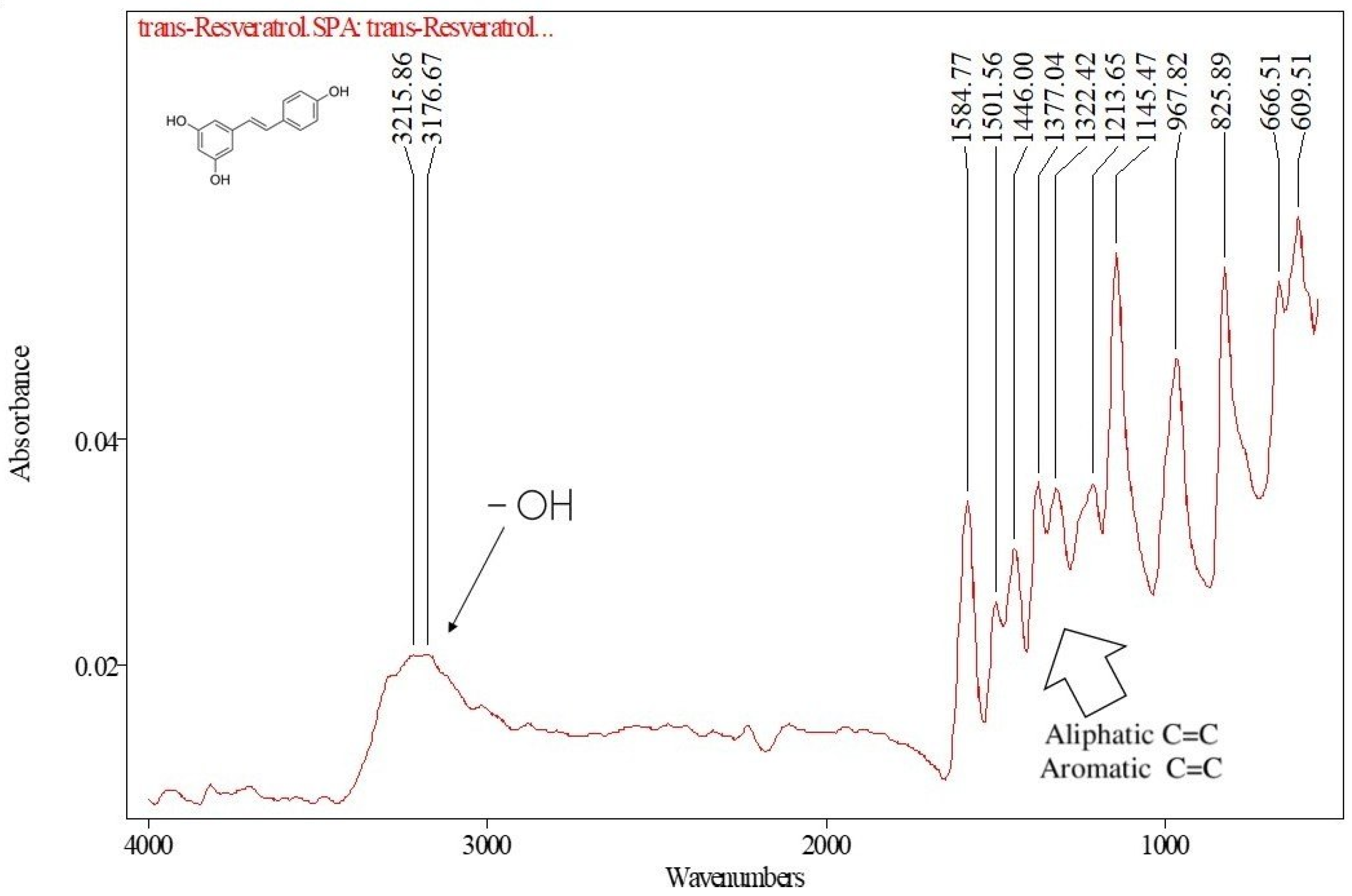
Figure 2.
Representative HPLC chromatogram of trans-resveratrol (306 nm); a single, well-resolved peak confirms method specificity.
Figure 2.
Representative HPLC chromatogram of trans-resveratrol (306 nm); a single, well-resolved peak confirms method specificity.
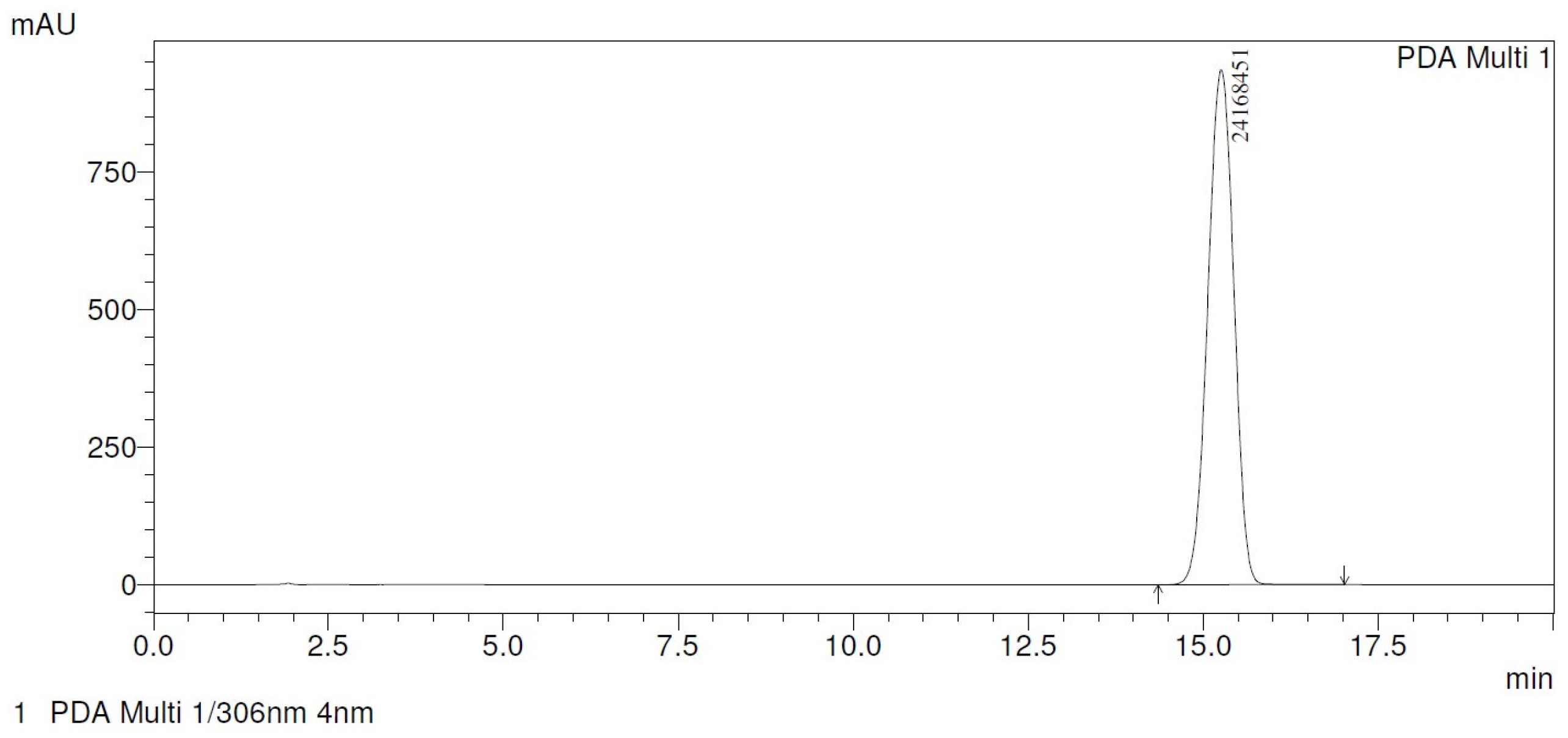
Figure 3.
Particle size (d₀.₅) of resveratrol-loaded (T13–T16, solid lines/filled symbols) and unloaded (T21–T24, dashed lines/open symbols) ethosomes over 180 days at 25 ± 2 °C.
Figure 3.
Particle size (d₀.₅) of resveratrol-loaded (T13–T16, solid lines/filled symbols) and unloaded (T21–T24, dashed lines/open symbols) ethosomes over 180 days at 25 ± 2 °C.
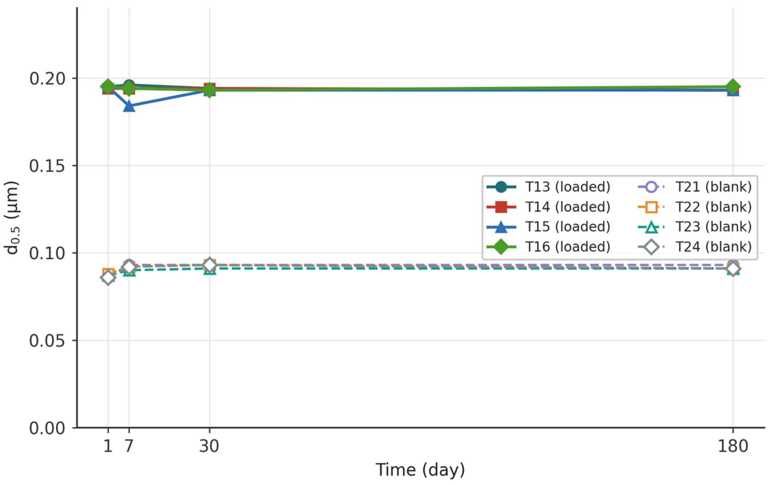
Figure 4.
Size-distribution uniformity of resveratrol-loaded (T13–T16, solid lines/filled symbols) and unloaded (T21–T24, dashed lines/open symbols) ethosomes over 180 days.
Figure 4.
Size-distribution uniformity of resveratrol-loaded (T13–T16, solid lines/filled symbols) and unloaded (T21–T24, dashed lines/open symbols) ethosomes over 180 days.
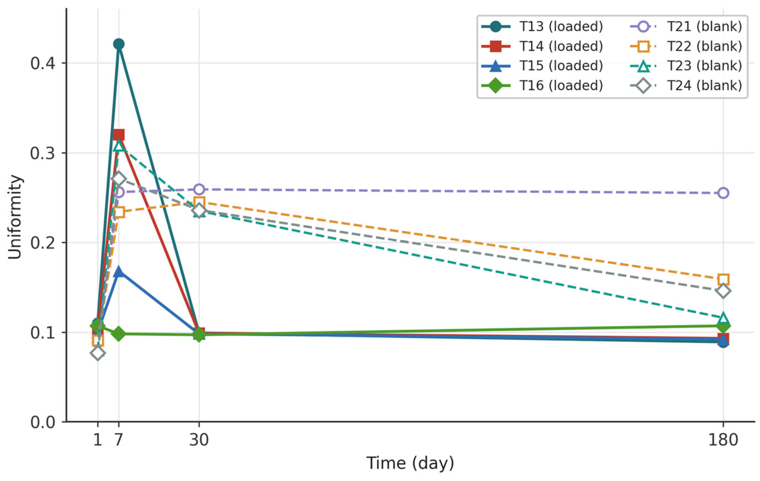
Figure 5.
Absolute zeta potential of resveratrol-loaded (T13–T16, solid) and unloaded (T21–T24, dashed) ethosomes over 30 days; the dotted line marks the 30 mV colloidal-stability threshold.
Figure 5.
Absolute zeta potential of resveratrol-loaded (T13–T16, solid) and unloaded (T21–T24, dashed) ethosomes over 30 days; the dotted line marks the 30 mV colloidal-stability threshold.
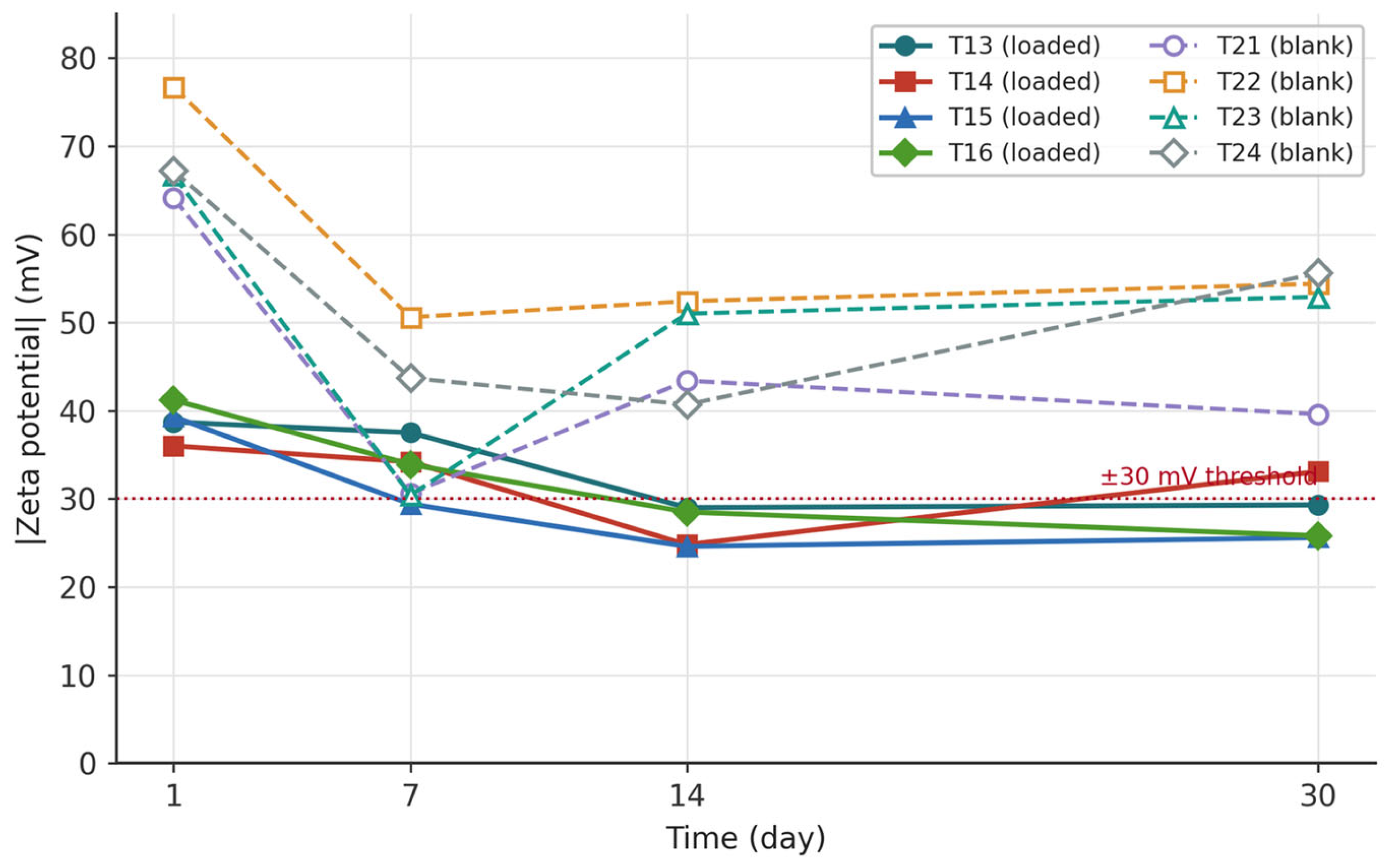
Figure 6.
Cryo-SEM images of (a,b) unloaded ethosomes (T24) at 10 µm and 5 µm and (c,d) resveratrol-loaded ethosomes (T16) at 10 µm and 5 µm, the latter with annotated morphometric area analysis.
Figure 6.
Cryo-SEM images of (a,b) unloaded ethosomes (T24) at 10 µm and 5 µm and (c,d) resveratrol-loaded ethosomes (T16) at 10 µm and 5 µm, the latter with annotated morphometric area analysis.
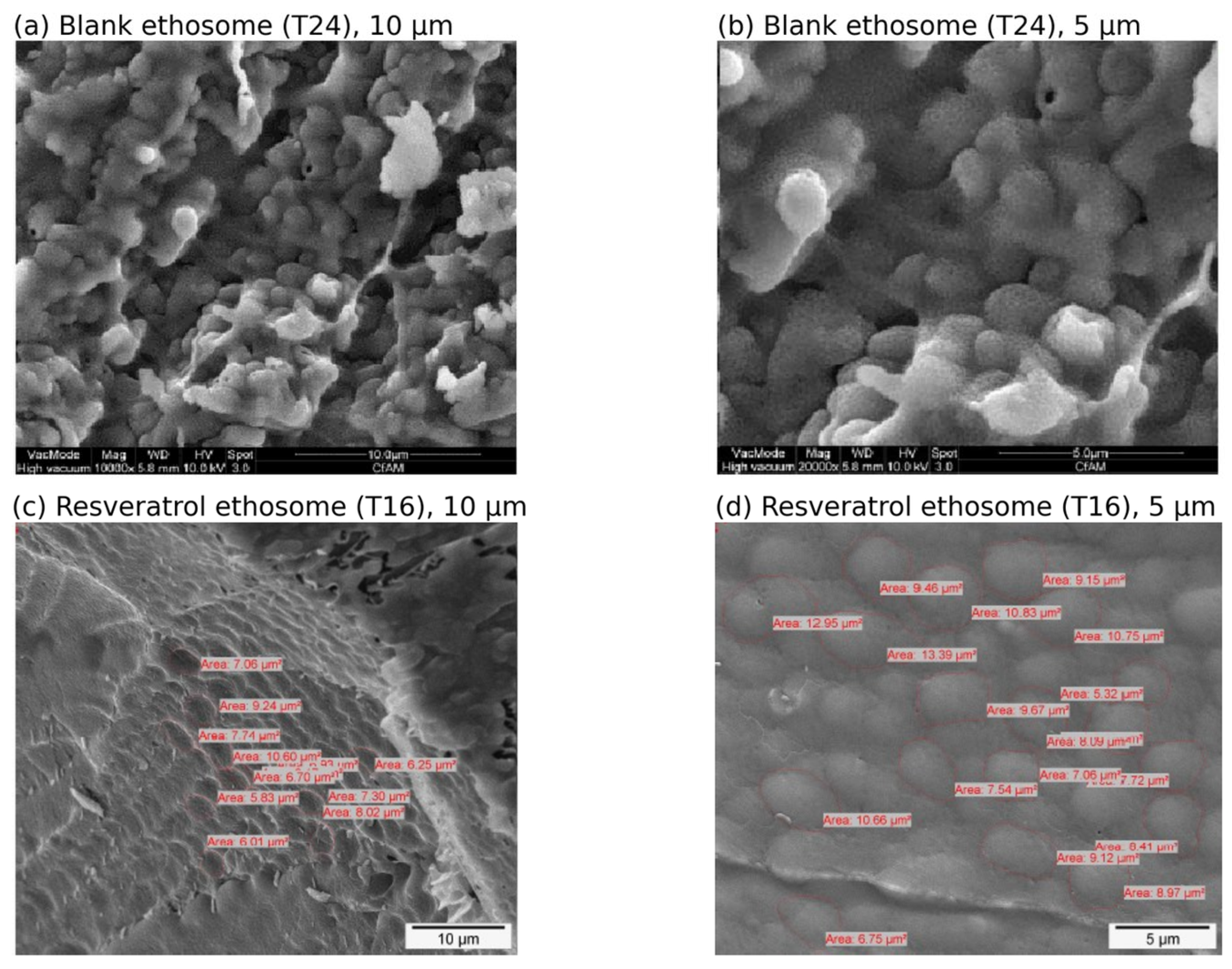
Figure 7.
FTIR spectra of loaded and unloaded (empty) ethosomal formulations, showing near-identical, phospholipid-dominated band patterns and no new covalent bands upon resveratrol loading.
Figure 7.
FTIR spectra of loaded and unloaded (empty) ethosomal formulations, showing near-identical, phospholipid-dominated band patterns and no new covalent bands upon resveratrol loading.
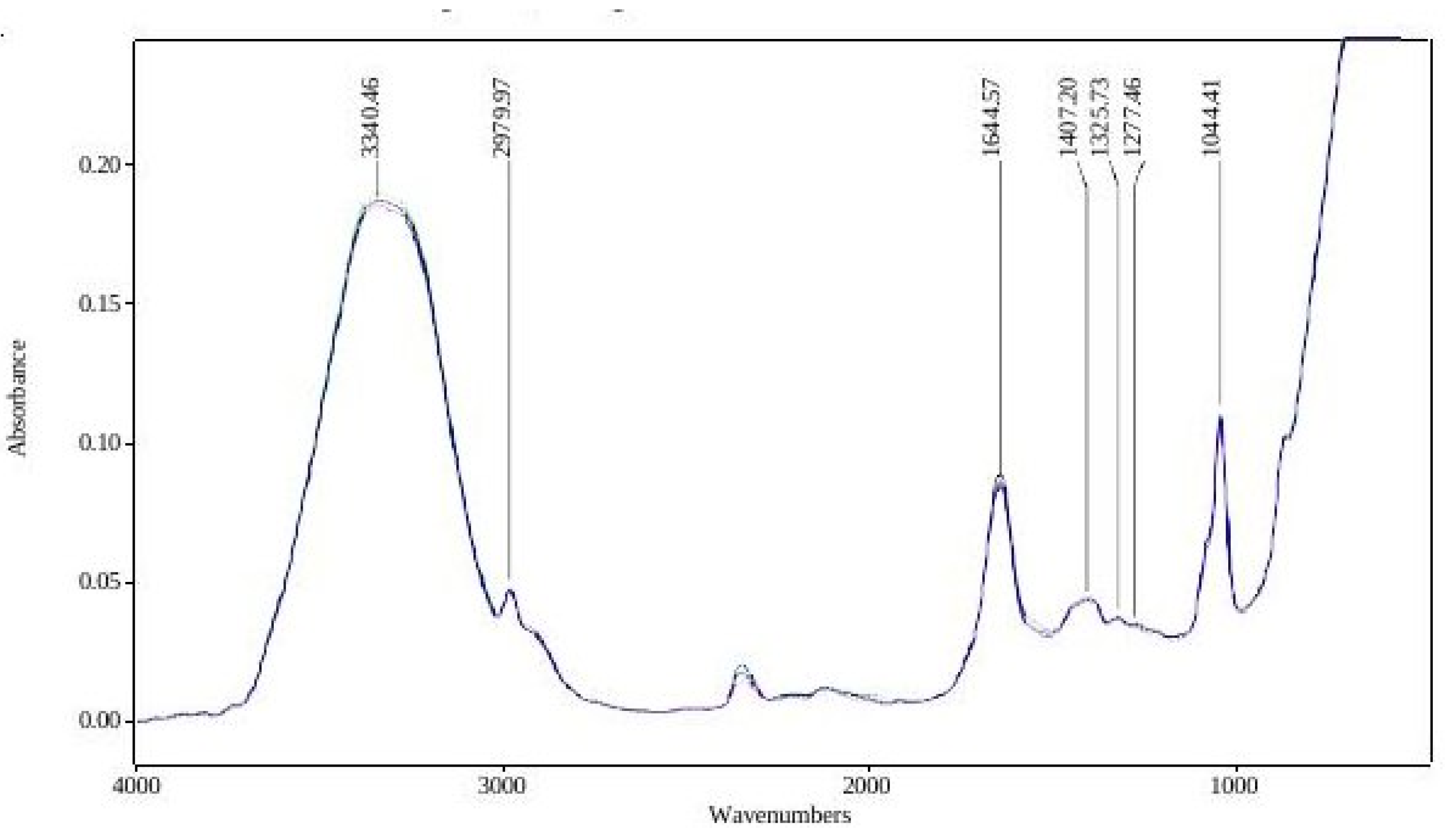
Figure 8.
HaCaT keratinocyte viability after 24 h exposure to pure trans-resveratrol, the resveratrol-loaded ethosome (T16), and the empty ethosome (T24) at 0.1–25% (v/v); mean ± SD.
Figure 8.
HaCaT keratinocyte viability after 24 h exposure to pure trans-resveratrol, the resveratrol-loaded ethosome (T16), and the empty ethosome (T24) at 0.1–25% (v/v); mean ± SD.
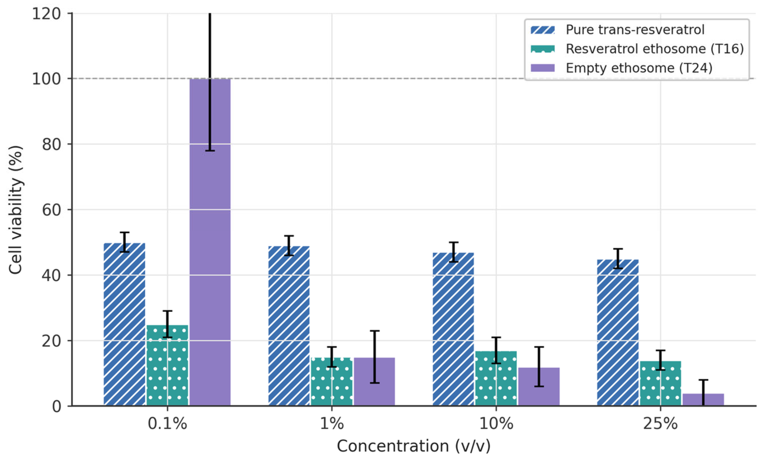
Figure 9.
Time-dependent in vitro release of trans-resveratrol from the T16 ethosome versus a 1.5% resveratrol solution across cellulose acetate and Strat-M membranes (Franz diffusion cells; mean ± SD).
Figure 9.
Time-dependent in vitro release of trans-resveratrol from the T16 ethosome versus a 1.5% resveratrol solution across cellulose acetate and Strat-M membranes (Franz diffusion cells; mean ± SD).
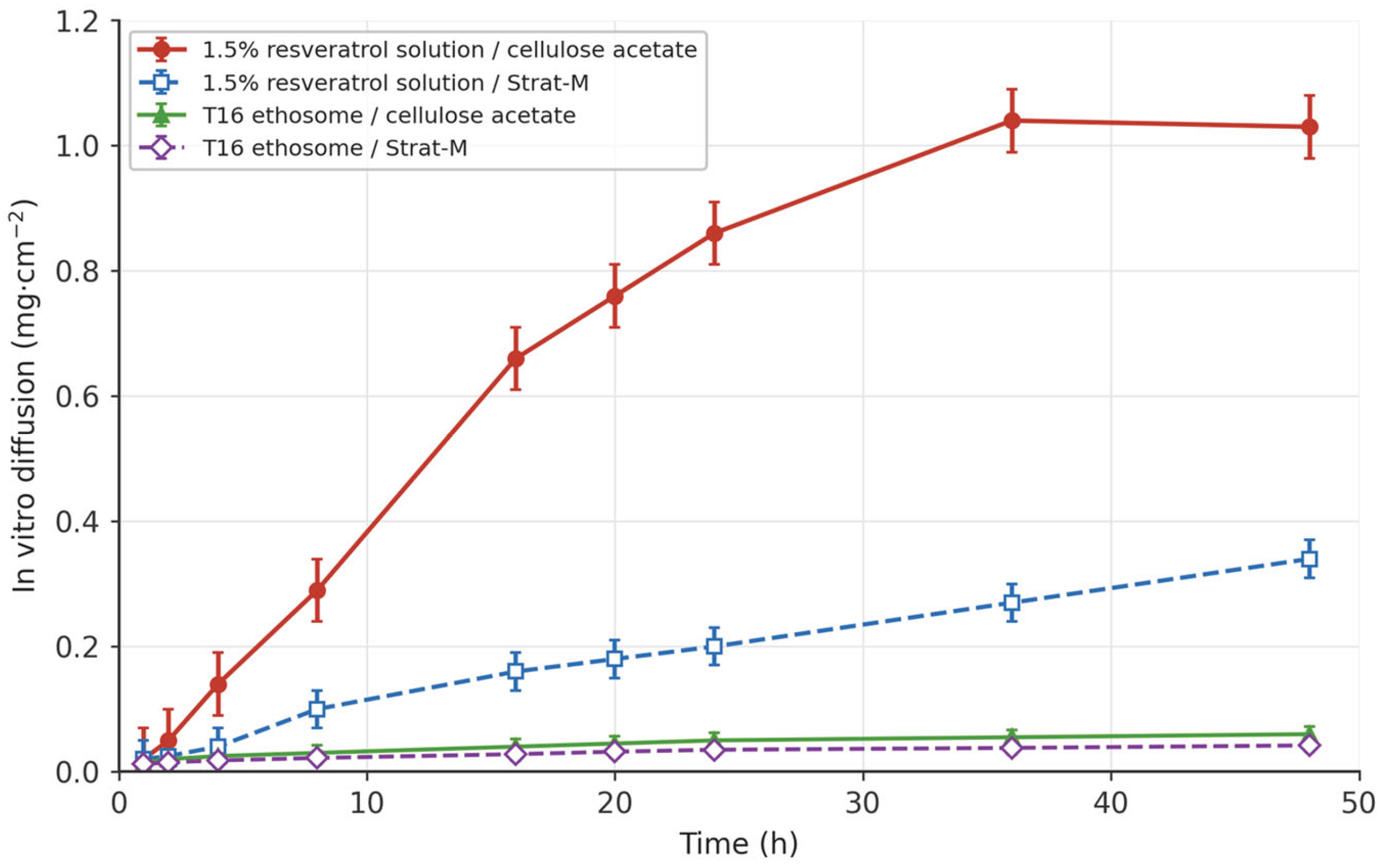

Table 1.
Composition of resveratrol-loaded (T13–T16) and unloaded (T21–T24) ethosome formulations. The optimized formulation (T16, bold) was carried forward for detailed characterization.
Table 1.
Composition of resveratrol-loaded (T13–T16) and unloaded (T21–T24) ethosome formulations. The optimized formulation (T16, bold) was carried forward for detailed characterization.
| Type | Code | Water (%) | Lecithin (%) (Lipoid P75) | Vitamin E (%) | Ethanol (%) | Resveratrol (%) | Cycle * |
|---|---|---|---|---|---|---|---|
| Loaded | T13 | 63.2 | 5 | 0.3 | 30 | 1.5 | 1 |
| Loaded | T14 | 63.2 | 5 | 0.3 | 30 | 1.5 | 3 |
| Loaded | T15 | 63.2 | 5 | 0.3 | 30 | 1.5 | 5 |
| Loaded | T16 | 63.2 | 5 | 0.3 | 30 | 1.5 | 7 |
| Unloaded | T21 | 64.7 | 5 | 0.3 | 30 | – | 1 |
| Unloaded | T22 | 64.7 | 5 | 0.3 | 30 | – | 3 |
| Unloaded | T23 | 64.7 | 5 | 0.3 | 30 | – | 5 |
| Unloaded | T24 | 64.7 | 5 | 0.3 | 30 | – | 7 |
* Number of passes through the high-pressure homogenizer interaction chamber (Microfluidics M110P).
Table 2.
Vesicle size (d₀.₅) and uniformity of resveratrol-loaded and unloaded ethosomes over 180 days (25 ± 2 °C).
Table 2.
Vesicle size (d₀.₅) and uniformity of resveratrol-loaded and unloaded ethosomes over 180 days (25 ± 2 °C).
| Code | Cycle * | d₀.₅ (µm) Day 1 | Day 7 | Day 30 | Day 180 | Unif. Day 1 | Day 7 | Day 30 | Day 180 |
|---|---|---|---|---|---|---|---|---|---|
| T13 | 1 | 0.195 | 0.196 | 0.194 | 0.193 | 0.110 | 0.421 | 0.099 | 0.089 |
| T14 | 3 | 0.194 | 0.194 | 0.194 | 0.193 | 0.102 | 0.320 | 0.099 | 0.093 |
| T15 | 5 | 0.195 | 0.184 | 0.193 | 0.193 | 0.100 | 0.168 | 0.098 | 0.092 |
| T16 | 7 | 0.195 | 0.194 | 0.193 | 0.195 | 0.107 | 0.098 | 0.097 | 0.107 |
| T21 | 1 | 0.086 | 0.093 | 0.093 | 0.093 | 0.089 | 0.256 | 0.259 | 0.255 |
| T22 | 3 | 0.088 | 0.092 | 0.093 | 0.091 | 0.091 | 0.234 | 0.245 | 0.159 |
| T23 | 5 | 0.087 | 0.090 | 0.091 | 0.091 | 0.080 | 0.308 | 0.235 | 0.116 |
| T24 | 7 | 0.086 | 0.092 | 0.093 | 0.091 | 0.077 | 0.271 | 0.236 | 0.146 |
* Number of passes through the high-pressure homogenizer interaction chamber (Microfluidics M110P).
Table 3.
pH, conductivity, density, and absolute zeta potential (ζ, mV) of resveratrol-loaded and unloaded ethosomes. Zeta potentials of phospholipid ethosomes are negative in sign; absolute values are shown in the table.
Table 3.
pH, conductivity, density, and absolute zeta potential (ζ, mV) of resveratrol-loaded and unloaded ethosomes. Zeta potentials of phospholipid ethosomes are negative in sign; absolute values are shown in the table.
| Code | pH | Conductivity (µS/cm) | Density (g/cm³) | ζ Day 1 | ζ Day 7 | ζ Day 14 | ζ Day 30 |
|---|---|---|---|---|---|---|---|
| T13 | 6.540 | 44.4 | 0.951 | 38.7 | 37.5 | 29.0 | 29.3 |
| T14 | 6.508 | 44.5 | 0.961 | 36.0 | 34.2 | 24.8 | 33.1 |
| T15 | 6.518 | 44.5 | 0.961 | 39.3 | 29.4 | 24.6 | 25.6 |
| T16 | 6.500 | 44.6 | 0.949 | 41.2 | 33.9 | 28.5 | 25.8 |
| T21 | 6.910 | 44.7 | 0.945 | 64.1 | 30.6 | 43.4 | 39.6 |
| T22 | 6.721 | 53.2 | 0.948 | 76.7 | 50.6 | 52.4 | 54.4 |
| T23 | 6.687 | 55.0 | 0.952 | 66.7 | 30.4 | 51.0 | 52.9 |
| T24 | 6.653 | 56.8 | 0.956 | 67.2 | 43.7 | 40.7 | 55.6 |
Table 4.
Encapsulated and unencapsulated trans-resveratrol in the optimized ethosome formulation (T16) on day 1 and after 180 days.
Table 4.
Encapsulated and unencapsulated trans-resveratrol in the optimized ethosome formulation (T16) on day 1 and after 180 days.
| Code | % Unencapsulated (Day 1) | % Unencapsulated (Day 180) | % Encapsulated (Day 1) | % Encapsulated (Day 180) |
|---|---|---|---|---|
| T16 (resveratrol) | 4.51 | 15.79 | 95.49 | 84.21 |
Table 5.
Mean aspect ratio and image-based size-dispersion (PDI) from cryo-SEM morphometric analysis.
Table 5.
Mean aspect ratio and image-based size-dispersion (PDI) from cryo-SEM morphometric analysis.
| Code | Mean Aspect Ratio * | Standard Deviation | PDI * |
|---|---|---|---|
| T16 (resveratrol) | 1.43 | 0.28 | 0.038 |
| T24 (unloaded) | 1.37 | 0.22 | 0.026 |
* Interpreted as morphological size-dispersion parameters, distinct from hydrodynamic PDI.
Disclaimer/Publisher’s Note: The statements, opinions and data contained in all publications are solely those of the individual author(s) and contributor(s) and not of MDPI and/or the editor(s). MDPI and/or the editor(s) disclaim responsibility for any injury to people or property resulting from any ideas, methods, instructions or products referred to in the content. |
© 2026 by the authors. Licensee MDPI, Basel, Switzerland. This article is an open access article distributed under the terms and conditions of the Creative Commons Attribution (CC BY) license (http://creativecommons.org/licenses/by/4.0/).
Copyright: This open access article is published under a Creative Commons CC BY 4.0 license, which permit the free download, distribution, and reuse, provided that the author and preprint are cited in any reuse.