

Submitted:
26 August 2026
Posted:
27 August 2026
You are already at the latest version
Abstract
Background: Nanotechnological surface modifications on titanium implants aim to optimize bone-tissue interaction, accelerate osseointegration, and prevent the release of toxic wear debris. This study evaluated the tissue response and biomechanical stability of advanced carbon- and metal-oxide-based nanocoatings in an in vivo model. Methods: Grade IV titanium implants were evaluated under four conditions using a split-body design in the distal femurs of 12 adult male New Zealand white rabbits: uncoated (control), coated with Graphene Diamond-like Carbon (DLC), Niobium via Chemical Vapor Deposition (CVD), and Niobium via Sol-Gel technique. Follow-up periods were set at 30 and 60 days. Analysis included a Reverse Torque Test (RTQ) for biomechanical stability and histopathological evaluation (H&E and Picrosirius Red under polarized light) to assess bone organization, osteoclastic proliferation, collagen matrix maturation, and coating delamination. Results: RTQ values significantly increased from 30 to 60 days across all groups (p < 0.05), with Graphene-DLC and uncoated titanium achieving the highest biomechanical anchorage. Graphene-DLC maintained excellent interfacial stability, accelerated bone mineralization (low osteoid volumes), and promoted intensive, physiologically normal bone coupling and lamellar remodeling at 60 days. Conversely, Niobium-CVD and Niobium-Gel coatings exhibited severe late-stage delamination, which released particulate debris, triggered chronic foreign body reactions, increased osteoclastic bone resorption, and compromised biomechanical interlocking. Conclusions: Graphene-DLC coatings successfully preserve structural integrity and effectively enhance functional osseointegration, matching or exceeding the biomechanical performance of conventional titanium. However, interface optimization is mandatory for Niobium-based coatings to prevent mechanical failure and debris-induced osteolysis.
Keywords:
osseointegration
; titanium
; graphene
; niobium compounds
; rabbits
1. Introduction
Nanotechnology is a cornerstone of modern medicine, involving the manipulation and control of matter at atomic and molecular scales. Its application in healthcare—often referred to as nanomedicine—is derived from the fundamental principles of nanoscience.
Due to its excellent biocompatibility, titanium remains the gold standard for implants. Current research increasingly focuses on surface functionalization to optimize bone-tissue interaction and durability. However, the release of titanium oxide debris from orthopedic prostheses raises toxicity concerns, necessitating the development of preventive coating strategies to enhance long-term interfacial stability and biological safety [1,2,3,4,5].
The surface functionalization of biomaterials using graphene and niobium-based materials has emerged as a high-performance strategy for enhancing osseointegration. Graphene derivatives facilitate osteogenesis by increasing surface area and protein adsorption, thereby accelerating mesenchymal stem cell differentiation. Integration of niobium oxide further enhances the electrochemical stability and biocompatibility of the implant. Moreover, this composite coating exhibits potent antimicrobial activity, effectively preventing peri-implant infections through physical membrane disruption and reactive oxygen species (ROS) generation, ensuring a sterile and regenerative interface [6,7,8,9].
The synergistic potential of integrating graphene with metal oxides as a dual-action approach to combating peri-implantitis is investigated by several authors. This combination not only optimizes the biological response at the bone-implant interface by promoting osteogenic differentiation but also establishes a robust barrier against bacterial colonization. This “dual-action” mechanism relies on graphene’s ability to serve as a nanostructured scaffold that enhances the controlled release of metal ions and generates localized oxidative stress, effectively disrupting pathogen membranes without compromising the viability of mammalian cells. This strategy represents a significant advancement in surface engineering, offering a multifunctional solution to increase the longevity of dental and orthopedic implants in clinically challenging environments [10,11,12].
Within this framework, the present study investigates advanced surface modification strategies through nanotechnology, specifically exploring coatings based on Graphene and Niobium Oxide. These films are synthesized using high-precision techniques: Diamond-Like Carbon (DLC), Chemical Vapor Deposition (CVD), and the Sol-Gel method. The primary objective is to evaluate the osseointegration kinetics of these three nanotechnological coatings in comparison to uncoated Grade IV commercially pure titanium. To achieve this, the following statistical hypotheses were established:
I. Regarding Implant Surface Performance:
H0 (Null Hypothesis): All implant surfaces (coated and uncoated) exhibit statistically similar biological behavior.
HA (Alternative Hypothesis): At least one nanotechnological coating demonstrates significantly different (superior) performance compared to the others.
II. Regarding Osseointegration Periods:
H0 (Null Hypothesis): Osseointegration rates remain constant and show no significant variation between the two experimental time points.
HA (Alternative Hypothesis): Osseointegration levels differ significantly across the evaluated time periods.
2. Materials and Methods
2.1. Materials and Implant Specifications
Grade IV titanium implants (7 × 2.5 mm, Cone Morse model—CM SCREW PLATINUM) were utilized in this study. The surface coating procedures were conducted at the Physical Chemistry Laboratories of CVD Coating, the Department of Aerospace Science and Technology (DCTA/INPE/ITA), and Department of Materials Engineering from the Federal University of Santa Catarina (UFSC).
2.2. Coating Methodology: Diamond-like Carbon (DLC)
The deposition of DLC films was performed using a magnetron sputtering system equipped with a high-vacuum chamber and pumping assembly. A Dual Pulsed Sputtering (DPS) source was employed to generate negative bias pulses with a constant amplitude at 160 kHz, modulated by a 2.5 kHz frequency. The power delivered to the plasma was regulated by the pulse-per-unit-time ratio. The magnetron sputtering configuration was optimized to maximize the effective power transmitted to the graphite target, thereby enhancing sputtering efficiency.
Deposition experiments were executed under two distinct power regimes: low effective power (70 W/0.15 A) and high effective power (140 W/0.2 A). A 5 cm diameter high-purity graphite disk served as the sputtering target. The deposition process was carried out in a controlled atmosphere consisting of an Argon (Ar) and Methane (CH4) gas mixture, regulated by mass flow controllers. The optimized deposition conditions were established at an Ar/CH4 ratio of 1:4, maintaining a total working pressure between 0.5 and 0.7 Pa. Substrate temperatures range was 150–300 °C using a specialized ceramic heating plate, with temperatures monitored via a thermocouple coupled directly to the sample holder. Under these specified parameters, the average deposition rate was approximately 3 nm/min, resulting in a final DLC coating thickness of approximately 5 µm on the titanium substrates. The methodology for depositions was based on the study of Marciano et al., (2011) [13].
2.3. Chemical Vapor Deposition (CVD) Coating
Niobium pentoxide (Nb2O5) films were separately deposited using a Rapid Thermal Processing (RTP) furnace (Annealsys RTP system). A crucible containing 10 g of Nb2O5 and Graphene Oxide (GO) nanoparticles (5–100 nm) was placed within the quartz tube. The system was preheated to 750 °C, reaching a peak deposition temperature of 950 °C. Nitrogen (N2, 1000 mL/min), Hydrogen (H2, 10 mL/min), and Methane (CH4, 5 mL/min) were utilized as carrier and cleaning gases for the drag deposition process, following the methodology described by Horst et al. (2021) [14].
2.4. Sol-Gel Coating Procedure
The synthesis of the coatings was performed using the sol-gel technique, employing a colloidal suspension of niobium nanoparticles (5–100 nm) as the primary precursor. This suspension was incrementally introduced into 2 mL of acrylic acid and subsequently diluted in a mixture of 20 mL of distilled water and 10 mL of ethanol. To ensure uniform dispersion and prevent nanoparticle agglomeration, the solution underwent ultrasonic processing (sonication) for 90 min. The titanium implants were then submerged in Petri dishes containing the prepared sol. The assembly was placed in a rotary evaporator and maintained at 180 °C for 24 h to achieve complete solvent evaporation and facilitate the gel-to-solid transition. Following this, the coated samples were thoroughly rinsed and subjected to a drying stage at 70 °C for 2 h, following the protocol established by Safavi et al. (2022) [9]. The resulting niobium-based films exhibited a thickness range between 450 nm and 2 µm. Figure 1 shows the implants.
2.5. Surgical Procedure and Implant Methodology
Following an established animal model protocol, four Grade IV titanium implants (7 × 2.5 mm, Cone Morse model—CM SCREW PLATINUM) were placed into the femoral condyles of each subject (rabbit). The experimental design included one uncoated control implant and three groups featuring the nano-coatings.
2.6. Animal Model and Ethical Considerations
The study utilized 12 adult male New Zealand white rabbits, aged between 6 and 12 months, with a mean body mass of 3.21 kg (range: 2.8–3.5 kg). Surgical procedures were conducted at the Nursery Vivarium of the University of Contestado (UNC), Mafra Campus. The experimental protocol was approved by the Institutional Animal Care and Use Committees (CEUA) under protocol numbers 03/24 (CEUA/UNC) and 9704270225 (CEUA/UNIFESP). The animals were housed in a climate-controlled environment in individual cages, with ad libitum access to water and a specific standardized diet.
2.7. Experimental Groups and Clinical Attrition
The subjects were randomly assigned to two follow-up periods: 30 days (n = 6) and 60 days (n = 6). During the initial 30-day interval, two losses were recorded: one instance of bone fracture and one mortality of unknown etiology within the first 10 days post-surgery. Consequently, the sample size was adjusted to five animals per group. These animals were originally included in the experimental design as a surplus to account for potential complications. For the subject that sustained a fracture, the “Humane Endpoint” (Sanitary Action Point) protocol was strictly observed to ensure animal welfare.
2.8. Anesthesia and Pre-Operative Protocol
The animals were initially weighed to calculate precise dosages. Pre-anesthetic medication (PAM) was administered intramuscularly (IM) and consisted of a combination of Midazolam (4 mg/kg), Acepromazine 0.2% (1 mg/kg), and Tramadol (2 mg/kg). Once a stable pre-anesthetic plane was achieved (approximately 30 min post-administration), general anesthesia was induced via IM injection of Xylazine 2% (5 mg/kg) and Ketamine (35 mg/kg).
2.9. Surgical Technique and Implant Distribution
Subjects were identified using ear tags. The surgical site preparation included trichotomy, followed by rigorous asepsis and antisepsis protocols, and the application of sterile surgical drapes. A lateral incision was made in the knee region, and an instrumental guide was positioned to ensure precision (Figure 2A,B). Osteotomy was performed using a 6 mm drill at low rotation under continuous saline irrigation to transfix the condyles. All implants were placed by the same surgeon following a standardized protocol to minimize inter-operator variability. The implants were distributed according to the following anatomical sites:
- ▪
- Right Lateral Condyle (RL): Uncoated control implant.
- ▪
- Right Medial Condyle (RM): Graphene-DLC coated implant.
- ▪
- Left Medial Condyle (LM): Niobium-CVD coated implant.
- ▪
- Left Lateral Condyle (LL): Niobium-Gel coated implant.
2.10. Post-Operative Care and Clinical Monitoring
Daily post-operative care consisted of surgical wound cleansing with sterile saline solution and antisepsis of the peri-incisional area using 2% alcoholic chlorhexidine. Clinical monitoring was performed every 8 h, during which the following parameters were recorded: spontaneous behavior, posture, respiratory patterns, coat condition, ocular and mucosal appearance, and overall body condition.
Pain management was achieved through the intramuscular (IM) administration of Tramadol (2 mg/kg, opioid analgesic) and Dipyrone (25 mg/kg, non-opioid analgesic/antipyretic). Surgical sutures were removed seven days post-operatively.
2.11. Euthanasia and Tissue Harvest
At the conclusion of the experimental periods (30 and 60 days), subjects were transported to the surgical suite for euthanasia. The pre-anesthetic protocol included Midazolam (4 mg/kg) and Acepromazine 0.2% (1 mg/kg) via IM injection. After 30 min, a deep anesthetic plane was induced with Xylazine 2% (5 mg/kg) and Ketamine (35 mg/kg) via IM, followed by an intravenous (IV) bolus of Propofol (30 mg/kg) to ensure cardiorespiratory arrest. Once death was confirmed, the anatomical regions containing the implants were harvested. Carcasses were removed and processed by a specialized biological waste management company.
2.12. Experimental Design and Bioethics
Each condyle was defined as a single experimental unit. A split-body design was utilized, in which all four implant types were placed within the same animal to neutralize the “Animal Effect” (inter-individual variability). This methodology was specifically selected to minimize the total number of animals required, adhering to the 3Rs principles (Replacement, Reduction, and Refinement) and the ethical requirements of the CEUA/UNC and CEUA/UNIFESP committees.
The experimental design of this study was strictly guided by the 3Rs principles (Replacement, Reduction, and Refinement) to ensure the highest standards of animal welfare. To address Reduction, a split-body experimental model was implemented, utilizing each of the four femoral condyles in a single subject as distinct experimental units. This approach allowed for the simultaneous evaluation of three different nanotechnological coatings against an internal control, effectively neutralizing inter-individual biological variability (the ‘animal effect’) while significantly minimizing the total number of animals required to achieve statistical power. Regarding Refinement, rigorous perioperative analgesia and a multi-modal anesthetic protocol were employed to eliminate pain and distress, ensuring a humane endpoint. Replacement was not feasible as the complex physiological environment of bone remodeling and the systemic response to multi-layered biomaterials cannot yet be fully replicated by in vitro or computational models.
The harvested samples were immersed in 10% buffered formalin in identified containers and transferred for histopathological analysis to the CEDAP Laboratory (Center for Anatomical Pathological Diagnostics), Joinville—SC, Brazil.
2.13. Biomechanical Analysis: Reverse Torque Test (RTQ)
Following a two-week fixation period in 10% formalin, the biomechanical stability of the bone-implant interface was assessed via a Reverse Torque Test (RTQ). The torque required to unscrew and dislodge the implants was measured using a calibrated digital torquemeter, with values recorded in Newton-centimeters (N/cm) as shown in Figure 3.
2.14. Histopathological and Histochemical Analysis
The samples were transferred to 10% buffered formalin for 48 h and subsequently underwent decalcification using formic acid. The histological analysis was performed by two independent pathologists, and scores were established by consensus. Tissue evaluation was conducted using two distinct staining protocols:
Hematoxylin and Eosin (H&E): This technique was employed to visualize the general tissue architecture and organ morphology. Hematoxylin-stained acidic structures (such as nuclei) blue-to-purple, while eosin-stained basic structures (such as cytoplasm and extracellular matrix) pink-to-red. Under optical microscopy, the samples were evaluated for callus formation, stromal reaction, implant osseointegration, and coating delamination (Figure 4A).
Picrosirius Red Staining: A histochemical evaluation was performed to characterize the collagen content under polarized light. This method distinguished between Type I collagen (established healing/mature bone), identified by orange-to-red birefringence, and Type III collagen (immature/recent collagen), identified by green birefringence (Figure 4B).
2.15. Histological Scoring and Semi-Quantitative Analysis
The histological sections were evaluated and scored based on the following parameters, ranging from the least favorable to the most favorable aspect:
1—Hematoxylin and Eosin (H&E) Analysis
A. Coating Delamination Presence of delamination with or without liberated nanoparticles:
0: Extensive; 1: Partial; 2: Focal; 3: Absent.
B. Bone Tissue Histological Structure Objective: To evaluate the peri-implant bone response.
(B1) Degree of bone organization: 0: Absent; 1: Disorganized; 2: Partially organized; 3: Well-organized.
(B2) Presence of osteoid: 0: Absent; 1: Present.
(B3) Osteoclastic proliferation: 0: Absent; 1: Discrete; 2: Moderate; 3: Marked.
(B4) Bone-to-Implant Contact (BIC): 0: None; 1: Focal (<25%); 2: Partial (25–50%); 3: Extensive (50–75%); 4: >75%.
(B5) Peri-implant interface thickness: Halo measurement within the Region of Interest (ROI) in millimeters (mm).
BIC refers to the percentage of the implant surface in direct contact with mineralized bone, as observed via histomorphometry.
C. Cartilaginous Proliferation and Healing Objective: To identify endochondral or fibrous repair pathways.
(C1) Predominant repair tissue: 1: Fibrous; 2: Cartilaginous; 3: Osseous.
(C2) Repair tissue maturation: 0: Absent; 1: Immature; 2: Partial; 3: Mature.
D. Vascular Response Objective: To evaluate neovascularization or microvascular alterations.
Neovascularization: 0: Absent; 1: Mild; 2: Moderate; 3: Intense.
E. Inflammatory Response Objective: To assess immunological response or material rejection.
(E1) Acute inflammatory infiltrate: 0: Absent; 1: Present.
(E2) Chronic inflammatory infiltrate: 0: Absent; 1: Mild; 2: Moderate; 3: Intense.
(E3) Presence of multinucleated giant cells: 0: Absent; 1: Present.
F. Bone Neoformation Objective: To verify osteoinduction and osteoconduction.
(F1) Maturity of newly formed bone: 1: Immature (osteoid); 2: Mixed; 3: Mature.
2—Picrosirius Red Staining Analysis
G. Type I and Type III Collagen Proliferation Objective: To evaluate fibrosis and extracellular matrix organization under polarized light microscopy.
(G1) Type I Collagen: 0: Absent; 1: Mild; 2: Moderate; 3: Intense.
(G2) Type III Collagen: 0: Absent; 1: Mild; 2: Moderate; 3: Intense.
(G3) Collagen Ratio: 1: Type I < Type III; 2: Type I = Type III; 3: Type I > Type III.
2.16. Statistical Analysis
The study followed an experimental design based on a 4 × 2 factorial scheme (four implant types and two osseointegration periods) with five replicates per group. Each animal was considered a replicate, and each femoral condyle was defined as a distinct experimental unit. Data analysis was performed using the Scheirer-Ray-Hare test, a non-parametric alternative to the two-way ANOVA, as the data did not meet the assumptions of normality required for parametric testing. Results were considered statistically significant when p < 0.05.
3. Results
3.1. Experimental Design and Animal Models
All rabbits were males aged between 6 and 12 months, with a mean weight of 3.21 kg (range: 2.8 to 3.5 kg). The screws were numbered for statistical analysis and corresponded to the following groups:
- ▪
- Implant/Screw P1 = Left Knee, Lateral Condyle = LL = NIOBIUM GEL
- ▪
- Implant/Screw P2 = Right Knee, Lateral Condyle = RL = UNCOATED
- ▪
- Implant/Screw P3 = Right Knee, Medial Condyle = RM = GRAPHENE DLC
- ▪
- Implant/Screw P4 = Left Knee, Medial Condyle = LM = NIOBIUM CVD
Analyses were performed based on the type of Implant/Screw (1, 2, 3, and 4) and the implantation period in the condyles (30 and 60 days).
3.2. Reverse Torque Test—RTQ
The evaluation of implant stability and the biomechanical quality of the bone-implant interface was quantitatively assessed using the reverse torque (RTQ) test (Figure 5). The force required to disrupt this interface, measured in Newton-centimeters (N/cm), serves as a direct indicator of the degree of osseointegration and mechanical interlocking achieved over time.
Reverse torque (RTQ) values significantly increased from 30 to 60 days across all groups (p < 0.05), reflecting the biological transition from mechanical primary stability to secondary osseointegration and bone matrix maturation.
At 60 days, P2 (uncoated titanium) and P3 (graphene-DLC) demonstrated the most prominent biomechanical anchorage. While P2 values confirm titanium as the biocompatible gold standard, P3 highlights the osteogenic potential of carbon-based nanomaterials. Graphene-DLC coatings optimize surface topography, energy, and wettability, accelerating plasma protein adsorption (fibronectin/vitronectin) and triggering bone marrow mesenchymal stem cell (BMSC) differentiation via the FAK/P38 signaling pathway [15,16,17]. The high RTQ required for P3 at 60 days confirms both the in vivo structural stability of the film and its capacity to stimulate a denser peri-implant architecture.
The lower biomechanical performance of the Niobium-based groups (CVD and GEL) during late healing is attributed to coating delamination, which compromised the mechanical continuity at the bone-implant interface. The time-dependent escalation of RTQ values, led by the P3 group, validates the hypothesis that graphene-DLC coatings effectively enhance and sustain functional osseointegration, matching or exceeding the biomechanical fixation of conventional titanium.
3.3. Laboratory Measurements
3.3.1. Coating Delamination
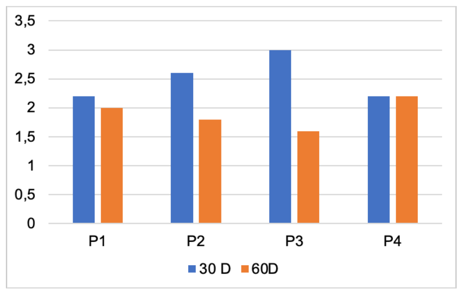
Coating delamination (Figure 6) significantly varied across implant types and timelines (p < 0.05), with overall degradation increasing at 60 days. P2 (uncoated titanium) and P3 (graphene-DLC) demonstrated superior interfacial stability, showing minimal particle release. This structural integrity prevents the detachment of free nanoparticles, avoiding foreign-body acute or chronic inflammatory responses that could jeopardize peri-implant bone maturation [18].
P1 (niobium GEL) and P4 (niobium CVD) exhibited severe delamination during late healing. This mechanical instability disrupts bone-implant continuity and releases particulate debris into the peri-implant bed. The resulting macro- and micro-instabilities impair osteogenesis and explain the lower reverse torque performance of the niobium groups. These findings highlight that rigorous interface optimization is mandatory to prevent coating failure and ensure safe, long-term clinical osseointegration [19].
3.3.2. Histological Structure of Bone Tissue
Histological analysis of peri-implant bone evaluates osseointegration quality and biomaterial-induced bone repair. Driven by mechanical stimuli, bone remodeling progressively replaces disorganized primary woven bone with oriented lamellar bone tissue to optimize occlusal load distribution. This process determines the Bone-to-Implant Contact (BIC) ratio, a critical quantitative parameter used to characterize the host tissue response and functional implant anchorage [20].
3.3.3. Degree of Bone Organization
The degree of bone organization (Figure 7) significantly progressed from unorganized woven bone at 30 days to highly oriented lamellar bone at 60 days, confirming active, mechanically-guided peri-implant remodeling (p < 0.05). At the 60-day mark, P2 (uncoated titanium) and P3 (graphene-DLC) induced the highest level of structural bone maturation. For the P3 group, this superior alignment and enhanced Bone-to-Implant Contact (BIC) validate the osteoinductive potential of the carbonaceous matrix [21], whose optimized topography and wettability accelerated orderly osteoblast orientation along loading axes. Conversely, P1 (niobium GEL) and P4 (niobium CVD) restricted bone maturation, leaving persistent woven bone at 60 days.
This impairment is directly attributed to severe coating delamination and subsequent particulate debris release, which disrupted the stable mechanical signaling required for lamellar remodeling and halted the bone healing cascade [22].
3.3.4. Presence of Osteoid
The presence of osteoid (Figure 8) showed no statistically significant differences across experimental periods or implant types (p > 0.05), with all groups displaying a partially to well-organized pattern. However, P3 (graphene-DLC) presented a lower presence of osteoid at both 30 and 60 days. In bone histomorphometry, osteoid represents the unmineralized, organic osteoblast-secreted matrix that precedes bone mineralization. A lower standing volume of osteoid, when paired with high reverse torque (RTQ) and superior bone organization, does not indicate impaired matrix deposition; rather, it signifies an accelerated bone maturation and mineralization rate [23].
This suggests that the graphene-DLC coating effectively shortens the lag phase between osteoid secretion and hydroxyapatite precipitation. By optimizing surface topography and wettability, the carbonaceous matrix promotes rapid calcium phosphate nucleation, shifting the tissue kinetics toward a mature, mineralized state faster than the other groups.
3.3.5. Osteoclastic Proliferation
Osteoclastic proliferation (Figure 9) significantly varied among the groups (p < 0.05), with overall activity predictably downregulating from 30 to 60 days as acute remodeling subsided.
At both timelines, P2 (uncoated titanium) and P3 (graphene-DLC) maintained significantly lower osteoclast counts. This suppressed osteoclastic activity indicates a highly biocompatible and stable interface. For P3, the graphene-DLC coating successfully prevents structural micro-instability and ion leakage, shielding the peri-implant environment from the macrophage-mediated pro-inflammatory cytokines (such as TNF-α and IL-1β) that drive osteoclast genesis. Consequently, the bone coupling mechanism shifted toward net bone formation, accelerating secondary stability [24].
P1 (niobium GEL) and P4 (niobium CVD) exhibited persistently high osteoclastic proliferation, particularly at 60 days. This pathological bone resorption is directly triggered by severe coating delamination. The release of unstable niobium particulate debris recruits and activates macrophages, initiating an aseptic inflammatory cascade. This foreign-body response upregulates the RANKL/RANK pathway, driving osteoclast differentiation and localized osteolysis. This continuous bone resorption explains the mechanical discontinuity and lower reverse torque values observed in the niobium-coated groups.
3.3.6. Bone-to-Implant Contact
Bone-to-Implant Contact (BIC) and osteoclastic proliferation (Figure 10) demonstrated distinct histomorphometric behaviors (p > 0.05). Across all time points, P3 (graphene-DLC) achieved BIC scores equal to or higher than P2 (uncoated titanium), confirming that the carbonaceous matrix maintains high cytocompatibility and effective secondary mechanical stability [25].
Crucially, P3 exhibited a marked increase in osteoclastic proliferation, reaching its maximum mean score at 60 days. In conventional implant healing, elevated osteoclast activity often indicates aseptic loosening or debris-induced osteolysis. However, in the case of P3, where high osteoclast counts explicitly coexist with stable BIC and excellent reverse torque, this phenomenon represents an accelerated, healthy bone coupling mechanism [23].
Because the graphene-DLC surface optimizes wettability and cellular anchoring, it drives rapid initial osteogenesis and mineralization. By day 60, the peri-implant microenvironment surrounding P3 enters an intensive mechanically-guided remodeling phase. Here, the proliferation of active osteoclasts is not a pathological foreign-body response, but a physiological requirement to resorb early, disorganized woven bone and swiftly replace it with a mature, oriented lamellar architecture, ensuring long-term functional anchorage.
Marked osteoclastic proliferation was observed across all experimental periods and implant types, with no statistically significant differences between groups or time points. At 60 days, the mean score for P3 (graphene DLC) reached the maximum degree of osteoclastic proliferation. Bone-to-implant contact (BIC) showed no statistically significant differences regarding implant types or experimental periods. Across all time points, the BIC scores for P3 (graphene DLC) were consistently equal to or higher than those for P2 (uncoated).
3.3.7. Thickness of the Bone-Implant Interface—Peri-Implant Halo (ROI)
The histomorphometric thickness of the bone-implant interface, measured as the peri-implant halo within the Region of Interest (ROI) (Figure 11), showed no statistically significant differences (p > 0.05) among the experimental periods or implant types.
However, P1 (niobium GEL) and P4 (niobium CVD) exhibited slightly higher mean halo values. In peri-implant histomorphometry, a widened interface or “halo” typically represents an unmineralized fibroblastic soft-tissue capsule or an expanded zone of granulation tissue rather than functional bone anchorage. This biological widening is a classic hallmark of mechanical instability or a subclinical foreign body reaction [26].
For P1 and P4, this expanded interface correlates directly with their severe coating delamination. The continuous release of niobium particulate debris induces localized aseptic inflammation, which upregulates osteoclastic resorption and halts osteoblast differentiation at the immediate interface [27]. Consequently, the establishment of an intimate bone-to-implant contact is physically disrupted, resulting in a thicker, fibrous, or less dense peri-implant halo and explaining the inferior biomechanical interlocking observed in the niobium-coated groups.
3.3.8. Cartilaginous Proliferation and Healing
The presence of fibrous, cartilaginous, or bone tissue, characterized based on morphology, allows for inferring whether repair occurred via an endochondral or fibrogenic pathway. Endochondral repair forms new bone from a cartilage template, which is subsequently replaced by trabecular and then lamellar bone, representing a more efficient mechanism of bone regeneration. Conversely, fibrous repair fails to mineralize and can lead to osseointegration failure, thereby compromising the stability and longevity of the implant.
3.3.9. Predominant Type of Scar Tissue
The histological evaluation of the predominant type of scar tissue (Figure 12) surrounding the peri-implant interface revealed critical variations in tissue quality and composition among the groups.
P3 (graphene-DLC) and P2 (uncoated titanium) exhibited a clear predominance of highly cellular, dense, and organized connective tissue, transitioning smoothly into mature bone. For P3, the structural and chemical inertness of the carbonaceous nanomaterial coating serves as an osteoconductive template [28]. By optimizing interfacial energy and eliminating cytotoxic metal ion dissolution, the graphene-DLC film prevents the formation of a thick, non-functional fibrous barrier. Instead, it encourages direct cellular attachment and alignment along the loading axes, reinforcing a stable structural bridge between the substrate and the advancing front of osseointegration.
P1 (niobium GEL) and P4 (niobium CVD) presented a persistent predominance of loose, disorganized connective tissue or thick, dense fibrous encapsulation at 60 days. This pathological tissue response is directly mediated by the severe delamination and subsequent release of unstable niobium wear particles into the peri-implant space. The presence of non-degradable metallic particulate debris triggers a chronic foreign body reaction (FBR) [29].
Local tissue macrophages phagocytose these particles, establishing a sustained pro-inflammatory microenvironment characterized by the release of reactive oxygen species (ROS) and destructive cytokines [30].
This inflammatory cascade blocks normal osteoblast differentiation and interrupts the biomechanical signaling required for lamellar bone maturation. To isolate the irritating particulate debris, host fibroblasts form an unmineralized capsule around the implant. This encapsulation explains the mechanical discontinuity, the widened peri-implant halo, and the lower torque values identified in the niobium-treated groups.
3.3.10. Maturation of Repair Tissue
The maturation of the peri-implant repair tissue (Figure 13) showed no statistically significant differences (p > 0.05) among the experimental periods or implant types, maintaining a stable biological pattern from 30 to 60 days.
Despite the lack of statistical variance, distinct chronological peaks in tissue maturation were qualitatively evident. P4 (niobium CVD) reached noticeable maturation early at 30 days. This accelerated initial phase suggests that the chemical composition and crystalline structure of niobium via chemical vapor deposition initially favor rapid focal adhesion and early protein adsorption, triggering a swift transition toward early bone matrix mineralization [31]. However, as previously discussed, this early advantage was not sustained over time due to later coating instability.
P3 (graphene-DLC) exhibited a delayed but progressive maturation peak at 60 days. This late-stage maturation aligns with the surface kinetics of carbon-based nanomaterials. Graphene-DLC surfaces modulate cellular behavior by initially allowing an intensive, prolonged proliferative phase of osteogenic lineages. Once an optimal cell density is established, the nano-topography and optimized wettability shift the tissue kinetics toward an organized, late-stage extracellular matrix mineralization. This explains the superior lamellar bone distribution and high torque values found in the graphene-DLC group at the final experimental timeline.
3.3.11. Vascular Response—Neovascularization
Neovascularization (Figure 14) provides vital metabolic support during bone healing by delivering the oxygen, nutrients, inflammatory cells, and progenitor cells necessary for osteogenesis and successful osseointegration. Consequently, poorly vascularized implant sites inherently compromise this biological cascade, presenting a greater challenge to bone regeneration than well-vascularized regions.
In this study, the lack of statistically significant differences across most groups and experimental periods indicates that all implant types maintained a biologically active, mild-to-moderate vascular response, confirming the absence of material-induced cytotoxicity or persistent ischemia. Notably, the lower vascular score observed for the graphene DLC screw at 60 days suggests an accelerated transition from active neoangiogenesis to tissue maturation and remodeling, signaling advanced late-stage biocompatibility and a potentially optimized healing trajectory.
No statistically significant differences were observed among the implant types or experimental periods within the groups. All groups exhibited scores above 1 (mild-to-moderate vascular response), except for screw 3 (graphene DLC) at 60 days.
3.3.12. Inflammatory Response
The initial stage to ensure osseointegration begins with the bleeding caused by implant insertion, which subsequently triggers the coagulation cascade. Characterizing the inflammatory response around metallic implants is crucial to evaluate and identify potential adverse reactions, such as implant rejection.
The presence of an acute inflammatory infiltrate may indicate an initial response to surgical trauma, whereas the persistence of a chronic infiltrate, particularly at moderate-to-intense levels, may reflect a failure in implant integration. The identification of macrophages and multinucleated giant cells is highly relevant; indeed, the ISO 10993-6 standard explicitly recommends analyzing these parameters as part of a scoring system for evaluating local tissue response following the implantation of medical devices.
3.3.13. Acute Inflammatory Infiltrate
Figure 15 demonstrates a dense acute inflammatory infiltrate, a hallmark of the innate immune response to acute tissue injury or pathogen invasion. The cellular profile is characteristically dominated by polymorphonuclear neutrophils (PMNs), which are recruited to the injury site via a well-orchestrated cascade involving local vasodilation, increased vascular permeability, and endothelial activation.
This allows PMNs to undergo margination, rolling, and integrin-mediated adhesion, followed by trans-endothelial migration into the extravascular space along a chemotactic gradient driven by mediators such as interleukin-8 (IL-8), complement fragment C5a, and leukotriene B4 (LTB4). Once localized, these activated neutrophils execute their primary effector functions through phagocytosis and the release of antimicrobial peptides, reactive oxygen species (ROS), and lysosomal enzymes. While this response is essential for neutralizing the offending agent, the non-specific release of these cytotoxic metabolites frequently induces collateral tissue damage and interstitial edema, defining the histopathological presentation of the acute inflammatory lesion [32].
3.3.14. Chronic Inflammatory Infiltrate
Figure 16 illustrates a chronic inflammatory infiltrate, typically characterized by a mononuclear cell profile predominantly composed of lymphocytes, macrophages, and plasma cells. Although no statistically significant differences were found among the groups regarding acute inflammation, a higher overall acute inflammatory response was observed at 30 days post-implantation for the P1 (niobium GEL) and P3 (graphene DLC) biomaterials, suggesting a prolonged recruitment phase. Conversely, the chronic inflammatory response showed no statistically significant differences across any of the experimental groups, with all cohorts trending toward absence and presenting scores below 1.
3.3.15. Presence of Multinucleated Giant Macrophages
Figure 17 documents the presence of multinucleated giant macrophages (foreign body giant cells), which typically form through the fusion of monocytes and macrophages in response to persistent, non-phagocytose foreign materials [35].
Quantitative analysis revealed no statistically significant differences among the experimental cohorts, demonstrating a generalized, baseline trend toward macrophage presence across all evaluated groups. Notably, at both the 30-day and 60-day intervals, the histological pattern observed for the P3 (graphene DLC) implant remained highly similar to that of the P2 (uncoated control) group. This comparable macrophage kinetic indicates that the addition of the graphene DLC coating does not exacerbate the biomaterial-directed foreign body reaction, reinforcing its interfacial biocompatibility and structural stability within the host tissue over extended implantation periods.
3.3.16. Bone New Formation: Maturity of Newly Formed Bone
Figure 18 evaluates the maturity of newly formed bone, a critical parameter for assessing the efficacy of biomaterials in promoting an organized, mineralized, and functional extracellular matrix during the osseointegration process.
Quantitative analysis revealed no statistically significant differences across the experimental periods or implant modalities. Notably, however, the P3 (graphene DLC) cohort consistently exhibited a higher mean maturity score at both the 30-day and 60-day intervals. This descriptive trend suggests that the carbon-based surface modification may accelerate the transition from woven bone to mature lamellar bone, potentially due to enhanced osteoblast differentiation and mineral deposition at the implant-tissue interface [36].
3.3.17. Type I and Type III Collagen Proliferation Under Picrosirius-Red Polarized Light Context
Picrosirius-red staining under polarized light microscopy is the gold-standard method to evaluate the organizational stage of collagen fibers in tissues and to quantify types I and III collagen.4 Type III collagen is predominant during the early stages of repair and wound healing, whereas type I prevails in mature and organized tissues. The predominance of type I collagen suggests a more stable microenvironment surrounding the implant.
Figure 19, Figure 20 and Figure 21 demonstrate the organizational remodeling of the peri-implant extracellular matrix through the quantification of Type I and Type III collagen fibers under polarized light microscopy. Regarding Type I collagen (Figure 20), a statistically significant difference was observed in mean values, with the P3 (graphene DLC) implant standing out prominently at the 30-day interval (p < 0.05).
For Type III collagen (Figure 20), which characterizes the early, provisional stages of tissue repair, the mean values at 30 days were significantly higher than at 60 days across the cohorts, with P3 showing a markedly prominent initial increase.
This synchronized upregulation of both structural proteins shifted the Type I/III collagen ratio (Figure 21), revealing a statistically significant difference in the 30-day mean values where P3 exhibited a superior organizational score (p < 0.05). Taken together, these findings indicate that the graphene DLC coating significantly accelerates early collagen synthesis and deposition, fostering a rapid transition toward a dense, Type I-dominated collagenous matrix that secures a more stable and biomechanically mature microenvironment surrounding the implant.
4. Discussion
Osseointegration was evaluated using a rabbit femoral condyle model under strict ethical control. Biomechanical assessment via reverse torque testing demonstrated uniform intergroup means, though future preservation methods under physiological conditions should be considered. Histopathological analysis through hematoxylin and eosin staining evaluated structural matrix features, inflammatory kinetics, and tissue integration. Additionally, polarized light microscopy of picrosirius-red stained sections differentiated established mature matrix formation from recent provisional fibroplasia. Statistical analysis was executed utilizing non-parametric methodology due to the non-normal distribution of the experimental data.
While titanium remains the standard material in implantology owing to its inherent biocompatibility, surface modifications are heavily researched to enhance interfacial capabilities. In this context, graphene nanomaterial coatings have emerged as a promising strategy to accelerate osteogenic differentiation and in vivo osseointegration. The diamond-like carbon (DLC) coating demonstrated structural stability, enhanced bone organization, and a low delamination rate comparable to uncoated titanium, aligning with its documented osteogenic and anti-biofilm properties. Furthermore, the DLC group showed similar macrophage and inflammatory kinetics to the control, confirming its remarkable biocompatibility, reduced adverse tissue reactions, and high corrosion resistance. Over extended experimental periods, the DLC coating promoted superior bone integration, accelerated neo-formed bone maturation, and enhanced mature matrix deposition, corroborating literature that highlights its durability under mechanical stress.
Niobium-based coatings exhibited high performance scores regarding tissue integration and biocompatibility, their overall efficacy was heavily compromised by coating delamination. Although niobium biomaterials possess reliable physical-mechanical properties, excellent bioactivity, and corrosion resistance, their practical biomedical application remains restricted by interface instability. These findings emphasize the critical necessity for strict control over the titanium-niobium interface to prevent clinical delamination.
5. Conclusions
This study demonstrates that surface modifications significantly influence the interfacial tissue response and osseointegration kinetics of titanium implants. The Graphene DLC (Diamond-like Carbon) coating exhibits outstanding therapeutic potential, promoting accelerated bone maturation, superior structural organization of the extracellular matrix, and a rapid transition toward a dense, Type I collagen-dominated microenvironment without exacerbating acute or chronic inflammatory pathways. While both Niobium-based coatings (CVD and GEL) display high bioactivity and favorable cellular compatibility, their clinical and structural efficacy is severely compromised by interface instability and a high rate of coating delamination.
Graphene DLC emerges as a highly promising, stable, and biocompatible nanomaterial candidate for enhancing the longevity of load-bearing biomedical implants, the practical application of Niobium coatings remains contingent upon developing stricter interfacial deposition controls to mitigate delamination under physiological stress.
In conclusion, while the animal model utilized in this study was appropriate, site randomization during implant fixation would be necessary in future protocols. To allow for a more comprehensive evaluation, it is recommended to conduct additional functional and biomechanical testing, as well as to explore the industrialization potential and clinical application of these coatings.
The reverse torque (RTQ) testing was considered valid due to the standardization applied across all implants; however, literature indicates that due to potential mechanical alterations, readings should be taken immediately or under physiological, non-fixative preservation.
Although the histological techniques applied were adequate, it is suggested to adopt larger, load-bearing animal models, implement automated histomorphometry, and utilize molecular biomarkers.
The results strongly suggest that the Graphene-DLC coating enhances osteogenesis, however the Niobium-based CVD and GEL coatings proved to be bioactive but unstable.
6. Patents
System, method, and packaging for electro-energized implants with promotion of tissue repair and osseointegration and corresponding packaging. BR 10 2026 019764 5.
Supplementary Materials
The following supporting information can be downloaded at the website of this paper posted on Preprints.org.
Author Contributions
For research articles with several authors, a short paragraph specifying their individual contributions must be provided. The following statements should be used “Conceptualization, M.A.S. and M.C.; methodology, M.A.S.; software and validation, C.A.D.; formal analysis, D.J.H.; investigation, M.A.S.; resources, D.J.H.; data curation, C.A.D.; writing—original draft preparation, M.A.S.; writing—review and editing, D.J.H.; visualization, D.J.H.; supervision, M.C.; project administration, M.C.; funding acquisition, M.A.S. All authors have read and agreed to the published version of the manuscript.” Please turn to the CRediT taxonomy for the term explanation. Authorship must be limited to those who have contributed substantially to the work reported.
Funding
This research received no external funding.
Institutional Review Board Statement
The animal study protocol was approved by the Institutional Review Board (or Ethics Committee) of CEUA/UNC and CEUA/UNIFESP (protocol code 03/24 and 9704270225).
Informed Consent Statement
Not applicable.
Data Availability Statement
Please refer to the “MDPI Research Data Policies” at https://www.mdpi.com/ethics for suggested Data Availability Statements, or provide details regarding where data supporting your reported results can be found.
Acknowledgments
The authors would like to thank the Department of Materials Science and Engineering (UFSC) and Department of Aerospace Science and Technology (DCTA/INPE/ITA).
Conflicts of Interest
The authors declare no conflicts of interest.
References
- James, J.R.; Kharat, A.; Chinnakutti, S.; Kamble, S.; Mandal, M.D. The Future of Dental Implants: A Narrative Review of Trends, Technologies, and Patient Considerations. Cureus 2025, 17, e90380. [Google Scholar] [CrossRef] [PubMed]
- Łosiewicz, B.; Osak, P.; Nowińska, D.; Maszybrocka, J. Developments in Dental Implant Surface Modification. Coatings 2025, 15, 109. [Google Scholar] [CrossRef]
- Marasli, C.; Katifelis, H.; Gazouli, M.; Lagopati, N. Nano-Based Approaches in Surface Modifications of Dental Implants: A Literature Review. Molecules 2024, 29, 3061. [Google Scholar] [CrossRef] [PubMed]
- Wu, N.; Gao, H.; Wang, X.; Pei, X. Surface modification of titanium implants by metal ions and nanoparticles for biomedical application. ACS Biomater. Sci. Eng. 2023, 9, 2970–2990. [Google Scholar] [CrossRef] [PubMed]
- Wu, J.; Liu, G.; Yang, Y.; Luo, J.; Xie, X. Nano-Modified Titanium Implant Surfaces for the Prevention and Treatment of Peri-Implantitis: A Review. Int. J. Nanomed. 2026, 21, 591723. [Google Scholar] [CrossRef] [PubMed]
- Rayannavar, S.; Mv, S.K.; Kamath, V.; Bembalgi, M.; Nayak, N.; Jodalli, P. Osseointegrative and antimicrobial properties of graphene oxide nano coated dental implants: A systematic review. F1000Research 2024, 13, 281. [Google Scholar] [CrossRef]
- Inchingolo, A.M.; Malcangi, G.; Inchingolo, A.D.; Mancini, A.; Palmieri, G.; et al. Potential of Graphene-Functionalized Titanium Surfaces for Dental Implantology: Systematic Review. Coatings 2023, 13, 725. [Google Scholar] [CrossRef]
- Dotta Maddalozzo, A.E.; Abdalam, J.; Santi, A.; Zorzi, J.E.; Vieira, L.; et al. Study of Niobium-Based Coatings and Silver Incorporation for Biomedical Applications. ACS Omega 2026, 11, 22044–22061. [Google Scholar] [CrossRef] [PubMed]
- Safavi, M.S.; Walsh, F.C.; Visai, L.; Khalil-Allafi, J. Progress in niobium oxide-containing coatings for biomedical applications: A critical review. ACS Omega 2022, 7, 9088–9107. [Google Scholar] [CrossRef] [PubMed]
- Upadhyay, R.K.; Dan, A.; Verma, A.K. Synergistic approach of graphene nanocomposite film for improved corrosion resistance and antibacterial properties. J. Alloys Compd. 2020, 842, 155878. [Google Scholar]
- Xiao, J.; Gao, X.; Xu, J.; Tan, J.; Zhang, X.; Zhang, H. Unveiling the Synergistic Effects in Graphene-Based Composites as a New Strategy for High Performance and Sustainable Material Development: A Critical Review. Sustainability 2025, 17, 10058. [Google Scholar] [CrossRef]
- Aboaziza, E.; Heakal, F.E.T.; Mofid, R. Impact of different niobium oxide nano-coatings on the corrosion and biocompatibility of dental Ti–6Al–4V alloy. Sci. Rep. 2024, 14, 21950. [Google Scholar]
- Marciano, F.R.; Lima-Oliveira, D.A.; Da-Silva, N.S.; Diniz, A.V.; Querido, S.M.; Trava-Airoldi, V.J. Antibacterial activity of diamond-like carbon films containing titanium nanoparticles. Appl. Surf. Sci. 2011, 257, 9857–9861. [Google Scholar] [CrossRef]
- Horst, D.J.; Duvoisin, C.A.; Vieira, R.A.; Arizpe, J.A.; Segovia, E.A.H.; Garcia-Garcia, A. Synthesis of 2D heterostructures: MOS2/GO and MOS2/graphene via Microdrop and CVD deposition. Int. J. Nanosci. 2021, 20, 1–8. [Google Scholar] [CrossRef]
- Nayak, T.R.; Andersen, H.; Makam, V.S.; Khaw, C.; Bae, S.; et al. Graphene for controlled and accelerated osteogenic differentiation of human mesenchymal stem cells. ACS Nano 2011, 5, 4670–4678. [Google Scholar] [CrossRef]
- Hauert, R. An overview on diamond-like carbon coatings in medical applications. Tribol. Int. 2004, 37, 991–1003. [Google Scholar] [CrossRef]
- Anselme, K. Osteoblast adhesion on biomaterials. Biomaterials 2000, 21, 667–681. [Google Scholar] [CrossRef] [PubMed]
- Łuczak, J.W.; Palusińska, M.; Matak, D.; Pietrzak, D.; Nakielski, P.; et al. The Future of Bone Repair: Emerging Technologies and Biomaterials in Bone Regeneration. Int. J. Mol. Sci. 2024, 25, 12766. [Google Scholar] [CrossRef] [PubMed]
- Mokeem, L.S.; Garcia, I.M.; Melo, M.A. Degradation and Failure Phenomena at the Dentin Bonding Interface. Biomedicines 2023, 11, 1256. [Google Scholar] [CrossRef] [PubMed]
- Iorio-Siciliano, V.; Blasi, A.; Isola, G.; Sculean, A.; Salvi, G.E.; Ramaglia, L. Resolution of peri-implant mucositis at tissue- and bone-level implants: A 6-month prospective controlled clinical trial. Clin. Oral Implant. Res. 2023, 34, 450–462. [Google Scholar] [CrossRef] [PubMed]
- Eivazzadeh-Keihan, R.; Maleki, A.; de la Guardia, M.; Bani, M.S.; Chenab, K.K.; et al. Carbon based nanomaterials for tissue engineering of bone: Building new bone on small black scaffolds: A review. J. Adv. Res. 2019, 18, 185–201. [Google Scholar] [CrossRef] [PubMed]
- Xu, L.; Chen, X.; Cao, Z.; Zheng, G.; Li, Z.; et al. Synergistic integration of biomaterials in orthopedic implantation for infection preventing and tissue engineering. Regen. Biomater. 2026, 13, rbag061. [Google Scholar] [CrossRef] [PubMed]
- Nikolova, M.P.; Apostolova, M.D. Advances in multifunctional bioactive coatings for metallic bone implants. Materials 2022, 16, 183. [Google Scholar] [CrossRef] [PubMed]
- Wang, H.; Wang, Y.; Li, X.; Cao, H.; Wang, C.; et al. 3D-printed titanium scaffolds coated with a multifunctional photothermal-responsive hydrogel promote osteoporotic bone defect repair. Mater. Today Bio 2026, 37, 102879. [Google Scholar] [CrossRef] [PubMed]
- Vandrovcová, M.; Bačáková, L. Adhesion, growth and differentiation of osteoblasts on surface-modified materials developed for bone implants. Physiol. Res. 2011, 60, 403–417. [Google Scholar] [CrossRef] [PubMed]
- Paternò Holtzman, L.; Malpassi, C.; Cuppini, M. Soft tissue management as part of peri-implantitis treatment: When, why and how? Dent. Rev. 2024, 4, 100147. [Google Scholar] [CrossRef]
- Almabrok, N.; Rani, K.G.A.; Kheder, W.; Alkawas, S.; Samsudin, A.R. The influence of titanium particles on the functionality of osteocytes in bone remodeling: An In-vitro study. PLoS ONE 2025, 20, e0334156. [Google Scholar] [CrossRef] [PubMed]
- Maher, N.; Mahmood, A.; Fareed, M.A.; Kumar, N.; Rokaya, D.; Zafar, M.S. An updated review and recent advancements in carbon-based bioactive coatings for dental implant applications. J. Adv. Res. 2024. [Google Scholar] [CrossRef] [PubMed]
- Carnicer-Lombarte, A.; Chen, S.T.; Malliaras, G.G.; Barone, D.G. Foreign body reaction to implanted biomaterials and its impact in nerve neuroprosthetics. Front. Bioeng. Biotechnol. 2021, 9, 622524. [Google Scholar] [CrossRef] [PubMed]
- Sun, K.Y.; Wu, Y.; Xu, J.; Xiong, W.; Xu, W.; et al. Niobium carbide (MXene) reduces UHMWPE particle-induced osteolysis. Bioact. Mater. 2022, 8, 435–448. [Google Scholar] [CrossRef] [PubMed]
- Li, Y.; et al. Biomineralized PEEK cages containing osteoinductive CaP bioceramics promote spinal fusion in goats. Bioact. Mater. 2025, 45, 128–147. [Google Scholar] [CrossRef] [PubMed]
- Chelangarimiyandoab, F.; Lavoie, J.C.; Flamand, N.; Cordat, E.; Breton, S. Molecular mechanisms of acute inflammation: Systemic responses and kidney-specific pathophysiology. Function 2026, 7, e0872025. [Google Scholar] [CrossRef] [PubMed]
- Liu, Z.; Zhao, M.C.; Yin, D.; Zhao, Y.C.; Atrens, A. Bio-functional niobium-based metallic biomaterials: Exploring their physicomechanical properties, biological significance, and implant applications. Acta Biomater. 2025, 192, 1–27. [Google Scholar] [CrossRef] [PubMed]
- Yang, L.; Shi, F.; Cao, F.; Wang, L.; She, J.; et al. Neutrophils in Tissue Injury and Repair: Molecular Mechanisms and Therapeutic Targets. MedComm (2020) 2025, 6, e70184. [Google Scholar] [CrossRef] [PubMed]
- Conner, S.G.; Carnielo, V.; Scialla, S.; Pardo, A.; De Matteis, V.; et al. A call for standardization: Evaluating different methodologies to induce in vitro foreign body giant cell formation for biomaterials research and design. Acta Biomater. 2025, 194, 32–48. [Google Scholar] [CrossRef] [PubMed]
- Li, Q.; Shen, A.; Wang, Z. Enhanced osteogenic differentiation of BMSCs and M2-phenotype polarization of macrophages on a titanium surface modified with graphene oxide for potential implant applications. RSC Adv. 2020, 10, 16537–16550. [Google Scholar] [CrossRef] [PubMed]
Figure 1.
Macro-photograph of the implants from left to right: without coating (pure Ti); Graphene-DLC, Niobium-CVD, and Niobium-sol-gel.
Figure 1.
Macro-photograph of the implants from left to right: without coating (pure Ti); Graphene-DLC, Niobium-CVD, and Niobium-sol-gel.
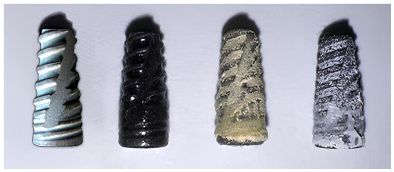
Figure 2.
(A). Osteotomy procedure performing drill passage with the assistance of the surgical guide. (B). Fixation of the implant into the left medial condyle. (C,D). Post-operative Anteroposterior (AP) and Lateral radiographic views of the right knee following implant placement.
Figure 2.
(A). Osteotomy procedure performing drill passage with the assistance of the surgical guide. (B). Fixation of the implant into the left medial condyle. (C,D). Post-operative Anteroposterior (AP) and Lateral radiographic views of the right knee following implant placement.
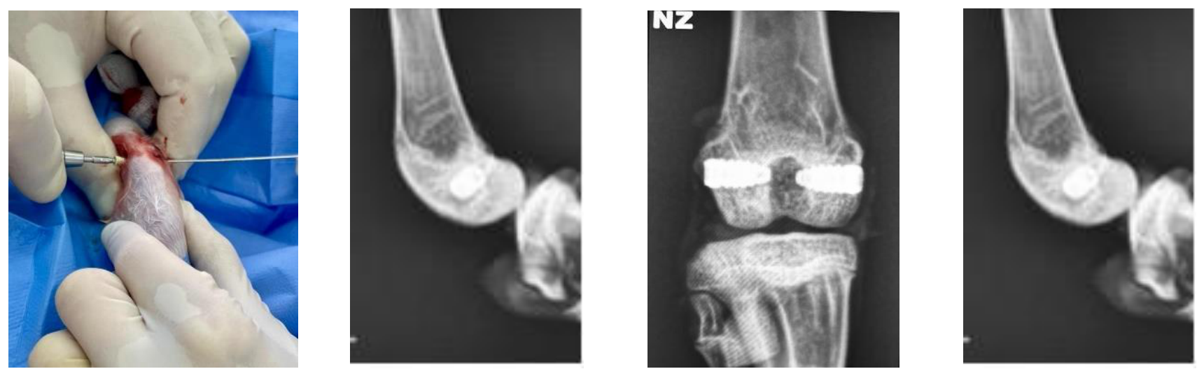
Figure 3.
Measurement of the removal torque required for implant.
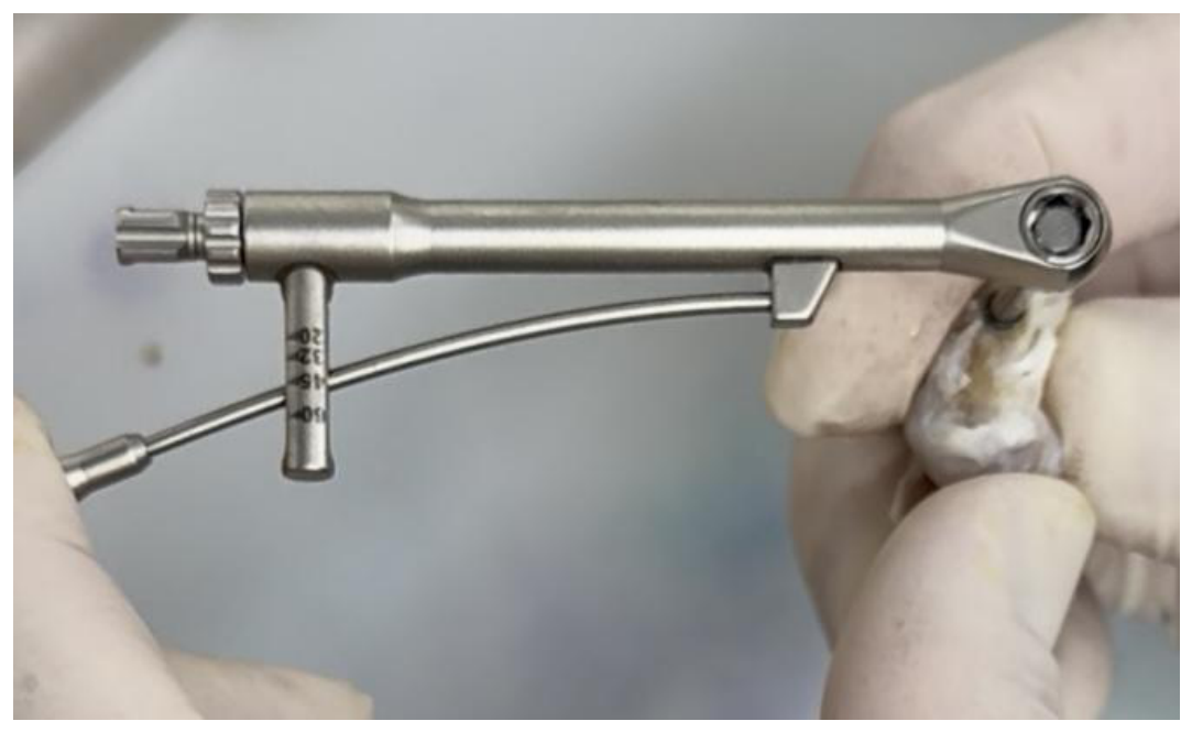
Figure 4.
(A). Representative histological section stained with Hematoxylin and Eosin (H&E). (B). Representative histological section stained with Picrosirius Red (observed under polarized light).
Figure 4.
(A). Representative histological section stained with Hematoxylin and Eosin (H&E). (B). Representative histological section stained with Picrosirius Red (observed under polarized light).
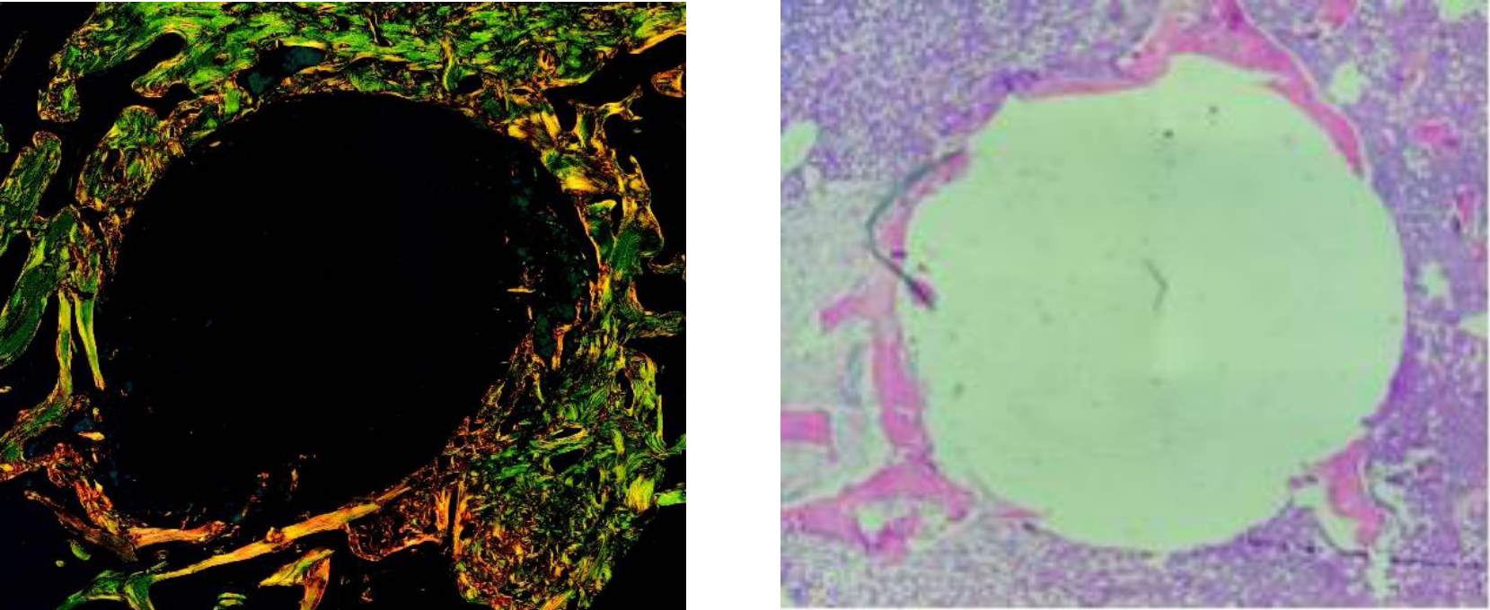
Figure 5.
Torque measurements (N/cm). Statistical differences were found among implant types, showing significance within the 60-day group. The mean value across the four implants was higher at 60 days compared to 30 days, with prominent results observed for P2 (uncoated) and P3 (graphene DLC).
Figure 5.
Torque measurements (N/cm). Statistical differences were found among implant types, showing significance within the 60-day group. The mean value across the four implants was higher at 60 days compared to 30 days, with prominent results observed for P2 (uncoated) and P3 (graphene DLC).
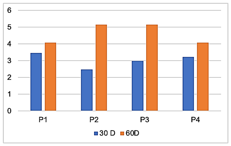
Figure 6.
Coating delamination measurements. The surface integrity of the implants is evaluated to verify the presence of free nanoparticles, given that unstable or bioactive coatings may release particles capable of triggering inflammation and impairing osseointegration.
Figure 6.
Coating delamination measurements. The surface integrity of the implants is evaluated to verify the presence of free nanoparticles, given that unstable or bioactive coatings may release particles capable of triggering inflammation and impairing osseointegration.
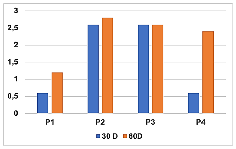
Figure 7.
Degree of bone organization.
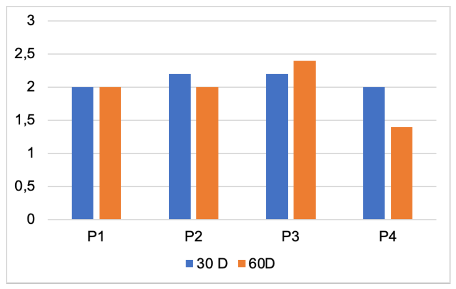
Figure 8.
Presence of osteoid.
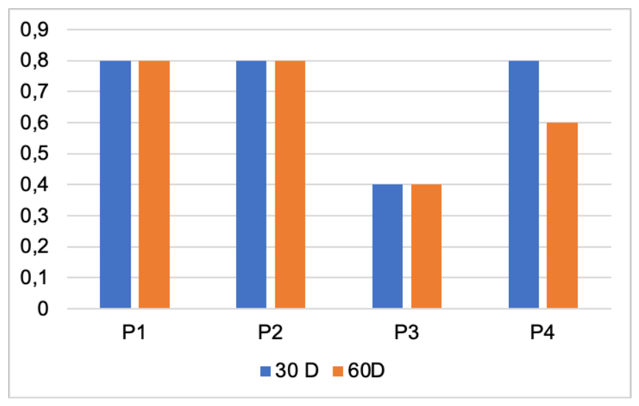
Figure 9.
Osteoclastic proliferation.
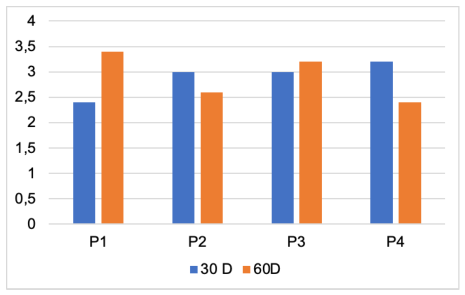
Figure 10.
Bone-to-implant contact (BIC).
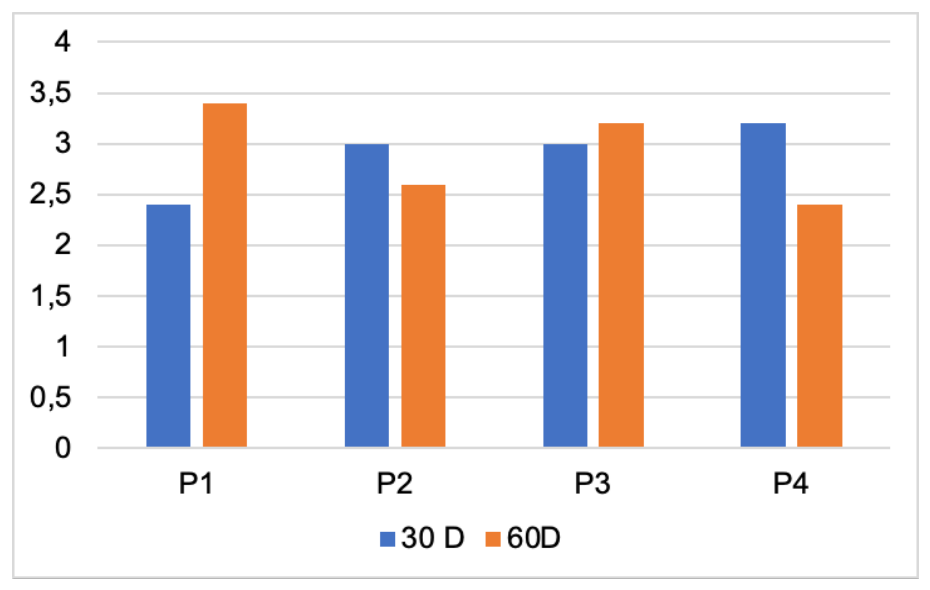
Figure 11.
Bone-implant interface thickness (ROI).
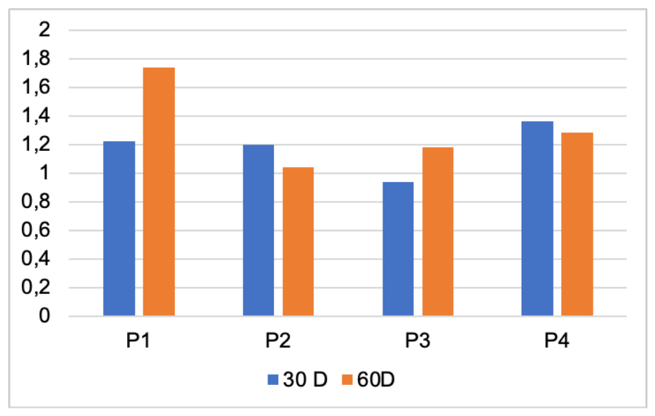
Figure 12.
Predominant tissue type.
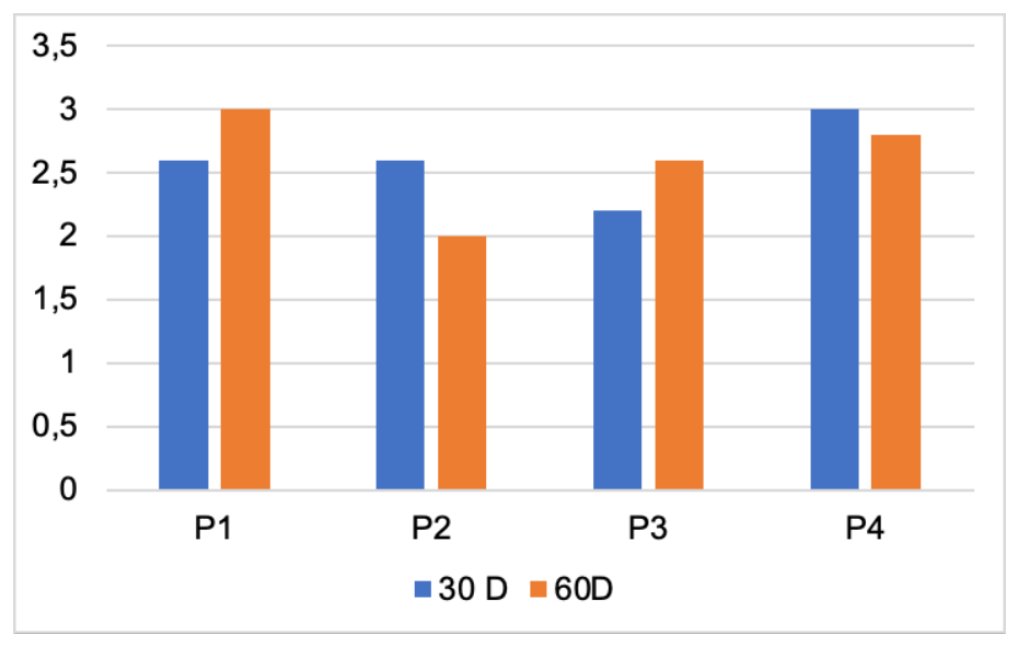
Figure 13.
Repair tissue maturation.
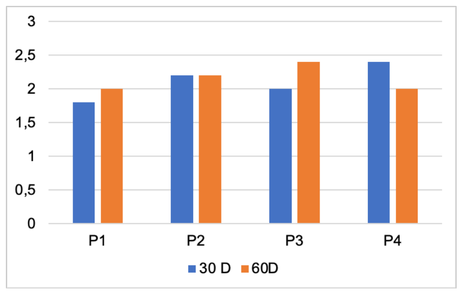
Figure 14.
Vascular response.
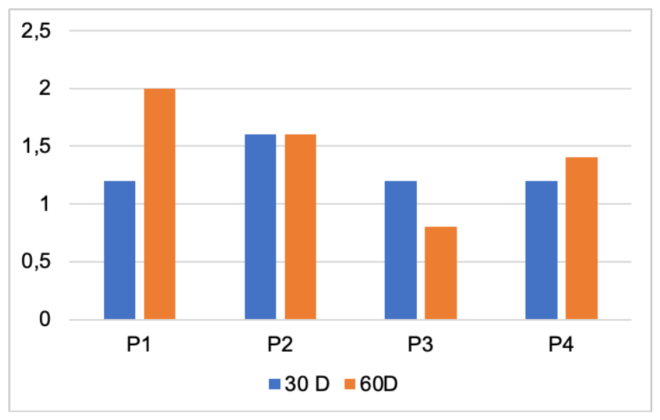
Figure 15.
Acute inflammatory infiltrate.
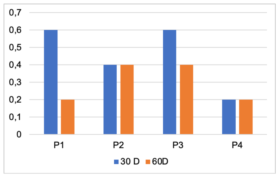
Figure 16.
Chronic inflammatory infiltrate.
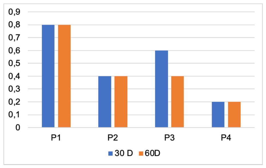
Figure 17.
Presence of multinucleated giant macrophages.
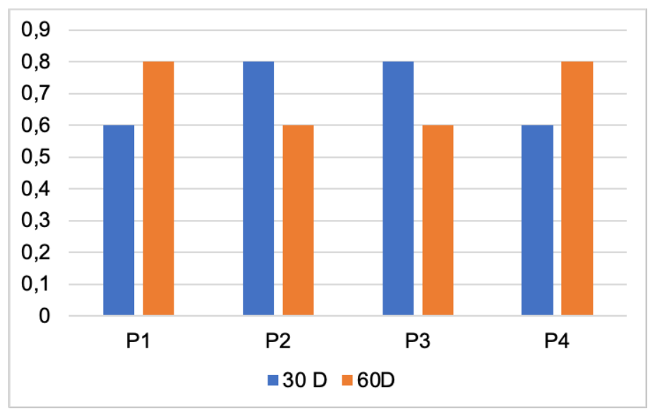
Figure 18.
Maturity of newly formed bone.
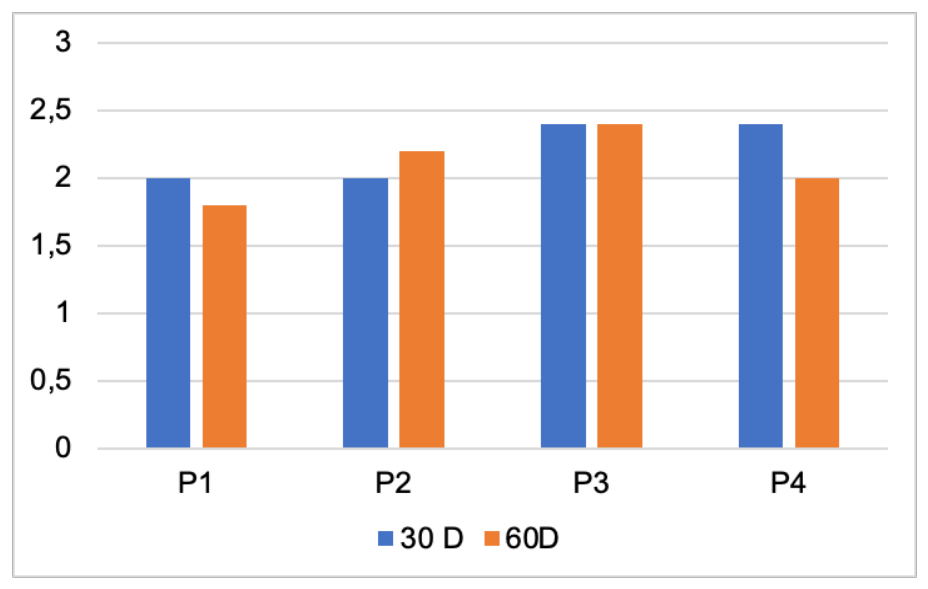
Figure 19.
Presence of type I collagen.
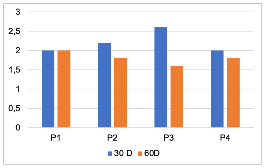
Figure 20.
Presence of type III collagen.
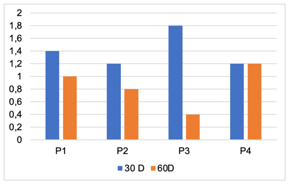
Figure 21.
Type I/III collagen ratio.
Disclaimer/Publisher’s Note: The statements, opinions and data contained in all publications are solely those of the individual author(s) and contributor(s) and not of MDPI and/or the editor(s). MDPI and/or the editor(s) disclaim responsibility for any injury to people or property resulting from any ideas, methods, instructions or products referred to in the content. |
© 2026 by the authors. Licensee MDPI, Basel, Switzerland. This article is an open access article distributed under the terms and conditions of the Creative Commons Attribution (CC BY) license (http://creativecommons.org/licenses/by/4.0/).
Copyright: This open access article is published under a Creative Commons CC BY 4.0 license, which permit the free download, distribution, and reuse, provided that the author and preprint are cited in any reuse.