

Submitted:
25 August 2026
Posted:
26 August 2026
You are already at the latest version
Abstract
Dengue virus (DENV) causes the most prevalent arboviral infection worldwide. Alt-hough classically regarded as an acute illness, growing evidence describes Post-Dengue Syndrome (PDS) as a chronic entity with profound sequelae. This narrative review an-alyzes its pathophysiology: the cytokine storm, antibody-dependent enhancement, and NS1-triggered molecular mimicry, together with the cells that execute them—dendritic cells, mononuclear phagocytes, NK cells, platelets, and complement on the innate side; plasmablast expansion, follicular helper T cell activation, defective regulatory control, and PD-L1/PD-1-mediated exhaustion on the adaptive side. Genetic susceptibility is examined through polymorphisms of HLA, FcγRIIa, Toll-like receptor, and cytokine genes. Multisystemic manifestations are described—persistent fatigue, fibromyal-gia-like myalgia and arthralgia, alopecia, brain fog, and mood disturbance—alongside neurological complications, categorized into direct neuroinvasion and post-infectious immune-mediated damage such as Guillain-Barré syndrome and encephalomyelitis. Recent population-based data linking dengue to acute neurological events and to later cognitive decline are examined critically, as is the unsettled question of whether post-dengue neuroinflammation amounts to neurodegeneration. Finally, the influence of age and sex is addressed, and therapeutic and research priorities are outlined, of-fering an immunoclinical perspective for the multidisciplinary management of these patients.
Keywords:
dengue
; post-dengue syndrome
; immunopathogenesis
; molecular mimicry
; innate immunity
; NK cells
; HLA
; T lymphocytes
; neuroinflammation
; cognitive decline
1. Introduction
Dengue virus (DENV) is an arbovirus in the family Flaviviridae, responsible for a clinical spectrum ranging from undifferentiated fever to severe presentations of dengue hemorrhagic fever and dengue shock syndrome [1]. This classic terminology—dengue fever, dengue hemorrhagic fever (DHF, graded I–IV), and dengue shock syndrome (DSS)—corresponds to the 1997 World Health Organization classification. In 2009, the WHO revised this scheme, replacing it with a classification based on clinical warning signs: dengue without warning signs, dengue with warning signs, and severe dengue [1]. Because the large majority of the genetic-association and pathophysiology studies discussed in this review were published under, or explicitly refer to, the earlier grading system, this article retains the classic DHF/DSS nomenclature when reporting original findings, while acknowledging that current WHO guidance frames these same clinical phenotypes within the revised warning-signs classification. Over recent decades, global incidence has increased dramatically, with millions of people exposed to prolonged sequelae after overcoming the acute phase of the illness [2]. Attention has traditionally focused on acute manifestations; however, a chronic clinical entity known as Post-Dengue Syndrome (PDS) has emerged, characterized by debilitating symptoms that persist for months or years after initial recovery [3,4,5]. This impression has recently been reinforced by a large population-based cohort study showing that dengue survivors carry a significantly elevated risk of new-incident autoimmune, endocrine, gastrointestinal, and renal complications for up to 300 days after the acute infection, underscoring that PDS is not a marginal phenomenon but a measurable, systemic burden on convalescent patients [6]. The parallel with other post-viral syndromes is difficult to ignore: the convalescent picture after dengue overlaps substantially with the one described after SARS-CoV-2 infection, and a direct comparison of the two infections shows that both combine ADE-prone antibody responses, endothelial injury, and a protracted inflammatory tail, while differing in tropism and in the kinetics of viral clearance [7]. Understanding why some patients recover fully while others develop this protracted syndrome requires an integrated view of the virus–host interaction: the immune mechanisms triggered during acute infection, the genetic background that shapes their intensity, and the resulting clinical and neurological expression. This review details the underlying immunological and genetic mechanisms, clinical symptomatology, and nervous system sequelae that define PDS, following the same logical sequence from mechanism to manifestation.
2. Materials and Methods
This is a narrative literature review; it was not registered as a systematic review or meta-analysis, and no PRISMA-type flow diagram was generated. Nonetheless, we followed a structured search strategy to identify the primary and secondary literature underlying each section.
Data sources. PubMed/MEDLINE and Scopus were used as the primary bibliographic databases, complemented by targeted searches in Google Scholar and, for regional and Spanish-language clinical reports, SciELO and LILACS. Reference lists of key articles and prior reviews were also hand-searched (snowball approach) to capture additional relevant studies, including several 2024–2026 publications not yet indexed under all search terms at the time of the initial search.
Search terms. Searches combined the terms “dengue,” “post-dengue syndrome,” “post-dengue fatigue syndrome,” “dengue immunopathogenesis,” “antibody-dependent enhancement,” “molecular mimicry,” “NS1 protein,” “blood–brain barrier,” “FcγRIIa,” “HLA dengue,” “dengue polymorphism,” “dengue neurological complications,” “post-dengue cognitive decline,” “dengue dementia,” “follicular helper T cells,” “PD-1/PD-L1 dengue,” “neonatal Fc receptor,” and “Toll-like receptor polymorphism dengue,” combined using Boolean operators (AND/OR) and adapted to the syntax of each database.
Time frame. The search covered January 2000 to August 2026, capturing both the foundational immunogenetic literature on dengue severity (much of which was published in the 2000s and early 2010s) and the most recent evidence on post-dengue syndrome, neurological sequelae, and long-term multi-organ outcomes (2024–2026).
Selection criteria. Original research articles, systematic reviews, meta-analyses, and case series/case reports (the latter used sparingly, mainly to illustrate rare neurological presentations such as isolated cranial nerve palsies) published in English or Spanish were considered eligible. Conference abstracts without peer-reviewed full text, non-peer-reviewed preprints, and articles not available in full text were excluded. Given the narrative nature of this review, the authors selected and synthesized studies based on relevance, methodological quality, and contribution to the mechanistic and clinical narrative, rather than through a predefined systematic screening protocol.
3. Immunological Mechanisms
To understand why PDS develops in only a subset of patients, it is first necessary to examine the immune events that unfold during the acute infection itself, since these same mechanisms set the stage for chronic disease. The immunopathogenesis of acute and chronic dengue is multifactorial. A principal feature is antibody-dependent enhancement (ADE), a phenomenon in which non-neutralizing antibodies generated during a primary infection facilitate entry of a heterologous viral serotype into monocytes, macrophages, and dendritic cells bearing Fc gamma receptors (FcγR) [8,9]. This amplifies viral replication and triggers a massive release of soluble factors known as the “cytokine storm,” involving increased interleukin-6 (IL-6), IL-8, IL-10, interferon-gamma (IFN-γ), and tumor necrosis factor-alpha (TNF-α), which are directly responsible for vascular endothelial dysfunction [10].
The ADE model does not account for one consistent observation. During acute dengue, the largest population of infected cells in the circulation is not the FcγR-bearing phagocyte but the B lymphocyte, even though the FcγRIIb expressed by B cells cannot mediate enhancement and instead drives anergy and apoptosis when engaged. This discrepancy is resolved by B cell receptor-dependent enhancement (BDE), in which DENV-reactive B cells internalize virus through their own antigen receptor rather than through an Fc receptor [11]. The mechanistic requirements have since been defined: entry depends on the specificity and affinity of the BCR for the viral envelope protein, requires proximal BCR signaling and dynamin-mediated endocytosis, involves trafficking to late endosomes, and results in productive infection of DENV-reactive B cells [12]. The implication for the present review is direct. The memory B cells that expand during infection, discussed in Section 5.1 as the source of persistent anti-NS1 specificities, are themselves susceptible to infection through the very receptor that defines their specificity, which offers a mechanism by which the humoral memory left behind by dengue may be qualitatively altered rather than merely enlarged.
Beyond the cytokine storm, long-term symptom persistence is strongly linked to molecular mimicry. DENV non-structural protein 1 (NS1) and the structural envelope (E) and premembrane (prM) proteins exhibit substantial structural homology with human proteins [13]. Cross-reactivity may result in autoantibodies that specifically target endothelial cells, platelets, and various coagulation molecules, including plasminogen, thrombin, and platelet factor 4. This activity can lead to cellular apoptosis and disrupt hemostatic function [14,15]. The repertoire of targets has been cataloged in detail: cross-reactive antibodies recognize plasminogen, thrombin, and other coagulation factors; platelet surface proteins; and endothelial antigens, inhibiting the coagulation cascade, promoting hyperfibrinolysis, and, through anti-endothelial cell antibodies, contributing to hepatic inflammation in severe infection [16]. Two mechanisms are particularly well documented for the platelet: anti-NS1 antibodies opsonize platelets and drive their phagocytosis by macrophages [17], and the degree of platelet activation itself determines the severity of the resulting thrombocytopenia [18]. Moreover, viral antigens have been detected in brain tissue, and viral RNA has been amplified from cerebrospinal fluid in patients presenting with encephalitis or encephalopathy, so the same cross-reactive repertoire may also engage neural and glial antigens, offering a direct link between mimicry and the neurological syndromes [19]. This persistent cross-reactivity prolongs the chronic post-viral inflammatory state and underlies tissue damage [3].
4. Innate Immune Response: Target Cells and Early Effector Mechanisms
The mechanisms discussed are not just theories; they involve specific cell populations, with genetic variants affecting molecules expressed by these cells. The innate immune response governs the early hours of infection and influences the subsequent adaptive response, warranting a detailed description before addressing B and T lymphocytes. This section focuses on what changes may persist after the acute phase of infection has resolved.
4.1. Dendritic Cells and Mononuclear Phagocytes
Cutaneous myeloid dendritic cells (DC) represent the initial cellular responders to dengue virus (DENV) following a mosquito bite. The principal entry receptor for DENV is DC-SIGN (CD209), which is why polymorphisms in the gene encoding this receptor frequently appear in the literature discussed in Section 6.3.1. Monocytes and macrophages are the second major reservoir and the cells in which antibody-dependent enhancement operates, since their FcγR internalizes virus–antibody complexes. Beyond serving as targets, these cells behave abnormally in patients: flow-cytometric analysis of monocytes and of myeloid and plasmacytoid DC from dengue patients shows increased TLR3 and TLR9 early in uncomplicated dengue but poor TLR3 and TLR9 stimulation in those with severe manifestations, together with differences in TLR2 expression and in the co-stimulatory molecules CD80 and CD86 between clinical groups [20]. The functional implication is that antigen-presentation quality is already compromised during the acute episode, precisely when the adaptive repertoire is being selected. Whether this presenting capacity is fully restored during convalescence, or whether a residual defect contributes to the altered memory compartment described in Section 5, has not been examined.
4.2. Natural Killer Cells and the NKG2D–MICB Axis
NK cells are among the first lymphocytes to respond to DENV, with early activation markers (CD69, HLA-DR, CD38, CD44) observed in patients during the initial days of illness. This early activation is linked to milder disease, indicating a protective role. [21]. A detailed analysis confirmed strong NK cell activation and proliferation during acute infection, dependent on IL-18 signaling. Responding cells showed a less mature phenotype with chemokine receptors indicating skin homing, with a subset detectable in the skin early in infection. [22]. The kinetics of responses in CD56bright and CD56dim subsets, stratified by NKG2A and CD57, are well characterized for the acute and immediate post-febrile phases.
One dimension, however, remains notably underexplored, and genetic evidence directly indicates its relevance. NKG2D (CD314) is the primary activating receptor through which natural killer (NK) cells, as well as CD8+ and γδ T cells, recognize stress-induced ligands, with MICA and MICB being the most prominent. Variants of the MICB gene are among the few loci consistently associated with dengue shock syndrome according to genome-wide analyses, as discussed in Section 6.3.4. Although the ligand has been genetically implicated, patients' receptor expression levels have been infrequently assessed. Furthermore, the potential for soluble MICA and MICB to be shed during acute infection, thereby downregulating NKG2D and diminishing cytotoxic function—a mechanism well established in other contexts—has not yet been investigated in dengue, particularly during convalescence. This represents a feasible research opportunity: the receptor can be quantified using conventional flow cytometry in conjunction with degranulation markers such as CD107a. A persistent deficit in NKG2D would provide a definitive cellular correlate for the extended post-infectious phase.
4.3. Unconventional and Innate-Like Lymphocytes
Between the innate and adaptive compartments are populations that activate rapidly and without conventional peptide–MHC restriction: γδ T cells, mucosal-associated invariant T (MAIT) cells, and NKT cells. All three have been reported to be activated in acute dengue, and their dysregulation contributes to the broader picture of disturbed immune cell responses in severe disease [23]. Innate cytokine profiling in patient cohorts has placed mediators of this compartment among the markers that discriminate clinical outcome [24]. NKT cells have since been shown to promote the Th1 bias that governs long-term protective antibody dynamics after DENV infection—a finding of particular interest here, because it links an innate-like population directly to the durability and quality of humoral memory [25]. Since CD1d restricts NKT activation, variation in this axis is a plausible contributor to post-dengue syndrome, and an analogous argument has been made for the persistence of altered innate compartments after other acute viral infections [7].
The γδ compartment adds a further layer. Analysis of T cell receptor repertoire dynamics in dengue patients shows that severity is accompanied by distinctive usage of TRGV and TRBV segments and by elongated δ-CDR3 sequences, a pattern consistent with specialized inflammatory function rather than with conventional antigen recognition [26]. Whether these repertoire features persist into convalescence, and whether they track with prolonged symptoms, has not been examined.
Investigating this subject is essential for understanding the role of these cells in dengue chronicity and potentially in sustaining the inflammatory response associated with new viral infections and their resolution.
4.4. Neutrophils and Neutrophil Extracellular Traps
Neutrophils are the most abundant circulating leukocyte and, until recently, the least examined in dengue. They are recruited early after infection, and DENV activates them to release myeloperoxidase and to form neutrophil extracellular traps (NETs); both processes have been shown to increase vascular permeability and to mediate the cardiac impairment described in severe disease. Neutrophil-derived soluble products, among them olfactomedin 4 and the soluble urokinase plasminogen activator receptor, have been proposed as biomarkers of clinical progression, and attenuating the neutrophil response has been suggested as a therapeutic avenue [27]. For a syndrome defined by persistent inflammation, this compartment is conspicuously unstudied: whether NET formation resolves with convalescence, or whether a low-grade NETotic state accompanies persistent symptoms, is unknown.
4.5. Complement, Platelets, and the Amplification of Vascular Injury
The innate contribution to dengue is not confined to cells that present antigen or kill. Circulating NS1 activates complement locally and systemically, generating anaphylatoxins and the terminal SC5b-9 complex in proportion to disease severity; these products contribute to the plasma leakage that defines severe dengue [28]. At the same time, the virus subverts the same system: flavivirus NS1 antagonizes complement component C4 and binds C4b-binding protein, attenuating activation on the surface of infected cells [29]. Complement therefore appears twice in the pathogenesis, as an effector of tissue injury and as a target of viral evasion.
Platelets, for their part, are not simply the cells whose count falls. DENV triggers assembly of NLRP3 inflammasomes in platelets, with caspase-1 activation and release of IL-1β-rich microparticles; these microparticles increase endothelial permeability in an IL-1-dependent manner, and inflammasome activation in patients correlates with clinical signs of vascular leak [30]. Read alongside the anti-platelet autoantibodies generated through molecular mimicry (Section 3), this establishes platelets as active immune effectors in dengue rather than passive casualties of the infection—a point of direct relevance to the persistent hemostatic and vascular complaints reported by some convalescent patients, and one that has not been followed beyond the acute phase.
5. Adaptive Immune Response: B- and T-Lymphocyte Dynamics from Acute to Convalescent Phase
Beyond the innate and humoral mechanisms described above, the cellular arms of adaptive immunity — B and T lymphocytes — undergo dramatic, time-dependent changes during dengue infection that are directly relevant to understanding why some patients transition into PDS while others recover without sequelae.
5.1. B-Lymphocyte Responses
During the acute febrile phase, DENV infection triggers one of the most striking plasmablast expansions described for any human viral infection: antibody-secreting plasmablasts can transiently account for up to 30–50% of circulating B lymphocytes, particularly during secondary heterotypic infection [31]. This burst is largely polyclonal and cross-reactive across serotypes, predominantly targeting the envelope (E) glycoprotein, and resolves rapidly, returning to baseline levels within about one month after discharge [31,32]. Longitudinal studies indicate that the memory B cell (MBC) compartment established during convalescence is largely distinct from, rather than directly descended from, this acute plasmablast population; MBCs preferentially target prM and non-structural proteins, including NS1, whereas plasmablasts are almost exclusively E-specific [32]. This divergence is directly relevant to PDS: because anti-NS1 memory B cell clones persist into convalescence and NS1 is the principal driver of the molecular-mimicry mechanisms described in Section 3, a sustained or dysregulated memory B cell response could plausibly help maintain production of cross-reactive autoantibodies well beyond viral clearance. However, this link has not yet been tested directly in patients with confirmed PDS [33].
Two further aspects of the B cell compartment remain poorly defined in dengue and deserve explicit mention. The first is the contribution of innate-like B cells. B-1-like and marginal-zone-like B cells are the main source of natural, polyreactive IgM in other systemic viral infections, and polyreactive IgM is precisely the isotype expected to participate in the early cross-reactive responses described above; these populations have nonetheless rarely been examined in acute or convalescent dengue, and their behavior in patients who go on to develop persistent symptoms is unknown. The second is prior exposure to other flaviviruses. In Latin America, where DENV and Zika virus circulate together, and yellow fever vaccination is widespread, the pre-existing memory pool is rarely dengue-specific: antibodies elicited by Zika virus cross-react extensively with DENV envelope and prM epitopes and can enhance DENV infection in vitro, and the converse also applies, so the individual serological history—and not merely the number of previous dengue episodes—conditions the quality of the response mounted during a new infection [34].
The help that sustains the plasmablast burst comes largely from circulating follicular helper T cells. Peripheral Tfh (pTfh) subsets expand and acquire an activated PD-1high CD38+ phenotype during acute DENV infection, reaching their highest frequencies during the critical phase; activated pTfh are more abundant in secondary than in primary infection, are more abundant in patients with more severe disease, and their frequency correlates with the magnitude of the plasmablast response [35]. Because Tfh function governs affinity maturation and the composition of the memory pool, this axis is a plausible—though so far untested—determinant of whether the convalescent repertoire remains protective or drifts toward the cross-reactive, potentially autoreactive specificities associated with persistent symptoms.
5.2. T-Lymphocyte Responses
T cell responses follow a parallel but mechanistically distinct trajectory, and given their centrality to dengue immunopathogenesis, they are considered here in more detail across five complementary lines of evidence: serotype cross-reactivity, cytotoxic function, regulatory control, checkpoint-mediated exhaustion, and the interface with innate and unconventional lymphocytes.
5.2.1. Antigen Cross-Reactivity
According to this hypothesis, secondary infection with a heterologous serotype preferentially reactivates lower-affinity, serotype-cross-reactive memory CD8+ T cells from the primary infection rather than generating an optimal, serotype-specific response; these cross-reactive cells display a skewed activation phenotype, characterized by high production of TNF-α and IFN-γ with reduced cytotoxic degranulation, and undergo substantial activation-induced apoptosis during acute illness [36]. This phenomenon was originally proposed as a contributor to the systemic vascular disturbances of severe dengue, since excessive cytokine production by aberrantly activated T cells could compound the endothelial damage attributed to NS1 and the cytokine storm described in Section 3. More recent, comprehensive epitope-mapping studies have partly revised this model: dengue-specific CD8+ T cell responses are strongly restricted by HLA class I genotype, and several of the HLA alleles associated with disease severity discussed in Section 6.1 are now understood to shape not only antibody-mediated but also cell-mediated antiviral immunity, with some cross-reactive T cell populations behaving in a protective rather than a purely pathological manner [37]. A further caveat is that this discussion is almost always framed around HLA class I restriction of CD8+ T cells, whereas class II molecules—which control the CD4+ response and are themselves associated with severity—present epitopes broadly shared with other flaviviruses, including Zika virus. In populations exposed to both viruses, the cellular memory recalled during a dengue episode need not be dengue-specific at all [34,37].
5.2.2. Cytotoxic Function and Epitope Hierarchy
The non-structural protein NS3 is the immunodominant target of the human CD8+ T cell response across multiple HLA backgrounds. Functional studies using an HLA-A11–restricted NS3 epitope showed that CD8+ (and CD4+) T cells from patients with uncomplicated dengue fever were predominantly degranulating and cytotoxic (CD107a-positive), whereas T cells from patients who progressed to DHF were skewed toward cytokine production without degranulation — a functional dichotomy that links the quality, and not merely the magnitude, of the T cell response to clinical severity [38]. CD4+ T cells contribute a complementary helper function, supporting both the B cell and CD8+ T cell responses described above, but a subset also acts as direct cytotoxic effectors against NS1- and NS2a-derived epitopes, adding a further, less-studied cytotoxic layer to CD4+ T cell biology in dengue [39]. This cytotoxic CD4+ compartment deserves more attention than it usually receives: DENV-specific CD4+ T cells expressing granzyme B and perforin, with a highly differentiated phenotype, have been characterized in individuals from hyperendemic areas, where they appear to be associated with protection rather than with pathology, and they represent a functional axis that conventional CD8+-centered epitope mapping does not capture [40].
5.2.3. Regulatory T Cells
CD4+CD25+FOXP3+ regulatory T cells (Tregs), whose normal role is to dampen excessive immune activation, are themselves dysregulated in acute dengue: Treg frequency and proliferation increase during acute infection, but these cells are abnormally activated, skewed toward a Th1-like phenotype, and functionally less suppressive than Tregs from healthy donors, particularly in patients who progress to more severe disease [41]. This suggests a failure of immune self-regulation that compounds, rather than restrains, the cytokine storm described in Section 3. Regulatory control, moreover, is not exercised by FOXP3+ Tregs alone. Dendritic cells, monocytes and macrophages, NK cells, γδ T cells, and NKT cells are all engaged early in dengue and shape the magnitude and quality of the subsequent adaptive response; their dysregulation in severe disease has been documented repeatedly, and it is at this innate–adaptive interface, rather than within any single cell type, that the balance between viral control and immunopathology appears to be decided [23].
5.2.4. T Cell Exhaustion and the Acute-to-Convalescent Transition
T cells in severe dengue also display features more typical of chronic viral infections, including elevated expression of the inhibitory receptors PD-1, TIM-3, and LAG-3, consistent with an exhaustion-like phenotype that correlates with reduced cytotoxic capacity [23]. Detailed kinetic studies show that CD4+ helper and CD8+ effector T cell activation peaks around defervescence and then declines over the following one to two weeks [42], but whether this decline is complete, or whether a residual, exhausted or persistently activated T cell population extends further into convalescence — as has been documented for weeks to months after other acute viral infections — is essentially unstudied in dengue specifically. This is a notable gap, since sustained low-grade T cell activation is one of the mechanisms proposed, albeit not yet confirmed, to underlie post-dengue fatigue syndrome [43]. Two recent lines of work provide a more concrete mechanistic basis for this checkpoint biology. Extracellular vesicles recovered from the plasma of patients with severe dengue carry PD-L1 and suppress CD4+ T cell responses through PD-L1/PD-1 engagement, which provides a circulating, cell-free route by which checkpoint signaling can be sustained systemically [44]. In parallel, work in a clinically relevant mouse model has shown that the conserved N-linked glycan at position 207 of NS1 restrains T cell mediated clearance: virus lacking this glycan is attenuated, produces milder lymphopenia and more functional DENV-specific CD8+ T cells, and is associated with reduced PD-L1 expression on innate immune cells and fewer PD-1+ T cells, while PD-1 blockade protects animals infected with wild-type virus [45]. Together, these findings identify the PD-L1/PD-1 axis as an active mechanism of dengue immunopathogenesis rather than a passive marker of exhaustion, and they raise the possibility—untested in humans—that the same axis contributes to the incomplete immune restoration of convalescence. The COVID-19 literature suggests a related and equally untested possibility: neutralizing autoantibodies against type I and type II interferons, together with functional deficiencies in IFN-α and IFN-γ signaling, are associated with impaired viral control and protracted disease after SARS-CoV-2 infection, and whether analogous anti-cytokine autoantibodies arise after dengue and contribute to chronicity has not been examined [7].
Taken together, the adaptive cellular response to DENV is intense and functionally heterogeneous at its acute peak — spanning protective cytotoxicity, dysregulated cross-reactivity, defective regulatory control, and exhaustion — but it leaves behind an altered, imperfectly characterized memory compartment in both the B and T cell lineages. Very few studies have examined how these adaptive compartments behave beyond the first weeks of convalescence. Essentially none have compared patients who go on to develop PDS with those who recover fully—a gap summarized in Table 1, which lists the populations discussed in this and the preceding section together with the markers used to identify them and the state of the evidence in each phase, and one that reinforces the mechanistic research priorities.
6. Genetic Susceptibility: FcγR and HLA
The intensity of the immune events described above—the cytokine storm, ADE, and molecular mimicry—varies across individuals and is substantially shaped by host genetics, which helps explain why only a fraction of infected persons progress to severe acute disease or PDS. Host genetics strongly determines the degree of dengue-related damage and its long-term sequelae [46]. Polymorphisms in the gene encoding the FcγRIIa receptor (also known as CD32) play a critical role in modulating the host immune response, determining susceptibility to severe forms of the disease and to the development of chronic sequelae in Post-Dengue Syndrome [3,46].
6.1. FcγR
FcγRIIa is an activating receptor present on the surface of phagocytic cells such as monocytes, macrophages, and dendritic cells, which transduces signals through an immunoreceptor tyrosine-based activation motif (ITAM) [47]. This receptor mediates antibody-dependent enhancement (ADE) by allowing binding and internalization of complexes formed by dengue virus and non-neutralizing antibodies. By facilitating viral entry into these target cells, it increases viral replication and infection severity [8].
FcγRIIa is not the only receptor that determines the fate of IgG–virus complexes. The neonatal Fc receptor (FcRn), classically described as the receptor that rescues IgG and albumin from degradation and extends their half-life several-fold, has emerged as an entry route exploited by structurally unrelated viruses, including echoviruses, astroviruses and—as shown by genome-wide CRISPR screening—the whole of the Arteriviridae family [48,49]. Two aspects of that work bear directly on dengue. The first is a negative result that should be stated plainly: in the same screening panel, yellow fever virus, a flavivirus, infected cells normally in the absence of FcRn, so there is no basis for proposing FcRn as an entry receptor for DENV, and none is proposed here.
The second is a corollary the authors themselves emphasize, and it is the one relevant to this review. Because FcRn continuously shuttles IgG and albumin between the cell surface and a non-degradative endosomal compartment, a virus that engages it in endothelial cells could be stored intracellularly alongside its physiological cargo and released slowly back into the circulation, constituting a semi-latent reservoir; conversely, antagonism of FcRn markedly shortens the half-life of circulating IgG and albumin. Persistence in the endothelium and disruption of IgG homeostasis relate to Post-Dengue Syndrome, which shows clinical correlates but lacks an established mechanism, with the endothelium being the most consistently injured by DENV. Neither possibility has been examined in dengue, and both are testable.
The clinical impact of this receptor is shaped by an important functional polymorphism at position 131 (nucleotide variation −393 A/G), which results in expression of either histidine (H) or arginine (R) [46]. The two variants generate markedly different phenotypes:
- Homozygous histidine genotype (HH131): a risk factor predisposing to susceptibility for dengue hemorrhagic fever (DHF). Individuals with this genotype show greater affinity for immunoglobulin IgG2, which triggers excessive monocyte activation and uncontrolled release of inflammatory mediators during clearance of dengue antigens. Patients carrying the HH131 genotype also have a significantly higher probability of experiencing persistent symptoms associated with Post-Dengue Syndrome, even up to two years after infection [3,46].
In sum, FcγRIIa polymorphisms directly modulate the immune system’s capacity to process virus–antibody complexes, either conditioning a state of sustained hyperinflammation and damage (histidine variant) or conferring resistance to shock and plasma leakage (arginine variant).
6.2. HLA System
Several studies have confirmed the role of human leukocyte antigen (HLA) alleles in both susceptibility and protection against severe and prolonged forms of the disease [52]. Alleles such as HLA-A*24, HLA-A*0203, and DRB1*11 predispose individuals to greater severity, whereas HLA-A*33, DRB1*03, and DRB1*09 appear to play a protective role [52,53]. A recent multi-population meta-analysis corroborates these associations while underscoring substantial regional heterogeneity: HLA-A*02 and HLA-A*03 increase susceptibility to dengue fever and dengue hemorrhagic fever, respectively, predominantly in Southeast Asian cohorts, whereas HLA-A*33 and HLA-B*44 exert a protective effect whose magnitude varies markedly by population, a finding with direct implications for genetic risk stratification as dengue’s geographic range expands under climate change [54]. These associations should be read with two caveats. Several of the alleles listed above are common, “public” alleles in the populations studied, so their apparent association with severity may partly reflect background allele frequency rather than a specific functional effect. Because class I and class II loci are inherited in linkage disequilibrium, the true unit of association is more likely the haplotype than any single allele [53,54]. Analyses reporting class I associations in isolation may therefore be capturing the effect of a linked class II allele. This distinction matters in settings where the same class II molecules present epitopes shared with other co-circulating flaviviruses [34].
6.3. Other Genetic Factors
Polymorphisms in the DC-SIGN gene (rs4804803 promoter), in NK-associated molecules (MICB), and in key cytokines such as TNF-α (−308A allele) also stand out, shaping cellular control and promoting pathogenic responses [55,56,57]. A broader case-control and meta-analytic evaluation of candidate genes in a pediatric cohort likewise confirms that this genetic layer contributes measurably to dengue severity in children specifically, reinforcing the relevance of these polymorphisms across age groups [58]. These are detailed below.
6.3.1. Viral Entry and Pathogen-Recognition Receptors
DC-SIGN (CD209): this receptor is essential for viral entry into myeloid dendritic cells, and the widely studied rs4804803 (−336 A/G) promoter polymorphism significantly influences disease severity [59,60]. Notably, its effect depends strongly on ethnic background. In Asian populations, the G allele is associated with higher receptor expression, increased production of cytokines (such as TNF-α, IL-12, and IP-10), and a significant increase in DHF risk. In South American populations, however, this same G allele acts as a protective factor against severe dengue [55,61,62].
CLEC5A: a receptor expressed on monocytes and macrophages. The TT genotype of the rs1285933 polymorphism has been strongly associated with dengue severity. Patients with this genotype secrete significantly higher levels of TNF during the critical phase of infection, worsening vascular permeability [57,63,64,65].
MBL2 (mannose-binding lectin 2): variations in this gene, which encodes a key molecule in pathogen recognition and complement activation, have been associated with an increased risk of thrombocytopenia and DHF [50,56].
Toll-like receptors (TLR): the TLR family sits upstream of most of the responses described above. A recent systematic review of eight case-control studies from India, Mexico, Colombia, and Indonesia, comprising 2,525 individuals, found that polymorphisms in TLR3, TLR4, TLR7, TLR8, and TLR9 are associated with susceptibility to dengue, and that all of these except TLR4 and TLR9 also correlate with severity or with specific clinical outcomes [66]. The involvement of TLR4 is mechanistically coherent with the preceding sections, since NS1 itself activates immune cells through TLR4, whereas the endosomal receptors TLR3, TLR7, TLR8, and TLR9 act as sensors of viral nucleic acid. Two caveats accompany these findings: the direction of effect differs between populations, with one TLR3 variant carrying risk through one allele in a Colombian cohort and through the opposite allele in an Indian one, and no individual association has yet been replicated consistently enough to support clinical risk stratification.
One locus in this family deserves particular attention here. TLR3 is expressed in astrocytes, microglia, and T cells, and its functional L412F variant, which impairs dsRNA sensing and reduces type I interferon production, has been examined specifically in patients with dengue encephalitis [67]. The same variant has been implicated in Japanese encephalitis and tick-borne encephalitis, suggesting a susceptibility pathway shared across neurotropic flaviviruses rather than a dengue-specific effect. This makes TLR3 a plausible genetic link between innate nucleic-acid sensing and the neurological syndromes discussed in Section 8, and one of the few points where the genetic and neurological literatures on dengue meet.
6.3.2. Cytokine and Chemokine Gene Polymorphisms
The development of the “cytokine storm” is also genetically predisposed:
TNF-α: the polymorphism at position −308A promotes high production of this vasoactive cytokine and represents an important risk factor for severity and hemorrhagic manifestations in American and Latin American patients. In contrast, the −238A allele has demonstrated a protective effect [53,62].
IL-10 and TGF-β1: the ACC/ATA haplotype in the IL-10 promoter (positions −1082/−819/−592) is a significant risk factor for DHF, favoring antibody-dependent enhancement (ADE). Likewise, the −509 CC genotype of the TGF-β1 gene is associated with greater susceptibility to dengue hemorrhagic fever, a risk that increases when combined with the +49G allele of the CTLA-4 gene [46,61,65].
JAK1: polymorphisms in this gene (TT genotypes in introns 1 and 3, and GG in intron 2), which is crucial for the interferon signaling pathway, are associated with the development of DHF [68,69,70].
Other variants. Beyond the loci detailed above, candidate-gene studies have reported associations for interleukin genes (IL-1β, IL-6, IL-8/CXCL8), chemokines such as CCL2/MCP-1, IFN-γ (+874 A/T), and genes governing complement activation and endothelial function. The catalogs compiled in recent reviews are considerably longer than can be usefully summarized here, and the recurring limitation is not the scarcity of candidate loci but the scarcity of replication across ethnically distinct cohorts [46,53,56].
6.3.3. Hemostasis and Endothelial Dysfunction Markers
Human Platelet Antigens (HPA): variations in the HPA promoter region influence autoantibody-induced cross-reactive thrombocytopenia. The HPA 1a/1a and HPA 2a/2b genotypes are more prevalent in DHF patients, whereas the HPA 1b genotype is strongly associated with dengue shock syndrome (DSS) [46,71,72].
6.3.4. Other Regulatory Factors Identified by GWAS
Vitamin D Receptor (VDR): vitamin D regulates immune interactions and can induce T-cell apoptosis after infection. The C allele at position −352 of the VDR promoter (associated with absence of the TaqI restriction site) confers clinical resistance to severe DHF [50,73].
MICB and PLCE1: variants in the MICB gene (associated with Natural Killer and T-cell activation) and in the PLCE1 gene (related to vascular endothelium), specifically the rs3132468 and rs3740360 genotypes, show a direct association with genetic susceptibility to dengue shock syndrome in children [56,74,75].
TAP proteins (Transporter associated with Antigen Processing): polymorphisms in the TAP1 gene (heterozygous at position 333, Ile/Val), which influence processing of viral peptides for presentation by MHC class I, increase the risk of DHF [9,69,71].
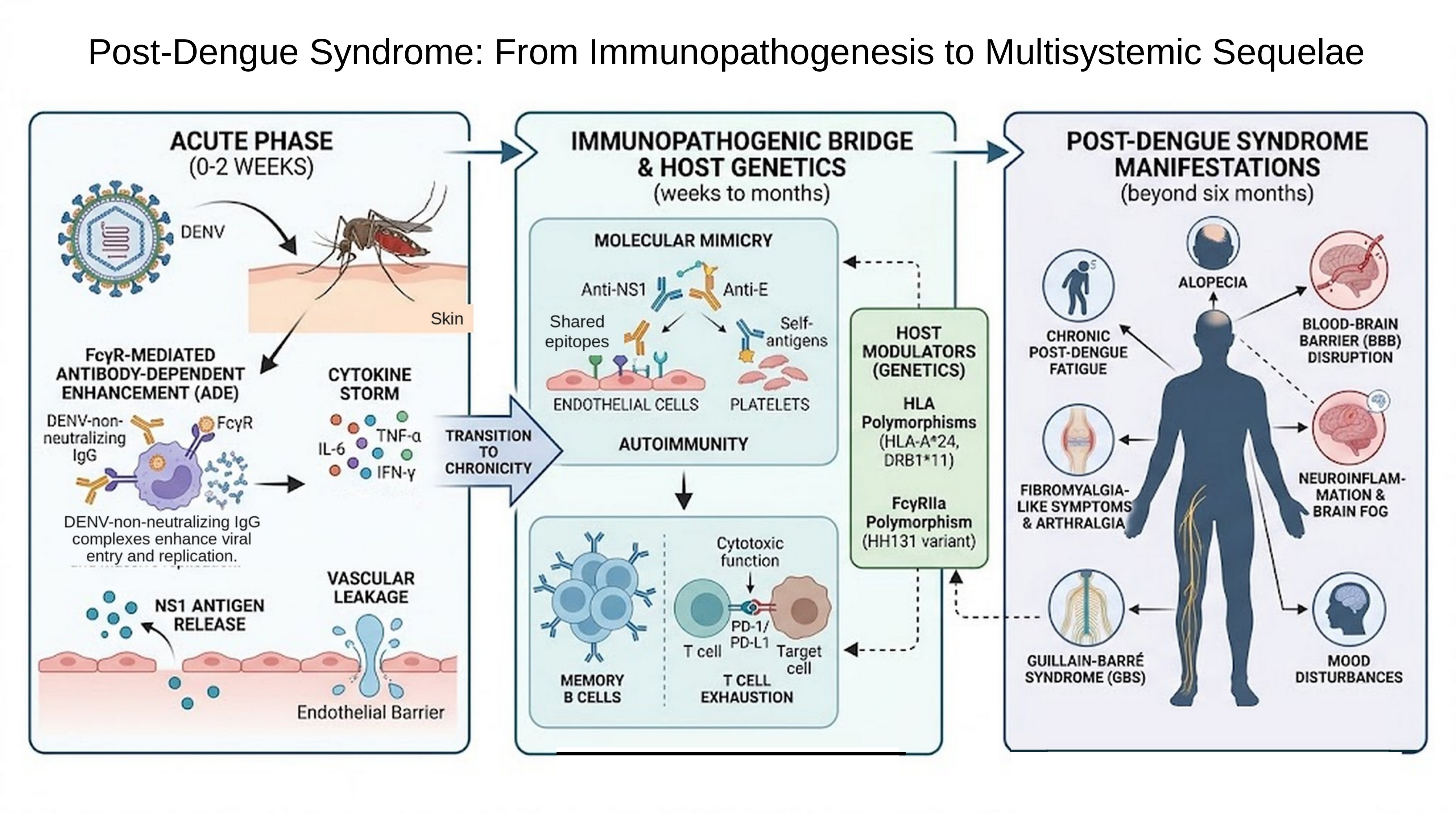
Taken together, Sections 3 to 6 describe one process rather than four. ADE and NS1 set the intensity of the acute response; dendritic cells, mononuclear phagocytes, NK cells, platelets, and complement determine the conditions under which that response is mounted; the adaptive compartment converts it into a memory repertoire that may remain cross-reactive and imperfectly regulated; and the host genotype modulates the amplitude of every one of these steps. From this integrated view follows the working hypothesis of the present review: Post-Dengue Syndrome is not a separate disease beginning where dengue ends, but the tail of this same process in patients whose immune system does not return to baseline. Figure 1 summarizes the sequence along the temporal axis that organizes the remainder of this review—acute phase, convalescence, and persistent disease—and connects each mechanistic stage with the clinical manifestations examined in Sections 7 to 9.
7. Antibody Fucosylation
A determinant of severity that is neither genetic nor cellular but structural deserves mention alongside the polymorphisms discussed above. The Fc glycan of IgG1 modulates affinity for FcγRIIIa, and IgG1 lacking core fucose binds this receptor with markedly higher avidity. In secondary dengue, the proportion of afucosylated anti-DENV IgG1 predicts progression to severe disease and thrombocytopenia [76]. The same feature operates in early life: the fucosylation state of maternally transferred anti-dengue IgG predicts infants' susceptibility to symptomatic dengue [77]. This adds a post-translational layer to the ADE argument developed in Section 3—what matters is not only how much cross-reactive IgG persists after infection, but how it is glycosylated—and whether an afucosylation signature persists into convalescence, or tracks with prolonged symptoms, has not been investigated.
8. Symptomatology and Post-Dengue Fatigue Syndrome
Having outlined the molecular and genetic machinery that drives PDS, we now turn to how these mechanisms translate into the clinical picture seen at the bedside. The clinical manifestations of PDS encompass physical, neurological, psychiatric, and autonomic disturbances. The principal symptom is profound fatigue, also described as post-infectious fatigue, present in up to 25% of hospitalized adults, which differs from ordinary tiredness because of its incapacitating impact on everyday activities [78,79]. Other reported symptoms include headache, persistent retro-orbital pain, intense myalgia, arthralgia, memory loss, concentration difficulties, and sleep disturbances (depression and anxiety). Statistically significant predictive factors for developing this syndrome include female sex, older age, initial presence of severe chills, and absence of the typical acute skin rash [78,79,80]. The autoimmune dimension of this convalescent phase has been tested at the population level, with an instructive result. In a Taiwanese cohort of 63,814 laboratory-confirmed dengue patients matched to 255,256 controls, dengue was associated with an increased short-term risk of autoimmune encephalomyelitis but not with the other autoimmune diseases examined, contrary to earlier reports [81]. The distinction matters for how PDS is framed: the evidence supports discrete, immune-mediated neurological complications rather than a generalized predisposition to autoimmunity, and the persistent systemic symptoms described in this section should not be equated with the onset of classical autoimmune disease.
There are significant differences in the development and severity of post-dengue syndrome according to sex, with women showing greater vulnerability and a higher risk of these chronic sequelae [43].
- Greater prevalence and severity of fatigue: female sex is an independent risk factor and a strong predictor for the development of post-infectious fatigue syndrome after dengue [82,83,84,85]. Several studies have shown that women are significantly more likely to meet clinical criteria for fatigue and report much higher mean exhaustion scores compared with men [86,87].
- Rheumatologic complications and physical limitations: joint and functional sequelae also disproportionately affect women. After the acute phase of illness, women report greater limitation, stiffness, and pain when performing everyday activities such as walking, standing, lying down, dressing, or climbing stairs. Similarly, the incidence of persistent post-dengue joint inflammation and pain is significantly higher in women [88]. Persistent post-dengue arthropathy has been attributed to autoantibody-mediated mechanisms rather than to ongoing viral replication: cross-reactive antibodies against endothelial and platelet antigens, and circulating immune complexes, have been detected months after viral clearance in patients with prolonged symptoms, which would also be consistent with the female predominance shared with classical autoimmune rheumatic disease [3,89].
Proposed mechanisms. The scientific literature attributes this greater predisposition of women to persistent symptoms to a combination of the following factors [86,90]:
- Biological and host factors: differences in genetic makeup, reproductive function, and hormonal imbalances are thought to play an important role in this vulnerability.
- Psychosocial and behavioral factors: differences in psychosomatic stress management may also influence predisposition to fatigue.
- Under-reporting in men: some authors note that part of the statistical gap between sexes may stem from men being more reluctant to report symptoms owing to cultural or gender norms in certain societies.
9. Neurological Sequelae and Nervous System Involvement
The nervous system warrants special attention due to severe complications from PDS. Recent studies confirm that neurological and psychiatric disorders are significant, yet likely under-recognized, consequences of dengue infection, occurring more frequently than previously thought. [91,92]. The incidence of nervous system complications from dengue ranges between 0.5% and 20% of cases [82,83,84]. Neurotropism and immune-mediated damage from DENV occur through three main mechanisms, leading to different clinical syndromes. The scale of the problem has recently been quantified. In a population-based cohort of 65,207 adults with confirmed dengue in Singapore, compared with more than 1.6 million uninfected individuals, the odds of a new neurological event within 30 days of infection were markedly elevated (adjusted odds ratio 9.69), with memory loss and movement disorders among the specific outcomes; the absolute excess burden was nevertheless small, since fewer than 1% of infected patients experienced such an event. Both figures must be kept in mind: dengue substantially raises relative risk while remaining, for most patients, neurologically uneventful [19].
9.1. Mechanisms of Nervous System Involvement
Direct neuroinvasion. Direct passage of the virus across the blood–brain barrier, or its transport via infected monocytes, induces meningoencephalitis, myelitis, and myositis [93,94,95]. Viral invasion promotes hyperintensities on magnetic resonance imaging in the thalamus, basal ganglia, and cerebellum [94]. Dengue virus non-structural protein 1 (NS1) profoundly and negatively affects the blood–brain barrier (BBB) through a combination of direct structural and indirect immune-mediated mechanisms, leading to vascular hyperpermeability and facilitating neuroinflammation. The principal mechanisms through which NS1 disrupts this barrier are:
- Cytokine storm and immune activation: NS1 acts as a potent immunological trigger by binding TLR4 on monocytes, macrophages, and endothelial cells. This interaction, together with activation of NK cells and T lymphocytes, provokes a massive release of pro-inflammatory cytokines such as IFN-γ, TNF-α, and interleukins IL-6, IL-8, and IL-12. High circulating concentrations of these cytokines directly damage blood–brain barrier integrity, allowing abnormal passage of immune mediators into brain tissue [96,97,98,99].
- Destruction of the endothelial glycocalyx and tight junctions: NS1 stimulates destructive enzymes such as heparanase, cathepsin L, and sialidase. These enzymes degrade sialic acid and destroy the endothelial glycocalyx (the protective layer lining blood vessels), producing severe endothelial dysfunction and consequent hyperpermeability [100]. NS1 additionally promotes expression of matrix metalloproteinases (such as MMP-9), which degrade critical structural proteins such as β-catenin and zonula occludens-1 (ZO-1). The reduction of these proteins disrupts the tight junctions that normally hold BBB endothelial cells firmly together [89,96,101,102].
- Autoimmune damage (molecular mimicry) and apoptosis: Due to the phenomenon of molecular mimicry, the patient's immune system produces anti-NS1 antibodies that exhibit cross-reactivity with the patient's own endothelial cells. These antibodies interact with the endothelium, causing cellular stress and triggering apoptosis through nitric oxide production. This endothelial cell death and dysfunction further undermine vascular integrity. [13,14,89,103].
Chronic and acute NS1-mediated disruption of the blood-brain barrier allows dengue virus and inflammatory mediators to enter the central nervous system, leading to severe outcomes like encephalitis and meningoencephalitis. This creates a chronic neuroinflammatory environment linked to long-term neuropsychiatric and cognitive issues in Post-Dengue Syndrome. The main sex-based differences in neurological manifestations are as follows:
- Transverse myelitis: A systematic review found that cases of this post-infectious inflammatory and demyelinating spinal cord condition were more common in men than women, at a ratio of 1.5 to 1 [94].
- Encephalitis: in prospective studies of large populations with dengue-related encephalitis, the proportion of affected men was 58% [94].
- Movement disorders: Emerging case reports and a recent review highlight dystonia, parkinsonism, chorea, tics, and ataxia as post-infectious complications of dengue encephalitis, expanding the recognized neurological manifestations beyond classical syndromes. [105]. Dengue-associated autoimmune encephalitis can lead to dramatic cases, such as a dystonic storm in a young adult, highlighting that post-infectious autoimmunity against neural antigens can cause hyperkinetic movement disorders, not just the typical demyelinating syndromes. [106].
Post-dengue fatigue syndrome is the only neuro-systemic condition after infection in which women show a significantly higher risk [104,107].
- Systemic and metabolic complications: these give rise to diffuse encephalopathy resulting from a combination of cerebral edema, hypoxia, organ failure (hepatic and renal), hyponatremia, and coagulopathy (hemorrhage and ischemic or hemorrhagic stroke). Another crucial systemic manifestation is hypokalemic paralysis, an acute neuromuscular disorder secondary to intracellular potassium shift or transient renal tubular acidosis [108,109].
- Immune-mediated post-infectious complications: these occur weeks to months into the convalescent phase as a result of cross-reactive autoimmune reactions (molecular mimicry against neural antigens). Clinically, they include Guillain-Barré syndrome (GBS), acute disseminated encephalomyelitis (ADEM), optic neuritis, cranial nerve palsies (oculomotor, abducens, facial), and neuromyelitis optica [108,109,110].
9.2. Neuroinflammation, Cognitive Sequelae, and the Question of Neurodegeneration
The mechanisms mentioned—barrier disruption, cytokine-driven neuroinflammation, and cross-reactive autoimmunity—pose an unresolved question: does the neuroinflammatory state resolve with recovery, or does it contribute to progressive cognitive decline? A systematic review of five population-based studies involving around 200,000 participants from Taiwan, Brazil, and France found short-term cognitive impairment—such as confusion and memory loss—in some dengue patients. There was also a potential link to longer-term dementia, including Alzheimer’s and vascular dementia, particularly in older adults. However, the authors stressed that the evidence is insufficient to establish a conclusive link. [111].
The most careful attempt to test that link argues for caution. A Taiwanese cohort of 37,928 laboratory-confirmed dengue patients aged 45 years or older, matched to 151,712 uninfected individuals, found only a slight increase in the incidence of dementia. The corresponding E-values were small enough that modest unmeasured confounding—differences in comorbidity, health-seeking behavior, or surveillance intensity—could account for the association entirely [112]. Post-dengue cognitive complaints are real and often categorized as brain fog, similar to other post-viral syndromes. However, their long-term trajectory and potential link to neurodegeneration remain unclear. Distinguishing neuroinflammation from neurodegeneration necessitates prospective cohorts with baseline and serial cognitive testing, neuroimaging, and biomarkers, rather than relying on current administrative database studies.
10. Differences by Age Group
Neurological and systemic manifestations of PDS vary by age, influencing both likelihood and specific phenotypes. In pediatric and adolescent patients, dengue encephalopathy and acute symptomatic seizures are notably more common [94,95]. Greater severity of primary capillary damage and differences in microvascular redistribution volume make infants especially vulnerable to ischemic or shock events [113]. In contrast, young or older adults show a greater predisposition to post-infectious demyelinating diseases (GBS, ADEM) and Post-Dengue Fatigue Syndrome, particularly in relation to comorbidities and baseline immune status [23,114]. Age, moreover, does not act on the nervous system alone; it acts on the immune system that responds to the virus. The pediatric immune system is highly plastic—broad naive repertoires, active thymic output, and a memory compartment still being assembled—which favors both vigorous, at times excessive, acute responses and a considerable capacity for functional recovery afterward. At the other extreme, immunosenescence and inflammaging narrow the repertoire, raise baseline inflammatory tone, and impair resolution in older adults. This contrast in immune plasticity offers a coherent explanation for the same age gradient observed at the bedside: encephalopathy and acute symptomatic seizures in the young, protracted fatigue and cognitive complaints in the old [23,114].
10.1. Encephalopathy and Encephalitis (Predominant in Pediatric and Adolescent Age Groups)
Encephalopathy is the most commonly described neurological syndrome in dengue, and its prevalence is significantly higher in children and adolescents. Within the pediatric population, infants (under one year of age) and children with obesity carry an especially high risk of developing this complication [115]. This heightened susceptibility to shock and encephalopathy in children is attributed to differences in baseline microvascular permeability compared with adults [116]. Additionally, infants born to dengue-immune mothers are at greatest risk of developing severe forms (dengue hemorrhagic fever and shock syndrome) during their first infection, between 6 and 9 months of age [113]. This results from an antibody-dependent enhancement (ADE) mechanism, in which passively transferred maternal antibodies fall to sub-neutralizing levels and facilitate viral replication [117]. Encephalitis from direct viral invasion also predominantly affects younger populations; several studies report that between 52% and 68% of patients with dengue encephalitis are under 15 years of age [93,94]. This vulnerability carries lasting consequences: a retrospective pediatric cohort found measurable long-term neurologic sequelae, most commonly cognitive and behavioral impairment, in nearly 40% of children discharged after dengue-associated acute encephalitis, reinforcing the need for structured follow-up and rehabilitation in this age group rather than assuming full recovery once the acute illness resolves [118].
10.2. Neuromuscular Complications: Myositis and Rhabdomyolysis
Notable contrasts exist in muscle involvement by age. In children, dengue-related muscle dysfunction or myositis typically presents as a relatively benign and self-limited condition (classically known as myalgia cruris epidemica), mainly involving calf pain and temporary difficulty walking [93,108]. In adult patients, by contrast, dengue-induced myositis tends to be a substantially more severe and aggressive entity, capable of triggering severe rhabdomyolysis, flaccid quadriparesis, and respiratory failure [93,94,108].
10.3. Myelitis
Unlike encephalopathy, which predominates in childhood, demyelinating spinal cord complications such as transverse myelitis (whether from acute direct invasion or post-infectious immune-mediated mechanisms) are more common in young adults, with a reported mean age of onset of 33 years in clinical cohorts [119]. Other autoimmune complications such as Guillain-Barré syndrome are frequently observed in adults, presenting weeks after the febrile phase resolves [120,121,122].
10.4. Post-Dengue Fatigue Syndrome (Commonly Found in Middle-Aged and Older Adults)
The development of persistent chronic symptoms, specifically Post-Dengue Fatigue Syndrome, is strongly and independently associated with older age. Older adults face a significantly higher risk of profound post-infectious fatigue that does not improve with rest, as well as memory loss, reasoning difficulties, hair loss, and persistent arthralgia. These chronic symptoms, which may last from several months to two years, impose severe physical and psychological limitations and are observed far more frequently in adults and older adults than in children [5,43,90,123,124].
11. Therapeutic Considerations and Future Perspectives
The evidence reviewed here converges on a coherent, if still incomplete, picture of Post-Dengue Syndrome as a genuine post-infectious entity rather than a diffuse or ill-defined complaint. Three lines of evidence support this view. First, the immunological mechanisms active during acute infection — antibody-dependent enhancement, the cytokine storm, and NS1-driven molecular mimicry — are not confined to the febrile phase; cross-reactive autoantibodies and endothelial injury plausibly persist beyond viral clearance, providing a mechanistic bridge to chronic symptoms [3,13,14,15]. Second, host genetics measurably shapes who develops severe or prolonged disease: FcγRIIa and HLA polymorphisms recur across genetically and geographically distinct cohorts — Vietnamese, Cuban, Thai, Brazilian, and Mexican — which strengthens confidence that these associations are biologically real rather than population-specific artifacts, even though effect sizes vary between studies [50,51,58,63,75]. Third, the clinical and epidemiological literature independently converges on the same risk architecture — female sex and older age for fatigue-predominant PDS, younger age for encephalopathy and direct neuroinvasion — suggesting that host factors operating at the level of both the adaptive immune system and end-organ vulnerability jointly determine phenotype [5,43,82,83,84,85,90,115,116,117,118,123,124].
At the same time, several limitations of the underlying evidence base deserve explicit comment, since they bound how confidently these findings can be generalized. Most of the genetic-association studies discussed in Section 6 are candidate-gene, case-control designs conducted in single, often small, ethnic cohorts; replication across populations has been inconsistent, and few genome-wide association studies have specifically targeted PDS rather than acute severe dengue [56]. The clinical literature on PDS itself is dominated by hospital-based cohorts, which likely overrepresent more severe acute presentations and may not capture the full spectrum of illness seen in ambulatory or asymptomatic infections. Sex- and age-based differences, while consistently reported, are drawn largely from observational studies without uniform case definitions for PDS or post-dengue fatigue syndrome, limiting direct comparison across studies and complicating pooled incidence estimates. Finally, because this is a narrative rather than a systematic review, article selection—while guided by the structured search strategy described in Section 2—inevitably involved subjective judgment regarding relevance and quality, and publication bias favoring positive or novel associations cannot be excluded.
Therapeutic considerations. No specific antiviral therapy exists for dengue, so PDS management is supportive and largely empirical. Key points include that immune-mediated neurological syndromes in the convalescent phase—such as Guillain-Barré syndrome, ADEM, transverse myelitis, and autoimmune encephalitis—should be managed like their idiopathic counterparts, using corticosteroids, intravenous immunoglobulin, or plasma exchange based on severity. Reported outcomes are generally favorable when treatment begins early, despite relying on case reports and small series. [106,119,121,122]. For the fatigue-predominant phenotype, no disease-modifying treatments exist. The management approach emphasizes a structured, symptom-guided activity framework that targets comorbid conditions such as sleep disturbances, pain, and mood-related symptoms. Additionally, it recognizes the biological underpinnings of these complaints as therapeutically significant. Third, the checkpoint biology described in Section 5.2 is the most concrete pharmacological lead to have emerged recently, since PD-1 blockade protects mice infected with wild-type DENV [45]; the distance between an experimental model of acute infection and a chronic post-infectious syndrome in humans is nevertheless considerable, and manipulating immune checkpoints in a disease already characterized by autoreactivity would demand careful justification. Vaccination adds another layer, since the same ADE mechanism that governs severity in secondary natural infection also shapes the risk profile of dengue vaccines in seronegative recipients, and whether vaccine-primed immunity modifies the incidence of PDS is unknown [117].
These gaps highlight priorities for future research and clinical practice. A standardized case definition for post-dengue syndrome (PDS) and post-dengue fatigue syndrome, validated across endemic regions, is needed for meaningful comparisons of incidence and risk factors. Population-based cohorts, like those reported in Singapore, should track multi-organ and neurological outcomes for at least a year post-infection to better understand the burden of PDS. Additionally, mechanistic studies linking genotype to autoantibody persistence or fatigue phenotypes are needed. Clinically, a structured post-dengue follow-up for women, older adults, and children recovering from encephalitis should be integrated into routine care in endemic areas, even in the absence of specific treatment options.
MICB variants are strongly associated with severe dengue and are the ligand for the NKG2D receptor on NK cells and cytotoxic lymphocytes. Investigating whether NKG2D expression and degranulation differ in convalescent patients, in relation to MICB genotype, soluble MICA/MICB levels, or symptom persistence, could link this genetic association to a measurable cellular phenotype. This approach addresses the issue of genotype being primarily connected to acute severity rather than disease persistence.
12. Conclusions
Post-Dengue Syndrome is viewed as a common outcome of an infection that affects multiple immune system pathways and may not fully resolve in some patients. Evidence links three factors: virological and innate mechanisms (including ADE and cytokine storms), an altered adaptive immune response (characterized by changes in plasmablasts and regulatory control), and genetic factors (like HLA haplotypes and cytokine variants) that influence individual responses. The resulting symptoms vary by sex and age, with fatigue in women and older adults, encephalopathy in children, and demyelinating syndromes in young adults. Clinicians in endemic areas should prioritize active follow-up during convalescence, especially for women, older adults, and children recovering from encephalitis. Research should focus on a validated case definition, prospective cohorts with immunological and cognitive assessments, and studies linking genotype to autoantibody persistence and fatigue. A significant question remains about whether identified mechanisms can be modified. As dengue expands due to climate change, post-infectious sequelae (PDS) must be formally recognized, rather than treated as an afterthought.
Author Contributions
Conceptualization, F.C. and A.G.; methodology, F.C., J.I. and A.G.; investigation and literature search, F.C., J.I., S.M. and I.B.; writing—original draft preparation, F.C. and J.I.; writing—review and editing, S.M., I.B., J.B.D.S. and A.G.; critical revision for immunological content, J.B.D.S.; S,M< project finance and management, supervision, A.G. All authors have read and agreed to the published version of the manuscript.
Funding
This research was funded by the National Fund for Science, Technology, and Innovation (FONACIT), an entity attached to the Ministry of Popular Power for Science and Technology of the Bolivarian Republic of Venezuela (MINCYT), and Corporación para el Desarrollo Científico (CODECYT). Project number 207. J.B.D.S. is partially financed by the National Institute of Virology and Bacteriology (Program EXCELES, ID Project No. LX22NPO5103)—funded by the European Union—Next Generation EU from the Ministry of Education, Youth and Sports of the Czech Republic (MEYS).
Data Availability Statement
No data were generated.
Conflicts of Interest
The authors declare no conflicts of interest.
References
- World Health Organization. Dengue Guidelines for Diagnosis, Treatment, Prevention and Control: New Edition; World Health Organization: Geneva, Switzerland, 2009. [Google Scholar]
- Bhatt, S.; Gething, P.W.; Brady, O.J.; Messina, J.P.; Farlow, A.W.; Moyes, C.L.; et al. The global distribution and burden of dengue. Nature 2013, 496, 504–507. [Google Scholar] [CrossRef] [PubMed]
- García, G.; González, N.; Pérez, A.B.; Sierra, B.; Aguirre, E.; Rizo, D.; et al. Long-term persistence of clinical symptoms in dengue-infected persons and its association with immunological disorders. Int. J. Infect. Dis. 2011, 15, e38–e43. [Google Scholar] [CrossRef] [PubMed]
- Tiga, D.C.; Undurraga, E.A.; Ramos-Castañeda, J.; Martínez-Vega, R.A.; Tschampl, C.A.; Shepard, D.S. Persistent symptoms of dengue: estimates of the incremental disease and economic burden in Mexico. Am. J. Trop. Med. Hyg. 2016, 94, 1085–1089. [Google Scholar] [CrossRef] [PubMed]
- Umakanth, M. Post dengue fatigue syndrome. Saudi J. Med. Pharm. Sci. 2017, 3, 858–861. [Google Scholar]
- Lim, J.T.; Wee, L.E.; Tan, W.Z.; Chiew, C.; Kurupatham, L.; Poh, C.; Md Suhaimi, N.-A.; Chua, H.Z.; Ng, L.C.; Chia, P.Y.; et al. Characterization of post-acute multi-organ sequelae following dengue infection. Clin. Microbiol. Infect. 2025, 31, 1500–1509. [Google Scholar] [CrossRef] [PubMed]
- García, A.H.; De Sanctis, J.B. Exploring the contrasts and similarities of dengue and SARS-CoV-2 infections during the COVID-19 era. Int. J. Mol. Sci. 2024, 25, 11624. [Google Scholar] [CrossRef] [PubMed]
- Halstead, S.B. Neutralization and antibody-dependent enhancement of dengue viruses. Adv. Virus Res. 2003, 60, 421–467. [Google Scholar] [CrossRef] [PubMed]
- Martina, B.E.; Koraka, P.; Osterhaus, A.D. Dengue virus pathogenesis: an integrated view. Clin. Microbiol. Rev. 2009, 22, 564–581. [Google Scholar] [CrossRef] [PubMed]
- Chaturvedi, U.C.; Agarwal, R.; Elbishbishi, E.A.; Mustafa, A.S. Cytokine cascade in dengue hemorrhagic fever: implications for pathogenesis. FEMS Immunol. Med. Microbiol. 2000, 28, 183–188. [Google Scholar] [CrossRef] [PubMed]
- Gebo, C.; Hardy, C.S.C.; McElvany, B.D.; Graham, N.R.; Lu, J.Q.; Moradpour, S.; et al. B cell receptor dependent enhancement of dengue virus infection. PLoS Pathog. 2024, 20, e1012683. [Google Scholar] [CrossRef] [PubMed]
- Madrigal, G.; Gebo, C.; McElvany, B.D.; Symeonides, M.; Diehl, S.A.; Waickman, A.T. Molecular and biophysical requirements for B cell receptor-dependent enhancement of dengue virus infection. J. Virol. 2026, e0221625. [Google Scholar] [CrossRef] [PubMed]
- Lin, Y.S.; Yeh, T.M.; Lin, C.F.; Wan, S.W.; Chuang, Y.C.; Hsu, T.K.; et al. Molecular mimicry between virus and host and its implications for dengue disease pathogenesis. Exp. Biol. Med. 2011, 236, 515–523. [Google Scholar] [CrossRef] [PubMed]
- Lin, C.F.; Lei, H.Y.; Shiau, A.L.; Liu, H.S.; Yeh, T.M.; Chen, S.H.; et al. Endothelial cell apoptosis induced by antibodies against dengue virus nonstructural protein 1 via production of nitric oxide. J. Immunol. 2002, 169, 657–664. [Google Scholar] [CrossRef] [PubMed]
- Chuang, Y.C.; Lin, Y.S.; Liu, H.S.; Yeh, T.M. Molecular mimicry between dengue virus and coagulation factors induces antibodies to inhibit thrombin activity and enhance fibrinolysis. J. Virol. 2014, 88, 13759–13768. [Google Scholar] [CrossRef] [PubMed]
- Ghorai, T.; Sarkar, A.; Roy, A.; Bhowmick, B.; Nayak, D.; Das, S. Role of auto-antibodies in the mechanisms of dengue pathogenesis and its progression: a comprehensive review. Arch. Microbiol. 2024, 206, 214. [Google Scholar] [CrossRef] [PubMed]
- Wan, S.W.; Yang, Y.W.; Chu, Y.T.; Lin, C.F.; Chang, C.P.; Yeh, T.M.; Anderson, R.; Lin, Y.S. Anti-dengue virus nonstructural protein 1 antibodies contribute to platelet phagocytosis by macrophages. Thromb. Haemost. 2016, 115, 646–656. [Google Scholar] [CrossRef] [PubMed]
- Ojha, A.; Nandi, D.; Batra, H.; Singhal, R.; Annarapu, G.K.; Bhattacharyya, S.; et al. Platelet activation determines the severity of thrombocytopenia in dengue infection. Sci. Rep. 2017, 7, 41697. [Google Scholar] [CrossRef] [PubMed]
- Wee, L.E.; Tan, W.Z.; Chow, J.Y.; Lim, J.T.; Chiew, C.; Chia, P.Y.; et al. Neurological events associated with acute dengue infection. JAMA Neurol. 2026, 83, 171–180. [Google Scholar] [CrossRef] [PubMed]
- Torres, S.; Hernández, J.C.; Giraldo, D.; Arboleda, M.; Rojas, M.; Smit, J.M.; Urcuqui-Inchima, S. Differential expression of Toll-like receptors in dendritic cells of patients with dengue during early and late acute phases of the disease. PLoS Negl. Trop. Dis. 2013, 7, e2060. [Google Scholar] [CrossRef] [PubMed]
- Azeredo, E.L.; De Oliveira-Pinto, L.M.; Zagne, S.M.; Cerqueira, D.I.S.; Nogueira, R.M.R.; Kubelka, C.F. NK cells, displaying early activation, cytotoxicity and adhesion molecules, are associated with mild dengue disease. Clin. Exp. Immunol. 2006, 143, 345–356. [Google Scholar] [CrossRef] [PubMed]
- Zimmer, C.L.; Cornillet, M.; Solà-Riera, C.; Cheung, K.-W.; Ivarsson, M.A.; Lim, M.Q.; et al. NK cells are activated and primed for skin-homing during acute dengue virus infection in humans. Nat. Commun. 2019, 10, 3897. [Google Scholar] [CrossRef] [PubMed]
- Yoo, J.S.; Shporn, O.Z.; Sklan, E.H. Dysregulated immune cell responses in severe dengue pathogenesis. Front. Immunol. 2025, 16, 1600999. [Google Scholar] [CrossRef] [PubMed]
- Pradeep, S.P.; Hoovina Venkatesh, P.; Manchala, N.R.; Vayal Veedu, A.; Basavaraju, R.K.; Selvasundari, L.; et al. Innate immune cytokine profiling and biomarker identification for outcome in dengue patients. Front. Immunol. 2021, 12, 677874. [Google Scholar] [CrossRef] [PubMed]
- Choi, Y.; Saron, W.A.; O’Neill, A.; Senanayake, M.; Wilder-Smith, A.; Rathore, A.P.; St John, A.L. NKT cells promote Th1 immune bias to dengue virus that governs long-term protective antibody dynamics. J. Clin. Invest. 2024, 134, e169251. [Google Scholar] [CrossRef] [PubMed]
- Khare, K.; Yadav, S.; Tarai, B.; Budhiraja, S.; Pandey, R. TCR repertoire dynamics and their responses underscores dengue severity. iScience 2024, 27, 110983. [Google Scholar] [CrossRef] [PubMed]
- Chua, C.L.L.; Morales, R.F.; Chia, P.Y.; Yeo, T.W.; Teo, A. Neutrophils—an understudied bystander in dengue? Trends Microbiol. 2024, 32, 1132–1142. [Google Scholar] [CrossRef] [PubMed]
- Avirutnan, P.; Punyadee, N.; Noisakran, S.; Komoltri, C.; Thiemmeca, S.; Auethavornanan, K.; et al. Vascular leakage in severe dengue virus infections: a potential role for the nonstructural viral protein NS1 and complement. J. Infect. Dis. 2006, 193, 1078–1088. [Google Scholar] [CrossRef] [PubMed]
- Avirutnan, P.; Fuchs, A.; Hauhart, R.E.; Somnuke, P.; Youn, S.; Diamond, M.S.; Atkinson, J.P. Antagonism of the complement component C4 by flavivirus nonstructural protein NS1. J. Exp. Med. 2010, 207, 793–806. [Google Scholar] [CrossRef] [PubMed]
- Hottz, E.D.; Lopes, J.F.; Freitas, C.; Valls-de-Souza, R.; Oliveira, M.F.; Bozza, M.T.; et al. Platelets mediate increased endothelium permeability in dengue through NLRP3-inflammasome activation. Blood 2013, 122, 3405–3414. [Google Scholar] [CrossRef] [PubMed]
- Wrammert, J.; Onlamoon, N.; Akondy, R.S.; Perng, G.C.; Polsrila, K.; Chandele, A.; et al. Rapid and massive virus-specific plasmablast responses during acute dengue virus infection in humans. J. Virol. 2012, 86, 2911–2918. [Google Scholar] [CrossRef] [PubMed]
- Priyamvada, L.; Cho, A.; Onlamoon, N.; Zheng, N.-Y.; Huang, M.; Kovalenkov, Y.; et al. B cell responses during secondary dengue virus infection are dominated by highly cross-reactive, memory-derived plasmablasts. J. Virol. 2016, 90, 5574–5585. [Google Scholar] [CrossRef] [PubMed]
- Waickman, A.T.; Victor, K.; Li, T.; Hatch, K.; Rutvisuttinunt, W.; Medin, C.; et al. Longitudinal analysis of acute and convalescent B cell responses in a human primary dengue serotype 2 infection model. EBioMedicine 2019, 41, 465–478. [Google Scholar] [CrossRef] [PubMed]
- Ngono, A.E.; Shresta, S. Immune response to dengue and Zika. Annu. Rev. Immunol. 2018, 36, 279–308. [Google Scholar] [CrossRef] [PubMed]
- Haltaufderhyde, K.; Srikiatkhachorn, A.; Green, S.; Macareo, L.; Park, S.; Kalayanarooj, S.; Rothman, A.L.; Mathew, A. Activation of peripheral T follicular helper cells during acute dengue virus infection. J. Infect. Dis. 2018, 218, 1675–1685. [Google Scholar] [CrossRef] [PubMed]
- Mongkolsapaya, J.; Dejnirattisai, W.; Xu, X.-N.; Vasanawathana, S.; Tangthawornchaikul, N.; Chairunsri, A.; et al. Original antigenic sin and apoptosis in the pathogenesis of dengue hemorrhagic fever. Nat. Med. 2003, 9, 921–927. [Google Scholar] [CrossRef] [PubMed]
- Weiskopf, D.; Angelo, M.A.; de Azeredo, E.L.; Sidney, J.; Greenbaum, J.A.; Fernando, A.N.; et al. Comprehensive analysis of dengue virus-specific responses supports an HLA-linked protective role for CD8+ T cells. Proc. Natl. Acad. Sci. USA 2013, 110, E2046–E2053. [Google Scholar] [CrossRef] [PubMed]
- Duangchinda, T.; Dejnirattisai, W.; Vasanawathana, S.; Limpitikul, W.; Tangthawornchaikul, N.; Malasit, P.; Mongkolsapaya, J.; Screaton, G. Immunodominant T-cell responses to dengue virus NS3 are associated with DHF. Proc. Natl. Acad. Sci. USA 2010, 107, 16922–16927. [Google Scholar] [CrossRef] [PubMed]
- Tian, Y.; Grifoni, A.; Sette, A.; Weiskopf, D. Human T cell response to dengue virus infection. Front. Immunol. 2019, 10, 2125. [Google Scholar] [CrossRef] [PubMed]
- Tian, Y.; Sette, A.; Weiskopf, D. Cytotoxic CD4 T cells: differentiation, function, and application to dengue virus infection. Front. Immunol. 2016, 7, 531. [Google Scholar] [CrossRef] [PubMed]
- Jayaratne, H.E.; Wijeratne, D.; Fernando, S.; Kamaladasa, A.; Gomes, L.; Wijewickrama, A.; Ogg, G.S.; Malavige, G.N. Regulatory T-cells in acute dengue viral infection. Immunology 2018, 154, 89–97. [Google Scholar] [CrossRef] [PubMed]
- Manh, D.H.; Weiss, L.N.; Thuong, N.V.; Mizukami, S.; Dumre, S.P.; Luong, Q.C.; et al. Kinetics of CD4+ T helper and CD8+ effector T cell responses in acute dengue patients. Front. Immunol. 2020, 11, 1980. [Google Scholar] [CrossRef] [PubMed]
- Garg, R. Post-dengue fatigue syndrome: a comprehensive review of an emerging clinical entity. Karnataka Med. J. 2024, 47, 52–55. [Google Scholar] [CrossRef]
- Kumari, S.; Bandyopadhyay, B.; Singh, A.; Aggarwal, S.; Yadav, A.K.; Vikram, N.K.; Guchhait, P.; Banerjee, A. Extracellular vesicles recovered from plasma of severe dengue patients induce CD4+ T cell suppression through PD-L1/PD-1 interaction. mBio 2023, 14, e0182323. [Google Scholar] [CrossRef] [PubMed]
- Idris, F.; Ooi, J.S.G.; Ting, D.H.R.; Tan, E.T.X.; Wan, C.; Benke, P.I.; et al. Glycans on non-structural protein 1 prevent premature T-cell mediated dengue virus clearance. EMBO Mol. Med. 2025, 17, 2995–3020. [Google Scholar] [CrossRef] [PubMed]
- Gupta, S.; Agarwal, A.; Biswas, D. Host genetic polymorphisms influencing susceptibility to dengue. DNA Cell Biol. 2018, 37, 1–3. [Google Scholar] [CrossRef] [PubMed]
- Nimmerjahn, F.; Ravetch, J.V. Fcgamma receptors as regulators of immune responses. Nat. Rev. Immunol. 2008, 8, 34–47. [Google Scholar] [CrossRef] [PubMed]
- Na, L.; Zheng, Y.; Yang, J.B.; Bao, H.L.; Tang, Y.D. The neonatal Fc receptor (FcRn): guardian or Trojan horse in viral infection? PLoS Pathog. 2025, 21, e1013285. [Google Scholar] [CrossRef] [PubMed]
- Shaw, T.M.; Huey, D.; Mousa-Makky, M.; et al. The neonatal Fc receptor (FcRn) is a pan-arterivirus receptor. Nat. Commun. 2024, 15, 6726. [Google Scholar] [CrossRef] [PubMed]
- Loke, H.; Bethell, D.; Phuong, C.X.; Day, N.; White, N.; Farrar, J.; et al. Susceptibility to dengue hemorrhagic fever in Vietnam: evidence of an association with variation in the vitamin D receptor and Fc gamma receptor IIa genes. Am. J. Trop. Med. Hyg. 2002, 67, 102–106. [Google Scholar] [CrossRef] [PubMed]
- García, G.; Sierra, B.; Pérez, A.B.; Aguirre, E.; Rosado, I.; González, N.; et al. Asymptomatic dengue infection in a Cuban population confirms the protective role of the RR variant of the FcγRIIa polymorphism. Am. J. Trop. Med. Hyg. 2010, 82, 1153–1156. [Google Scholar] [CrossRef] [PubMed]
- Chen, Y.; Liao, Y.; Yuan, K.; Wu, A.; Liu, L. HLA-A, -B, -DRB1 alleles as genetic predictive factors for dengue disease: a systematic review and meta-analysis. Viral Immunol. 2019, 32, 121–130. [Google Scholar] [CrossRef] [PubMed]
- Stephens, H.A. HLA and other gene associations with dengue disease severity. Curr. Top. Microbiol. Immunol. 2010, 338, 99–114. [Google Scholar] [CrossRef] [PubMed]
- Ghosh, A.G.; Kim, H.L.; Khor, S.-S. HLA alleles and dengue susceptibility across populations in the era of climate change: a comprehensive review. Front. Immunol. 2025, 16, 1473475. [Google Scholar] [CrossRef] [PubMed]
- Ren, J.; Wang, Z.; Chen, E. Different associations between DC-SIGN promoter −336G/A (rs4804803) polymorphism with severe dengue in Asians and South-Central Americans: a meta-analysis. Int. J. Environ. Res. Public Health 2019, 16, 1475. [Google Scholar] [CrossRef] [PubMed]
- Khor, C.C.; Chau, T.N.; Pang, J.; Davila, S.; Long, H.T.; Ong, R.T.; et al. Genome-wide association study identifies susceptibility loci for dengue shock syndrome at MICB and PLCE1. Nat. Genet. 2011, 43, 1139–1141. [Google Scholar] [CrossRef] [PubMed]
- Fernández-Mestre, M.T.; Gendzekhadze, K.; Rivas-Vetencourt, P.; Layrisse, Z. TNF-alpha-308A allele, a possible severity risk factor of hemorrhagic manifestation in dengue fever patients. Tissue Antigens 2004, 64, 469–472. [Google Scholar] [CrossRef] [PubMed]
- Xavier-Carvalho, C.; Gibson, G.; Brasil, P.; Ferreira, R.X.; de Souza Santos, R.; Gonçalves Cruz, O.; et al. Single nucleotide polymorphisms in candidate genes and dengue severity in children: a case-control, functional and meta-analysis study. Infect. Genet. Evol. 2013, 20, 197–205. [Google Scholar] [CrossRef] [PubMed]
- Sakuntabhai, A.; Turbpaiboon, C.; Casademont, I.; Chuansumrit, A.; Lowhnoo, T.; Kajaste-Rudnitski, A.; et al. A variant in the CD209 promoter is associated with severity of dengue disease. Nat. Genet. 2005, 37, 507–513. [Google Scholar] [CrossRef] [PubMed]
- Wang, L.; Chen, R.F.; Liu, J.W.; Lee, I.K.; Lee, C.P.; Kuo, H.C.; et al. DC-SIGN (CD209) promoter −336 A/G polymorphism is associated with dengue hemorrhagic fever and correlated to DC-SIGN expression and immune augmentation. PLoS Negl. Trop. Dis. 2011, 5, e934. [Google Scholar] [CrossRef] [PubMed]
- Chen, S.T.; Lin, Y.L.; Huang, M.T.; Wu, M.F.; Cheng, S.C.; Lei, H.Y.; et al. CLEC5A is critical for dengue-virus-induced lethal disease. Nature 2008, 453, 672–676. [Google Scholar] [CrossRef] [PubMed]
- Acioli-Santos, B.; Segat, L.; Dhalia, R.; Brito, C.A.A.; Braga-Neto, U.M.; Marques, E.T.A.; et al. MBL2 gene polymorphisms protect against development of thrombocytopenia associated with severe dengue phenotype. Hum. Immunol. 2008, 69, 122–128. [Google Scholar] [CrossRef] [PubMed]
- Vejbaesya, S.; Luangtrakool, P.; Luangtrakool, K.; Kalayanarooj, S.; Vaughn, D.W.; Endy, T.P.; et al. TNF and LTA gene, allele, and extended HLA haplotype associations with severe dengue virus infection in ethnic Thais. J. Infect. Dis. 2009, 199, 1442–1448. [Google Scholar] [CrossRef] [PubMed]
- Perez, A.B.; Sierra, B.; Garcia, G.; Aguirre, E.; Babel, N.; Alvarez, M.; et al. Tumor necrosis factor-alpha, transforming growth factor-beta1, and interleukin-10 gene polymorphisms: implication in protection or susceptibility to dengue hemorrhagic fever. Hum. Immunol. 2010, 71, 1135–1140. [Google Scholar] [CrossRef] [PubMed]
- Chen, R.F.; Wang, L.; Cheng, J.T.; Chuang, H.; Chang, J.C.; Liu, J.W.; et al. Combination of CTLA-4 and TGF-beta1 gene polymorphisms associated with dengue hemorrhagic fever and virus load in a dengue-2 outbreak. Clin. Immunol. 2009, 131, 404–409. [Google Scholar] [CrossRef] [PubMed]
- Candrasari, D.S.; Purwosatrio, P.G.; Purnomosari, D.; Laksanawati, I.S.; Nirwati, H. Host genetic factors modulating dengue virus: a systematic review of TLR polymorphisms. BMC Infect. Dis. 2026, 26, 399. [Google Scholar] [CrossRef] [PubMed]
- Verma, R.; Pandey, A.K.; Chakraborty, R.; Prakash, S.; Jain, A. Toll-like receptor 3 genetic polymorphism in dengue encephalitis. J. Fam. Med. Prim. Care 2024, 13, 2397–2403. [Google Scholar] [CrossRef] [PubMed]
- Silva, L.K.; Blanton, R.E.; Parrado, A.R.; Melo, P.S.; Morato, V.G.; Reis, E.A.; et al. Dengue hemorrhagic fever is associated with polymorphisms in JAK1. Eur. J. Hum. Genet. 2010, 18, 1221–1227. [Google Scholar] [CrossRef] [PubMed]
- Soundravally, R.; Hoti, S.L. Immunopathogenesis of dengue hemorrhagic fever and shock syndrome: role of TAP and HPA gene polymorphism. Hum. Immunol. 2007, 68, 973–979. [Google Scholar] [CrossRef] [PubMed]
- Sierra, B.; Perez, A.B.; Vogt, K.; Garcia, G.; Schmolke, K.; Aguirre, E.; Alvarez, M.; Volk, H.D.; Guzman, M.G. MCP-1 and MIP-1α expression in a model resembling early immune response to dengue. Cytokine 2010, 52, 175–183. [Google Scholar] [CrossRef] [PubMed]
- Snyder, J.T.; Alexander-Miller, M.A.; Berzofsky, J.A.; Belyakov, I.M. Molecular mechanisms and biological significance of CTL avidity. Curr. HIV Res. 2003, 1, 287–294. [Google Scholar] [CrossRef] [PubMed]
- Sosothikul, D.; Seksarn, P.; Pongsewalak, S.; Thisyakorn, U.; Lusher, J. Activation of endothelial cells, coagulation and fibrinolysis in children with dengue virus infection. Thromb. Haemost. 2007, 97, 627–634. [Google Scholar] [CrossRef]
- Tsai, T.T.; Chuang, Y.J.; Lin, Y.S.; Wan, S.W.; Chen, C.L.; Lin, C.F. An emerging role for the anti-inflammatory cytokine interleukin-10 in dengue virus infection. J. Biomed. Sci. 2013, 20, 40. [Google Scholar] [CrossRef] [PubMed]
- Sierra, B.; Alegre, R.; Pérez, A.B.; et al. HLA-A, -B, -C, and -DRB1 allele frequencies in Cuban individuals with antecedents of dengue 2 disease: advantages of the Cuban population for HLA studies of dengue virus infection. Hum. Immunol. 2007, 68, 531–540. [Google Scholar] [CrossRef] [PubMed]
- LaFleur, C.; Granados, J.; Vargas-Alarcon, G.; Ruiz-Morales, J.; Villarreal-Garza, C.; Higuera, L.; et al. HLA-DR antigen frequencies in Mexican patients with dengue virus infection: HLA-DR4 as a possible genetic resistance factor for dengue hemorrhagic fever. Hum. Immunol. 2002, 63, 1039–1044. [Google Scholar] [CrossRef] [PubMed]
- Bournazos, S.; Vo, H.T.M.; Duong, V.; Auerswald, H.; Ly, S.; Sakuntabhai, A.; Dussart, P.; Cantaert, T.; Ravetch, J.V. Antibody fucosylation predicts disease severity in secondary dengue infection. Science 2021, 372, 1102–1105. [Google Scholar] [CrossRef] [PubMed]
- Thulin, N.K.; Brewer, R.C.; Sherwood, R.; Bournazos, S.; Edwards, K.G.; Ramadoss, N.S.; et al. Maternal anti-dengue IgG fucosylation predicts susceptibility to dengue disease in infants. Cell Rep. 2020, 31, 107642. [Google Scholar] [CrossRef] [PubMed]
- Umakanth, M. Post Dengue Fatigue Syndrome (PDFS) among dengue IgM-antibody positive patients at Batticaloa Teaching Hospital, Sri Lanka. Open Access Libr. J. 2018, 5, e4798. [Google Scholar]
- Seet, R.C.; Quek, A.M.; Lim, E.C. Post-infectious fatigue syndrome in dengue infection. J. Clin. Virol. 2007, 38, 1–6. [Google Scholar] [CrossRef] [PubMed]
- Hickie, I.; Davenport, T.; Wakefield, D.; Vollmer-Conna, U.; Cameron, B.; Vernon, S.D.; et al. Post-infective and chronic fatigue syndromes precipitated by viral and non-viral pathogens: prospective cohort study. BMJ 2006, 333, 575. [Google Scholar] [CrossRef] [PubMed]
- Shih, H.-I.; Chi, C.-Y.; Tsai, P.-F.; Wang, Y.-P.; Chien, Y.-W. Re-examination of the risk of autoimmune diseases after dengue virus infection: a population-based cohort study. PLoS Negl. Trop. Dis. 2023, 17, e0011127. [Google Scholar] [CrossRef] [PubMed]
- De Holanda, A.; et al. Dengue fever presenting as acute cerebellar ataxia: case report and literature review. J. Neurovirol. 2022, 28, 460–463. [Google Scholar] [CrossRef] [PubMed]
- Herath, H.M.M.; Hewavithana, J.S.; De Silva, C.M.; Kularathna, O.A.R.; Weerasinghe, N.P. Cerebral vasculitis and lateral rectus palsy—two rare central nervous system complications of dengue fever: two case reports and review of the literature. J. Med. Case Rep. 2018, 12, 100. [Google Scholar] [CrossRef] [PubMed]
- Kulkarni, R.; Pujari, S.; Gupta, D. Neurological manifestations of dengue fever. Ann. Indian Acad. Neurol. 2021, 24, 693–702. [Google Scholar] [CrossRef] [PubMed]
- Carod-Artal, F.J. Neurological complications associated with dengue virus infection. Rev. Neurol. 2019, 69, 113–122. [Google Scholar] [PubMed]
- Condé, A.; Maillard, O.; Rodrigo, C.; et al. Post-infectious fatigue and depression following dengue: a systematic review and meta-analysis of associated factors. Rev. Med. Virol. 2026, e70164. [Google Scholar] [CrossRef] [PubMed]
- Guzman, M.; Harris, E. Dengue. Lancet 2014, 385, 453–465. [Google Scholar] [CrossRef] [PubMed]
- Zambrano, L.I.; Fuentes-Barahona, I.C.; Portillo-Pineda, R.; Aguilar-Ponce; et al. Assessment of post-dengue rheumatic symptoms using the WOMAC and DAS-28 questionnaires in a Honduran population after a four-month follow-up. Trop. Med. Infect. Dis. 2022, 7, 394. [Google Scholar] [CrossRef] [PubMed]
- Wan, S.W.; Lin, C.F.; Yeh, T.M.; Liu, C.C.; Liu, H.S.; Wang, S.; et al. Autoimmunity in dengue pathogenesis. J. Formos. Med. Assoc. 2013, 112, 3–11. [Google Scholar] [CrossRef] [PubMed]
- Sigera, P.C.; Rajapakse, S.; Weeratunga, P.; De Silva, N.L.; Gomes, L.; Malavige, G.N.; Rodrigo, C.; Fernando, S.D. Dengue and post-infection fatigue: findings from a prospective cohort—the Colombo Dengue Study. Trans. R. Soc. Trop. Med. Hyg. 2021, 115, 669–676. [Google Scholar] [CrossRef] [PubMed]
- Lin, H.C.; Chou, H.P.; Chiang, Y.C.; et al. Neurological or psychiatric disorders after dengue fever. JAMA Netw. Open 2024, 7, e2410075. [Google Scholar] [CrossRef] [PubMed]
- Fong, S.L.; Wong, K.T.; Tan, C.T. Dengue virus infection and neurological manifestations: an update. Brain 2024, 147, 830–838. [Google Scholar] [CrossRef] [PubMed]
- Verma, R.; Sharma, P.; Garg, R.K.; Atam, V.; Singh, M.K.; Mehrotra, H.S. Neurological complications of dengue fever: experience from a tertiary center of north India. Ann. Indian Acad. Neurol. 2011, 14, 272–278. [Google Scholar] [CrossRef] [PubMed]
- Lora-Andosilla, M.; Almanza-Hurtado, A.; Rodríguez-Yáñez, T.; Martínez-Ávila, M.C.; Dueñas-Castell, C. Encephalitis as a neurological complication of dengue. Rev. Chil. Infectol. 2022, 39, 91–94. [Google Scholar]
- Hasan, M.M.; Rahman, M.M.; Rahman, M.; Hoda, M.D.; Rashid, M.A.; Shumi, S.S. Neurological manifestations among dengue patients in an endemic population: insights from a tertiary care hospital in Bangladesh. J. Med. Coll. Women Hosp. 2026, 22, 156–164. [Google Scholar] [CrossRef]
- Velandia-Romero, M.L.; Calderón-Peláez, M.A.; Castellanos, J.E. In vitro infection with dengue virus induces changes in the structure and function of the mouse brain endothelium. PLoS ONE 2016, 11, e0157786. [Google Scholar] [CrossRef] [PubMed]
- Modhiran, N.; Watterson, D.; Muller, D.A.; Panetta, A.K.; Sester, D.P.; Liu, L.; et al. Dengue virus NS1 protein activates cells via Toll-like receptor 4 and disrupts endothelial cell monolayer integrity. Sci. Transl. Med. 2015, 7, 304ra142. [Google Scholar] [CrossRef] [PubMed]
- Yang, Z.S.; Baua, A.D.; Hemdan, M.S.; Assavalapsakul, W.; Wang, W.H.; Lin, C.Y.; et al. Dengue virus infection: a systematic review of pathogenesis, diagnosis and management. J. Infect. Public Health 2025, 18, 102982. [Google Scholar] [CrossRef] [PubMed]
- Bhatt, P.; Sabeena, S.P.; Varma, M.; Arunkumar, G. Current understanding of the pathogenesis of dengue virus infection. Curr. Microbiol. 2021, 78, 17–32. [Google Scholar] [CrossRef] [PubMed]
- Puerta-Guardo, H.; Glasner, D.R.; Harris, E. Dengue virus NS1 disrupts the endothelial glycocalyx, leading to hyperpermeability. PLoS Pathog. 2016, 12, e1005738. [Google Scholar] [CrossRef] [PubMed]
- Pan, P.; Li, G.; Shen, M.; Yu, Z.; Ge, W.; Lao, Z.; et al. DENV NS1 and MMP-9 cooperate to induce vascular leakage by altering endothelial cell adhesion and tight junction. PLoS Pathog. 2021, 17, e1008603. [Google Scholar] [CrossRef] [PubMed]
- Roy, S.K.; Bhattacharjee, S. Dengue virus: epidemiology, biology, and disease aetiology. Can. J. Microbiol. 2021, 67, 687–702. [Google Scholar] [CrossRef] [PubMed]
- Halstead, S.B. Controversies in dengue pathogenesis. Paediatr. Int. Child Health 2012, 32 (Suppl. 1), 5–9. [Google Scholar] [CrossRef] [PubMed]
- Misra, U.K.; Kalita, J.; Mani, V.E.; Chauhan, P.S.; Kumar, P. Central nervous system and muscle involvement in dengue patients: study from a tertiary care center. J. Clin. Virol. 2015, 72, 146–151. [Google Scholar] [CrossRef] [PubMed]
- Rosca, E.C.; Garg, D.; Perez-Lloret, S.; et al. Movement disorders after dengue virus infection: a scoping review. Mov. Disord. 2025, 40, 583–604. [Google Scholar] [CrossRef] [PubMed]
- Sidharth, S.; Vibha, D.; Singh, R.K.; Tripathi, M.; Elavarasi, A.; Gaikwad, S.B.; et al. Dengue-associated autoimmune encephalitis presenting as a dystonic storm in a young male. J. Neurosci. Rural Pract. 2025, 16, 300–303. [Google Scholar] [CrossRef]
- D’Souza, P.R.; D’Silva, D.S. Psychiatric sequelae of dengue: a review of the interface. J. Trop. Med. 2025, 2025, 7136558. [Google Scholar] [CrossRef] [PubMed]
- Trivedi, S.; Chakravarty, A. Neurological manifestations of dengue virus infection. Curr. Neurol. Neurosci. Rep. 2022, 22, 515–529. [Google Scholar] [PubMed]
- Murthy, J.M. Neurological complications of dengue infection. Neurol. India 2010, 58, 581–584. [Google Scholar] [CrossRef] [PubMed]
- Hasliza, A.H.; Tohid, H.; Loh, K.Y.; Santhi, P. Post dengue neurological complication. Malays. Fam. Physician 2015, 10, 49–51. [Google Scholar] [PubMed]
- Thangavelu, L.; Abdelwahab, S.I.; Farasani, A.; Ballal, S.; Bansal, P.; Nathiya, D.; et al. Risk of cognitive decline among patients with dengue virus infection: a systematic review. Int. J. Neuropsychopharmacol. 2024, 27, pyae053. [Google Scholar] [CrossRef] [PubMed]
- Chien, Y.-W.; Shih, H.-I.; Wang, Y.-P.; Chi, C.-Y. Re-examination of the risk of dementia after dengue virus infection: a population-based cohort study. PLoS Negl. Trop. Dis. 2023, 17, e0011788. [Google Scholar] [CrossRef] [PubMed]
- Kliks, S.C.; Nimmanitya, S.; Nisalak, A.; Burke, D.S. Evidence that maternal dengue antibodies are important in the development of dengue hemorrhagic fever in infants. Am. J. Trop. Med. Hyg. 1988, 38, 411–419. [Google Scholar] [CrossRef] [PubMed]
- Hu, Y.S.; Lo, Y.T.; Yang, Y.C.; Wang, J.L. Frailty in older adults with dengue fever. Medicina 2024, 60, 537. [Google Scholar] [CrossRef] [PubMed]
- Pourzangiabadi, M.; Najafi, H.; Fallah, A.; Goudarzi, A.; Pouladi, I. Dengue virus: etiology, epidemiology, pathobiology, and developments in diagnosis and control—a comprehensive review. Infect. Genet. Evol. 2025, 127, 105710. [Google Scholar] [CrossRef] [PubMed]
- Tsheten, T.; Clements, A.C.A.; Gray, D.J.; Adhikary, R.K.; Furuya-Kanamori, L.; Wangdi, K. Clinical predictors of severe dengue: a systematic review and meta-analysis. Infect. Dis. Poverty 2021, 10, 123. [Google Scholar] [CrossRef] [PubMed]
- Aynekulu Mersha, D.G.; van der Sterren, I.; van Leeuwen, L.P.M.; Langerak, T.; Hakim, M.S.; Martina, B.; et al. The role of antibody-dependent enhancement in dengue vaccination. Trop. Dis. Travel Med. Vaccines 2024, 10, 22. [Google Scholar] [CrossRef] [PubMed]
- Srivastava, N.; Mankal, R.; Beniwal, R.; Agarwal, A.; Alam, U.; Pandey, A.K.; Kant, R.; Mittal, M. Neurologic sequelae after encephalitis associated with dengue virus in children. Open Forum Infect. Dis. 2025, 12, ofaf521. [Google Scholar] [CrossRef] [PubMed]
- Frigo Pires, B.; Goulart Nunes de Souza, C.; Coviello Mendes de Campos, E.; Rajab, T.; Ferreira de Albuquerque, M.; et al. Post-dengue transverse myelitis: a challenging case of neurological and therapeutic evidence. Oxf. Med. Case Rep. 2026, 2, 148–150. [Google Scholar] [CrossRef] [PubMed]
- Nasir, M.; Irfan, J.; Asif, A.B.; Khan, Q.U.; Anwar, H. Complexities of dengue fever: pathogenesis, clinical features and management strategies. Discoveries 2024, 12, e189. [Google Scholar] [CrossRef] [PubMed]
- Ralapanawa, D.M.P.U.K.; Kularatne, S.A.M.; Jayalath, W.A.T.A. Guillain-Barré syndrome following dengue fever and literature review. BMC Res. Notes 2015, 8, 729. [Google Scholar] [CrossRef] [PubMed]
- Dalugama, C.; Shelton, J.; Ekanayake, M.; Gawarammana, I.B. Dengue fever complicated with Guillain-Barré syndrome: a case report and review of the literature. J. Med. Case Rep. 2018, 12, 137. [Google Scholar] [CrossRef] [PubMed]
- Dinakaran, D.; Sreeraj, V.S.; Venkatasubramanian, G. Dengue and psychiatry: manifestations, mechanisms, and management options. Indian J. Psychol. Med. 2022, 44, 429–435. [Google Scholar] [CrossRef] [PubMed]
- Hitani, A.; Yamaya, W.; To, M.; et al. A case of dengue fever and subsequent long-lasting depression accompanied by alopecia in a Japanese traveler returning from Bali, Indonesia. Kansenshogaku Zasshi 2015, 89, 279–282. [Google Scholar] [CrossRef] [PubMed]
Figure 1.
Integrated model of Post-Dengue Syndrome (PDS) pathogenesis, from acute infection to persistent disease. Left panel, acute phase (0–2 weeks): following the mosquito bite, non-neutralizing immunoglobulin G (IgG) from a previous heterotypic infection binds dengue virus (DENV) and promotes antibody-dependent enhancement (ADE) through the Fc gamma receptor (FcγR) in monocytes and macrophages, amplifying replication; the resulting cytokine storm—interleukin 6 (IL-6), tumour necrosis factor alpha (TNF-α) and interferon gamma (IFN-γ)—together with the release of circulating non-structural protein 1 (NS1) antigen, disrupts the endothelial barrier and produces vascular leakage. Centre panel, immunopathogenic bridge (weeks to months): antibodies directed against NS1 (anti-NS1) and against the viral envelope protein (anti-E) recognize epitopes shared with host structures, generating autoreactivity against endothelial cells and platelets; in parallel, memory B cells persist, and T cells acquire an exhausted phenotype in which engagement of programmed cell death protein 1 (PD-1) by its ligand (PD-L1) limits cytotoxic function. Host modulators—human leukocyte antigen (HLA) polymorphisms (HLA-A*24, HLA-DRB1*11) and the histidine/histidine variant at position 131 of the Fc gamma receptor IIa (FcγRIIa HH131)—act on the amplitude of these events rather than on any single one of them. Right panel, Post-Dengue Syndrome (beyond six months): the clinical expression of the process, comprising chronic fatigue, fibromyalgia-like symptoms and arthralgia, alopecia, mood disturbance, blood–brain barrier (BBB) disruption with neuroinflammation and brain fog, and immune-mediated neurological complications such as Guillain-Barré syndrome (GBS). Solid arrows denote the temporal progression and the mechanisms established in patients; dashed arrows denote proposed relationships inferred from acute-phase data but not demonstrated during convalescence.
Figure 1.
Integrated model of Post-Dengue Syndrome (PDS) pathogenesis, from acute infection to persistent disease. Left panel, acute phase (0–2 weeks): following the mosquito bite, non-neutralizing immunoglobulin G (IgG) from a previous heterotypic infection binds dengue virus (DENV) and promotes antibody-dependent enhancement (ADE) through the Fc gamma receptor (FcγR) in monocytes and macrophages, amplifying replication; the resulting cytokine storm—interleukin 6 (IL-6), tumour necrosis factor alpha (TNF-α) and interferon gamma (IFN-γ)—together with the release of circulating non-structural protein 1 (NS1) antigen, disrupts the endothelial barrier and produces vascular leakage. Centre panel, immunopathogenic bridge (weeks to months): antibodies directed against NS1 (anti-NS1) and against the viral envelope protein (anti-E) recognize epitopes shared with host structures, generating autoreactivity against endothelial cells and platelets; in parallel, memory B cells persist, and T cells acquire an exhausted phenotype in which engagement of programmed cell death protein 1 (PD-1) by its ligand (PD-L1) limits cytotoxic function. Host modulators—human leukocyte antigen (HLA) polymorphisms (HLA-A*24, HLA-DRB1*11) and the histidine/histidine variant at position 131 of the Fc gamma receptor IIa (FcγRIIa HH131)—act on the amplitude of these events rather than on any single one of them. Right panel, Post-Dengue Syndrome (beyond six months): the clinical expression of the process, comprising chronic fatigue, fibromyalgia-like symptoms and arthralgia, alopecia, mood disturbance, blood–brain barrier (BBB) disruption with neuroinflammation and brain fog, and immune-mediated neurological complications such as Guillain-Barré syndrome (GBS). Solid arrows denote the temporal progression and the mechanisms established in patients; dashed arrows denote proposed relationships inferred from acute-phase data but not demonstrated during convalescence.
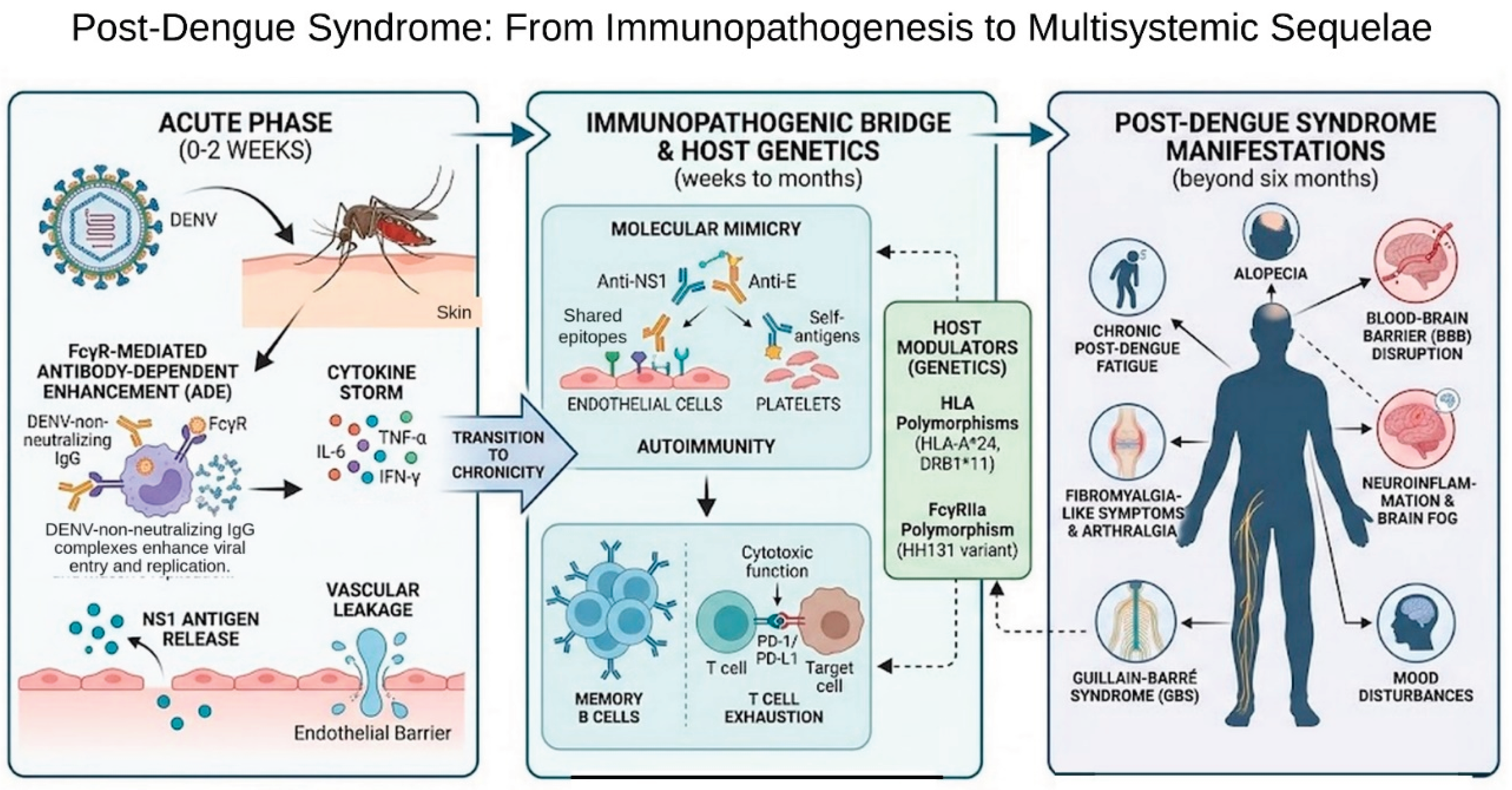

Table 1.
Immune populations implicated in dengue immunopathogenesis.
| Compartment/population | Markers commonly used | Acute phase | Convalescence and PDS |
| Myeloid and plasmacytoid DC, monocytes | CD11c, CD123, HLA-DR, CD14, CD80/CD86, DC-SIGN (CD209), TLR2/3/4/9 | Primary targets: ADE via FcγR; altered TLR expression and co-stimulation [20] | Not characterized |
| NK cells | CD56, CD16, CD69, CD38, NKG2A, CD57, NKG2D (CD314), CD107a | Early activation associated with mild disease; IL-18-driven proliferation; skin-homing imprint [22] | Essentially unstudied; NKG2D–MICB axis untested in patients |
| γδ T, MAIT and NKT cells | Vδ2 TCR, CD161, Vα7.2, CD1d multimers | Activation reported in small cohorts [24,25,26] | Not characterized |
| Plasmablasts | CD19, CD20low, CD27hi, CD38hi | Expansion up to 30–50% of circulating B cells, mainly E-specific [31] | Return to baseline within about one month; link to autoantibody persistence untested |
| Memory B cells | CD19/CD20, CD27, IgD, NS1 and prM specificity | Distinct from the plasmablast pool; prM- and NS1-directed | Persist; contribution to cross-reactive autoantibodies hypothesized, not demonstrated |
| Peripheral follicular helper T cells | CD4, CXCR5, PD-1high, CD38 | Expanded and activated; correlate with plasmablasts and with severity [35] | Not characterized |
| CD8+ effector T cells | CD3, CD8, CD69, CD137, CD107a, granzyme B, IFN-γ | NS3-dominated response; functional quality tracks clinical severity [38] | Activation declines after defervescence; later kinetics unknown |
| Cytotoxic CD4+ T cells | CD3, CD4, CD107a, granzyme B, perforin | Associated with protection in hyperendemic settings | Not characterized |
| Regulatory T cells | CD4, CD25high, CD127low, FOXP3 | Expanded but functionally impaired and Th1-skewed [41] | Not characterized |
| CD4+ helper subsets (Th1, Th2, Th17) | CD4, IFN-γ (Th1), IL-4 (Th2), IL-17 (Th17) | Th1 bias promoted by NKT cells and linked to protective antibody dynamics; Th2 and Th17 skewing reported in severe disease [25] | Not characterized |
| Checkpoint/exhaustion axis | PD-1, TIM-3, LAG-3, PD-L1 (including on extracellular vesicles) | Elevated in severe disease; PD-L1/PD-1 drives premature T cell apoptosis [44,45] | Proposed contributor to chronicity; untested in humans |
| Platelets and complement | CD41, CD62P, NLRP3/IL-1β; C3a, C5a, SC5b-9 | Inflammasome activation, IL-1β microparticles, NS1-driven complement activation [30] | Persistence unknown |
Disclaimer/Publisher’s Note: The statements, opinions and data contained in all publications are solely those of the individual author(s) and contributor(s) and not of MDPI and/or the editor(s). MDPI and/or the editor(s) disclaim responsibility for any injury to people or property resulting from any ideas, methods, instructions or products referred to in the content. |
© 2026 by the authors. Licensee MDPI, Basel, Switzerland. This article is an open access article distributed under the terms and conditions of the Creative Commons Attribution (CC BY) license (http://creativecommons.org/licenses/by/4.0/).
Copyright: This open access article is published under a Creative Commons CC BY 4.0 license, which permit the free download, distribution, and reuse, provided that the author and preprint are cited in any reuse.