

Submitted:
25 August 2026
Posted:
26 August 2026
You are already at the latest version
Abstract
Image-guided surgery has become an integral component of modern veterinary surgery using advanced imaging modalities, as computed tomography, magnetic resonance imaging, ultrasonography, and fluoroscopy. Recently, extended reality (XR) technologies have emerged as promising tools to enhance image-guided surgical procedures by improving three-dimensional anatomical understanding, surgical navigation, and intraoperative decision-making. Although XR has gained considerable attention in human surgery, its application in veterinary surgery remains at an early stage. This narrative review aimed to provide a comprehensive overview of the current evidence regarding XR technologies in small animal surgery, with particular emphasis on their integration with preoperative and intraoperative imaging modalities. The available literature was reviewed to evaluate current clinical applications, technological developments and future perspectives. Although several studies have explored the use of XR technologies for veterinary education, anatomical training, and diagnostic imaging instruction, relatively few investigations have assessed their direct application in clinical surgical practice. Potential applications have been described in orthopedic, oncologic, neurologic, cardiovascular, and minimally invasive procedures. Current evidence suggests that XR has considerable potential to improve image-guided veterinary surgery by enhancing surgical planning, anatomical understanding, and procedural guidance. Further prospective clinical studies are required to establish its clinical effectiveness, cost-effectiveness, and impact on surgical outcomes.
Keywords:
virtual reality
; augmented reality
; mixed reality
; veterinary surgery
1. Introduction
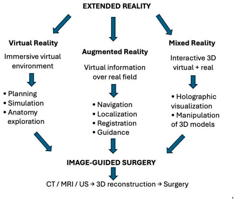
Image-guided surgery relies on the integration of imaging data into the surgical workflow, allowing clinicians to visualize anatomical structures and pathological lesions before and during a surgical procedure [1,2,3,4,5,6,7,8,9,10]. Despite these advances, surgeons are still often required to mentally translate two-dimensional imaging information into a three-dimensional (3D) understanding of complex anatomical relationships. Recent developments in digital visualization technologies have led to the emergence of extended reality (XR), an umbrella term encompassing virtual reality (VR), augmented reality (AR), and mixed reality (MR). These technologies aim to bridge the gap between medical imaging and surgical execution by providing immersive, interactive, and spatially intuitive visualization of patient-specific anatomy [11,12]. VR creates a fully digital environment in which users can interact with 3D anatomical models derived from imaging datasets. AR overlays virtual information onto the real-world environment, enabling surgeons to visualize relevant anatomical structures while maintaining direct interaction with the patient. MR further expands these capabilities by allowing virtual objects to be anchored within and interact dynamically with the physical environment [11,12,13]. Collectively, these technologies provide different levels of interaction between virtual content and the physical environment, making them suitable for different stages of the surgical workflow.
In human medicine, XR technologies have experienced rapid growth over the past decade and have been investigated in numerous surgical disciplines, including neurosurgery, orthopedic surgery, cardiovascular interventions, maxillofacial surgery, hepatobiliary surgery, and surgical oncology [11,13,14,15]. Reported benefits can include improved anatomical understanding, enhanced surgical planning, reduced operative times, increased procedural accuracy, and improved educational opportunities for trainees [11,13,14]. Furthermore, the combination of XR with surgical navigation systems, artificial intelligence, robotic platforms, and advanced image-processing algorithms is increasingly being investigated as a component of precision surgery [13,14,15,16,17].
In contrast, the application of XR technologies in veterinary medicine remains limited. Although recent reviews have summarized the use of XR technologies in veterinary medicine, they have primarily focused on educational applications and anatomical teaching rather than on their integration within image-guided surgical workflows [18,19].
Therefore, the purpose of this review is to provide a comprehensive overview of the current applications of XR technologies in image-guided veterinary surgery. Emphasis is placed on the integration of XR with contemporary imaging modalities, its potential role in surgical planning and intraoperative guidance, current limitations hindering widespread adoption, and future developments. By critically examining the available evidence, this review aims to highlight both the opportunities and challenges associated with the implementation of XR technologies in veterinary surgical practice.
2. Materials and Methods
This narrative review was conducted following the general methodological principles proposed for narrative literature reviews [20]. A comprehensive search of the veterinary literature was performed using the PubMed, Scopus, and Web of Science databases up to May 2026 to identify publications addressing the application of XR technologies in veterinary surgery.
The following keywords and combinations of terms were used: (“extended reality” OR “virtual reality” OR “augmented reality” OR “mixed reality”) AND (“veterinary” OR “canine” OR “dog” OR “feline” OR “cat”) AND (“surgery” OR “surgical” OR “image-guided surgery” OR “navigation” OR “intervention”). Original articles, case reports, pilot studies, technical reports, and review articles published in English were considered. Priority was given to studies describing the clinical application of XR technologies in small animal surgery, including preoperative planning, intraoperative navigation, and image-guided interventions.
Given the limited number of clinically oriented studies currently available, publications describing XR-based surgical simulation and procedural training were also considered when they provided insights into technologies directly applicable to image-guided surgical workflows. Studies exclusively focused on general veterinary education, anatomy teaching, or non-surgical applications were considered only when they contributed to understanding the current development and potential clinical translation of XR technologies.
The selected literature was analyzed qualitatively, and studies were grouped according to their principal contribution to clinical application, technological development, or surgical education and simulation.
3. Current Evidence on Extended Reality in Image-Guided Small Animal Surgery
The literature search identified a growing body of publications addressing XR technologies in veterinary medicine. Most of the available literature focused on educational applications, anatomical visualization, simulation-based training, and technical development of immersive visualization systems. In contrast, relatively few studies described the direct clinical application of XR in small animal surgery.
The publications considered in this review encompassed original research, technical reports, pilot studies, case reports, and reviews. Clinical applications were primarily related to preoperative surgical planning, intraoperative navigation, image-guided interventional procedures, and selected neurosurgical, cardiovascular, orthopedic, and oncologic applications. VR, AR, and MR represent distinct but partially overlapping approaches within the XR spectrum, with different potential roles across the surgical workflow (Figure 1).
Given the heterogeneity of the available veterinary literature in terms of study design, technologies employed, and clinical objectives, a qualitative narrative synthesis was considered the most appropriate approach for critically discussing the available evidence. Educational studies directly related to surgical simulation, image-guided procedures, or operative workflow were included to illustrate the current maturity of XR technologies and their potential role in facilitating future clinical translation.
Studies describing direct clinical applications are summarized in Table 1, whereas educational and simulation-based applications are discussed separately, as they represent an important developmental step toward the future implementation of XR-assisted surgery.

Table 2.
Original studies investigating extended reality technologies for image-guided veterinary surgery.
Table 2.
Original studies investigating extended reality technologies for image-guided veterinary surgery.
| Authors (year) | Sample | XR technology | Imaging/data source | Surgical application |
Main findings |
|---|---|---|---|---|---|
| Zhou et al. (2017) | Canine experimental model |
AR-assisted robotic surgery |
Preoperative imaging 3D reconstruction |
Mandibular angle split osteotomy |
AR-assisted robotic guidance enabled accurate localization and execution of predefined osteotomy planes, demonstrating the feasibility of AR-guided robotic surgery |
| Li et al. (2021) | Canine experimental model |
AR navigation | Patient-specific CT | Pulmonary nodule localization before thoracoscopic surgery |
Patient-specific 3D reconstructions were registered with the surgical field, allowing accurate localization of pulmonary targets and reducing localization errors |
| George et al. (2023) | Canine experimental model |
Holographic MR display | Fluorescence and multispectral imaging |
Fluorescence-guided surgery |
Real-time fluorescence information was displayed holographically, enabling spatial visualization of surgical targets and demonstrating feasibility for intraoperative guidance |
| Stoner et al. (2025) | 5 dogs | Interactive stereoscopic 3D visualization |
ECG-gated CT angiography |
Assessment and planning of ASD intervention |
Interactive 3D visualization facilitated detailed assessment of ASD morphology and spatial relationships and supported procedural planning |
| Cupido et al. (2025) | 1 dog | VR + AR | CT and echocardiography |
Surgical correction of complex vascular anomaly |
VR facilitated preoperative planning, while AR provided intraoperative anatomical guidance, supporting successful surgical correction |
| Tipirneni et al. (2026) | Canine experimental model |
AR glasses | 3D canine head model |
Spatial tracking and surface annotation |
AR improved real-time spatial tracking and enabled direct surface-level annotations, supporting potential applications in surgical navigation |
* XR, extended reality; AR, augmented reality; VR, virtual reality; MR, mixed reality; CT, computed tomography; ASD, atrial septal defect; 3D, three-dimensional.
3.1. Current Clinical Applications of Extended Reality in Small Animal Surgery
3.1.1. Extended Reality for Preoperative Surgical Planning
Preoperative planning currently represents the most advanced and clinically mature application of XR technologies in veterinary surgery. The conversion of computed tomography (CT) and magnetic resonance imaging (MRI) datasets into interactive 3D environments enables surgeons to visualize patient-specific anatomy in a more intuitive and immersive manner than conventional multiplanar imaging. Rather than replacing standard imaging modalities, XR complements existing diagnostic workflows by improving spatial perception, facilitating multidisciplinary communication, and enhancing surgical strategy development before entering the operating room [18,19,21].
Cardiovascular surgery represents one of the earliest areas in which immersive visualization demonstrated potential clinical value in veterinary medicine. Stoner et al. reported an early application of stereoscopic 3D visualization for evaluating atrial septal defects in five dogs. The interactive visualization platform enabled clinicians to manipulate patient-specific cardiac reconstructions in real time, substantially improving the perception of intracardiac anatomy compared with conventional two-dimensional echocardiographic images. Although the system was primarily designed as a diagnostic and planning tool rather than an intraoperative navigation platform, the study highlighted the potential of immersive visualization to facilitate therapeutic decision-making in complex congenital cardiovascular disease [22].
Saunders et al. demonstrated the potential of CT angiography-derived immersive 3D visualization for the evaluation of canine patent ductus arteriosus morphology. Using EchoPixel True 3D together with conventional volume-rendering software, the authors generated interactive patient-specific reconstructions that allowed free manipulation of cardiovascular anatomy, facilitating comparison with conventional angiographic and echocardiographic views. Although the technology was not used for real-time intraoperative navigation, the study highlighted the potential value of immersive XR visualization for procedural planning, device selection, and preoperative understanding of complex cardiovascular anatomy, representing an early example of XR-assisted planning in veterinary interventional cardiology [23].
The first clinical implementation of a complete XR workflow in small animal surgery was later reported by Cupido et al., who described the management of a dog presenting with a complex congenital vascular anomaly. Following CT angiography, patient-specific vascular structures were segmented to generate an immersive 3D model that could be explored interactively before surgery. The XR environment enabled surgeons to appreciate the complex anatomical relationships between the anomalous vessels and surrounding structures, facilitating surgical planning and improving intraoperative orientation. Although limited to a single case report, this study demonstrated the clinical feasibility of integrating XR into the surgical management of canine patients and represented an important proof-of-concept for future clinical applications [24].
3.1.2. Extended Reality for Intraoperative Navigation and Surgical Guidance
While preoperative planning currently represents the most mature application of XR in veterinary medicine, the integration of XR technologies into the intraoperative environment remains at an early stage of development. Intraoperative XR aims to provide surgeons with real-time visualization of patient-specific anatomical information by superimposing virtual structures onto the operative field or by displaying holographic anatomical models synchronized with the patient’s position. These systems seek to overcome one of the principal limitations of conventional image-guided surgery, namely the need for surgeons to repeatedly shift their attention between the surgical field and external imaging displays.
One of the first veterinary studies specifically investigating intraoperative AR was recently published by Tipirneni et al., who evaluated the use of AR smart glasses for spatial tracking and intraoperative annotation during surgical procedures. The system allowed virtual annotations to be projected directly within the surgeon’s field of view while simultaneously tracking the position of surgical instruments. The authors demonstrated that direct AR-assisted visualization was not only technically feasible but significantly improved precision, yielding a lower mean distance error compared to traditional screen transfer and increasing accurate area coverage without compromising procedural speed. Although the study primarily focused on technical validation rather than clinical outcome assessment, it highlighted the potential of wearable AR devices to enhance intraoperative guidance in veterinary surgery [25].
To date, intraoperative AR applications in small animal surgery have focused on both rigid maxillofacial structures and soft-tissue interventions. Zhou et al. pioneered an in vivo craniomaxillofacial application by evaluating an AR-assisted robotic system for mandibular osteotomy. Four novice surgeons performed 20 bilateral drillings on two live dogs using a stereoscopic head-mounted display (nVisor ST60) and a 7-degree-of-freedom robotic arm. To protect the inferior alveolar nerve, a haptic force-feedback algorithm automatically halted drilling upon bone penetration. Postoperative analysis demonstrated high technical accuracy, with mean entrance and target errors of 1.04 ± 0.19 mm and 1.22 ± 0.24 mm, respectively. However, limitations included a small sample size, potential drilling-induced thermal damage, and a rigid human-computer interface [26]. Similarly, Li et al. investigated an AR navigation system for pulmonary nodule localization using a canine experimental model. Patient-specific CT images were used to generate virtual 3D reconstructions that were accurately registered with the surgical field through an AR navigation platform. The system enabled precise localization of pulmonary targets before thoracoscopic surgery, reducing localization errors while demonstrating excellent spatial correspondence between virtual and real anatomical structures. Although performed in an experimental setting, this work represents one of the most advanced examples of AR-assisted surgical navigation currently available in veterinary-related research [27].
George et al. developed a MR fluorescence-guided surgical platform integrating a bioinspired multispectral imaging sensor with a holographic head-mounted display. Following validation on phantoms and experimental cancer models, the system was evaluated during surgical treatment of canine head and neck tumors. Fluorescent information was projected directly into the surgeon’s field of view as holographic overlays, improving visualization of fluorescent targets while integrating seamlessly into the surgical workflow. This study represents one of the first peer-reviewed demonstrations of MR-assisted image-guided oncologic surgery in veterinary patients [28].
3.1.3. Simulation and Training as a Bridge to Clinical Translation
Although the primary focus of this review is image-guided surgery, XR-based education and procedural training are included because they currently represent the most extensively investigated application of XR technologies in veterinary medicine. In contrast to direct clinical applications, which remain limited to a small number of pilot studies and case reports, educational applications of VR, AR, and MR have been more extensively explored for surgical simulation, procedural rehearsal, anatomical orientation, and image-guided skills acquisition [18,19].
Immersive VR environments enable repeated practice of laparoscopic and endoscopic procedures in a controlled setting without compromising animal welfare. Several studies evaluating laparoscopic ovariectomy simulators and basic laparoscopic skills have reported improvements in hand-eye coordination, instrument manipulation, procedural efficiency, and operator confidence among veterinary students and trainees [29,30,31,32,33,34,35]. Interestingly, a direct technological comparison by Fransson et al. investigated the construct and concurrent validity of instrument motion metrics in veterinary trainees, demonstrating that AR simulators were superior to VR systems in assessing complex skills like path length and economy of motion, whereas the VR system was validated primarily for time scores [36].
Beyond minimally invasive surgery, XR technologies have also been applied to image-guided procedural training. AR and MR platforms have been explored for image-guided procedural training, including ultrasound-guided regional anesthesia, ultrasonography, and diagnostic imaging interpretation by overlaying relevant anatomical information onto physical simulators or real-world training environments. These systems can enhance spatial orientation, facilitate needle trajectory planning, and improve the correlation between imaging findings and underlying anatomy, thereby supporting the acquisition of image-guided procedural skills [18,31,37,38].
XR-based simulation also offers important advantages within the framework of competency-based veterinary education [39]. Many contemporary XR platforms provide objective performance metrics, immediate feedback, and repeated opportunities for procedural repetition, while contributing to the implementation of the 3Rs (Replacement, Reduction, and Refinement) by reducing the need for live animals during preclinical training. However, most veterinary XR education studies are still limited by small sample sizes, heterogeneous study designs, and outcome measures based primarily on user satisfaction or simulator performance [40]. Objective assessments of long-term knowledge retention, transfer of skills to clinical surgery, and educational cost-effectiveness remain scarce [39,41]. In this context, Hunt et al. conducted a randomized controlled trial involving 44 veterinary students to evaluate whether integrating a stereoscopic VR application into a traditional surgical curriculum improved performance during their first canine ovariohysterectomy. Interestingly, the study found no significant differences in surgical performance scores or operative times between the VR and control groups, highlighting that simply adding immersive tools to an already comprehensive curriculum may not automatically translate into superior hands-on clinical skills [42]. These features are particularly relevant for competency-based surgical curricula, where standardized assessment and repeated deliberate practice are fundamental components of skill acquisition.
Although these educational applications are primarily educational rather than clinical, they reproduce the same digital workflow used in XR-assisted surgery, including image segmentation, 3D reconstruction, spatial registration, and immersive visualization; thereby facilitating the acquisition of technical and cognitive skills that are directly transferable to image-guided surgical procedures. For these reasons, surgical education currently represents the principal gateway for the clinical implementation of XR technologies in veterinary medicine, providing the technological framework and user familiarity necessary for their future integration into image-guided surgical practice.
3.1.4. Current Clinical Evidence: Strengths and Remaining Gaps
Despite the increasing interest in XR technologies, the body of clinical evidence available in small animal surgery is still remarkably limited [22,23,24,30]. Furthermore, most studies have primarily focused on technical feasibility rather than patient-centered clinical outcomes [24,30].
Current evidence nevertheless suggests several potential advantages of XR-assisted surgery. The available studies indicate that immersive visualization can improve surgeons’ perception of complex 3D anatomy, facilitate interpretation of advanced imaging datasets, and increase confidence during surgical planning and intraoperative image-guided procedures [22,24]. These benefits appear particularly relevant for anatomically complex cases involving congenital vascular anomalies, neurosurgical procedures, surgical oncology, thoracic surgery, and image-guided interventions [22,24,30].
However, several important limitations continue to hinder widespread clinical implementation. Accurate image registration presents significant technical challenges, particularly in soft tissues that undergo deformation during surgery. In addition, maintaining stable tracking throughout an operative procedure remains difficult because of patient positioning changes, respiratory motion, and intraoperative tissue manipulation. Hardware costs, limited software interoperability, prolonged preparation times, and the absence of standardized clinical workflows further restrict routine adoption [14,18,30]. Finally, no prospective randomized clinical studies have yet demonstrated that XR significantly improves operative efficiency, surgical accuracy, complication rates, or postoperative outcomes in veterinary patients [22,23,24,30].
4. Discussion
The present review shows that XR technologies have entered the field of veterinary surgery, although their clinical implementation remains at an early stage. While the number of publications has steadily increased over the last decade, the available literature is characterized by a predominance of proof-of-concept studies, technical feasibility reports, and isolated clinical cases rather than prospective clinical investigations [18,19]. Consequently, current evidence supports the technical feasibility of XR-assisted veterinary surgery but is still insufficient to establish its routine clinical value.
An important finding emerging from the present review is the imbalance between educational and clinical applications. Most veterinary publications focus on simulation, anatomical visualization, and procedural training, whereas only a limited number describe the direct use of XR during surgical planning or intraoperative image-guided procedures. This distribution probably reflects the natural evolution of emerging surgical technologies, where educational implementation generally precedes widespread clinical adoption. The current evolution of XR in veterinary surgery follows a progressive pathway, with educational simulation representing the most mature application, followed by preoperative planning and, more recently, early clinical implementation of image-guided surgical procedures [18,22,24,25]. Nevertheless, it warrants careful consideration that the pedagogical efficacy of XR in veterinary training is highly task-dependent and not universally superior to traditional methods. For instance, while immersive platforms like MR head-mounted displays significantly enhance student satisfaction and technical proficiency in procedures requiring precise depth and angular perception, such as orthopedic screw fixation, they can offer limited benefits in delicate microsurgical tasks like corneal suturing [40]. Furthermore, a randomized controlled trial evaluating stereoscopic VR video applications failed to demonstrate a measurable improvement in objective surgical performance scores or operative times during live canine ovariohysterectomies compared to conventional training [42]. This suggests that simply introducing immersive visual tools into an already rigorous curriculum does not automatically translate into enhanced tactile proficiency or superior hands-on clinical skills. Consequently, future veterinary educational frameworks should focus on integrating high-fidelity simulators that combine visual immersion with advanced haptic feedback, ensuring that XR implementation addresses specific cognitive or spatial training deficits rather than serving as a generic replacement for traditional instructional modalities.
One of the most striking observations emerging from this review is the considerable gap between human and veterinary medicine regarding the clinical implementation of XR technologies. Several factors likely contribute to this discrepancy [11,12,13,14,15,16,17,43]. The veterinary healthcare market is considerably smaller, limiting commercial investment in dedicated XR technologies. Furthermore, the relatively low number of highly complex surgical procedures performed within individual veterinary centers makes prospective multicenter clinical trials difficult to conduct. Finally, successful development of XR systems requires close collaboration among veterinary surgeons, radiologists, biomedical engineers, and computer scientists, multidisciplinary teams that could be relatively uncommon in veterinary institutions.
Beyond its direct surgical applications, immersive visualization can also enhance communication with pet owners by facilitating the explanation of complex anatomy, planned surgical procedures, and expected outcomes through patient-specific 3D models. Although this potential has been primarily described in human medicine, similar benefits can be anticipated in veterinary practice, particularly during preoperative counselling and the informed consent process [11].
Despite encouraging technological developments, several important barriers continue to hinder widespread clinical implementation of XR. Accurate image registration is one of the principal technical limitations, particularly during soft tissue surgery where respiratory motion, tissue deformation, and surgical manipulation continuously modify anatomical relationships [18]. In addition, maintaining accurate spatial correspondence between virtual models and the operative field remains challenging during dynamic surgical procedures, highlighting the need for more robust tracking algorithms and real-time registration techniques before routine clinical adoption [18,32]. Generation of patient-specific 3D models continue to be a time-consuming process because image segmentation, registration, and model refinement often require substantial manual input from experienced operators before surgical visualization can be achieved [11,33]. In addition, the high costs associated with advanced imaging equipment, dedicated software, and head-mounted displays currently limit the implementation of XR technologies primarily to referral hospitals, academic institutions, and specialized research centers [18]. Finally, the quality of the available evidence remains relatively low. Most veterinary studies evaluate technical feasibility rather than clinically meaningful outcomes such as operative time, surgical precision, complication rates, or long-term patient outcome. Consequently, prospective comparative clinical studies remain one of the major priorities for future veterinary XR research [19].
The findings of the present review clearly identify several priorities for future investigation. Prospective multicenter clinical trials should evaluate objective surgical outcomes, including operative time, procedural accuracy, postoperative complications, and learning curves. Standardized reporting guidelines would facilitate comparison among studies while improving the overall quality of evidence [19]. Future technological developments are expected to focus on improving image registration accuracy, reducing workflow complexity, enhancing real-time visualization, and facilitating integration between XR platforms and surgical navigation systems. Advances in artificial intelligence can further automate image segmentation and registration, potentially improving the clinical feasibility of XR-assisted procedures [11,18]. Finally, stronger collaboration between veterinary surgeons, biomedical engineers, imaging specialists, and computer scientists will be essential for translating promising technological innovations into clinically validated surgical tools. Such multidisciplinary collaboration has been recognized as a key prerequisite for the successful development and clinical implementation of XR technologies in surgery [11].
5. Conclusions
Extended reality is transforming veterinary image-guided surgery by converting conventional two-dimensional imaging into immersive, patient-specific 3D environments that improve anatomical understanding and support surgical decision-making. Current evidence demonstrates promising applications in preoperative planning, intraoperative guidance, image-guided interventions, and surgical education. However, clinical adoption remains limited, with most available studies consisting of technical reports, pilot investigations, or isolated case reports.
The widespread implementation of XR in veterinary surgery will require robust prospective clinical studies, standardized workflows, and technological advances that improve image registration, workflow efficiency, and system interoperability. As these challenges are progressively addressed, XR has the potential to become an integral component of precision veterinary surgery, supporting more accurate planning, safer procedures, and improved patient-specific surgical care.
Author Contributions
Conceptualization, D.C, F.V., A.C. and A.B.; methodology, D.C, F.V., A.C. and A.B.; software, D.C, F.V., A.C. and A.B.; validation, D.C, F.V., A.C. and A.B.; formal analysis, D.C, F.V., A.C. and A.B.; investigation, D.C, F.V., A.C. and A.B.; resources, D.C, F.V., A.C. and A.B.; data curation, D.C, F.V., A.C. and A.B.; writing—original draft preparation, D.C, F.V., A.C. and A.B.; writing—review and editing, D.C, F.V., A.C. and A.B. All authors have read and agreed to the published version of the manuscript.
Funding
This research received no external funding.
Institutional Review Board Statement
Not applicable.
Informed Consent Statement
Not applicable.
Data Availability Statement
Not applicable.
Conflicts of Interest
The authors declare no conflicts of interest.
References
- Privitera, L.; Paraboschi, I.; Dixit, D.; Arthurs, O.J.; Giuliani, S. Image-guided surgery and novel intraoperative devices for enhanced visualisation in general and paediatric surgery: A review. Innov. Surg. Sci. 2021, 6, 161–172. [Google Scholar] [CrossRef] [PubMed]
- Azagury, D.E.; Dua, M.M.; Barrese, J.C.; Henderson, J.M.; Buchs, N.C.; Ris, F.; Cloyd, J.M.; Martinie, J.B.; Razzaque, S.; Nicolau, S.; et al. Image-guided surgery. Curr. Probl. Surg. 2015, 52, 476–520. [Google Scholar] [CrossRef] [PubMed]
- Vignoli, M.; Saunders, J.H. Image-guided interventional procedures in the dog and cat. Vet. J. 2011, 187, 297–303. [Google Scholar] [CrossRef] [PubMed]
- Avner, A.; Herrtage, M.E.; Segal, U. Ultrasound guided trans-hepatic coil embolization of an intrahepatic portosystemic shunt in a dog. Isr. J. Vet. Med. 2019, 74, 3–8. [Google Scholar]
- Caivano, D.; Birettoni, F.; Marchesi, M.C.; Moretti, G.; Corda, A.; Petrescu, V.F.; Porciello, F.; Bufalari, A. Septic pericarditis and cardiac tamponade caused by migrating intrathoracic grass awn in an English setter dog. Isr. J. Vet. Med. 2019, 74, 82–87. [Google Scholar]
- Griffin, M.A.; Culp, W.T.N.; Buote, N.J. The role of minimally invasive surgery in oncology—Part 1: Laparoscopy. Vet. Oncol. 2025, 2, 2. [Google Scholar] [CrossRef]
- Griffin, M.A.; Culp, W.T.N.; Buote, N.J. The role of minimally invasive surgery in oncology—Part 2: Thoracoscopy. Vet. Oncol. 2025, 2, 3. [Google Scholar] [CrossRef]
- Nagaya, T.; Nakamura, Y.A.; Choyke, P.L.; Kobayashi, H. Fluorescence-guided surgery. Front. Oncol. 2017, 7, 314. [Google Scholar] [CrossRef] [PubMed]
- Bufalari, A.; Moretti, G.; Monti, E.; Garofanini, L.; Angeli, G.; Porciello, F.; Marchesi, M.C.; Caivano, D. Intracardiac Porcupine Quill Migration in a Dog: Multimodality Imaging Findings and Surgical Management. Vet. Sci. 2022, 9, 700. [Google Scholar] [CrossRef] [PubMed]
- Sasaki, K.; Medan, M.S.; Azuma, T.; Kawabata, K.; Shimoda, M.; Umemura, S. Effect of echo-guided high-intensity focused ultrasound ablation on localized experimental tumors. J. Vet. Med. Sci. 2006, 68, 1069–1074. [Google Scholar] [CrossRef] [PubMed]
- Zhang, J.; Lu, V.; Khanduja, V. The impact of extended reality on surgery: a scoping review. Int. Orthop. 2023, 68, 611–621. [Google Scholar] [CrossRef] [PubMed]
- Morimoto, T.; Kobayashi, T.; Hirata, H.; Otani, K.; Sugimoto, M.; Tsukamoto, M.; Yoshihara, T.; Ueno, M.; Mawatari, M. XR (Extended Reality: Virtual Reality, Augmented Reality, Mixed Reality) Technology in Spine Medicine: Status Quo and Quo Vadis. J. Clin. Med. 2022, 11, 470. [Google Scholar] [CrossRef] [PubMed]
- Khubzan, W.D.; Alharati, S.A.; Aljuaid, R.W.; Algethami, D.S.; Alotibi, S.S.; Alotaibi, H.M.; Aljuaid, S.S.; Almalki, R.S.; Mahfouz, M.E.M. The digital evolution of surgical planning: A systematic review of immersive and interactive technologies. Front. Surg. 2026, 13, 1764132. [Google Scholar] [CrossRef] [PubMed]
- Ma, L.; Huang, T.; Wang, J.; Liao, H. Visualization, registration and tracking techniques for augmented reality guided surgery: A review. Phys. Med. Biol. 2023, 68, 045001. [Google Scholar] [CrossRef] [PubMed]
- Murphy, E.; Cahill, R.A. Technological advances in intra-operative navigation: Integrating fluorescence, extended reality, and artificial intelligence. J. Clin. Med. 2025, 14, 8574. [Google Scholar] [CrossRef] [PubMed]
- Nowak, R.; Nowak, M.; Rękas, M.; Ali, M.J. Intraoperative use of mixed reality (MR) in humans across surgical disciplines: A major review. 3D Print. Med. 2025, 11, 59. [Google Scholar] [CrossRef] [PubMed]
- Li, Y.; Li, M.; Zheng, S.; Yang, L.; Peng, L.; Fu, C.; Chen, Y.; Wang, C.; Chen, C.; Li, B.; Xiong, B.; Breschi, S.; Liu, Y.; Shidujaman, M.; Piazzolla, P.; Zhang, Y.; De Momi, E.; van Eijk, D. Exploring the dynamics of user experience and interaction in XR-enhanced robotic surgery: A systematic review. Front. Virtual Real. 2024, 5, 1461105. [Google Scholar] [CrossRef]
- Aghapour, M.; Bockstahler, B. State of the art and future prospects of virtual and augmented reality in veterinary medicine: A systematic review. Animals 2022, 12, 3517. [Google Scholar] [CrossRef] [PubMed]
- Daungsupawong, H.; Wiwanitkit, V. Virtual reality: Applications in veterinary science. Vet. Med. Sci. 2026, 12, e70847. [Google Scholar] [CrossRef] [PubMed]
- Green, B.N.; Johnson, C.D.; Adams, A. Writing narrative literature reviews for peer-reviewed journals: Secrets of the trade. J. Chiropr. Med. 2006, 5, 101–117. [Google Scholar] [CrossRef] [PubMed]
- Klasen, J.R.S.; Thatcher, G.P.; Bleedorn, J.A.; Soukup, J.W. Virtual surgical planning and 3D printing: Methodology and applications in veterinary oromaxillofacial surgery. Front. Vet. Sci. 2022, 9, 971318. [Google Scholar] [CrossRef] [PubMed]
- Stoner, C.H.; Wesselowski, S.; Gordon, S.G.; Saunders, A.B. Application of an interactive, stereoscopic three-dimensional viewer to evaluate atrial septal defects in five dogs. J. Vet. Cardiol. 2025, 62, 164–169. [Google Scholar] [CrossRef] [PubMed]
- Saunders, A.B.; Doocy, K.R.; Birch, S.A. A pictorial view of the three-dimensional representation and comparative two-dimensional image orientation derived from computed tomography angiography in a dog with a patent ductus arteriosus. J. Vet. Cardiol. 2019, 21, 34–40. [Google Scholar] [CrossRef] [PubMed]
- Cupido, S.; Valeri, F.; Nicoli, S.; Bargellini, P.; Caivano, D.; Birettoni, F.; Bortolotti, A.; Rishniw, M.; Porciello, F. A new technology for a novel clinical approach in a dog with a complex vascular anomaly: The “extended reality”. Vet. Res. Commun. 2025, 49, 100. [Google Scholar] [CrossRef] [PubMed]
- Tipirneni, Y.; Arzi, B.; Blandino, A.; Skouritakis, C.T.; Goldschmidt, S. Augmented reality glasses improve real-time spatial tracking and surface-level annotations on the canine head. Am. J. Vet. Res. 2026, 87, 1–9. [Google Scholar] [CrossRef] [PubMed]
- Zhou, C.; Zhu, M.; Shi, Y.; Lin, L.; Chai, G.; Zhang, Y.; Xie, L. Robot-assisted surgery for mandibular angle split osteotomy using augmented reality: Preliminary results on clinical animal experiment. Aesthetic Plast. Surg. 2017, 41, 1228–1236. [Google Scholar] [CrossRef] [PubMed]
- Li, C.; Zheng, Y.; Yuan, Y.; Li, H. Augmented reality navigation-guided pulmonary nodule localization in a canine model. Transl. Lung Cancer Res. 2021, 10, 4152–4160. [Google Scholar] [CrossRef] [PubMed]
- George, M.B.; Lew, B.; Liang, Z.; Blair, S.; Zhu, Z.; Cui, N.; Ludwig, J.; Zayed, M.; Selmic, L.; Gruev, V. Fluorescence-guided surgical system using holographic display: From phantom studies to canine patients. J. Biomed. Opt. 2023, 28, 096003. [Google Scholar] [CrossRef] [PubMed]
- Hottmann, N.M.; Fransson, B.A. Effects of short-course virtual reality or video-box training on basic laparoscopic skills and simulated surgical performance in veterinary students. Vet. Surg. 2025, 54, 860–871. [Google Scholar] [CrossRef] [PubMed]
- Fransson, B.A.; Ragle, C.A. Assessment of laparoscopic skills before and after simulation training with a canine abdominal model. J. Am. Vet. Med. Assoc. 2010, 236, 1079–1084. [Google Scholar] [CrossRef] [PubMed]
- Fransson, B.A.; Ragle, C.A.; Bryan, M.E. A laparoscopic surgical skills assessment tool for veterinarians. J. Vet. Med. Educ. 2010, 37, 304–313. [Google Scholar] [CrossRef] [PubMed]
- Guaraná, J.B.; Aytaç, G.; Müller, A.F.; Thompson, J.; Freitas, S.H.; Lee, U.Y.; Lozanoff, S.; Ferrante, B. Extended reality veterinary medicine case studies for diagnostic veterinary imaging instruction: Assessing student perceptions and examination performance. Anat. Histol. Embryol. 2023, 52, 101–114. [Google Scholar] [CrossRef] [PubMed]
- Micșa, C.; Vițălaru, B.A.; Dragomirișteanu, I.; Bîrțoiu, I.A. Different training methods used to improve laparoscopic skills in veterinary medicine. Alveus 2023, 33, 13–20. [Google Scholar]
- Danner, A.L.; Song, M.K.; Fracka, A.B.; Dickson, R.; Fransson, B. Validity assessment for veterinary use of human laparoscopic cholecystectomy training models. Am. J. Vet. Res. 2026, 87, ajvr.25.07.0264. [Google Scholar] [CrossRef] [PubMed]
- Redander, J.; Enlund, K.; Höglund, O.V. The development of a novel virtual reality simulation module for canine laparoscopic ovariectomy. Acta Vet. Scand. 2025, 67, 14. [Google Scholar] [CrossRef] [PubMed]
- Fransson, B.A.; Ragle, C.A. Instrument motion metrics for laparoscopic skills assessment in virtual reality and augmented reality simulators. Vet. Surg. 2016, 45, 920–928. [Google Scholar] [CrossRef] [PubMed]
- Hung, C.Y.; Chang, K.V.; Ozcakar, L.; Hsiao, M.Y.; Wang, T.G.; Chen, W.S. Ultrasound-guided injection techniques for the low back and hip joint. Chang Gung Med. J. 2011, 34, 543–557. [Google Scholar]
- McGaffey, M.; Shafford, H.L.; Meadows, L.; Fox, S.; Boettcher, L.M.; Hostutler, R.A. Assessment of an ultrasound skill simulator in teaching ultrasound-guided paracentesis: A prospective observational study. J. Vet. Med. Educ. 2024, 51, 180–189. [Google Scholar] [CrossRef] [PubMed]
- Zhang, J.; Lauinger, A.; Shrivastava, R.; Falak, A.; Abraskin, K.; Burnette, P.; Galvez, R. Use of extended reality head-mounted displays in healthcare education: A scoping review. Front. Med. 2026, 13, 1798546. [Google Scholar] [CrossRef] [PubMed]
- Sasaki, N.; Lee, S. Educational dissemination in the digital age through the TranspaVet project: Evaluation of students’ satisfaction and instructional efficacy. J. Vet. Med. Educ. 2025, 52, 74–82. [Google Scholar]
- Oviedo-Peñata, C.A.; Tapia-Araya, V.; Lemos, Z.; Riaño-Benavides, C.E.; Case, J.B.; Maldonado-Estrada, J.G. Validation of training and acquisition of surgical skills in minimally invasive surgery in small animal surgery: A comprehensive review. Front. Vet. Sci. 2020, 7, 306. [Google Scholar] [PubMed]
- Hunt, J.A.; Heydenburg, M.; Anderson, S.L.; Thompson, R.R. Does virtual reality training improve veterinary students’ first canine surgical performance? Vet. Rec. 2020, 186, 562. [Google Scholar] [CrossRef] [PubMed]
- Saito, E.; Hasegawa, T.; Kitamura, M.; Yamaguchi, S.; Suzuki, T. Clinical implementation of extended reality (XR) technologies: Bridging the gap between human and veterinary medicine. J. Vet. Med. Sci. 2022, 84, 1245–1254. [Google Scholar]
Figure 1.
Extended reality technologies in image-guided veterinary surgery. Schematic representation of virtual reality, augmented reality, and mixed reality and their main applications in surgical planning and intraoperative guidance. CT, computed tomography; MRI, magnetic resonance imaging, US, ultrasound; 3D, three-dimensional.
Figure 1.
Extended reality technologies in image-guided veterinary surgery. Schematic representation of virtual reality, augmented reality, and mixed reality and their main applications in surgical planning and intraoperative guidance. CT, computed tomography; MRI, magnetic resonance imaging, US, ultrasound; 3D, three-dimensional.
Disclaimer/Publisher’s Note: The statements, opinions and data contained in all publications are solely those of the individual author(s) and contributor(s) and not of MDPI and/or the editor(s). MDPI and/or the editor(s) disclaim responsibility for any injury to people or property resulting from any ideas, methods, instructions or products referred to in the content. |
© 2026 by the authors. Licensee MDPI, Basel, Switzerland. This article is an open access article distributed under the terms and conditions of the Creative Commons Attribution (CC BY) license.
Copyright: This open access article is published under a Creative Commons CC BY 4.0 license, which permit the free download, distribution, and reuse, provided that the author and preprint are cited in any reuse.