

Submitted:
25 August 2026
Posted:
26 August 2026
You are already at the latest version
Abstract
Introduction: Vitiligo is a common pigmentary disorder, which is characterized by discrete depigmented patches or macules of different shapes and sizes, caused by the destruction of melanocytes. Although pruritus has been reported in a selected number of patients with vitiligo, the condition is normally asymptomatic. Objectives: The study aimed to determining the frequency of pruritus in patients with vitiligo and to identify the factors related to manifestation of pruritus. Design and methods: A prospective cross-sectional observational design was used in this study. Presence of symptomatic itch and the intensity of this symptom were evaluated among 59 patients with vitiligo who presented in the study period at Khartoum Dermatology Teaching Hospital and Omdurman Military Hospital in Sudan. Results: The study population consisted of 59 vitiligo patients, among them 33 (55.9%) were male and 26 (44.1%) were female, with a mean age of 30.15 ± 16.67 years. Itch was reported by 21 patients (35.6%), and 38 (64.4%) had no history of pruritus associated with vitiligo. Pruritus was the initial manifestation of vitiliginous lesions in most patients (66.7%), but in 33.3% it occurred after the lesions appeared. The itch intensity was 4 on a 10-point Visual Analogue Scale (VAS), with mild, moderate, and severe itch being reported in 42.9, 38.1, and 19% of patients respectively. In the majority of cases itch was intermittent (52.3%), then daily (33.3%), persistent itch (9.6%), and nocturnal itch (4.8%). The most frequently reported aggravating factors were hot weather. Conclusion: Itch was present in vitiligo patients with different levels of severity. Hot weather is the major aggravating factor. It usually preceded the onset of vitiligo lesions, and it was reported to be more intense in males compared females.
Keywords:
vitiligo
; itch
; pruritus
; itch severity
; visual analogue scale
Introduction
Vitiligo is a common pigmentary disorder, which is characterized by discrete depigmented patches or macules of different shapes and sizes, caused by the destruction of melanocytes [1,2]. It affects about 1-4% of the world population and its clinical picture demonstrates a large degree of heterogeneity, in particular, in people with darker skin coloring [3].
This disorder causes severe cosmetic disfigurement. In some cultures, vitiligo patients are abandons in society casing both emotional and physical consequences [2,4]. The resulting impact in the quality of life is closely related to the development of the disease [5].
Vitiligo has an etiopathogenic multifactorial characteristic, including genetic, immunologic, autoimmune, neurogenic, and environmental factors. Immune system dysregulation is one of the pathogenic mechanisms that are reported in many studies [6,7,8]. The inheritance pattern of the disease is complex and it is probably polygenic with variable penetrance [9].
Vitiligo clinically appearances as depigmented chalky-white or dusky macules and patches. These are commonly round or oval lesions with scalloped edges, and the size spans between a few millimeters to several centimeters involving both the skin and mucosal membranes. The onset of lesions is usually asymptomatic, although few patients may have preceded itching or burning. The disease has a progressive pattern as well as remission and exacerbation that are associated with triggering stimuli [10].
Treatment is often recommended early, because vitiligo has a significant effect on patients’ quality of life. The goal of treatment must focus more on the disease stabilization and re-pigmentation, which should be clearly explained to the patients. The majority of modern treatments are immunomodulatory and lead to satisfactory stabilization of the disease; however, the stimulation of melanocyte precursor (e.g., by UV exposure) is useful. The possibility of re-pigmentation depends on the location of the anatomical sites. The recent treatment modalities are directed on preventing the progression of the disease and achievement of re-pigmentation. Even though many therapeutic agents have been reported, they have not shown the same level of effectiveness. The most commonly used therapies include photo-chemotherapy (PUVA), topical corticosteroids and topical immunomodulators. Topical JAK (Janus kinase) inhibiters (ruxolitinib cream 1.5%) is the first FDA approved treatment for vitiligo especially the non-segmental type. There are also the combination regimens of multiple modalities, and in some severe cases depigmentation of normal skin may be the option [1,6]. Combination therapies usually have greater effectiveness than the monotherapy. The reported modalities include phototherapy in combination with topical agents [11], systemic antioxidant adjuncts [12], and systemic immunosuppressives combined with phototherapy [13]. Surgery is still an option when the patient has segmental vitiligo or refractory localized lesion once it is ensured that vitiligo is stable at least one year.
The pathogenesis of the pruritus in vitiligo (when present) is still not fully understood. The hypotheses proposed include neurogenic theory, in which the release of neuropeptides by dermal nerve endings induced by various stimuli, but more studies are required to explain such complex itch mechanisms in vitiligo [14]. Although pruritus has been reported in a selected number of patients with vitiligo, the condition is normally asymptomatic [15].
There is very little data on the prevalence and nature of pruritus in vitiligo. The current research aims were to determining the frequency of pruritus in patients with vitiligo and to identify the factors related to manifestation of pruritus.
Material and Methods
Study Population: This prospective, observational, cross-sectional study was conducted at Khartoum Dermatology Teaching Hospital and Omdurman Military Hospital between February and August 2022 and involved all the patients who were clinically diagnosed with vitiligo and attended the hospitals of choice within the study period. The exclusion criteria were: vitiligo patients with other skin diseases that can cause itching and patients with itching related to therapy that appeared when they began vitiligo treatment.
Data collection: Dermatological examination was conducted after a full history. The patients with Vitiligo who have been given the consent and who met the inclusion criteria were recruited into the study. Data were collected from vitiligo patients by direct interview, using a prepared questionnaire that contained visual analogue. Visual Analogue Scale (VAS) is a severity scale tool used to measure pruritus on a 10 point scale such that 0 indicates no itch and 10 the worst imaginable itch. The Visual Analogue Scale (VAS) is a validated and commonly used patient-reported outcome measure to assess subjective symptoms, particularly pruritus intensity [16].
Statistical Analysis: The data analysis was done using SPSS version 22. Descriptive statistics were calculated in frequencies and percentages for categorical data, the state of central tendency measures (mean) and dispersion measures (standard deviation) to continuous variables. To establish the significance of differences the relevant test of chi-square tests and t-tests were used with the significance level of p < 0.05.
Results
The study population consisted of 59 vitiligo patients who fulfilled the inclusion criteria. Of them, 33 were males (55.9%), and 26 were females (44.1%). The mean age of participants was 30.15 (±16.67) years and the most common age group was above 30 years age as shown in Figure 1.
The duration of vitiligo varies, most commonly more than 5 years. The mean duration was 7.5years (±9.85) as shown in Figure 2.
Concerning localization of initial vitiligo lesions, in 21 patients the initial lesion was on head and neck (35.6%),16 patients on legs (27.1%), 8 patients on hands (13.6%), 5 patients on feet (8.5%), 4 patients on arms (6.8%) ,3 patients on trunk (5.1%), one patient on forearms (1.7%) and other patients on genital area (1.7%) (Table 1).
Only 10 patients reported family history of vitiligo, and 83.1% had no family history.
Regarding itching, 21 patients presented with different intensities of itch (35.6%), the rest of patients 38 (64.4%) did not report any vitiligo associated history of itch. In most of patients with itching (14 patients, 66.7%), itching appears before vitiligo lesions, and in the remaining 7 patients (33.3%) itching started after appearance of the vitiligo (Figure 3).
Concerting the severity of itch, the median intensity was 4 (on VAS of 1 to 10-points) in vitiligo patients (Figure 4).
This is compared to VAS 5 score that is considered as equivalent to itch caused by insect bite reaction. Severe itch was reported in 4 patients (19%). Itch of moderate and mild intensity was documented in 8 (38.1%) and 9 (42.9%) of patients, respectively. Itch was described to be more intense in males (14 patients) when compared to the 7 females counterpart (33.3% versus 18.6%) p = 0.217 (Table 2)
Concerning the frequency of itching, it was intermittent in 11 patients (52.3%), sometimes during every day in 7 patients (33.3%), present always (persistent) in two patients (9.6%), and one patient have itch mainly at night (4.8%).
Koebner phenomenon was presented in 15 patients (25.4%) and 44 patients have no koebnerization (74.6%). There was no correlation between itch and Koebner, that 5 patients (23.8%) had itch with Koebner and 16 (76.2%) patients had itch without Koebner (p = 0.832, not significant) (Table 3).
In 7 patient itching was aggravated by hot weather (33.3%), 4 by sunlight (19%), one patient by soap and perfumes (4.8%) and 9 patients noticed no aggravating factors (42.9%) (Table 4).
In 6 patients itching was relieved by topical steroids (28.6%), 2 by shower (9.5%), one by oral antihistamine (4.8%) and other one by cold environment (4.8%).
The rest of participants (11, 52.4%) had no relieving factors (Table 5).
Discussion
Pruritus is considered as the most common skin-related symptom. It usually develops from a primary cutaneous disease but may be a symptom of a systemic disease in number of affected individuals, particularly the generalized form. In 2007, the International Forum of the Study of Itch proposed a clinical classification scheme of chronic pruritus [17]. This system has three large categories of pruritus: chronic pruritus of lesional (inflamed) skin, chronic pruritus of non-lesional (non-inflamed) skin, and chronic pruritus with secondary scratch induced lesions (e.g., prurigo nodularis, lichen simplex chronicus). After initial assessment, the etiology of the pruritus will be categorized into dermatologic, systemic, neurologic, psychogenic, mixed, and other/unknown. In contrast, pruritus that is related to vitiligo is infrequently covered in literature.
In the current study, pruritus was found to be common with 35.6% in comparison with previous studies which gave a lower prevalence of pruritus. Vachiramon et al. have described pruritus in 20.2% of vitiligo patients [14]. The increased prevalence herein in our study could be explained by ethnic differences in research subjects and environmental triggering conditions especially climate differences. Because pruritus can also develop due to treatment of topical therapy or phototherapy, these patients were not included in our study as to minimize the likelihood of confounding factors. We believe that pruritus affecting vitiliginous lesions can represent an intrinsic manifestation of vitiligo itself. In the literature, certain systemic symptoms were correlated with rare forms of vitiligo, e.g mixed vitiligo [3].
The intensity of itch can be assessed in a number of ways. In this study, the ratings were done on the 10- point VAS self-evaluation scale. Study results showed the median intensity of itch to be 4, compared to median VAS=5 that is equivalent to the itch caused by arthropod bite. Nevertheless, itch intensity is extremely hard to compare with other types of dermatoses and no reported data is currently available. Despite the fact that a similar VAS score had been used in the previous research by Vasanop Vachiramon, the severity of itch in vitiligo was 5 in thier study [14].
Itch may add a psychological distress to patient and increase the severity of a number of dermatoses such as vitiligo. Therefore, we highlighted the importance of itch consideration in vitiligo patients which is not well appreciated.
In terms of factor that affected itch, sunlight and hot environment were identified as a significant aggravating factor of itch in the study group of vitiligo patients (33.3%) possibly due to high temperatures in our tropical climate.
Conclusions
Itch is present in vitiligo patients with different levels of severity and the hot weather is the major aggravating factor. In most of patients itching usually precedes the onset of vitiligo lesions, and it is more intense in males compared females.
Ethical Approval
Ethical approval was obtained from Research Ethics Committee form Sudan Medical Specialization Board. Ethical consent was also taken from all patients before enrollment in the study.
Consent for Publication
The authors agree to the final version submitted in the journal.
Funding
No, sources of funding are reported.
Acknowledgments
We thank the patients at Khartoum Dermatology and Omdurman Military Hospitals in SUDAN for providing consent for their involvement in the study and for the publication of their clinical details.
Conflicts of Interest
The authors declare no conflict of interest, financial or otherwise.
References
- AlGhamdi, K.M. A survey of vitiligo management among dermatologists in Saudi Arabia. J. Eur. Acad. Dermatol. Venereol. 2009, 23(11), 1282–8. [Google Scholar] [CrossRef] [PubMed]
- Hartmann, A.; Bröcker, E.B.; Becker, J.C. Hypopigmentary skin disorders: current treatment options and future directions. Drugs 2004, 64(1), 89–107. [Google Scholar] [CrossRef] [PubMed]
- Shamad, Mahdi M.A.; Siddig, Mohammed Tageldin. Characteristics and types of vitiligo in patients with skin of color. Am. J. Med. Med. Sci. 2025, 15(2), 307–310. [Google Scholar]
- Nordlund, J.J. The epidemiology and genetics of vitiligo. Clin. Dermatol. 1997, 15(6), 875–8. [Google Scholar] [CrossRef] [PubMed]
- Thabit, Sarah Fuad; Basrawi, Basel Adel; Eltayeb, Alamin Mustafa; Hassan, Amira Ibrahim; Shamad, Mahdi. Quality of life among Sudanese patients with vitiligo. Am. J. Dermatol. Venereol. 2025, 14(1), 1–5. [Google Scholar]
- Nordlund, J.J.; Ortonne, J.P.; LePoole, I.C. Vitiligo vulgaris. In The Pigmentary System: Physiology and Pathophysiology; Nordlund, J.J., Boissy, R.E., Hearing, V.J., et al., Eds.; Blackwell: Oxford, 2006; pp. 551–598. [Google Scholar]
- Kovacs, S.O. Vitiligo. J. Am. Acad. Dermatol. 1998, 38 5 Pt 1, 647–66. [Google Scholar] [CrossRef] [PubMed]
- Budania, A.; Parsad, D.; Kanwar, A.J.; Dogra, S. Comparison between autologous noncultured epidermal cell suspension and suction blister epidermal grafting in stable vitiligo: a randomized study. Br. J. Dermatol. 2012, 167(6), 1295–301. [Google Scholar] [CrossRef] [PubMed]
- Koga, M.; Tango, T. Clinical features and course of type A and type B vitiligo. Br. J. Dermatol. 1988, 118(2), 223–8. [Google Scholar] [CrossRef] [PubMed]
- Kathuria, S.; Khaitan, B.K.; Ramam, M.; Sharma, V.K. Segmental vitiligo: a randomized controlled trial to evaluate efficacy and safety of 0.1% tacrolimus ointment vs 0.05% fluticasone propionate cream. Indian J. Dermatol. Venereol. Leprol. 2012, 78(1), 68–73. [Google Scholar] [CrossRef] [PubMed]
- Passeron, T.; Ostovari, N.; Zakaria, W.; Fontas, E.; Larrouy, J.C.; Lacour, J.P.; Ortonne, J.P. Topical tacrolimus and the 308-nm excimer laser: a synergistic combination for the treatment of vitiligo. Arch. Dermatol. 2004, 140(9), 1065–9. [Google Scholar] [CrossRef] [PubMed]
- Middelkamp-Hup, M.A.; Bos, J.D.; Rius-Diaz, F.; Gonzalez, S.; Westerhof, W. Treatment of vitiligo vulgaris with narrow-band UVB and oral Polypodium leucotomos extract: a randomized double-blind placebo-controlled study. J. Eur. Acad. Dermatol. Venereol. 2007, 21(7), 942–50. [Google Scholar] [CrossRef] [PubMed]
- Radakovic-Fijan, S.; Fürnsinn-Friedl, A.M.; Hönigsmann, H.; Tanew, A. Oral dexamethasone pulse treatment for vitiligo. J. Am. Acad. Dermatol. 2001, 44(5), 814–7. [Google Scholar] [CrossRef] [PubMed]
- Vachiramon, V.; Onprasert, W.; Harnchoowong, S.; Chanprapaph, K. Prevalence and Clinical Characteristics of Itch in Vitiligo and Its Clinical Significance. BioMed Res. Int. 2017, 2017, 5617838. [Google Scholar] [CrossRef] [PubMed]
- Fitzpatrick, T.B. Hypomelanosis. South Med. J. 1964, 57, 995–1005. [Google Scholar] [CrossRef] [PubMed]
- Reich, A.; Chatzigeorkidis, E.; Zeidler, C.; Osada, N.; Furue, M.; Takamori, K.; Ebata, T.; Augustin, M.; Szepietowski, J.C.; Ständer, S. Tailoring the Cut-off Values of the Visual Analogue Scale and Numeric Rating Scale in Itch Assessment. Acta Derm. Venereol. 2017, 97(6), 759–760. [Google Scholar] [CrossRef] [PubMed]
- Ständer, S.; Weisshaar, E.; Mettang, T.; Szepietowski, J.C.; Carstens, E.; Ikoma, A.; Bergasa, N.V.; Gieler, U.; Misery, L.; Wallengren, J.; Darsow, U.; Streit, M.; Metze, D.; Luger, T.A.; Greaves, M.W.; Schmelz, M.; Yosipovitch, G.; Bernhard, J.D. Clinical classification of itch: a position paper of the International Forum for the Study of Itch. Acta Derm. Venereol. 2007, 87(4), 291–4. [Google Scholar] [CrossRef] [PubMed]
Figure 1.
Age groups in study population.
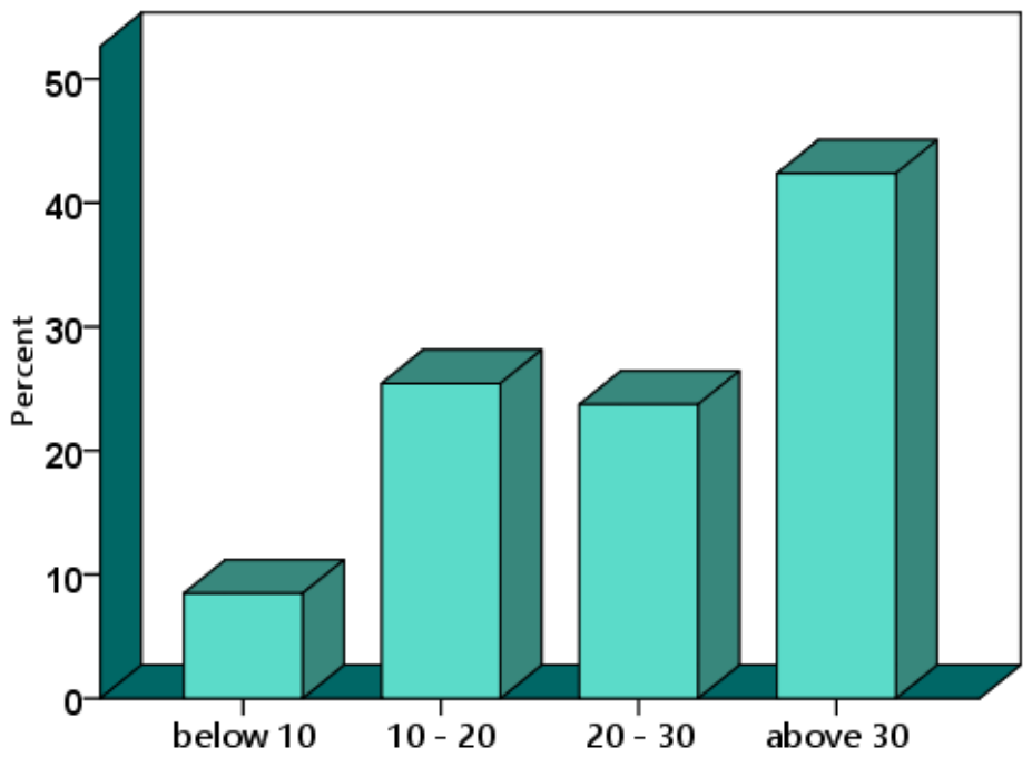
Figure 2.
Duration of vitiligo in the study population.
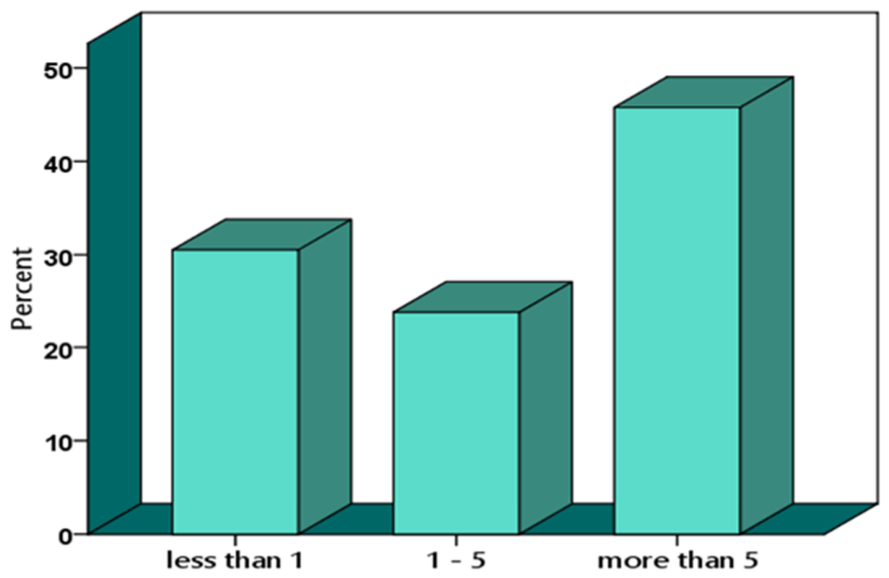
Figure 3.
Presence of itch in the study population.
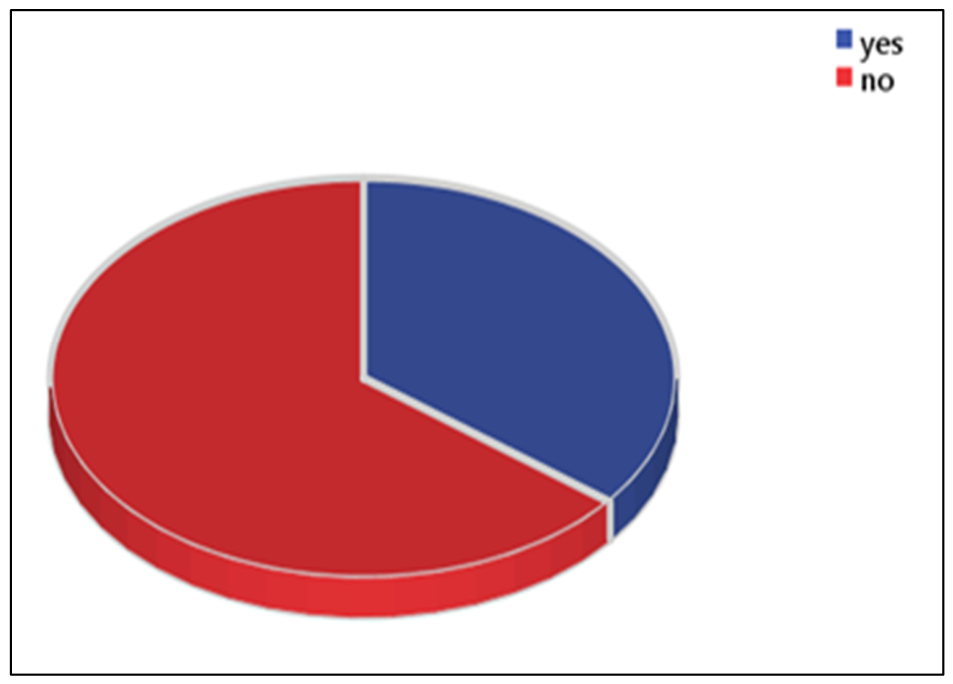
Figure 4.
Intensity of itch in the study population.
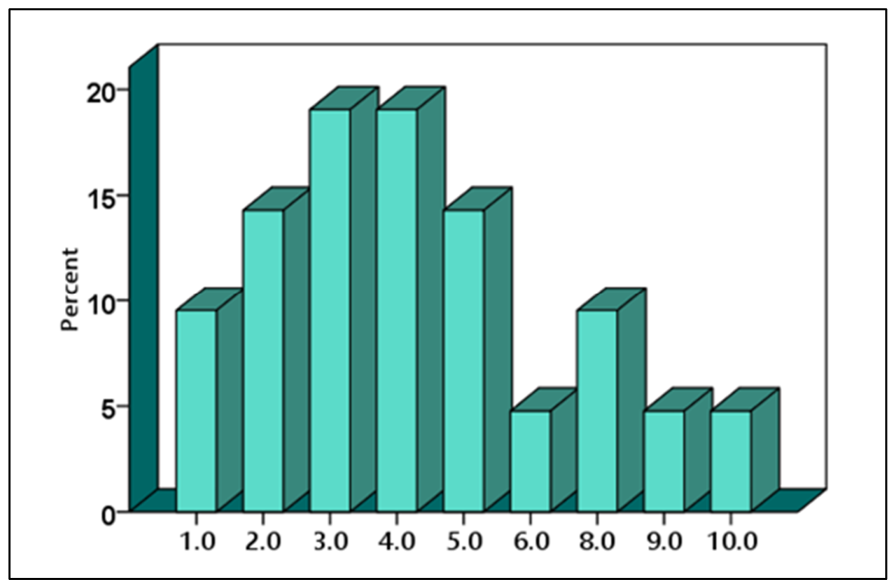

Table 1.
Initial location of vitiligo in the study population.
| Frequency | Percent | |
|---|---|---|
| head and neck | 21 | 35.6 |
| Trunk | 4 | 6.8 |
| Arms | 4 | 6.8 |
| Hands | 10 | 16.9 |
| Legs | 13 | 22.0 |
| Genital | 2 | 3.4 |
| Feet | 5 | 8.5 |
| Total | 59 | 100.0 |
Table 2.
Correlation between gender distribution and presence of itch.
| Gender | Presence of Itch | Total (%) | |
|---|---|---|---|
| Yes | No | ||
| Male | 14 | 19 | 33 (55.9%) |
| Female | 7 | 19 | 26 (44.1%) |
| Total | 21 | 38 | 59 (100%) |
Table 3.
Correlation between presence of koebner and presence of itch.
| Presence of Koebner | Total (%) | |||
|---|---|---|---|---|
| Yes | No | |||
| Presence of Itch | Yes | 5 | 16 | 21 (35.6%) |
| No | 10 | 28 | 38 (64.4%) | |
| Total (%) | 15 (25.4%) | 44 (74.6%) | 59 (100%) | |
Table 5.
Percentage of Relieving Factors of Itch in study population.
| Number | Percentage | |
|---|---|---|
| Topical steroid | 6 | 28.6 |
| Shower | 2 | 9.5 |
| Oral antihistamines | 1 | 4.8 |
| Cold environments | 1 | 4.8 |
| No | 11 | 52.4 |
| Total | 21 | 100.0 |
Disclaimer/Publisher’s Note: The statements, opinions and data contained in all publications are solely those of the individual author(s) and contributor(s) and not of MDPI and/or the editor(s). MDPI and/or the editor(s) disclaim responsibility for any injury to people or property resulting from any ideas, methods, instructions or products referred to in the content. |
© 2026 by the authors. Licensee MDPI, Basel, Switzerland. This article is an open access article distributed under the terms and conditions of the Creative Commons Attribution (CC BY) license (http://creativecommons.org/licenses/by/4.0/).
Copyright: This open access article is published under a Creative Commons CC BY 4.0 license, which permit the free download, distribution, and reuse, provided that the author and preprint are cited in any reuse.