

Submitted:
21 August 2026
Posted:
26 August 2026
You are already at the latest version
Abstract
CYP17A1 inhibition suppresses adrenal androgen precursor production in advanced prostate cancer, but steroidal and non-steroidal inhibitors may differ in their selectivity towards 17α-hydroxylase and 17,20-lyase activities. Here we compare abiraterone and seviteronel using enzyme assays, adrenal steroidomics, structural analysis, molecular-dynamics simulations and prostate cancer cell models. Abiraterone was the more potent inhibitor of both CYP17A1 activities, whereas seviteronel showed weaker absolute potency but a higher lyase/hydroxylase selectivity index. In NCI-H295R cells, abiraterone caused marked pregnenolone and progesterone accumulation with broad depletion of downstream steroids. Seviteronel produced a more restricted shift, preserving partial 17α-hydroxylated flux while suppressing androgen output. Structural analysis showed that abiraterone combines heme coordination with a steroidal Asn202 anchor, whereas seviteronel uses Val482-backbone anchoring and a distinct non-steroidal contact distribution. Replicate simulations identified greater conformational heterogeneity of the central I-helix region in the seviteronel-bound system, although these dynamics do not establish the mechanism of catalytic selectivity. In prostate cancer models, both compounds preferentially reduced viability in androgen-responsive cells and increased sensitivity to ferroptosis-inducing lipid-peroxidation stress. These findings distinguish the endocrine, structural and redox pharmacology of two CYP17A1-targeted agents.
Keywords:
castration-resistant prostate cancer
; steroidogenesis
; liquid chromatography-mass spectrometry
; molecular dynamics simulations
; seviteronel
; cytochrome P450
; CYP17A1
1. Introduction
Prostate cancer remains a leading cause of cancer-related mortality in men, with its growth and progression being critically dependent on androgen receptor (AR) signaling. Androgen deprivation therapy (ADT) has long been the standard treatment for advanced disease. ADT reduces systemic androgen signaling, but residual adrenal and intratumoral androgen metabolism can sustain AR activity. While initially effective, a substantial proportion of advanced tumors ultimately progress to a more aggressive state known as castration-resistant prostate cancer (CRPC) 1,2. It is now well-established that CRPC is not truly hormone-independent; rather, it develops sophisticated mechanisms to reactivate AR signaling despite systemic androgen depletion. In CRPC, residual androgen signaling is sustained largely through uptake and downstream intracrine conversion of adrenal precursors such as DHEA and androstenedione, with variable and often limited contribution from complete de novo steroidogenesis within prostate cancer cells 3,4. This precursor-dependent intracrine metabolism allows cancer cells to produce sufficient levels of testosterone and dihydrotestosterone (DHT) to sustain AR-mediated proliferation and survival, rendering initial ADT ineffective and creating an urgent need for therapies that can reduce adrenal precursor supply and/or inhibit downstream intracrine androgen conversion.
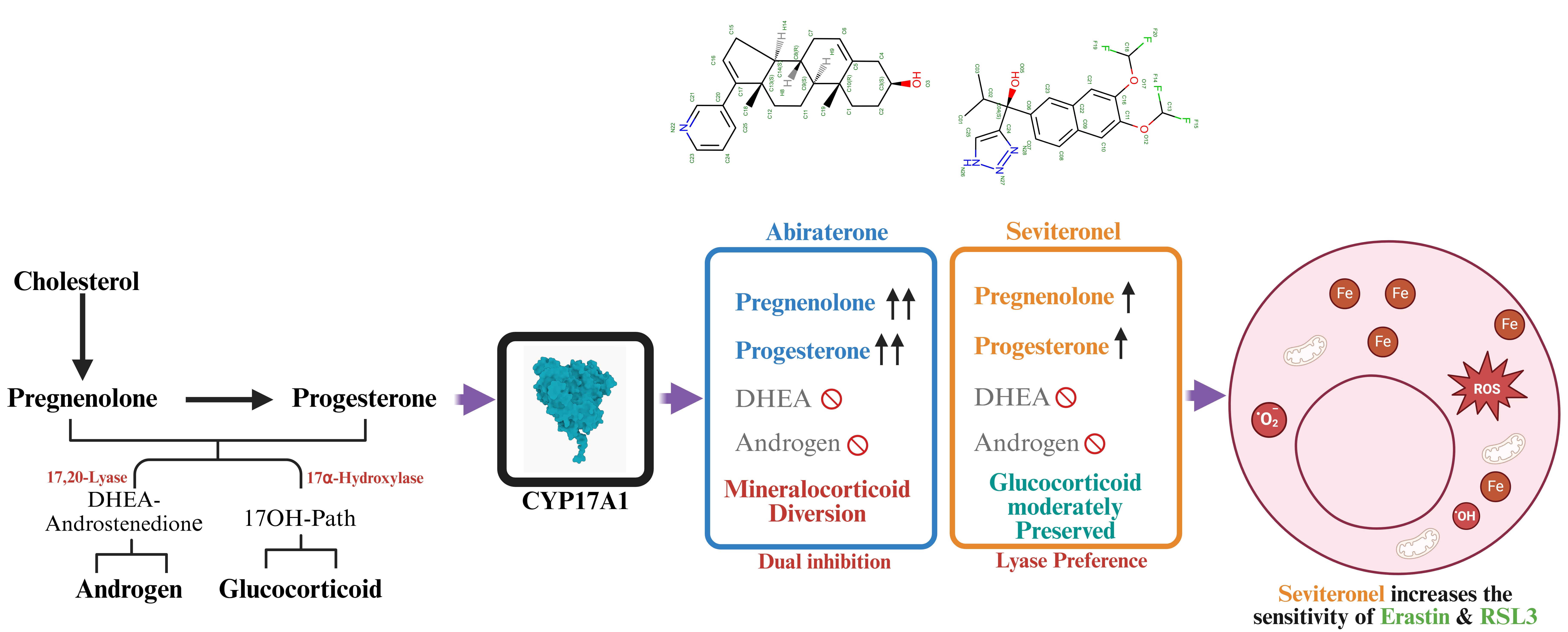
CYP17A1 is the key adrenal and gonadal enzyme that determines the supply of 17α-hydroxylated and C19 androgen precursors available for downstream intracrine metabolism in prostate cancer 5. CYP17A1 catalyzes two sequential reactions essential for converting pregnane precursors into androgens: an initial 17α-hydroxylase activity, which converts pregnenolone and progesterone into 17α-hydroxypregnenolone and 17α-hydroxyprogesterone, respectively, and the physiologically dominant human 17,20-lyase reaction converts 17α-hydroxypregnenolone to DHEA; conversion of 17α-hydroxyprogesterone to androstenedione is inefficient. CYP17A1 has become a major therapeutic target in CRPC due to its key role 6. Abiraterone acetate, a prodrug of abiraterone, was the first potent CYP17A1 inhibitor approved for CRPC 7. It effectively inhibits CYP17A1, resulting in significant suppression of adrenal and gonadal androgen generation, thereby reducing the precursor pool available for tumor intracrine metabolism. Because abiraterone also inhibits 17α-hydroxylase activity, cortisol synthesis is suppressed, ACTH rises, and mineralocorticoid precursors accumulate, contributing to hypertension, hypokalaemia, and fluid retention and necessitating glucocorticoid co-administration 7-10 (Figure 1).
In parallel with persistent AR reactivation, increasing evidence suggests that CRPC cells undergo adaptive metabolic reprogramming that influences susceptibility to regulated cell death pathways, including ferroptosis 11-14, an iron-dependent form of non-apoptotic cell death 15,16. AR signaling regulates lipid metabolism and antioxidant programs that can influence susceptibility to lipid peroxidation and ferroptosis. AR regulates the transcription of genes that are involved in de novo lipogenesis, fatty acid elongation, and lipid remodeling. This regulation alters the composition of membranes and the pool of substrates that are sensitive to peroxidation 17,18. In CRPC progression, the AR-dependent metabolic pathways can influence glutathione metabolism, lipid composition and susceptibility to lipid peroxidation 13,14.
The clinical success of abiraterone acetate has led to the development of next-generation CYP17A1 inhibitors that are more selective 19-22. Structurally, abiraterone mimics the steroid nucleus, employing a C17-pyridyl group to coordinate with the heme iron of CYP17A1 6,23. While potent, this structural mimicry results in non-selective inhibition of both 17α-hydroxylase and 17,20-lyase, leading to mineralocorticoid excess and typically requires glucocorticoid co-administration 7-9. Seviteronel is a non-steroidal triazole-containing inhibitor designed to favor 17,20-lyase inhibition over 17α-hydroxylase blockade 24-26, however, an earlier side-by-side study using purified-enzyme did not reproduce marked lyase selectivity for S-seviteronel 27. This lyase-selectivity is intended to preserve sufficient cortisol synthesis to reduce the need for glucocorticoid co-administration and associated endocrine complications 28. Despite these theoretical differences, a side-by-side quantitative comparison of their inhibitory profiles across the adrenal pathway remains incomplete. Furthermore, the metabolic impact of using a steroidal versus a non-steroidal scaffold on the steroid flux in human adrenal models has not been fully characterized. A second pharmacological distinction is tissue distribution. Unlike abiraterone, which is generally associated with a lower CNS adverse-event profile than CNS-penetrant AR inhibitors, seviteronel has been described as blood–brain-barrier penetrant and has been explored in AR-positive glioblastoma models 29. This property may be therapeutically relevant for CNS disease but may also contribute to neurological tolerability limitations 30.
This study fills these gaps by examining the biochemical differences between abiraterone and seviteronel. We further tested whether these agents alter the sensitivity of androgen-responsive prostate cancer cells to lipid-peroxidation stress. By assessing inhibitory potencies against CYP17A1 and off-target effects on CYP21A2, we map the distinct biochemical signatures of these inhibitors and define structural and functional correlates of their divergent pharmacological profiles.
2. Materials and Methods
2.1. Chemicals and Reagents
Abiraterone acetate was obtained from MedChemExpress (Lucerna Chem AG, Lucerne, Switzerland). Radiolabeled substrates, including progesterone [4-14C] (55 mCi/mmol), 17α-hydroxypregnenolone [21-3H] (15 Ci/mmol), and [3H]-17α-hydroxyprogesterone (60-120 Ci/mmol), were purchased from American Radiolabeled Chemicals (St. Louis, MO, USA). Unlabeled steroids (pregnenolone, progesterone, 17α-hydroxyprogesterone, 17α-hydroxypregnenolone), resazurin sodium salt, dimethyl sulfoxide (DMSO), and other analytical-grade reagents were obtained from Sigma-Aldrich (St. Louis, MO, USA). Organic solvents were supplied by Carl Roth GmbH (Karlsruhe, Germany). Activated charcoal was purchased from Merck (Darmstadt, Germany). Silica gel 60 aluminum-backed TLC plates were obtained from Macherey-Nagel (Oensingen, Switzerland). Phosphor imaging screens were acquired from Fujifilm (Dielsdorf, Switzerland). 2,2’-bipyridine (CAS No: 366-18-7), FIN56 (CAS No: 1083162-61-1), RSL3-analog (CAS No: 937043-70-8), Cisplatin (CAS No: 14913-33-8), liproxstatin-1 (CAS No: 950455-15-9), Deferoxamine (CAS No: 70-51-9), ferrostatin-1 (CAS No: 347174-05-4), and Erastin (CAS No: 571203-78-6) were obtained from Molport (Riga, Latvia). All compounds were dissolved in distilled water, DMSO, or ethanol as appropriate, and final solvent concentrations in cell culture did not exceed 0.1% (v/v).
2.2. Cell Lines and Culture Conditions
Human prostate cancer cell lines LNCaP (CRL-1740), VCaP (CRL-2876), PC-3 (CRL-1435), and DU-145 (HTB-81), as well as the non-malignant prostate epithelial cell line RWPE-1 (CRL-3607), were obtained from ATCC. LNCaP and PC-3 cells were cultured in RPMI-1640 supplemented with 10% fetal bovine serum (FBS), 2 mM L-glutamine, 10 mM HEPES, 1 mM sodium pyruvate, and 1% penicillin-streptomycin. VCaP and DU-145 cells were maintained in DMEM containing 10% FBS, 1 mM sodium pyruvate, and 1% penicillin-streptomycin. RWPE-1 cells were cultured in keratinocyte serum-free medium supplemented with bovine pituitary extract (0.05 mg/mL), recombinant human epidermal growth factor (5 ng/mL), and antibiotics (100 U/mL penicillin; 100 μg/mL streptomycin). Human adrenocortical NCI-H295R cells (CRL-2128) were grown in DMEM/Ham’s F-12 medium supplemented with 5% NU-I serum, insulin-transferrin-selenium supplement, 15 mM HEPES, and antibiotics. For ACTH–MC2R reporter assays, OS3 cells were cultured in Ham’s F10 with 15% horse serum, 5% fetal calf serum, penicillin (100 U/ml), and streptomycin (100 μg/ml) at 37 C in 5% CO2. All cells were maintained at 37 °C in a humidified incubator with 5% CO₂. Experiments were performed using cells below passage 30.
2.3. Resazurin Reduction Cell Viability Assay
Cell viability was evaluated using the resazurin reduction assay as previously described 19,22,31,32. Briefly, DU-145, 22Rv1, LNCaP, PC-3, and RWPE-1 cells (1 × 10⁴ cells/well; VCaP 1.2 × 10⁴ cells/well) were seeded in 96-well plates and allowed to adhere overnight. Cells were treated with test compounds (10 μM) for 24 or 48 h. Following incubation with resazurin (0.05 mg/mL) for 4 h, fluorescence was measured (λex 550 nm; λem 590 nm). Data represent the mean of at least three independent experiments performed in triplicate, with viability expressed relative to vehicle controls.
2.4. Ferroptosis Assays
Prostate cancer cell lines were maintained in RPMI-1640 supplemented with 10% fetal bovine serum and 1% penicillin-streptomycin at 37 °C in a humidified 5% CO2 atmosphere. Cell viability was assessed using the Dojindo CCK-8 assay kit. Briefly, cells were seeded at a density of 10 × 103 cells per well in 96-well plates and allowed to adhere overnight before treatment. To induce ferroptosis, cells were treated with various concentrations of the system Xc- inhibitor Erastin (1-50 µM) or the GPX4 inhibitor (1S,3R)-RSL3 (0.01-3 µM) for 24-48 h. For specific challenge experiments, cells were treated with Fin56 (5 µM), a type 3 ferroptosis inducer that promotes GPX4 degradation and CoQ10 depletion 33. In combination experiments, seviteronel and abiraterone were co-administered with Erastin or RSL3 to assess enhanced combination effects. To test whether cytotoxicity involved lipid-peroxidation-dependent mechanisms, cells were co-treated with ferrostatin-1 (Fer-1, 2 µM) or liproxstatin-1 (Liprox, 1 µM) 34. These lipophilic antioxidants were added simultaneously with the ferroptosis inducers. For recovery assays, cells were first exposed to inducers for 24 h, followed by a 48 h recovery phase in the presence of inhibitors.
2.5. Apoptosis Assay (Annexin V binding Assay)
LNCaP cells were seeded at a density of 1.0 × 10⁴ cells in a 96-well black microplate with a clear bottom and cultured overnight in a normoxic incubator. Cells were treated with seviteronel or abiraterone for 24 h in a final volume of 100 µL. Sixty microlitres of Annexin V working solution (Dojindo) were added to each well. The microplate was incubated in the dark at room temperature for 15 minutes. The fluorescence intensity was measured using a fluorescence microplate reader (bottom reading, Ex/Em = 488 nm/525 nm). To obtain the experimental reading, the blank value was subtracted from each sample’s measured value.
2.6. Detection of Intracellular Labile Iron Pool (LIP)
Intracellular ferrous iron (Fe2+) was detected using a Fe²⁺-responsive fluorescent probe FerroOrange (Dojindo, Japan) 35. LNCaP cells were seeded at a density of 10,000 cells per dish in glass-bottom confocal dishes and allowed to adhere for 24 h. Following adhesion, cells were treated with abiraterone, seviteronel, or the indicated combinations for 24 h. For positive control experiments, cells were incubated with 100 µmol/L ammonium iron (II) sulfate for 30 min at 37 °C to induce exogenous iron overload. To validate probe specificity via iron chelation, cells were co-treated with the membrane-permeable iron chelator 2,2’-bipyridyl (50 µmol/L) for 30 min prior to imaging. Deferoxamine (DFO) was included as an iron-chelation control. The growth medium was removed, and cells were washed three times with HBSS to eliminate background fluorescence from extracellular iron. Cells were then incubated with FerroOrange in serum-free medium for 30 min in a humidified incubator (37 °C, 5% CO₂). For nuclear visualization, cells were counterstained with Hoechst 33342 (1 µg/mL) for 10 min during the final stage of incubation. Live-cell imaging was performed using a 40X or 60X oil-immersion objective of a Nikon W1 LIPSI spinning disk inverted microscope (NA 1.4) using the following excitation and emission parameters: Hoechst 33342 (Nuclei), λex = 405, λem = 420-480 nm; FerroOrange (Fe2+), λex = 543 nm (or 561 nm), and λem = 570-620 nm. All imaging parameters, including laser power, pinhole size (1.0 Airy Unit), and gain settings, were kept constant across all experimental groups to ensure valid comparative analysis. Images were processed using ImageJ/FIJI. Line-scan analysis was performed by drawing a linear ROI across representative cells to generate spatial fluorescence intensity profiles. To examine subcellular distribution of the labile iron pool, fluorescence line scan analysis was performed. The relative intensities of FerroOrange (red) and Hoechst 33342 (blue) were plotted as a function of distance (µm), demonstrating that the FerroOrange signal is localized primarily within the extranuclear cytoplasmic compartment.
2.7. Wound-Healing (Scratch) Migration Assay
Cell migration was evaluated via a wound-healing assay as previously described 22,31, with modifications for live-cell imaging. DU-145 cells were seeded in 24-well plates to achieve 85–90% confluence. To separate migration from proliferation, cells were pre-treated with mitomycin C (5 μg/mL, 2 h) prior to wound induction. Following a uniform scratch, cells were treated with test compounds (10 μM) or vehicle control (0.1% DMSO/ethanol). Kinetic monitoring was performed using the CellogerMini Plus system (Curiosis, Seoul, Republic of Korea), capturing phase-contrast images every 4 h for 48 h at fixed coordinates. The wound area was quantified using integrated automated segmentation software and manually verified. Results were normalized to vehicle controls across three independent experiments (n=3). Parallel viability assays confirmed that migratory inhibition was not due to cytotoxicity.
2.8. Steroidogenic Enzyme Activity and Profiling
To distinguish direct enzymatic inhibition from indirect modulation of the pathway, we evaluated compound effects using both the endogenous human adrenocortical NCI-H295R model and recombinant HEK293T overexpression systems. NCI-H295R cells, selected for their robust expression of the steroidogenic machinery, including CYP17A1 and CYP21A2, were seeded at 5 × 10⁵ cells/well and equilibrated in serum-free medium for 2 h to deplete exogenous steroids. Cells were pre-incubated with seviteronel (10 μM), vehicle (0.1% DMSO/ethanol), or abiraterone as a positive control for 4 h before substrate addition. In parallel, HEK293T cells were transiently transfected or modified with lentivirus to overexpress human CYP21A2, HSD3B2, or CYP19A1 to confirm enzyme-specific inhibition in an isolated genetic background, with assays performed 48 h post-transfection as previously described 36-39.
Enzymatic activities were quantified using optimized radiometric assays with [¹⁴C]- or [³H]-labeled substrates (1 μM) to ensure linear reaction kinetics. For CYP17A1 17α-hydroxylase and CYP21A2 activities, cells were incubated with [¹⁴C]-progesterone or [³H]-17α-hydroxyprogesterone, respectively; 17α-hydroxylase assays were supplemented with Trilostane (10 μM) to prevent metabolic shunting through 3β-HSD. Following organic extraction (ethyl acetate: isooctane, 1:1 v/v), steroids were resolved by thin-layer chromatography (TLC) and visualized via phosphor imaging, with activity calculated as the percentage of substrate-to-product conversion relative to total recovered radioactivity. Conversely, CYP17A1 (17,20-lyase) and CYP19A1 (aromatase) activities were determined by measuring the stoichiometric release of tritiated water from [³H]-17α-hydroxypregnenolone or [³H]-androstenedione. Following dextran-coated charcoal treatment to remove unreacted steroids, the aqueous [³H]₂O product was quantified by liquid scintillation counting. For HSD3B2 activity, transfected HEK293T cells were incubated with [³H]-DHEA, which was converted to androstenedione and analyzed by TLC and phosphorimaging.
Comprehensive steroidomic profiling was performed by ultra-high-performance liquid chromatography coupled to high-resolution mass spectrometry (LC-MS). NCI-H295R cells were treated with compounds for 4 hours and then stimulated with 1 μM pregnenolone for another 4 hours to increase biosynthetic flux 19,22,31,32. Analyte concentrations were determined using external calibration curves and normalized to total cellular protein content across three independent experiments.
2.9. Quantitative Real-Time PCR (qRT-PCR)
Total RNA was extracted from cells using the Direct-zol RNA Kit according to the manufacturer’s instructions. RNA concentration and purity were assessed using a spectrophotometer (A260/280 ratio). For each sample, 500 ng of total RNA was reverse-transcribed into cDNA using the iScript cDNA Synthesis Kit in a final volume of 20 µL. Real-time PCR was performed using SYBR Green Master Mix on a GeneExplorer GE-969 system. Each 20 µL reaction contained cDNA and 200 nM of gene-specific primers. The thermal cycling conditions were an initial denaturation at 95 °C for 3 min, followed by 40 cycles of denaturation at 95 °C for 15 s, primer annealing at 60 °C for 30 s, and extension at 72 °C for 30 s. A melting curve analysis was performed at the end of each run to ensure primer specificity and the absence of primer dimers. The relative gene expression was calculated using the 2^−ΔΔCt method. GAPDH was utilized as the internal reference gene for normalization. Steroidogenesis data: Represent the mean of two independent biological replicates (n = 2), each performed in technical duplicate. Statistical significance was assessed via a one-sample t-test against a theoretical mean of 1.0 (DMSO control). Ferroptosis-associated qPCR data were treated as exploratory and interpreted as pathway-level trends rather than definitive transcriptional endpoints. Primer sequences are provided in Supplementary Table S1.
2.10. Cell Cycle Analysis
LNCaP cells were treated with test compounds for 24 h. Cells were harvested by trypsinization, washed with PBS, and fixed in 70% ice-cold ethanol overnight at -20 °C to ensure adequate DNA preservation. Before analysis, cells were washed and incubated with staining solutions containing propidium iodide (50 μg/mL) and RNase A (100 μg/mL) for 30 min at room temperature in the dark. DNA content was measured by flow cytometry, and at least 10,000 events per sample were acquired. Doublets were excluded based on forward- and side-scatter pulse geometry. Cell cycle distribution (G₀/G₁, S, and G₂/M phases) was quantified in FlowJo (v10.8). Results were expressed as the percentage of the total single-cell population 40.
3. ACTH Treatment and Dual-Luciferase Quantitation
Two days after transient transfection in 12-well plates, the culture medium was replaced, and cells were exposed to 10-10 M Adrenocorticotropic Hormone (ACTH; Sigma-Aldrich) for 18 h at 37 °C under 5% CO2. Control wells received an equivalent volume of vehicle. Cells were subsequently washed with PBS and harvested in passive lysis buffer. Firefly and Renilla luciferase signals were determined sequentially using a Dual-Luciferase Assay Kit (Promega), as previously outlined by Hirsch et al. 41. Relative luciferase activity was calculated as the ratio of firefly luminescence to Renilla luminescence to normalize transfection efficiency.
3.1. Statistical Analysis
All data are presented as mean ± standard deviation (SD) from at least three independent biological experiments, each performed with technical replicates, unless otherwise indicated. Graphical representations and statistical analyses were conducted using GraphPad Prism (v10.0, GraphPad Software, San Diego, CA, USA) and R (R Foundation for Statistical Computing, Vienna, Austria). Before hypothesis testing, data distribution was assessed for normality using the Shapiro-Wilk test, and homogeneity of variances was evaluated using Levene’s test. When both assumptions were satisfied, parametric tests were applied. Comparisons among three or more groups were performed using one-way analysis of variance (ANOVA), followed by Tukey’s multiple-comparisons post hoc test to control type I error across pairwise comparisons. For experiments involving two independent variables (e.g., treatment and time), two-way ANOVA was used where appropriate, followed by Tukey’s or Šidák’s post hoc correction for multiple comparisons. In cases where assumptions for parametric testing were not met, non-parametric alternatives (Kruskal-Wallis’ test followed by Dunn’s post hoc test) were applied. For pairwise comparisons, unpaired and two-tailed Student’s t-tests were used unless otherwise specified. IC₅₀ values were calculated using nonlinear regression analysis with a four-parameter logistic (4PL) curve-fitting model. Correlation analyses were performed using Pearson’s correlation coefficient for normally distributed data or Spearman’s rank correlation for non-normal distributions. All statistical tests were two-tailed, and p-values < 0.05 were considered statistically significant. Exact p-values are reported where possible. Significance levels are denoted as follows: *p < 0.05, **p < 0.01, ***p < 0.001, and ****p < 0.0001. Sample sizes were based on prior experimental experience and comparable published studies.
3.2. Computational Details
Protein structures were extracted from the Protein Data Bank 42 and prepared for the MD simulations using the Protein Preparation Procedure in Maestro 43, which checks for structural errors, optimizes hydrogen-bonding networks and resolves steric clashes. For the abiraterone structure (3RUK) only the C and D chains were used as chains with missing residues were excluded 23. For the non-steroidal inhibitors only the C chains with (S)-orteronel (5IRQ) and (S)-seviteronel (5IRV), respectively, contained all residues 27. To enable a direct comparison between the structures, compared systems were standardized to the same residue range. Accordingly, Q503 in 5IRQ and S30 and Q503 in 5IRV were deleted. The final structures contained 472 residues from Leu31 to Ala502. In the results we refer to these structures as 3RUK_C, 3RUK_D, 5IRQ_C and 5IRV_C, respectively.
The Desmond program (Desmond Molecular Systems, D. E. Shaw Research, New York, NY, USA, 2009, v8.2.133) available as part of the Schrodinger system (Schrodinger Suite 2025-2, Schrödinger, LLC, 2023) were used for the molecular dynamics (MD) simulations. The proteins were embedded in a predefined orthorhombic box of SPC water molecules with at least 10 Å between the protein and the boundary of the box and neutralized with proper charged ions. The 5IRV_C systems contained a total of 75912 atoms with 45 ligand atoms, 7570 protein atoms, 22741 water molecules (68223 atoms) and 73 atoms from heme group and one chloride ion. The other systems contained a similar number of atoms. Prior to the MD production runs, the systems were equilibrated by the Desmond standard step-wise equilibration protocol comprising a short minimization of water and ions to optimize the contact between the protein and the surrounding solvent, a short minimization of the entire system to remove clashes, a short simulation at 10 K of the solvent atoms, two short simulations with NVT and NPT constraints, respectively, followed by a short unrestricted NPT simulation of the entire system. Each system was subjected to four simulations for generating four independent replicate trajectories. Production runs were 100 ns at 300 K using the OPLS4 force field 44. For each run 100 frames were selected for subsequent analyses.
The Principal Component (PC) analysis was performed via a KNIME workflow 45. Originally, both the Phi, Psi and Omega backbone torsional angles were determined, but the Omega angles were discarded due to a high degree of correlation. The final PC analysis was performed on 41600 data points from four simulations of four systems with 100 frames for each simulation. Each frame was characterised by the Phi and Psi angles for 13 residues. Structures were displayed with the Pymol program (PyMOL Molecular Graphics System, Schrödinger, LLC, v.3.1.5.1).
4. Results
4.1. Distinct Steroidogenic Signatures of Abiraterone and Seviteronel Reveal Partitioned 17,20-Lyase Inhibition
Abiraterone and seviteronel were evaluated for their effects on CYP17A1, CYP21A2, HSD3B2, and CYP19A1 activities in steroidogenesis (Figure 2). We first assessed inhibition of CYP17A1 hydroxylase and 17,20-lyase activities at a fixed concentration (10 µM), followed by a dose-response analysis (Figure 2A-C). From the initial screening, a clear functional divergence between the two clinically relevant inhibitors emerged at CYP17A1. Abiraterone showed near-complete inhibition of the hydroxylase reaction at 10 µM (Figure 2D), reducing activity to ~5-10% of DMSO (****P < 0.0001), whereas seviteronel retained substantial residual activity (~60-70%). This result translated into a marked difference in potency, with abiraterone exhibiting submicromolar inhibition (IC₅₀ = 0.05 µM) and seviteronel being approximately 170-fold weaker (IC₅₀ = 8.6 µM). Furthermore, inhibition of CYP17A1 lyase activity showed a distinct profile (17,20). Both compounds significantly reduced the lyase activity at 10 µM (Figure 2F; ~25-35% residual activity, ****P < 0.0001 versus control), and no statistically significant difference was observed between them under these assay conditions. However, dose–response analysis showed that abiraterone was modestly more potent against 17,20-lyase activity than seviteronel (IC₅₀, 0.67 µM versus 1.2 µM). Thus, abiraterone was more potent in absolute terms against both CYP17A1 activities, whereas seviteronel displayed lyase-biased partitioning relative to its much weaker hydroxylase inhibition. For effects on CYP21A2 hydroxylation, both abiraterone and seviteronel reduced the enzyme activity to 10 µM (Figure 2F), with abiraterone showing a significant (****P < 0.0001) inhibitory effect (~20-25% residual activity) compared to seviteronel (~30-40% residual activity). This result was consistent with the IC₅₀ values (4.6 µM for abiraterone versus 9.8 µM for seviteronel), indicating approximately twofold greater potency for abiraterone (Figure 3, Table 1). Neither compound significantly inhibited CYP19A1 activity (Figure 2G), with both remaining comparable to the control, while the reference inhibitor anastrozole produced robust suppression (~25-30% residual activity, ****P < 0.0001), confirming assay sensitivity and selectivity. This absence of aromatase inhibition indicates that neither compound directly blocked aromatase activity under these assay conditions. With HSD3B2, the conversion of DHEA to androstenedione was more strongly inhibited by abiraterone (Figure 2H), which reduced activity to ~10-15% of control, whereas seviteronel retained substantially higher activity (~55-65%) (****P < 0.0001), suggesting a broader upstream blockade by abiraterone at the level of Δ⁵ to Δ⁴ steroid conversion.
4.2. Differential Endocrine Signatures of Abiraterone and Seviteronel Reveal Distinct Metabolic Targets in Human Adrenal Cells
To determine how distinct CYP17A1 inhibition profiles translate into the steroidogenic output, we performed comprehensive steroid profiling in NCI-H295R human adrenal cells following treatment with seviteronel and abiraterone. Quantification of protein-normalized steroid levels revealed a marked redistribution of flux across the pathway, consistent with CYP17A1 blockade and secondary effects on adjacent enzymatic steps. Under basal (DMSO) conditions, NCI-H295R cells maintained balanced flux through the Δ5 and Δ4 pathways, reflected by low levels of pregnenolone (13.3 nmol/g protein) and progesterone (2.1 nmol/g protein), with efficient downstream conversion to 17α-hydroxylated intermediates and androgen precursors (Table 2). Inhibition of CYP17A1 markedly disrupted this equilibrium. Abiraterone treatment resulted in a significant accumulation of proximal substrates, with pregnenolone increasing to 222.6 nmol/g protein and progesterone to 138.0 nmol/g protein (both p < 0.0001 vs DMSO) (Figure 4, Supplementary Table S2). Seviteronel induced a similar but attenuated effect, elevating pregnenolone to 82.3 nmol/g protein and progesterone to 65.1 nmol/g protein (p < 0.001 vs DMSO; p < 0.01 vs abiraterone) (Figure 4), indicating a comparatively weaker upstream constraint on steroid flux. A significant reduction in CYP17A1-dependent products accompanied this substrate accumulation. 17α-hydroxyprogesterone was strongly reduced by abiraterone but increased under seviteronel, consistent with preserved hydroxylase flux and a downstream bottleneck beyond 17α-hydroxylation. The impact on 17,20-lyase activity was even more pronounced: DHEA levels were reduced to near-undetectable levels with abiraterone (p < 0.0001 vs DMSO), whereas seviteronel maintained a small but measurable residual production (p < 0.001 vs DMSO; p < 0.05 vs abiraterone). A similar pattern was observed for androstenedione, which was comparably suppressed by both inhibitors. Together, these findings indicate that abiraterone strongly suppresses both hydroxylase and lyase activities, whereas seviteronel preferentially suppresses 17,20-lyase activity while preserving hydroxylase function. The consequences of these differences are evident when considering adrenal, androgen, and cortisol pathways. Abiraterone inhibited DHEA and androstenedione production, consistent with marked suppression of measured androgen output, but this was accompanied by marked suppression of 17α-hydroxylated intermediates, indicating restricted flux toward cortisol biosynthesis. Conversely, seviteronel significantly reduced androgen output while maintaining detectable levels of 17α-hydroxyprogesterone, suggesting that entry into the glucocorticoid pathway is not fully abrogated. This profile supports a more lyase-selective mode of action, functionally separating androgen suppression from complete inhibition of cortisol precursor generation. The redistribution of steroid intermediates also provides insight into upstream and parallel enzymatic activity. The disproportionate progesterone accumulation is consistent with continued HSD3B2-mediated conversion. This effect is most pronounced under abiraterone, where the magnitude of progesterone levels suggests efficient conversion of pregnenolone despite upstream accumulation. The DOC response further separated the two inhibitors. Seviteronel increased DOC despite partial hydroxylase preservation, consistent with altered partitioning into the mineralocorticoid branch. In contrast, abiraterone reduced DOC despite progesterone accumulation, consistent with broader pathway suppression that includes CYP21A2 inhibition.
4.3. Machine Learning and Dimensionality Reduction Define Distinct Steroidogenic Signatures
To characterize the biological divergence between abiraterone and seviteronel, machine learning and dimensionality reduction were applied to steroidomic profiles (Supplementary Figure S1 A-D). An exploratory Random Forest analysis separated treatment groups in this dataset and identified DOC and 17α-hydroxyprogesterone as major discriminatory features (Supplementary Figure S1 B). The high importance of DOC highlights the unique mineralocorticoid shunt induced by seviteronel. On the other hand, 17α-hydroxyprogesterone reflects seviteronel’s preserved 17α-hydroxylase throughput, a metabolic flux entirely abolished by abiraterone. 3D PCA and hierarchical clustering confirmed this separation (Supplementary Figure S1 A & C), with PC1 explaining > 80% of the variance.
The spatial distance between treatment nodes indicates that these drugs exert qualitatively distinct impacts on the steroidome, rather than merely differences in potency. Heatmap analysis further defined two distinct metabolic signatures: an abiraterone steroidogenic blockade, characterized by a cluster of upstream precursors (pregnenolone, progesterone) coupled with a collapse of all downstream products, and a seviteronel-selective androgen blockade, characterized by robust accumulation of DOC and 17α-hydroxyprogesterone alongside effective androgen depletion. The analyses support distinct treatment-associated steroid profiles, with stronger upstream precursor accumulation under abiraterone and greater preservation of 17α-hydroxyprogesterone and DOC under seviteronel. To support this biochemical link, Pearson correlation matrices were generated (Supplementary Figure S1D & E; Table 3). In the seviteronel group, the negative DOC–androgen association is consistent with altered partitioning toward the mineralocorticoid branch.
4.4. Structural Comparison of Abiraterone and Seviteronel Bound to CYP17A1
To define the structural basis for the divergent inhibitory profiles, we compared the crystallographic CYP17A1 complexes with abiraterone (PDB: 3RUK) 23 and VT-464/seviteronel (PDB: 5IRV) 27. Both inhibitors coordinate the catalytic heme iron through a nitrogen-containing heterocycle, consistent with the classical type II binding mode of CYP17A1 inhibitors. However, despite sharing this conserved iron-coordination mechanism, the two ligands differ in scaffold architecture, active site orientation, and protein contact networks, (Supplementary Figure S2 Table S3).
In the abiraterone-bound structure, the ligand adopts a steroidal substrate-like binding mode. The pyridine nitrogen coordinates the heme iron at approximately 2.04 Å, while the 3β-hydroxyl group forms a hydrogen bond with Asn202 (O3-Asn202 OD1, 2.73 Å), corresponding to the canonical steroid-recognition interaction reported in the CYP17A1 complexes with substrates 46. The steroid nucleus is further stabilized by extensive hydrophobic interactions with residues Ala113, Phe114, Ile205, Ile206, Leu209, Ala302, Thr306, Ala367, Ile371, Val482, and Val483. Additional nearby contacts involving Asp298 and Glu305 are observed in the interaction diagram but appear secondary to the conserved Asn202 interaction. Collectively, these interactions support a steroid-mimetic binding mode in which axial heme coordination is reinforced by the conserved Asn202 hydrogen bond and extensive hydrophobic complementarity surrounding the steroid core (Figure 5A, C and Supplementary Table S3).
In contrast, seviteronel adopts a distinct non-steroidal binding mode while preserving direct heme coordination. The triazole N27 nitrogen coordinates the heme iron at approximately 2.23 Å. Unlike abiraterone, seviteronel lacks the steroidal 3β-hydroxyl–Asn202 hydrogen-bonding. Instead, the strongest direct polar interaction identified in the contact analysis is between the ligand hydroxyl oxygen O05–Val482 backbone carbonyl (approximately 2.94 Å). The triazole ring is positioned adjacent to Thr306, with close contacts to the Thr306 hydroxyl group, while additional contacts are observed near Asn202 and Asp298 through the difluoromethoxy substituents. Because organic fluorine atoms are generally poor hydrogen-bond acceptors 47, these interactions are more appropriately interpreted as close electrostatic or van der Waals contacts rather than classical hydrogen bonds. The non-steroidal scaffold is further surrounded by residues including Ala105, Ala113, Phe114, Ile205, Ile206, Arg239, Gly301, Ala302, Thr306, Ala367, Ile371, Val482, and Val483 (Figure 5B and 5D), resulting in a different interaction network than that observed for abiraterone.
Comparison of the two crystal structures therefore demonstrates that abiraterone and seviteronel differ not only in chemical scaffold but also in their active-site anchoring strategies. Abiraterone combines heme coordination with the conserved Asn202 steroid-recognition interaction and compact steroid-core packing characteristic of substrate-like inhibitors. Seviteronel retains iron coordination but replaces the steroidal anchoring interaction with an alternative network centered on Val482 and residues surrounding the I-Helix, including Thr306 (Figure 5 and Supplementary Figure S2).
4.5. Molecular Dynamics Simulations
To further explore possible differences between Abiraterone and Seviteronel binding to CYP17A1 we performed a series of molecular dynamics (MD) simulations of the 3D structures of these complexes (3RUK and 5IRV, respectively). For comparison, we have included the 3D structure of CYP17A1-Orteronel complex (5IRQ), to enable a more general study of the binding differences between steroidal and non-steroidal CYP17A1 inhibitors. To enable a one-to-one comparison of the structural changes the MD simulations were performed on structures of equal length (number of residues) and without gaps, which would impose unrealistic flexibility in some regions.
The selection and preparation of the CYP17A1 complexes subjected to MD simulations are described in detail in the Methods section. The C- and D-chains of the abiraterone complex (3RUK_C and 3RUK_D) and the C-chains of the seviteronel and orteronel complexes (5IRV_C and 5IRQ_C, respectively) all containing 472 residues from Leu31 to Ala502 were selected. Four MD simulations on 100 ns on each complex with different initially assigned velocities were considered more appropriate that one more long MD simulations. MD simulations on cytochrome P450 complexes are generally stable during MD simulations at 300 K, but in our case we were interested in identifying minor, perhaps subtle differences between the steroidal and non-steroidal complexes. A comparison of the RMSD values for the MD simulations (Figure 6) shows that there are only minor differences between the Abiraterone simulations (3RUK_C and 3RUK_D) and the orteronel simulations (5IRQ_C), whereas the seviteronel simulations (5IRV_C) showed a broader RMSD distribution. The differences between each of the four simulations confirm that MD simulations with different initial starting velocities can be considered as individual experiments.
Whereas the RMSD values provide information about the overall performance of the individual simulations, the RMSF describe the effect on each residue in a simulation. Inspection of the individual RMSF plots (not shown), did not reveal any significant differences between the MD simulations, probably because the RMSF plot primarily identifies the most flexible part of the structures, loops and N- and C-terminal parts, which are comparable for the present systems. Since we were interested in differences between the steroidal and non-steroidal systems, we averaged the RMSF values for each of the four complexes and subsequently calculated the differences between the average RMSF values for each of the abiraterone complexes and the orteronel and seviteronel complexes to identify possible differences between the steroidal and non-steroidal MD simulations (Figure 7).
The fluctuations shown in the RMSF difference plots (Figure 7) are much smaller than the fluctuations observed in the individual RMSF plots, which confirms that by this approach we could identify more subtle differences between the simulations. It should be mentioned that we have not calculated all possible combinations but limited the analysis to differences between the four examples shown in Figure 7. The regions highlighted in Figure 7 have been subjected to a visual comparison based on the corresponding experimentally determined 3D structures (Figure 8).
Region A comprised by Lys136-Gln140 (KDGDQ) is a surface loop between two helices. Although the loop contains a potentially flexible Gly138 the major difference between the four X-ray structures is in the side-chain orientations (Supplementary Figure S3). Region B from Val218 to Asn226 (VPWKLIFPN) is a sequence located on the surface with nearly overlapping backbone atoms and only minor different side chain conformations (Supplementary Figure S4). Region C from Asp274 to Asp281 (DNGNAGPD) is a highly flexible surface loop with two Glycine residues connecting Helix F and Helix G. The F/G loop is similar in the two 3RUK structures, nearly identical in 5IRQ_C and 5IRV_C, but significantly different between the steroidal and non-steroidal structures (Supplementary Figure S5). Different conformations of the F/G loop have been associated with formation of a peripheral ligand binding site in CYP17A1 27.
Region D from Ile299 to Gly303 (IFGAG) constitutes the middle part of I-Helix. In 5IRV_C, this part of the helix and the ligand Seviteronel is moved approximately 1.2-1.6 Å relative to the rest of the protein and the heme group. The ligand, Seviteronel, is L-shaped and to achieve a proper directionality of the interaction between the nitrogen lone pair on the triazole ring and the Fe atom and maintain the close contact between I-Helix and the naphthalene ring in Seviteronel, Seviteronel and the GAG part of I-Helix is moved away from the heme group (Figure 9).
Region E from Gly444 to Arg449 is in direct contact the heme group, but the backbone as well as sidechains of this moiety seems unaffected. The only difference between the four crystal structures is the orientation of the vinyl (-CH=CH2) sidechain in the heme group (Supplementary Figure S6). Region F from Asp487 to Lys490 (DSFK) is loop on the surface with the side chains of the three polar residues exposed to solvent (Supplementary Figure S7). Finally, the RMSF difference plots (Figure 7) showed some minor differences between Region E and Region F. This part of the structures, referred to as Region DE, is a long stretch located on the surface of the proteins. Although one could expect it to be rather flexible, it only displays minor differences between the four crystal structures (Supplementary Figure S8). The above visual inspection of regions identified from the RMSF differences plot (Figure 7) is based the X-ray structures and, accordingly, only compares the starting points for the MD simulations. Nevertheless, they point out that Region D, especially the GAG (Gly301-Ala302-Gly303) part of I-Helix, could be associated with differences in binding mode between abiraterone and seviteronel due to its proximity to both the ligands and the heme group.
4.6. Principal Component Analysis
To investigate the potential effect of abiraterone and seviteronel on I-Helix, we determined the backbone torsional angles (Phi and Psi) for the central part of the helix, i.e. IGDIFGAGVETTT, comprising a total of 13 residues, the GAG moiety and five additional residues before and after.
A Principal Component (PC) analysis based on these Phi and Psi angles of all the frames from the MD simulations of the four systems (see Methods for details) showed a distinct difference between the two steroidal systems (3RUK_C and 3RUK_D) compared to the two non-steroidal systems (5IRQ_C and 5IRV_C) (Figure 10). The most important conclusion is that the steroidal and non-steroidal CYP17A1-inhibitor complexes behave significantly different during the MD simulations. The steroidal complexes are stable and do not visit different conformational states during our two times four 100 ns MD simulations (except for five out of a total of 800 frames as shown in Figure 10) of the 3RUK_C and 3RUK_D systems.
Contrary to the steroidal systems, the non-steroidal systems visited several additional and different conformational states during the MD simulations. For seviteronel only 25% of the conformations observed corresponded to the dominant conformations of the steroidal systems and another approximately 25% to the conformation observed in a few cases for the 3RUK_C system. The remaining 50% of the conformations with seviteronel the central part of I-Helix displayed a relatively high degree of flexibility and several different conformational states were detected.
In the PC analysis we did not distinguish between the individual MD runs, but the heatmap based on the Phi and Psi backbone torsional angles of the 13 central residues in I-Helix (Figure 11) allows a direct comparison of the MD simulations. From the heatmap, it is clear that the MD simulations of the abiraterone containing systems are stable for all four runs for both systems. The situation is substantially different for the non-steroidal systems.
The non-steroidal inhibitors apparently impose some flexibility in the protein structure. The flexibility is different between the Orteronel (5IRQ_C) and Seviteronel (5IRV_C) systems and between the different MD runs, but in all cases the flexibility are centred around the FGAGV motif in I-Helix. In two MD runs, 5IRQ_C run 4 and 5IRV_C run 1, the non-steroidal systems behave like the steroidal systems without displaying any flexibility in the I-Helix. The remaining six non-steroidal MD runs are all different. In the 5IRQ_C run 1 the system visit three different conformational states, one similar to the steroidal systems, in run 2 the system do not maintain one specific conformation but display a considerable flexibility of the FGA part of I-Helix, in run 3 the system initially behave like run 1, but after one third of the time it reverts to the conformation observed for the steroidal systems.
In the 5IRV_C the three runs 2, 3 and 4 are different and the flexibility is primarily associated with different parts of I-Helix, the FG, FGA and GV residues, respectively. For each system, the four simulations are based on the same PDB structure prepared by the Maestro Protein Preparation Protocol and subsequently subjected to the Desmond equilibration protocol (see Methods for details). After the equilibration and before the production runs, the initial conformations are different, which means that the conformational changes take place already during the equilibration process and that the system remain stable during the production runs (0-100 ns). A more detailed inspection of the heatmaps reveal that the primarily flexibility of the FGAGV motif is associated with fluctuations in the Psi torsional angles of GD. The flexibility identified from the heatmaps were not obvious from inspection of the individual RMSF plots probably, as already pointed out, because these primarily identify the most flexible parts of the structures, whereas differences in the more conserved parts of the structures like I-Helix are difficult to identify. We have used the heatmaps to identify representative conformations of the I-Helix from the non-steroidal MD simulations to compare with the conformation being dominant in the steroidal MD simulations.
The difference between the four 5IRV_C simulations are obvious both by comparison of the individual frames (Figure 12A) and from the average values of the backbone torsional angles Phi and Psi (Figure 12B). In run1, the Phi and Psi values do not change, similar to the corresponding values in the simulations 3RUK_C and 3RUK_D for the steroidal inhibitor abiraterone. In run2 the major changes in in the FG residues (Phe300 Psi and Gly301 Phi torsional angles), in run 3 the FGA residues are involved in the conformational change (Phe300 Psi, Gly301 Phi and Psi, and Ala 302 Phi torsional angles) and in run 4 the conformational change is associated with the GV residues (Gly303 Psi and Val 304 Phi torsional angles).
The MD simulations provide some clear answers but also raise several new questions. We observe a distinct difference between the steroidal and non-steroidal complexes with the non-steroidal inhibitors, seviteronel and orteronel, inducing a conformational change of flexibility in the CYP17A1 proteins, whereas the abiraterone complexes remain unaffected. The flexibility in the non-steroidal complexes is associated with a conformational change in I-Helix, which is in direct contact with the inhibitors.
Abiraterone is both inhibiting the 17α-hydroxylase and 17,20-lyase reactions, whereas the seviteronel and orteronel are less potent, but more selective inhibitors for the 17,20-lyase reaction. It has been suggested that CYP17A1 should undergo a conformation change from the 17α-hydroxylase active conformation to the 17,20-lyase conformation. One can speculate on this involves the flexibility of I-Helix observed in the MD simulations. Abiraterone can be viewed as a steroid with a heme-coordinating moiety (pyridine) and, accordingly, it has a high degree of 3D similarity to the substrates for both the 17α-hydroxylase and 17,20-lyase reactions. Seviteronel and most of the non-steroidal inhibitors, which are biased for the 17,20-lyase reaction, do not be as complementary to the CYP17A1 active site as abiraterone and may therefore impose the conformational change in the CYP17A1 protein towards the 17,20-lyase active form.
4.7. Effects of Abiraterone and Seviteronel on Cell Viability, Selectivity, and Migration
We next asked whether these pharmacological differences were associated with distinct responses in prostate cancer models differing in androgen dependence. Cell viability assays demonstrated clear differences in both potency and cellular context dependence at 24 and 48 hours (Figure 13A and Table S4). In androgen-sensitive models (LNCaP, VCaP, 22Rv1), both drugs reduced viability, but to different degrees. At 24 hours, abiraterone reduced viability to ~60-63% across these lines, whereas seviteronel produced a comparable but slightly less consistent effect (~61-66%). By 48 hours, the divergence became more apparent: abiraterone maintained or enhanced its cytotoxic effect (LNCaP ~45.4%, VCaP ~58.1%, 22Rv1 ~62.9%), while seviteronel showed a more modest reduction (generally ~60-67%). These differences were statistically significant, indicating that abiraterone was more effective over time in androgen-responsive cell models. Androgen-insensitive models (PC-3 and DU145) were largely refractory to both seviteronel and abiraterone. Viability remained high (>80–95%) at both time points, with only small drops (PC-3 ~80.5% at 24 h and ~83.2% at 48 h with Abiraterone; DU145 ~95–96% under both treatments). This highlights the dependence of drug efficacy on androgen receptor signaling and supports a link between androgen-axis dependence and sensitivity to these agents, without implying robust endogenous CYP17A1 activity in the prostate cancer cells. The non-malignant RWPE-1 cells provided important comparators for therapeutic selectivity. Abiraterone had minimal impact on viability (~94-96%), whereas seviteronel produced a slightly greater reduction (~87-93%), though still modest compared to cancer cells. Cisplatin, included as a control, showed marked non-selective toxicity across all cell types, reinforcing the relative specificity of CYP17A1-targeted agents. These observations are reflected in the selectivity index (SI), calculated relative to RWPE-1 cells (Supplementary Figure S9). At 24 hours, both abiraterone and seviteronel demonstrated favorable selectivity (SI ~1-1.6) across most cancer cell lines. At 48 hours, seviteronel maintained selectivity in several androgen-sensitive models (LNCaP SI ~1.3 and VCaP SI ~1.44), whereas abiraterone showed a modest increase (LNCaP SI ~2.12), reflecting stronger suppression of tumor cells than normal cells.
To explore the mechanisms driving these changes in viability, we performed cell cycle analysis (Figure 13E). Both abiraterone and seviteronel treatments resulted in a significant shift in cell cycle distribution compared to the control. Specifically, we observed a marked decrease in the proportion of cells in S-phase, accompanied by a significant accumulation in G1 and G2. While both agents exerted G1/G2 arrest, seviteronel showed a more pronounced reduction in the S-phase population (p < 0.001), suggesting a potent inhibition of DNA synthesis. This cytostatic effect was accompanied by the induction of cellular stress and apoptosis, as measured using the Dojindo Annexin V Plate kit for Apoptosis (Figure 13F). Both abiraterone and seviteronel significantly increased the RFU signal compared to the DMSO control (p < 0.0001). While seviteronel and abiraterone showed comparable induction levels, both were significantly lower than the high-toxicity control, cisplatin. These data suggest that the cell cycle arrest and increased Annexin V signal may contribute to the reduced viability (Figure 13A).
Finally, we assessed the impact of these agents on cell migration using wound healing assays (Figure 13B & C). Both abiraterone and seviteronel significantly impaired migratory capacity compared to control, as evidenced by delayed wound closure over 48 hours. The control group exhibited rapid closure, reaching near completion within ~40 hours, whereas both treatments substantially slowed down this process. seviteronel consistently showed the strongest inhibition of migration, with greater retention of the wound area across all time points, while abiraterone had an intermediate effect. Quantification using T50 (time to 50% wound closure) further highlighted these differences. Control cells reached T50 at approximately 10-11 hours, whereas abiraterone and seviteronel delayed it to ~14 and ~13 hours, respectively. These increases were statistically significant (p < 0.05-0.01), confirming that both drugs impair migratory dynamics. Because migration was assessed in DU-145 cells, these effects should be interpreted as androgen-axis-associated or off-target pharmacological effects rather than direct CYP17A1-dependent steroidogenesis.
4.8. CYP17A1 Inhibitor Exposure Sensitizes Prostate Cancer Cells to Ferroptosis-Inducing Lipid-Peroxidation Stress
The potent reduction in cell viability observed following seviteronel and abiraterone treatments (Figure 14A, B) suggested engagement of a non-apoptotic regulated cell death pathway. We next tested whether inhibitor exposure altered sensitivity to ferroptosis-inducing stress. Our data reveal that seviteronel and abiraterone have enhanced combination effect with classical ferroptosis inducers (FINs). Specifically, the combination of these agents with erastin, which inhibits the system XC- cystine/glutamate antiporter, resulted in a more pronounced loss of viability than erastin monotherapy (Figure 14A). This effect was mirrored when using an RSL3-analog (Figure 14B), which targets the glutathione peroxidase 4 (GPX4) enzyme 48. The heightened sensitivity of PCa cells to these combinations is consistent with increased dependence on the glutathione-GPX4 lipid-peroxide detoxification axis.
To test whether the cytotoxicity was lipid-peroxidation dependent, we conducted a series of pharmacological rescue and challenge experiments. The addition of the lipophilic radical-trapping antioxidants Fer-1 and Liprox significantly attenuated the cytotoxic effects of seviteronel, abiraterone, and their combinations. This rescue supports a contribution of lipid-peroxidation-dependent toxicity in this model (LNCaP). Conversely, treatment with Fin56, which facilitates GPX4 degradation and depletes coenzyme Q10, further sensitized cells to the treatments (Figure 14A, B), consistent with increased dependence on GPX4-regulated lipid-peroxide buffering.
4.9. FerroOrange Imaging Argues Against Inhibitor-Induced Expansion of Labile Ferrous Iron
To further investigate whether the pro-ferroptotic effects of seviteronel and abiraterone involve alterations in intracellular ferrous iron availability, we quantified the labile Fe²⁺ pool using the Fe²⁺-selective fluorescent probe FerroOrange. Ferroptosis is dependent on iron-catalyzed lipid peroxidation, where labile Fe²⁺ serves as a substrate for Fenton chemistry and promotes the generation of lipid-reactive oxygen species 49-51. FerroOrange staining revealed a basal cytosolic Fe²⁺ signal in untreated LNCaP cells, whereas cells lacking FerroOrange exhibited negligible background fluorescence, confirming probe specificity (Figure 15). Treatment with abiraterone or seviteronel did not noticeably increase FerroOrange fluorescence intensity or alter its intracellular distribution compared with control conditions. Quantitative analysis of fluorescence intensity further confirmed these observations (Supplementary Figure S10), demonstrating that neither abiraterone nor seviteronel significantly increased intracellular labile Fe²⁺ levels relative to DMSO-treated cells. In contrast, conditions that directly modulate cellular iron availability produced the expected changes in FerroOrange fluorescence. Iron loading with AFS increased fluorescence intensity, whereas iron chelation with DFO reduced the detectable Fe²⁺ signal, thereby validating the assay’s responsiveness. Similarly, RSL3 treatment increased FerroOrange fluorescence, consistent with enhanced iron-dependent oxidative stress during ferroptosis induction, whereas inhibition of ferroptosis with ferrostatin-1 partially attenuated this effect (Supplementary Figure S10). Together, the confocal imaging (Figure 15) and quantitative fluorescence analysis (Supplementary Figure S10) demonstrate that seviteronel and abiraterone do not promote ferroptosis by increasing intracellular labile Fe²⁺ accumulation. Instead, these CYP17A1 inhibitors appear to sensitize cells to ferroptosis, potential mechanisms include altered antioxidant or lipid-peroxidation thresholds, which were not directly resolved by the present experiments.
4.10. Transcriptional Reprogramming of Steroidogenesis and Ferroptotic Sensitivity
To elucidate the molecular basis of the biochemical shifts observed following CYP17A1 inhibition, we performed quantitative RT-PCR (qPCR) analysis of key steroidogenic and ferroptotic regulatory genes. Our results reveal a distinct divergence between the targeted transcriptional disruption induced by seviteronel and the relative maintenance of baseline expression under abiraterone treatment (Figure 16). In seviteronel-treated cells, the observed suppression of androgenic and corticosteroid pathways is reflected in the significant attenuation of CYB5A and POR (Figure 16A). As CYB5A strongly augments human CYP17A1 17,20-lyase activity, its downregulation provides a mechanistic rationale for the selective blockade of androgen synthesis. Conversely, the Abiraterone profile (Figure 16C) demonstrates no significant deviations from baseline for these cofactors, suggesting that abiraterone’s broader inhibitory effects do not stem from a reduction of the underlying transcriptional machinery, but rather from direct enzymatic competition. The subtle upregulation of CYP11A1 in the abiraterone group may indicate a compensatory response to the localized metabolic bottleneck created by non-selective CYP17A1 inhibition. A defining feature of the cellular response to both inhibitors is the induction of a ferroptosis-associated stress-response signature, though the magnitude of this response is treatment-dependent (Figure 16B & 16D). We observed induction of AKR1C3, with ~15-fold upregulation in seviteronel-treated cells compared to ~3-fold in the abiraterone group. While AKR1C3 is a recognized mediator of intracrine androgen bypass, it is also a key component of the antioxidant response. Its substantial upregulation likely represents an adaptive cellular attempt to mitigate rising oxidative stress and is associated with lipid-peroxidation stress 52,53.
The shift toward a ferroptosis-associated stress response is supported by CHAC1 induction and increased SLC7A11, suggesting an adaptive response to oxidative or glutathione-related stress 54. Crucially, the downregulation of ACSL3 in the seviteronel group (Figure 16B) may contribute to altered susceptibility to lipid peroxidation. The loss of ACSL3 reduces the incorporation of protective monounsaturated fatty acids (MUFAs), thereby lowering the kinetic barrier for the peroxidation of polyunsaturated fatty acids (PUFAs) 55. These transcriptional changes suggest that seviteronel induces a stronger adaptive stress response than abiraterone under these conditions. The induction of AKR1C3 in the seviteronel group likely represents a critical resistance node. By upregulating AKR1C3, the cell initiates a dual-purpose escape mechanism: restoring intracrine androgen synthesis while fortifying antioxidant defenses. AKR1C3 induction is an exploratory candidate response that could be tested in future inhibitor-combination studies 56.
4.11. Seviteronel and Abiraterone Suppress ACTH-Induced Transcription Downstream of the MC2R-cAMP Axis
Binding of adrenocorticotropic hormone (ACTH) to the melanocortin 2 receptor (MC2R) initiates heterotrimeric G-protein dissociation, driving Gα-mediated activation of adenylyl cyclase and subsequent cyclic AMP (cAMP) synthesis from ATP (Figure 17A). This cascade activates protein kinase A (PKA), which phosphorylates steroidogenic acute regulatory protein (StAR) and hormone-sensitive lipase (HSL) to modulate steroidogenesis, while concurrently phosphorylating the transcription factor cAMP response element-binding protein (CREB) to stimulate target gene expression (Figure 17A). To quantify the functional impact of small-molecule interventions on this transcriptional network, we measured relative luciferase reporter activity under stimulated and treated conditions (Figure 17B). Cells treated with the DMSO vehicle control maintained robust transcriptional activation comparable to the ACTH-stimulated baseline (mean relative luciferase activity [RLU] = 0.16 versus 0.19; P > 0.05, two-tailed Student’s t-test; Figure 17B). By contrast, administration of seviteronel significantly suppressed reporter expression relative to the DMSO vehicle control (mean RLU = 0.09; Figure 17B), demonstrating a marked reduction in downstream transcriptional output. This inhibitory effect was further pronounced in the abiraterone-treated cohort, which exhibited the lowest reporter induction (mean RLU = 0.05; Figure 17B). Both seviteronel and abiraterone treatments resulted in highly significant transcriptional suppression when compared directly against the primary ACTH-stimulated state (Figure 17B).
These findings indicate that abiraterone has a stronger effect on ACTH-stimulated transcriptional output in this peripheral MC2R-cAMP-CREB reporter system. Seviteronel differs from abiraterone in that it is blood-brain-barrier penetrant and has androgen receptor antagonist activity; therefore, CNS adverse events reported clinically for seviteronel are more likely to reflect central exposure, central AR-pathway modulation, and stronger inhibition of MCR.
5. Discussion
The functional integrity of the ACTH-MC2R signaling axis in OS3 cells provides an important context for interpreting the effects of CYP17A1 inhibition. ACTH stimulation activates the canonical MC2R-Gs-adenylyl cyclase-cAMP/PKA pathway, resulting in CREB-dependent transcriptional activation of steroidogenic regulators, including StAR and hormone-sensitive lipase (HSL), which are essential for adrenal steroid hormone biosynthesis 5,57,58. Both seviteronel and abiraterone significantly attenuated ACTH-induced luciferase activity, indicating that pharmacological inhibition of CYP17A1 is accompanied by suppression of downstream steroidogenic transcription. The greater reduction in reporter activity observed with abiraterone is consistent with its broader inhibition of both 17α-hydroxylase and 17,20-lyase activities, whereas the more moderate effect of seviteronel aligns with its preferential inhibition of 17,20-lyase while preserving partial hydroxylase function. Our data suggest that the differences observed in steroid hormone production are attributable to distinct modulation of steroidogenic signaling downstream of ACTH receptor activation rather than to impaired responsiveness of the MC2R pathway itself.
The ACTH-reporter data also help separate peripheral endocrine effects from CNS tolerability. In our OS3 reporter system, abiraterone produced a stronger attenuation of ACTH-stimulated transcriptional output than seviteronel, consistent with its broader suppression of adrenal steroidogenesis. However, seviteronel is a blood–brain-barrier-penetrant CYP17 lyase inhibitor with androgen receptor antagonist activity, which has motivated its repurposing in AR-positive glioblastoma models. Central exposure provides a plausible explanation for clinical reports of tremor, fatigue, concentration impairment, mental status changes, delirium, and confusional state during seviteronel treatment. Thus, the endocrine pharmacology and CNS tolerability of these agents should be considered in future modifications: abiraterone may impose stronger suppression of the adrenal pathway and ACTH-axis perturbation, whereas seviteronel may carry greater CNS liability because it can access the brain and interfere with both MCR- and AR-dependent neural signaling.
The present study delineates mechanistic and functional divergences between abiraterone and seviteronel that extend beyond simple potency. Our data support a model in which these agents differentially partition CYP17A1 catalytic functions, thereby imposing unique metabolic constraints on prostate cancer cells. At the enzymatic level, abiraterone’s near-complete suppression of both 17α-hydroxylase and 17,20-lyase activity necessitates exogenous glucocorticoid co-administration to prevent compensatory ACTH-driven mineralocorticoid excess 59. However, seviteronel demonstrates a biochemical preference for inhibiting 17,20-lyase activity while sparing sufficient hydroxylase function to maintain cortisol homeostasis 25. This selectivity mirrors the regulatory role of cytochrome b5, which naturally biases CYP17A1 toward lyase activity in the zona reticularis 60,61. Structural biology suggests this divergence stems from the inhibitor’s orientation relative to the heme iron; seviteronel likely exploits a binding pose that disrupts the lyase-favoring interaction with redox partners 46. Consequently, seviteronel provides a targeted reduction in C19 androgen precursors dehydroepiandrosterone (DHEA) and androstenedione without the systemic metabolic volatility associated with CYP17A1 blockade 6.
These enzymatic differences are faithfully recapitulated in steroidogenic flux. The profound accumulation of pregnenolone and progesterone under abiraterone treatment indicates a near-complete blockade of CYP17A1-mediated metabolism, effectively creating a bottleneck at the entry point to both androgen and cortisol biosynthesis. Conversely, the more moderate accumulation observed with seviteronel suggests partial preservation of hydroxylase throughput. Importantly, the near-ablation of DHEA and androstenedione production by abiraterone, relative to the residual levels maintained under seviteronel, reveals a critical pharmacological distinction: abiraterone enforces a comprehensive shutdown of androgen synthesis, whereas seviteronel permits a low-level flux that may be sufficient to sustain certain physiological processes while still limiting tumor-promoting androgen signaling. This distinction has important implications for endocrinology. The marked suppression of 17α-hydroxylated intermediates by abiraterone implies a restriction of substrate availability for cortisol biosynthesis, consistent with the well-documented requirement for glucocorticoid co-administration in clinical settings to mitigate mineralocorticoid excess . The steroid profiles presented here strongly support a redistribution of flux toward the mineralocorticoid pathway under conditions of upstream blockade, likely driven by increased substrate availability for CYP21A2. Seviteronel, by preserving partial hydroxylase activity, appears to maintain a more balanced partitioning between androgen and glucocorticoid pathways, thereby reducing the extent of endocrine disruption. This functional selectivity may represent a meaningful therapeutic advantage, particularly in contexts where long-term hormonal homeostasis is a consideration.
The structural comparison provides insight into the distinct binding modes of abiraterone and seviteronel within the CYP17A1 active site and helps to contextualize their different pharmacological profiles 23,46,60,62-65. Abiraterone adopts a classical steroidal binding mode in which the pyridine nitrogen coordinates the catalytic heme iron, while the 3β-hydroxyl group forms a hydrogen bond with Asn202. The steroidal tetracyclic scaffold is further stabilized by extensive hydrophobic interactions with residues surrounding the I-Helix, including Ala302 and neighboring hydrophobic residues. This binding mode closely resembles the orientation of endogenous steroid substrates and is consistent with the potent inhibition of both the 17α-hydroxylase and 17,20-lyase activities of CYP17A1 reported in biochemical and clinical studies 46. Thus, abiraterone functions as a steroid-mimetic active-site inhibitor rather than as a selectively lyase-directed inhibitor. Seviteronel adopts a structurally distinct, non-steroidal binding mode while retaining the conserved type II interaction with the heme iron through its triazole nitrogen. In contrast to abiraterone, seviteronel lacks the canonical steroidal 3β-hydroxyl-Asn202 hydrogen-bonding interaction. Instead, structural analysis indicates that the ligand is stabilized by a polar interaction with the Val482 backbone carbonyl together with contacts involving residues surrounding I-Helix, including Thr306, Gly301, and Ala302. Additional close contacts involving the difluoromethoxy substituents are observed near Asn202, Asp298, and Arg239; however, these interactions are more appropriately interpreted as weak electrostatic or van der Waals contacts rather than classical hydrogen bonds. Collectively, these observations indicate that seviteronel engages the CYP17A1 active site through an alternative interaction network rather than through direct steroid mimicry. Importantly, crystallographic structures should not be interpreted as demonstrating the molecular basis of lyase selectivity. In the original biochemical characterization of seviteronel, only modest selectivity for inhibition of the 17,20-lyase reaction was observed under purified-enzyme conditions, and the authors emphasized that apparent selectivity depends on experimental variables, including substrate concentration, cytochrome b5 content, electron-transfer partner ratios, and assay conditions 27. Consequently, the greater lyase-to-hydroxylase selectivity observed in the present study is best viewed as a functional pharmacological phenotype that likely reflects the combined influence of ligand-binding geometry, catalytic environment, substrate availability, and cellular steroidogenic context, rather than the effect of a single protein-ligand interaction. The crystal structures provide a framework consistent with these functional differences but do not, by themselves, establish the mechanism responsible for catalytic selectivity. Within this framework, the endocrine effects of abiraterone are consistent with broad inhibition of the active site. Conserved heme coordination, the Asn202 hydrogen bond, and extensive hydrophobic packing around the steroid nucleus support efficient inhibition of both CYP17A1 catalytic activities. Seviteronel, although maintaining heme coordination, redistributes its interactions toward Val482 and residues lining I-Helix, including Thr306, Gly301, and Ala302, while engaging additional residues through a more dispersed non-steroidal interaction network. This alternative binding mode is consistent with the higher lyase/hydroxylase selectivity index in the present assays, in which androgen synthesis was suppressed while 17α-hydroxylated steroid production was comparatively preserved. Although these structural differences do not by themselves explain catalytic partitioning, they provide a plausible molecular basis for the distinct pharmacological profiles of the two inhibitors.
Crystal structures are indeed extremely important for deducing the molecular mechanisms for biological molecules, but crystallography may only to some extent describe mechanisms where flexibility and conformational changes are important. It has been suggested that a conformational change of the CYP17A1 structure observed by crystallography is required in order to achieve the lyase active form of CYP17A1 66 67 Our MD simulations are an attempt to explore the conformational space of CYP17A1 in presence of different inhibitors, abiraterone and seviteronel. The MD simulations show a clear and distinct difference between the systems with a steroidal and a non-steroidal inhibitor. The greater conformational heterogeneity observed in the seviteronel-bound trajectories raises the hypothesis that ligand-dependent I-helix dynamics may contribute to catalytic partitioning.
The cellular consequences of these metabolic perturbations are equally instructive. The sustained reduction in viability in androgen-responsive models is consistent with disruption of androgen-axis signaling and precursor-dependent intracrine metabolism, rather than necessarily reflecting direct CYP17A1 inhibition within these cells 68. However, the comparable early effects and the preserved selectivity indices of seviteronel suggest that maximal pathway blockade is not a prerequisite for anti-proliferative success. Instead, reaching a specific androgen deprivation threshold appears sufficient to impair growth, beyond which further suppression primarily increases systemic mineralocorticoid toxicity rather than therapeutic gain 10.
Central to this divergence is the differential impact on cell cycle and motility. Both agents induce a cytostatic phenotype, characterized by G1/G2 accumulation and S-phase depletion. The stronger anti-migratory effect of seviteronel in DU-145 cells suggests activity beyond the canonical inhibition of adrenal CYP17A1 and should be interpreted as a pharmacological phenotype warranting further mechanistic validation. Recent evidence indicates that seviteronel, unlike abiraterone, can downregulate AR-V7 expression and activity, which is a known driver of both taxane resistance and increased metastatic flux 25,69. Furthermore, the divergent metabolic constraints imposed by these agents extend into redox biology. While abiraterone’s blockade induces a metabolic shock, seviteronel’s selective throughput may trigger a more targeted ROS signature. This oxidative shift likely activates the Nrf2/HO-1 antioxidant pathway, suggesting a coordinated stress response that modulates cell fate 70. The superior anti-migratory profile of seviteronel may therefore stem from its dual role: restricting the C19 steroid pool while simultaneously inducing a redox state that is incompatible with the cytoskeletal remodeling required for invasion 71.
Another finding of this study is the convergence of CYP17A1 inhibition on ferroptosis, an iron-dependent form of regulated cell death driven by lipid peroxidation 16. The synergistic interaction between these inhibitors and classical ferroptosis inducers, Erastin (a System xCT inhibitor) and RSL3 (a GPX4 inhibitor), reveals a previously unrecognized vulnerability: the coupling of steroid metabolism to cellular redox homeostasis. The combination data indicate that both inhibitors lower the threshold for ferroptosis-inducing stress. Rescue by ferrostatin-1/liproxstatin-1 implicates lipid-peroxidation-dependent toxicity, while FerroOrange imaging argues against drug-induced expansion of the labile Fe²⁺ pool. These data support ferroptosis sensitization rather than autonomous ferroptosis induction 72,73. Lowering the threshold for ferroptotic entry may provide a novel rationale for combining androgen deprivation with pro-ferroptotic agents in advanced prostate cancer 74.
These metabolic shifts are mirrored at the transcriptional level, where the divergence between abiraterone and seviteronel becomes most pronounced. Seviteronel triggers a broad reduction of the steroidogenic transcriptome, including downregulation of CYB5A, reinforcing its biochemical blockade across multiple regulatory layers. This is accompanied by a induction of the integrated stress response, evidenced by the upregulation of CHAC1 (a glutathione-degrading enzyme) and a compensatory increase in SLC7A11 (the cystine/glutamate antiporter). The dramatic induction of AKR1C3 under seviteronel suggests an adaptive program that may paradoxically drive resistance by facilitating intratumoral androgen synthesis and modulating redox balance 75. On the other hand, abiraterone elicits a more restrained transcriptional signature, preserving core steroidogenic components while still activating key ferroptosis-associated pathways. This balanced profile suggests that selective inhibition of 17,20-lyase generates sufficient oxidative flux to sensitize cells to ferroptosis without triggering a homeostatic collapse in androgen-sensitive cells. Furthermore, seviteronel’s downregulation of ACSL3, an enzyme that promotes the incorporation of ferroptosis-resistant MUFAs, may alter the balance of lipid species that influence susceptibility to peroxidation 55.
Our data support a conceptual framework in which CYP17A1 inhibitors operate along a spectrum of metabolic disruption. Together, the data support a model in which abiraterone produces broader steroidogenic suppression, whereas seviteronel produces weaker absolute CYP17A1 inhibition but greater lyase/hydroxylase partitioning. The prostate cancer cell data further suggest that these agents can modify sensitivity to lipid-peroxidation stress. By positioning ferroptosis as a candidate downstream vulnerability of steroidogenic therapy, our findings provide a rational basis for combining CYP17A1 inhibitors with pro-ferroptotic or redox-active compounds to overcome resistance in advanced prostate cancer 74. Future studies should test whether ferroptosis sensitization is reproduced in organoid, xenograft, or ex vivo models and whether it depends on GPX4, SLC7A11, AKR1C3, or lipid-remodeling pathways. By delineating the molecular crosstalk among androgen receptor variants (AR-V7), iron transporters such as FTH1, and the GPX4 antioxidant axis, future studies could define whether redox-state or lipid-remodeling markers predict response to combinations of steroidogenic inhibitors and ferroptosis-inducing agents 74.
6. Conclusions
Abiraterone and seviteronel differ in absolute potency, catalytic partitioning and their effects on adrenal steroid output. Abiraterone strongly inhibits both CYP17A1 activities and produces broad steroidogenic suppression, whereas seviteronel shows weaker absolute inhibition but a higher lyase/hydroxylase selectivity index with partial preservation of 17α-hydroxylated flux. Structural analysis distinguishes the steroidal Asn202-anchored binding mode of abiraterone from the Val482-backbone anchoring and distributed non-steroidal contacts of seviteronel. Molecular-dynamics simulations further identified ligand-associated differences in helix-I conformational heterogeneity, although their relationship to catalytic selectivity remains to be established. In prostate cancer models, both compounds preferentially reduced viability in AR-positive cells and increased susceptibility to ferroptosis-inducing lipid-peroxidation stress without detectably expanding the labile Fe²⁺ pool. These findings define distinct pharmacological profiles and support further evaluation of how adrenal precursor suppression, tumor intracrine metabolism and redox state influence responses to CYP17A1-targeted therapy.
Author Contributions
Conceptualization, A.V.P.; Formal analysis, J.Y., T.D.T., F.S.J., A.V.P.; Funding acquisition, A.V.P.; Investigation, J.Y., T.D.T., K.A., S.S.; A.M.; F.S.J., A.V.P.; Project administration, A.V.P.; Supervision, A.V.P.; Visualization, J.Y., T.D.T., K.A., S.S.; A.M.; F.S.J., A.V.P.; Writing – original draft, J.Y., T.D.T., K.A., S.S.; A.M.; F.S.J., A.V.P.; Writing – review and editing, J.Y., T.D.T., F.S.J., A.V.P.
Funding
This research was supported by grants from the Swiss Cancer Research [KFS-5557-02-2022] and the Swiss National Science Foundation [204518] to A.V.P. J.Y. was partially funded by a Swiss Government Excellence Scholarship (ESKAS) grant number 2022.0470.
Data Availability Statement
All source data supporting the results are in the figures, or supplementary figures and tables.
Acknowledgments
We thank Prof. Mark Rubin (Department of Biomedical Research, University of Bern, Switzerland) for providing PC3, VCaP, DU145, and RWPE-1 cell lines.
Conflicts of Interest
The authors report there are no competing interests to declare
Declaration of generative AI and AI-assisted technologies in the manuscript preparation process
During the preparation of this work the authors used Google Gemini and ChatGPT to check the language and grammar and check for journal specific formatting requirements. After using these tools/services, the authors reviewed and edited the content as needed and take full responsibility for the content of the published article.
References
- Watson, P. A., Arora, V. K. & Sawyers, C. L. Emerging mechanisms of resistance to androgen receptor inhibitors in prostate cancer. Nat Rev Cancer 15, 701–711. (2015). [CrossRef]
- Shiota, M., Endo, S., Blas, L., Fujimoto, N. & Eto, M. Steroidogenesis in castration-resistant prostate cancer. Urol Oncol 41, 240–251. (2023). [CrossRef]
- Montgomery, R. B. et al. Maintenance of intratumoral androgens in metastatic prostate cancer: a mechanism for castration-resistant tumor growth. Cancer Res 68, 4447–4454. (2008). [CrossRef]
- Cai, C. & Balk, S. P. Intratumoral androgen biosynthesis in prostate cancer pathogenesis and response to therapy. Endocr Relat Cancer 18, R175–182. (2011). [CrossRef]
- Miller, W. L. & Auchus, R. J. The molecular biology, biochemistry, and physiology of human steroidogenesis and its disorders. Endocr Rev 32, 81–151. (2011). [CrossRef]
- Wrobel, T. M. et al. Non-steroidal CYP17A1 Inhibitors: Discovery and Assessment. J Med Chem 66, 6542–6566. (2023). [CrossRef]
- de Bono, J. S. et al. Abiraterone and increased survival in metastatic prostate cancer. N Engl J Med 364, 1995–2005. (2011). [CrossRef]
- Fizazi, K. et al. Abiraterone plus Prednisone in Metastatic, Castration-Sensitive Prostate Cancer. N Engl J Med 377, 352–360. (2017). [CrossRef]
- Malikova, J. et al. CYP17A1 inhibitor abiraterone, an anti-prostate cancer drug, also inhibits the 21-hydroxylase activity of CYP21A2. J Steroid Biochem Mol Biol 174, 192–200. (2017). [CrossRef]
- Attard, G. et al. Clinical and biochemical consequences of CYP17A1 inhibition with abiraterone given with and without exogenous glucocorticoids in castrate men with advanced prostate cancer. J Clin Endocrinol Metab 97, 507–516. (2012). [CrossRef]
- Zhang, H. et al. Cyclin-dependent kinase 12 deficiency reprogrammes cellular metabolism to alleviate ferroptosis potential and promote the progression of castration-resistant prostate cancer. Clin Transl Med 14, e1678. (2024). [CrossRef]
- Wei, G. et al. SREBF1-based metabolic reprogramming in prostate cancer promotes tumor ferroptosis resistance. Cell Death Discov 11, 75. (2025). [CrossRef]
- Lv, D. et al. Precision targeting of androgen receptor-ferroptosis crosstalk in prostate cancer: From mechanisms to therapeutic strategies. Pharmacol Res 219, 107915. (2025). [CrossRef]
- Liang, J. et al. Ferroptosis landscape in prostate cancer from molecular and metabolic perspective. Cell Death Discov 9, 128. (2023). [CrossRef]
- Stockwell, B. R. et al. Ferroptosis: A Regulated Cell Death Nexus Linking Metabolism, Redox Biology, and Disease. Cell 171, 273–285. (2017). [CrossRef]
- Dixon, S. J. et al. Ferroptosis: an iron-dependent form of nonapoptotic cell death. Cell 149, 1060–1072. (2012). [CrossRef]
- Swinnen, J. V. et al. Androgens, lipogenesis and prostate cancer. J Steroid Biochem Mol Biol 92, 273–279. (2004). [CrossRef]
- Butler, L. M., Centenera, M. M. & Swinnen, J. V. Androgen control of lipid metabolism in prostate cancer: novel insights and future applications. Endocr Relat Cancer 23, R219–227. (2016). [CrossRef]
- Wrobel, T. M. et al. Exploring the Potential of Sulfur Moieties in Compounds Inhibiting Steroidogenesis. Biomolecules 13. (2023). [CrossRef]
- Rafferty, S. W., Eisner, J. R., Moore, W. R., Schotzinger, R. J. & Hoekstra, W. J. Highly-selective 4-(1,2,3-triazole)-based P450c17a 17,20-lyase inhibitors. Bioorg Med Chem Lett 24, 2444–2447. (2014). [CrossRef]
- Bonomo, S. et al. Promising Tools in Prostate Cancer Research: Selective Non-Steroidal Cytochrome P450 17A1 Inhibitors. Sci Rep 6, 29468. (2016). [CrossRef]
- Wrobel, T. M. et al. Pyridine indole hybrids as novel potent CYP17A1 inhibitors. J Enzyme Inhib Med Chem 40, 2463014. (2025). [CrossRef]
- DeVore, N. M. & Scott, E. E. Structures of cytochrome P450 17A1 with prostate cancer drugs abiraterone and TOK-001. Nature 482, 116–119. (2012). [CrossRef]
- Bardia, A. et al. Phase 1 study of seviteronel, a selective CYP17 lyase and androgen receptor inhibitor, in women with estrogen receptor-positive or triple-negative breast cancer. Breast Cancer Res Treat 171, 111–120. (2018). [CrossRef]
- Toren, P. J. et al. Anticancer activity of a novel selective CYP17A1 inhibitor in preclinical models of castrate-resistant prostate cancer. Mol Cancer Ther 14, 59–69. (2015). [CrossRef]
- Gupta, S. et al. Phase I Study of Seviteronel, a Selective CYP17 Lyase and Androgen Receptor Inhibitor, in Men with Castration-Resistant Prostate Cancer. Clinical cancer research : an official journal of the American Association for Cancer Research 24, 5225–5232. (2018). [CrossRef]
- Petrunak, E. M., Rogers, S. A., Aube, J. & Scott, E. E. Structural and Functional Evaluation of Clinically Relevant Inhibitors of Steroidogenic Cytochrome P450 17A1. Drug Metab Dispos 45, 635–645. (2017). [CrossRef]
- Maity, S. N. et al. Targeting of CYP17A1 Lyase by VT-464 Inhibits Adrenal and Intratumoral Androgen Biosynthesis and Tumor Growth of Castration Resistant Prostate Cancer. Sci Rep 6, 35354. (2016). [CrossRef]
- Werner, C. K. et al. Expression of the Androgen Receptor Governs Radiation Resistance in a Subset of Glioblastomas Vulnerable to Antiandrogen Therapy. Mol Cancer Ther 19, 2163–2174. (2020). [CrossRef]
- Madan, R. A. et al. Phase 2 Study of Seviteronel (INO-464) in Patients With Metastatic Castration-Resistant Prostate Cancer After Enzalutamide Treatment. Clin Genitourin Cancer 18, 258–267 e251. (2020). [CrossRef]
- Yakubu, J. et al. Nanoparticles with curcumin and piperine modulate steroid biosynthesis in prostate cancer. Sci Rep 15, 13613. (2025). [CrossRef]
- Sharma, K. et al. Effect of Essential Oil Components on the Activity of Steroidogenic Cytochrome P450. Biomolecules 14. (2024). [CrossRef]
- Shimada, K. et al. Global survey of cell death mechanisms reveals metabolic regulation of ferroptosis. Nature chemical biology 12, 497–503. (2016). [CrossRef]
- Zilka, O. et al. On the Mechanism of Cytoprotection by Ferrostatin-1 and Liproxstatin-1 and the Role of Lipid Peroxidation in Ferroptotic Cell Death. ACS Cent Sci 3, 232–243. (2017). [CrossRef]
- Hirayama, T. et al. A universal fluorogenic switch for Fe(ii) ion based on N-oxide chemistry permits the visualization of intracellular redox equilibrium shift towards labile iron in hypoxic tumor cells. Chem Sci 8, 4858–4866. (2017). [CrossRef]
- Parween, S. et al. Molecular Basis of CYP19A1 Deficiency in a 46,XX Patient With R550W Mutation in POR: Expanding the PORD Phenotype. J Clin Endocrinol Metab 105. (2020). [CrossRef]
- Udhane, S. S., Pandey, A. V., Hofer, G., Mullis, P. E. & Fluck, C. E. Retinoic acid receptor beta and angiopoietin-like protein 1 are involved in the regulation of human androgen biosynthesis. Sci Rep 5, 10132. (2015). [CrossRef]
- Rodriguez Castano, P., Parween, S. & Pandey, A. V. Bioactivity of Curcumin on the Cytochrome P450 Enzymes of the Steroidogenic Pathway. Int J Mol Sci 20. (2019). [CrossRef]
- Bouchoucha, N. et al. Characterization of a novel CYP19A1 (aromatase) R192H mutation causing virilization of a 46,XX newborn, undervirilization of the 46,XY brother, but no virilization of the mother during pregnancies. Mol Cell Endocrinol 390, 8–17. (2014). [CrossRef]
- Pozarowski, P. & Darzynkiewicz, Z. Analysis of cell cycle by flow cytometry. Methods Mol Biol 281, 301–311. (2004). [CrossRef]
- Hirsch, A. et al. Loss of the C terminus of melanocortin receptor 2 (MC2R) results in impaired cell surface expression and ACTH insensitivity. J Clin Endocrinol Metab 96, E65–72. (2011). [CrossRef]
- Berman, H. M. et al. The Protein Data Bank. Nucleic Acids Res 28, 235–242. (2000). [CrossRef]
- Sastry, G. M., Adzhigirey, M., Day, T., Annabhimoju, R. & Sherman, W. Protein and ligand preparation: parameters, protocols, and influence on virtual screening enrichments. J Comput Aided Mol Des 27, 221–234. (2013). [CrossRef]
- Lu, C. et al. OPLS4: Improving Force Field Accuracy on Challenging Regimes of Chemical Space. J Chem Theory Comput 17, 4291–4300. (2021). [CrossRef]
- Berthold, M. R. et al. KNIME - the Konstanz information miner. ACM SIGKDD Explorations Newsletter 11, 26–31. (2009). [CrossRef]
- Petrunak, E. M., DeVore, N. M., Porubsky, P. R. & Scott, E. E. Structures of human steroidogenic cytochrome P450 17A1 with substrates. J Biol Chem 289, 32952–32964. (2014). [CrossRef]
- Vulpetti, A. & Dalvit, C. Hydrogen Bond Acceptor Propensity of Different Fluorine Atom Types: An Analysis of Experimentally and Computationally Derived Parameters. Chemistry 27, 8764–8773. (2021). [CrossRef]
- Li, M. et al. RSL3 enhances the antitumor effect of cisplatin on prostate cancer cells via causing glycolysis dysfunction. Biochem Pharmacol 192, 114741. (2021). [CrossRef]
- Maeda, Y. et al. Iron-catalyzed oxidative stress compromises cancer promotional effect of BRCA2 haploinsufficiency through mitochondria-targeted ferroptosis. Redox Biol 85, 103739. (2025). [CrossRef]
- Khan, A., Huo, Y., Guo, Y., Shi, J. & Hou, Y. Ferroptosis is an effective strategy for cancer therapy. Med Oncol 41, 124. (2024). [CrossRef]
- Hong, Y. et al. Liposome-coated nanoparticle triggers prostate cancer ferroptosis through synergetic chemodynamic-gas therapy. Nanoscale Adv 6, 524–533. (2024). [CrossRef]
- Chen, J. et al. AKR1C3 suppresses ferroptosis in hepatocellular carcinoma through regulation of YAP/SLC7A11 signaling pathway. Mol Carcinog 62, 833–844. (2023). [CrossRef]
- Adeniji, A. O., Chen, M. & Penning, T. M. AKR1C3 as a target in castrate resistant prostate cancer. J Steroid Biochem Mol Biol 137, 136–149. (2013). [CrossRef]
- Dixon, S. J. et al. Pharmacological inhibition of cystine-glutamate exchange induces endoplasmic reticulum stress and ferroptosis. eLife 3, e02523. (2014). [CrossRef]
- Magtanong, L. et al. Exogenous Monounsaturated Fatty Acids Promote a Ferroptosis-Resistant Cell State. Cell Chem Biol 26, 420–432 e429. (2019). [CrossRef]
- Liedtke, A. J. et al. Development of Potent and Selective Indomethacin Analogues for the Inhibition of AKR1C3 (Type 5 17β-Hydroxysteroid Dehydrogenase/Prostaglandin F Synthase) in Castrate-Resistant Prostate Cancer. Journal of Medicinal Chemistry 56, 2429–2446. (2013). [CrossRef]
- Miller, W. L. Steroid hormone synthesis in mitochondria. Mol Cell Endocrinol 379, 62–73. (2013). [CrossRef]
- Miller, W. L. & Bose, H. S. Early steps in steroidogenesis: intracellular cholesterol trafficking. J Lipid Res 52, 2111–2135. (2011). [CrossRef]
- Attard, G. et al. Phase I clinical trial of a selective inhibitor of CYP17, abiraterone acetate, confirms that castration-resistant prostate cancer commonly remains hormone driven. Journal of clinical oncology : official journal of the American Society of Clinical Oncology 26, 4563–4571. (2008). [CrossRef]
- Pandey, A. V. & Miller, W. L. Regulation of 17,20 lyase activity by cytochrome b5 and by serine phosphorylation of P450c17. J Biol Chem 280, 13265–13271. (2005). [CrossRef]
- Auchus, R. J. & Rainey, W. E. Adrenarche - physiology, biochemistry and human disease. Clin Endocrinol (Oxf) 60, 288–296. (2004). [CrossRef]
- Akhtar, M. K., Kelly, S. L. & Kaderbhai, M. A. Cytochrome b(5) modulation of 17alpha hydroxylase and 17-20 lyase (CYP17) activities in steroidogenesis. J Endocrinol 187, 267–274. (2005). [CrossRef]
- Auchus, R. J. The genetics, pathophysiology, and management of human deficiencies of P450c17. Endocrinol Metab Clin North Am 30, 101–119, vii. (2001). [CrossRef]
- Auchus, R. J., Lee, T. C. & Miller, W. L. Cytochrome b5 augments the 17,20-lyase activity of human P450c17 without direct electron transfer. J Biol Chem 273, 3158–3165. (1998). [CrossRef]
- Miller, W. L. & Tee, M. K. The post-translational regulation of 17,20 lyase activity. Mol Cell Endocrinol 408, 99–106. (2015). [CrossRef]
- Estrada, D. F., Skinner, A. L., Laurence, J. S. & Scott, E. E. Human cytochrome P450 17A1 conformational selection: modulation by ligand and cytochrome b5. J Biol Chem 289, 14310–14320. (2014). [CrossRef]
- Guengerich, F. P., Wilkey, C. J., Glass, S. M. & Reddish, M. J. Conformational selection dominates binding of steroids to human cytochrome P450 17A1. J Biol Chem 294, 10028–10041. (2019). [CrossRef]
- Norris, J. D. et al. Androgen receptor antagonism drives cytochrome P450 17A1 inhibitor efficacy in prostate cancer. J Clin Invest 127, 2326–2338. (2017). [CrossRef]
- Antonarakis, E. S. et al. AR-V7 and resistance to enzalutamide and abiraterone in prostate cancer. N Engl J Med 371, 1028–1038. (2014). [CrossRef]
- Bellezza, I. et al. ROS-independent Nrf2 activation in prostate cancer. Oncotarget 8, 67506–67518. (2017). [CrossRef]
- Martin, S. K., Kamelgarn, M. & Kyprianou, N. Cytoskeleton targeting value in prostate cancer treatment. Am J Clin Exp Urol 2, 15–26 (2014).(2014). [CrossRef]
- Yang, W. S. et al. Regulation of ferroptotic cancer cell death by GPX4. Cell 156, 317–331. (2014). [CrossRef]
- Ghoochani, A. et al. Ferroptosis Inducers Are a Novel Therapeutic Approach for Advanced Prostate Cancer. Cancer Res 81, 1583–1594. (2021). [CrossRef]
- Liu, C. et al. Intracrine Androgens and AKR1C3 Activation Confer Resistance to Enzalutamide in Prostate Cancer. Cancer Res 75, 1413–1422. (2015). [CrossRef]
Figure 1.
Steroidogenic pathways and chemical structures of CYP17A1 inhibitors. (A) Schematic representation of human steroidogenesis showing the glucocorticoid, mineralocorticoid and androgen pathways and the major enzymatic steps catalysed by CYP11A1, CYP17A1, HSD3B2, CYP21A2, CYP11B1, CYP11B2 and CYP19A1. CYP17A1 catalyses 17α-hydroxylation of pregnenolone and progesterone and the physiologically dominant 17,20-lyase conversion of 17α-hydroxypregnenolone to DHEA. (B) Two-dimensional chemical structures of abiraterone and seviteronel (VT-464) showing the atom-numbering scheme used in the CYP17A1 ligand-contact analysis. Relevant atoms include the abiraterone heme-coordinating pyridyl nitrogen N22 and hydroxyl oxygen O3, and the seviteronel heme-coordinating triazole nitrogen N27 and hydroxyl oxygen O05.
Figure 1.
Steroidogenic pathways and chemical structures of CYP17A1 inhibitors. (A) Schematic representation of human steroidogenesis showing the glucocorticoid, mineralocorticoid and androgen pathways and the major enzymatic steps catalysed by CYP11A1, CYP17A1, HSD3B2, CYP21A2, CYP11B1, CYP11B2 and CYP19A1. CYP17A1 catalyses 17α-hydroxylation of pregnenolone and progesterone and the physiologically dominant 17,20-lyase conversion of 17α-hydroxypregnenolone to DHEA. (B) Two-dimensional chemical structures of abiraterone and seviteronel (VT-464) showing the atom-numbering scheme used in the CYP17A1 ligand-contact analysis. Relevant atoms include the abiraterone heme-coordinating pyridyl nitrogen N22 and hydroxyl oxygen O3, and the seviteronel heme-coordinating triazole nitrogen N27 and hydroxyl oxygen O05.
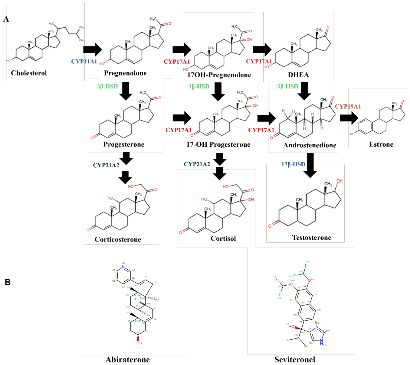
Figure 2.
Abiraterone and seviteronel differentially inhibit steroidogenic enzyme activities. (A-C) Representative thin-layer chromatography (TLC) assays showing dose-dependent inhibition of steroidogenic enzymes. (A) CYP17A1 hydroxylase, (B) CYP21A2, and (C)HSD3B2. Increasing inhibitor concentration reduces substrate-to-product conversion, as evidenced by a decrease in product band intensity. (D-H) Quantification of enzyme activities expressed as a percentage of control (DMSO). (D) CYP17A1 hydroxylase, (E) CYP17A1 lyase, (F) CYP21A2, (G) CYP19A1, and (H) HSD3B2. Data are presented as mean ± SEM from independent experiments. Statistical analysis was performed using one-way ANOVA with multiple comparisons. Significance is indicated as ****P < 0.0001, **P < 0.01, and ns (not significant).
Figure 2.
Abiraterone and seviteronel differentially inhibit steroidogenic enzyme activities. (A-C) Representative thin-layer chromatography (TLC) assays showing dose-dependent inhibition of steroidogenic enzymes. (A) CYP17A1 hydroxylase, (B) CYP21A2, and (C)HSD3B2. Increasing inhibitor concentration reduces substrate-to-product conversion, as evidenced by a decrease in product band intensity. (D-H) Quantification of enzyme activities expressed as a percentage of control (DMSO). (D) CYP17A1 hydroxylase, (E) CYP17A1 lyase, (F) CYP21A2, (G) CYP19A1, and (H) HSD3B2. Data are presented as mean ± SEM from independent experiments. Statistical analysis was performed using one-way ANOVA with multiple comparisons. Significance is indicated as ****P < 0.0001, **P < 0.01, and ns (not significant).
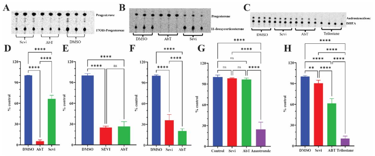
Figure 3.
Concentration-dependent inhibition of CYP17A1 and CYP21A2 by abiraterone and seviteronel. Concentration–response curves for inhibition of (A) CYP17A1 17α-hydroxylase, (B) CYP17A1 17,20-lyase and (C) CYP21A2 activities by abiraterone and seviteronel. Curves were fitted using four-parameter logistic regression. Abiraterone inhibited CYP17A1 17α-hydroxylase with an IC₅₀ of 0.05 µM compared with 8.6 µM for seviteronel. For CYP17A1 17,20-lyase, IC₅₀ values were 0.67 µM for abiraterone and 1.2 µM for seviteronel. CYP21A2 IC₅₀ values were 4.6 µM for abiraterone and 9.8 µM for seviteronel. Data are shown as mean ± SEM from independent experiments.
Figure 3.
Concentration-dependent inhibition of CYP17A1 and CYP21A2 by abiraterone and seviteronel. Concentration–response curves for inhibition of (A) CYP17A1 17α-hydroxylase, (B) CYP17A1 17,20-lyase and (C) CYP21A2 activities by abiraterone and seviteronel. Curves were fitted using four-parameter logistic regression. Abiraterone inhibited CYP17A1 17α-hydroxylase with an IC₅₀ of 0.05 µM compared with 8.6 µM for seviteronel. For CYP17A1 17,20-lyase, IC₅₀ values were 0.67 µM for abiraterone and 1.2 µM for seviteronel. CYP21A2 IC₅₀ values were 4.6 µM for abiraterone and 9.8 µM for seviteronel. Data are shown as mean ± SEM from independent experiments.
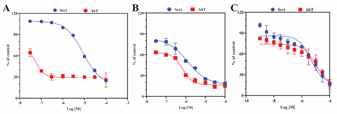
Figure 4.
Abiraterone and seviteronel generate distinct adrenal steroidogenic profiles. (A) Protein-normalized steroid concentrations in NCI-H295R cells following treatment with vehicle (DMSO), seviteronel or abiraterone. Shown are pregnenolone, progesterone, 17α-hydroxyprogesterone, DHEA and androstenedione. Abiraterone produced greater accumulation of upstream pregnane precursors, whereas both inhibitors reduced androgen output. (B) Steroid product-to-precursor ratios used as surrogate indices of CYP17A1 pathway flux. The 17α-hydroxylase index is calculated as 17α-hydroxyprogesterone/progesterone. The 17,20-lyase index should be calculated as DHEA/17α-hydroxypregnenolone if the revised analysis uses the measured direct Δ5 substrate; otherwise it should be explicitly described as a composite pathway-output ratio rather than a direct lyase index. Data are shown as mean ± SEM from independent experiments. Statistical comparisons were performed by one-way ANOVA with the multiple-comparison procedure specified in Methods. **P < 0.01; ****P < 0.0001; ns, not significant.
Figure 4.
Abiraterone and seviteronel generate distinct adrenal steroidogenic profiles. (A) Protein-normalized steroid concentrations in NCI-H295R cells following treatment with vehicle (DMSO), seviteronel or abiraterone. Shown are pregnenolone, progesterone, 17α-hydroxyprogesterone, DHEA and androstenedione. Abiraterone produced greater accumulation of upstream pregnane precursors, whereas both inhibitors reduced androgen output. (B) Steroid product-to-precursor ratios used as surrogate indices of CYP17A1 pathway flux. The 17α-hydroxylase index is calculated as 17α-hydroxyprogesterone/progesterone. The 17,20-lyase index should be calculated as DHEA/17α-hydroxypregnenolone if the revised analysis uses the measured direct Δ5 substrate; otherwise it should be explicitly described as a composite pathway-output ratio rather than a direct lyase index. Data are shown as mean ± SEM from independent experiments. Statistical comparisons were performed by one-way ANOVA with the multiple-comparison procedure specified in Methods. **P < 0.01; ****P < 0.0001; ns, not significant.
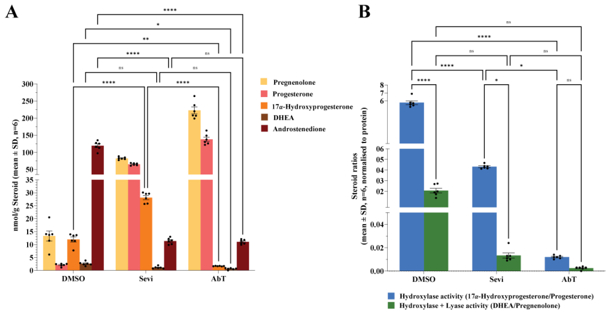
Figure 5.
Distinct CYP17A1 active-site interactions of abiraterone and seviteronel. Structural comparison of CYP17A1 bound to abiraterone and seviteronel. (A,B) Active-site views of CYP17A1 bound to (A) abiraterone (PDB 3RUK) and (B) seviteronel (PDB 5IRV). Key protein residues are shown as sticks with carbon atoms in white, heme carbon atoms in green and ligand carbon atoms in yellow; heteroatoms are coloured by element. Helix I is shown as a cartoon. Abiraterone coordinates the heme iron through pyridyl nitrogen N22 and forms the canonical 3β-hydroxyl–Asn202 interaction. Seviteronel coordinates the heme through triazole nitrogen N27 and adopts an alternative polar anchoring geometry involving its hydroxyl oxygen O05 and the Val482 backbone carbonyl, with additional close contacts around Thr306 and the helix-I region. (C,D) Two-dimensional ligand-interaction diagrams for (C) abiraterone and (D) seviteronel. Background shading denotes solvent exposure. Residue classes and interaction types are indicated by the colour key; π–π interactions and metal coordination are indicated by green and black lines, respectively. Contact maps represent geometrically identified interactions and do not indicate interaction energies.
Figure 5.
Distinct CYP17A1 active-site interactions of abiraterone and seviteronel. Structural comparison of CYP17A1 bound to abiraterone and seviteronel. (A,B) Active-site views of CYP17A1 bound to (A) abiraterone (PDB 3RUK) and (B) seviteronel (PDB 5IRV). Key protein residues are shown as sticks with carbon atoms in white, heme carbon atoms in green and ligand carbon atoms in yellow; heteroatoms are coloured by element. Helix I is shown as a cartoon. Abiraterone coordinates the heme iron through pyridyl nitrogen N22 and forms the canonical 3β-hydroxyl–Asn202 interaction. Seviteronel coordinates the heme through triazole nitrogen N27 and adopts an alternative polar anchoring geometry involving its hydroxyl oxygen O05 and the Val482 backbone carbonyl, with additional close contacts around Thr306 and the helix-I region. (C,D) Two-dimensional ligand-interaction diagrams for (C) abiraterone and (D) seviteronel. Background shading denotes solvent exposure. Residue classes and interaction types are indicated by the colour key; π–π interactions and metal coordination are indicated by green and black lines, respectively. Contact maps represent geometrically identified interactions and do not indicate interaction energies.
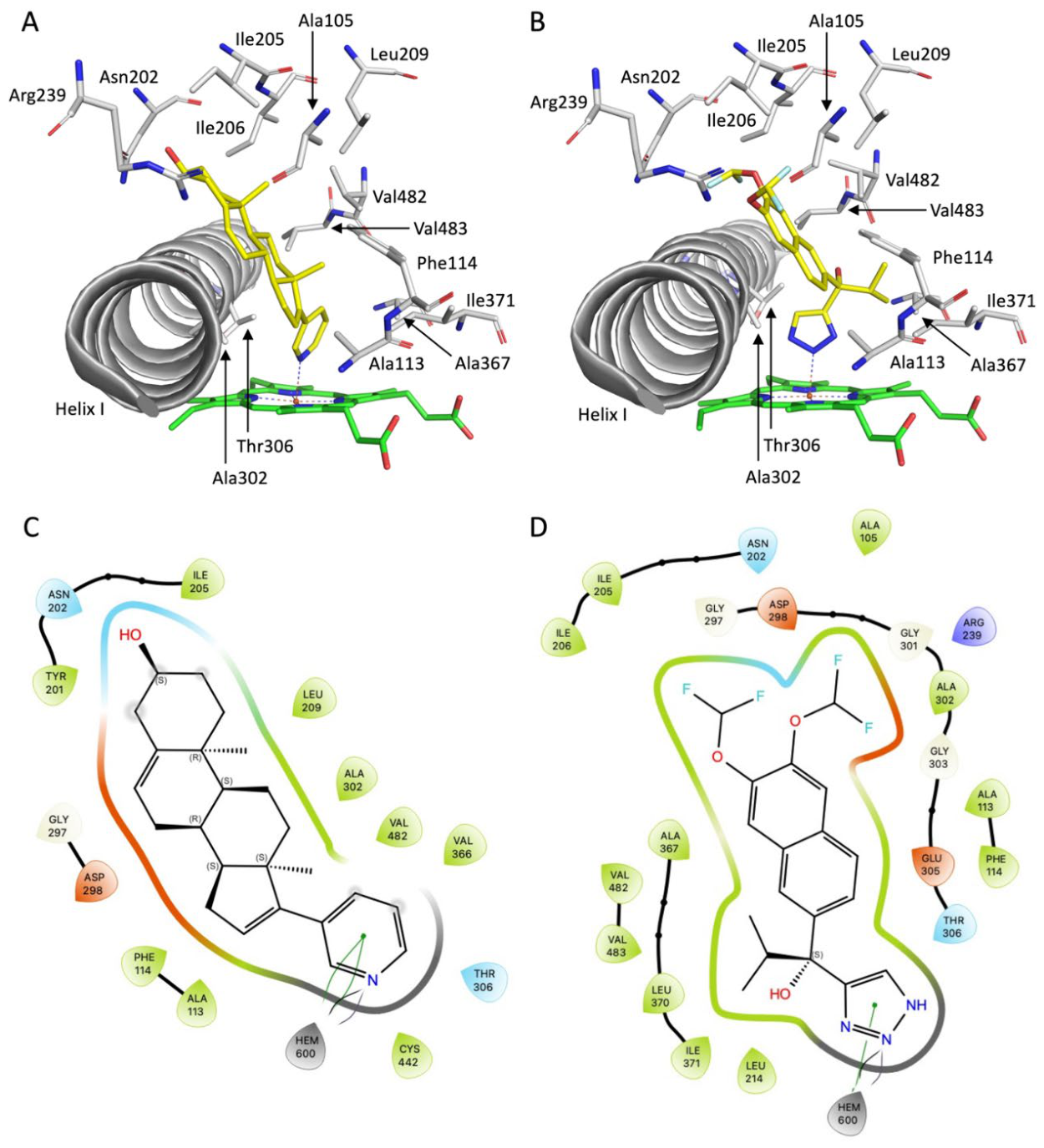
Figure 6.
Replicate molecular-dynamics simulations show broader RMSD distributions for seviteronel-bound CYP17A1. Violin plots showing distributions of protein backbone root-mean-square deviation (RMSD) values during four independent 100-ns molecular-dynamics trajectories for CYP17A1 complexes 3RUK_C and 3RUK_D (abiraterone), 5IRQ_C (orteronel) and 5IRV_C (seviteronel). Within each complex, the four violins correspond to independently initiated replicate trajectories 1–4. The broader distribution observed in some seviteronel-bound trajectories indicates greater conformational sampling but should not by itself be interpreted as protein instability.
Figure 6.
Replicate molecular-dynamics simulations show broader RMSD distributions for seviteronel-bound CYP17A1. Violin plots showing distributions of protein backbone root-mean-square deviation (RMSD) values during four independent 100-ns molecular-dynamics trajectories for CYP17A1 complexes 3RUK_C and 3RUK_D (abiraterone), 5IRQ_C (orteronel) and 5IRV_C (seviteronel). Within each complex, the four violins correspond to independently initiated replicate trajectories 1–4. The broader distribution observed in some seviteronel-bound trajectories indicates greater conformational sampling but should not by itself be interpreted as protein instability.
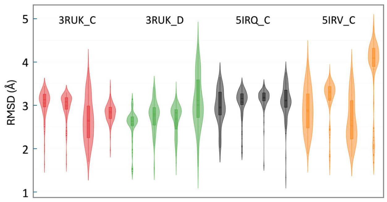
Figure 7.
Residue-level RMSF differences identify regions with ligand-associated conformational variation. Difference profiles calculated from mean residue-level root-mean-square fluctuation (RMSF) values for the steroidal abiraterone-bound complexes and the non-steroidal orteronel- and seviteronel-bound complexes. Dotted vertical lines identify regions A–F selected for subsequent structural comparison. Residue positions correspond to CYP17A1 residues Leu31–Ala502. The y-axis represents differences in RMSF in Å. The analysis highlights local regions showing differential conformational sampling between the compared ligand-bound systems rather than formal residue-by-residue statistical significance.
Figure 7.
Residue-level RMSF differences identify regions with ligand-associated conformational variation. Difference profiles calculated from mean residue-level root-mean-square fluctuation (RMSF) values for the steroidal abiraterone-bound complexes and the non-steroidal orteronel- and seviteronel-bound complexes. Dotted vertical lines identify regions A–F selected for subsequent structural comparison. Residue positions correspond to CYP17A1 residues Leu31–Ala502. The y-axis represents differences in RMSF in Å. The analysis highlights local regions showing differential conformational sampling between the compared ligand-bound systems rather than formal residue-by-residue statistical significance.
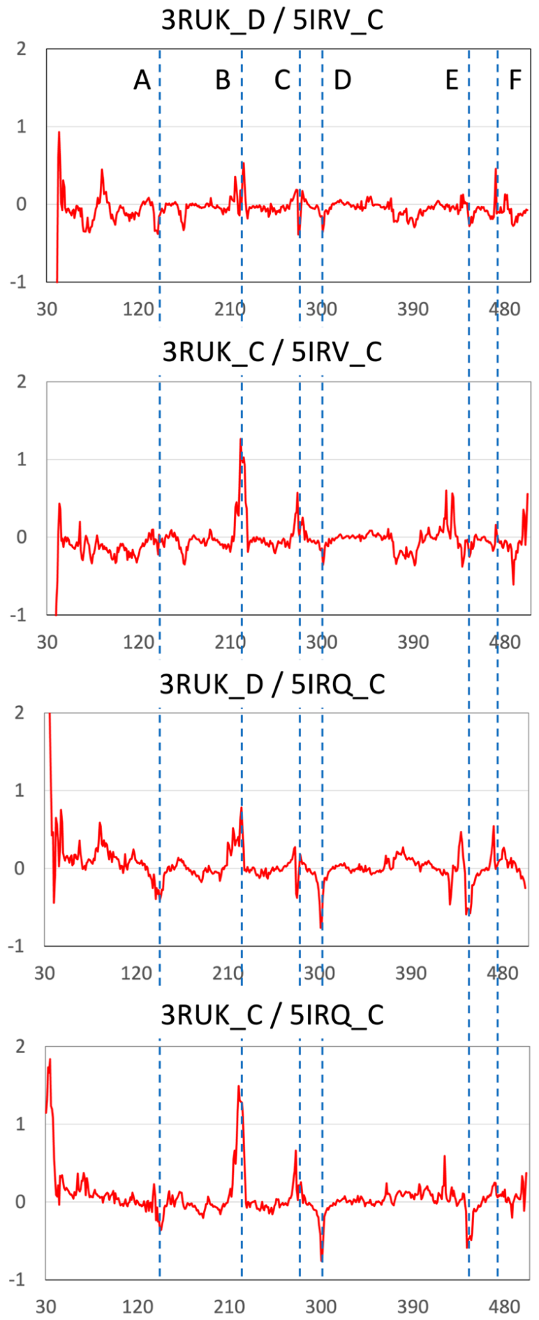
Figure 8.
Structural locations of CYP17A1 regions identified by RMSF-difference analysis. Superposition of the CYP17A1 complexes with Abiraterone (3RUK_C and 3RUK_D), Orteronel (5IRQ_C) and Seviteronel (5IRV_C). Proteins are shown as cartoons and colored wheat, palegreen, lightblue and palecyan, respectively, except for the regions A-F, which are colored orange. Heme and ligands are shown as sticks with C-atoms colored green and yellow, respectively, and hetero atoms are colored by element.
Figure 8.
Structural locations of CYP17A1 regions identified by RMSF-difference analysis. Superposition of the CYP17A1 complexes with Abiraterone (3RUK_C and 3RUK_D), Orteronel (5IRQ_C) and Seviteronel (5IRV_C). Proteins are shown as cartoons and colored wheat, palegreen, lightblue and palecyan, respectively, except for the regions A-F, which are colored orange. Heme and ligands are shown as sticks with C-atoms colored green and yellow, respectively, and hetero atoms are colored by element.
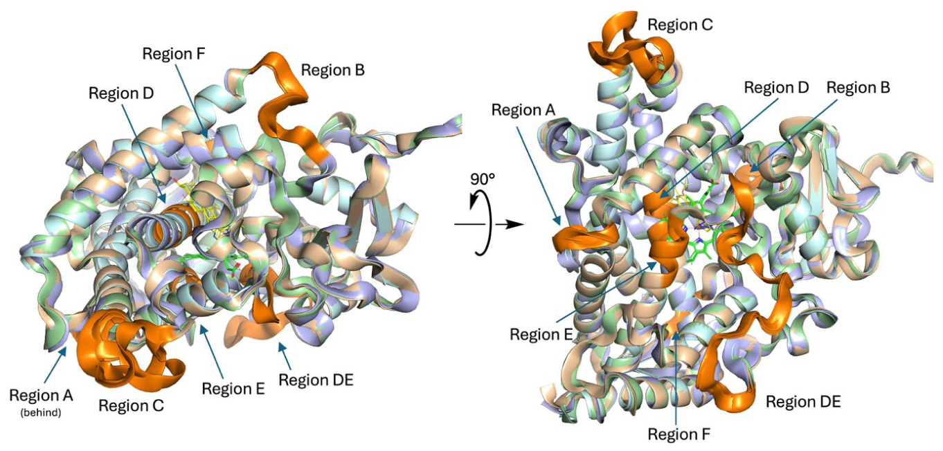
Figure 9.
Seviteronel is associated with displacement of the central helix-I region in the crystallographic comparison. Comparison of the Region D in the four CYP17A1 complexes. From left to right the location of Region D in the overall structure of CYP17A1 followed by a zoom in on Region D in I-Helix with Abiraterone and Seviteronel shown as stick models, and finally an illustration of the shift of residues Ile299-Gly303 from the Abiraterone (3RUK) to the Seviteronel (5IRV) structures. Proteins are shown as cartoons and colored wheat, pale green, light blue and pale cyan, respectively, except for the regions A-F, which are colored orange. Heme and ligands are shown as sticks with C-atoms colored green and yellow, respectively, and heteroatoms coloured by element.
Figure 9.
Seviteronel is associated with displacement of the central helix-I region in the crystallographic comparison. Comparison of the Region D in the four CYP17A1 complexes. From left to right the location of Region D in the overall structure of CYP17A1 followed by a zoom in on Region D in I-Helix with Abiraterone and Seviteronel shown as stick models, and finally an illustration of the shift of residues Ile299-Gly303 from the Abiraterone (3RUK) to the Seviteronel (5IRV) structures. Proteins are shown as cartoons and colored wheat, pale green, light blue and pale cyan, respectively, except for the regions A-F, which are colored orange. Heme and ligands are shown as sticks with C-atoms colored green and yellow, respectively, and heteroatoms coloured by element.
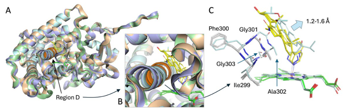
Figure 10.
Principal-component analysis reveals distinct helix-I conformational sampling among CYP17A1 complexes. Principal Component plot for the four systems 3RUK_C (blue), 3RUK_D (green), 5IRQ_C (yellow) and 5IRV_C (red) based on Phi and Psi values from 13 residues in I-Helix with a total of 400 frames for each system. The dotted circles correspond to conformations similar to the conformation in the X-ray structures. The numbers refer to number of frames with similar conformations. The x- and y-axes correspond to the first and second principal components, respectively.
Figure 10.
Principal-component analysis reveals distinct helix-I conformational sampling among CYP17A1 complexes. Principal Component plot for the four systems 3RUK_C (blue), 3RUK_D (green), 5IRQ_C (yellow) and 5IRV_C (red) based on Phi and Psi values from 13 residues in I-Helix with a total of 400 frames for each system. The dotted circles correspond to conformations similar to the conformation in the X-ray structures. The numbers refer to number of frames with similar conformations. The x- and y-axes correspond to the first and second principal components, respectively.
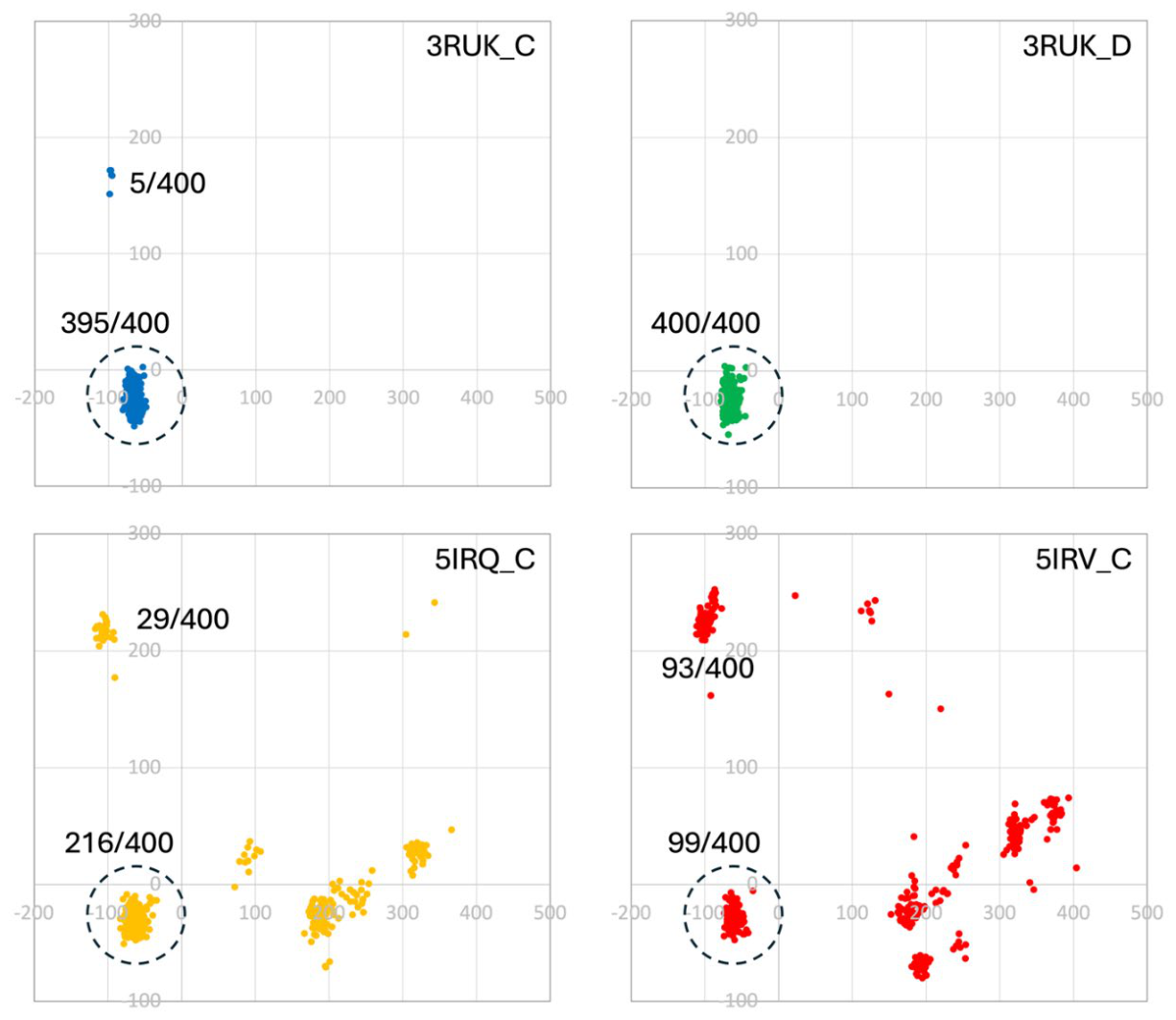
Figure 11.
Helix-I torsion-angle heatmaps reveal trajectory-dependent conformational heterogeneity. Heatmaps of backbone φ and ψ torsional angles for 13 central helix-I residues during four replicate molecular-dynamics trajectories of 3RUK_C and 3RUK_D (abiraterone), 5IRQ_C (orteronel) and 5IRV_C (seviteronel). Residue sequences are shown below the corresponding heatmaps. The plots permit direct comparison of torsional-angle states among individual trajectories and highlight heterogeneous sampling around the central FGAGV-containing region in several non-steroidal inhibitor-bound trajectories.
Figure 11.
Helix-I torsion-angle heatmaps reveal trajectory-dependent conformational heterogeneity. Heatmaps of backbone φ and ψ torsional angles for 13 central helix-I residues during four replicate molecular-dynamics trajectories of 3RUK_C and 3RUK_D (abiraterone), 5IRQ_C (orteronel) and 5IRV_C (seviteronel). Residue sequences are shown below the corresponding heatmaps. The plots permit direct comparison of torsional-angle states among individual trajectories and highlight heterogeneous sampling around the central FGAGV-containing region in several non-steroidal inhibitor-bound trajectories.
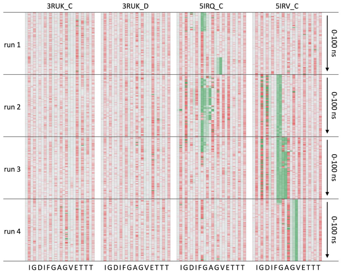
Figure 12.
Replicate seviteronel-bound simulations sample distinct helix-I conformations. (A) Representative helix-I conformations after 50 ns from four independently initiated 5IRV_C molecular-dynamics trajectories: run 1, white; run 2, green; run 3, blue; and run 4, yellow. The helix-I sequence is shown below the structural comparison; residues highlighted in red correspond to the central region included in the principal-component analysis in Figure 10. (B) Mean backbone φ and ψ torsional angles for the corresponding central helix-I residues in trajectories 1–4. Differences among the four trajectories illustrate replicate-dependent conformational sampling within the seviteronel-bound system.
Figure 12.
Replicate seviteronel-bound simulations sample distinct helix-I conformations. (A) Representative helix-I conformations after 50 ns from four independently initiated 5IRV_C molecular-dynamics trajectories: run 1, white; run 2, green; run 3, blue; and run 4, yellow. The helix-I sequence is shown below the structural comparison; residues highlighted in red correspond to the central region included in the principal-component analysis in Figure 10. (B) Mean backbone φ and ψ torsional angles for the corresponding central helix-I residues in trajectories 1–4. Differences among the four trajectories illustrate replicate-dependent conformational sampling within the seviteronel-bound system.
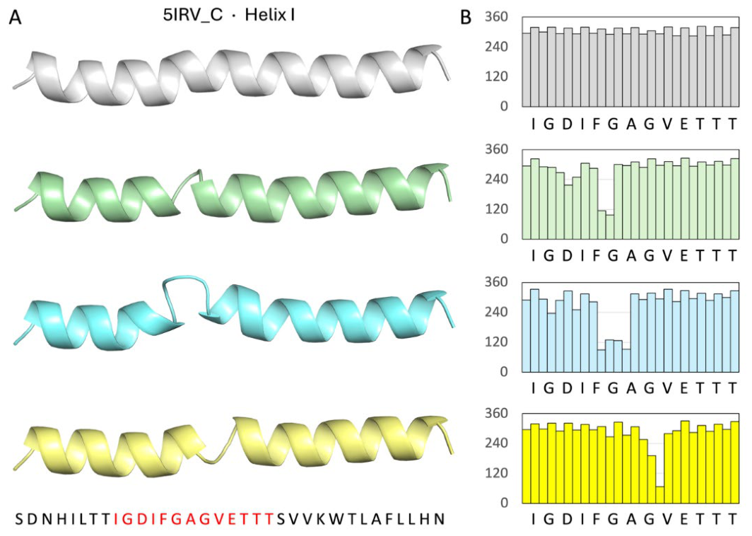
Figure 13.
Differential effects of abiraterone and seviteronel on prostate cancer cell viability, migration and cell-cycle profiles. (A) Cell Viability Heatmap: Percentage of viable cells following 24- and 48-hour treatment with abiraterone (Abiraterone), cisplatin, or seviteronel across androgen-sensitive (LNCaP, VCaP) AR-positive/castration-resistant (22Rv1), androgen-insensitive (PC-3, DU-145), and normal (RWPE-1) cell lines. The values are the mean ± the standard deviation. (B) Wound Healing Kinetics: Representative time-course of wound closure over 60 hours in DU-145 cells treated with abiraterone or seviteronel. Data points represent % wound area remaining (mean ± SEM). (C) Migration Quantification (T50): Bar graph showing the time required to reach 50% wound closure (T50). *p < 0.05, **p < 0.01 vs. control. (D) Cell Cycle Analysis: Distribution of cells in G1, G2, and S phases following treatment. Bars represent the percentage of the total population in each phase. seviteronel and abiraterone significantly reduced the S-phase population while inducing G1/G2 arrest. **p < 0.01, ***p < 0.001 vs. control. (E) Apoptosis Assay: Induction of Apoptotic markers measured by Annexin V fluorescence (RFU 488/525). Both agents significantly increased signaling compared to DMSO. ****p < 0.0001; ns = not significant.
Figure 13.
Differential effects of abiraterone and seviteronel on prostate cancer cell viability, migration and cell-cycle profiles. (A) Cell Viability Heatmap: Percentage of viable cells following 24- and 48-hour treatment with abiraterone (Abiraterone), cisplatin, or seviteronel across androgen-sensitive (LNCaP, VCaP) AR-positive/castration-resistant (22Rv1), androgen-insensitive (PC-3, DU-145), and normal (RWPE-1) cell lines. The values are the mean ± the standard deviation. (B) Wound Healing Kinetics: Representative time-course of wound closure over 60 hours in DU-145 cells treated with abiraterone or seviteronel. Data points represent % wound area remaining (mean ± SEM). (C) Migration Quantification (T50): Bar graph showing the time required to reach 50% wound closure (T50). *p < 0.05, **p < 0.01 vs. control. (D) Cell Cycle Analysis: Distribution of cells in G1, G2, and S phases following treatment. Bars represent the percentage of the total population in each phase. seviteronel and abiraterone significantly reduced the S-phase population while inducing G1/G2 arrest. **p < 0.01, ***p < 0.001 vs. control. (E) Apoptosis Assay: Induction of Apoptotic markers measured by Annexin V fluorescence (RFU 488/525). Both agents significantly increased signaling compared to DMSO. ****p < 0.0001; ns = not significant.
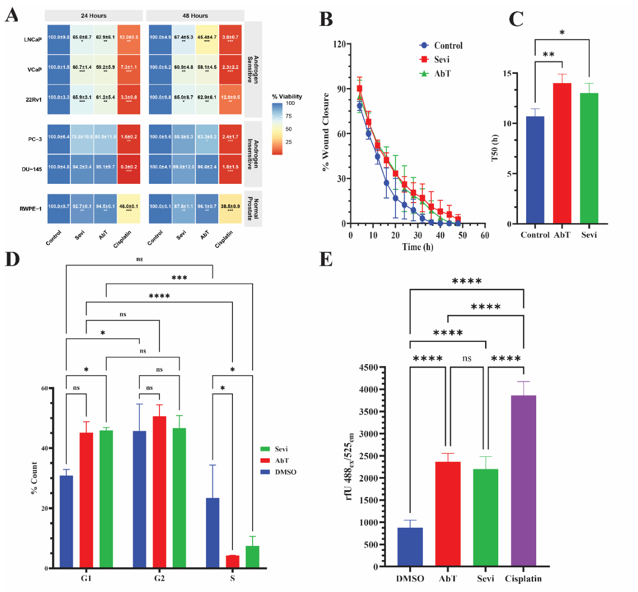
Figure 14.
Abiraterone and seviteronel increase sensitivity to ferroptosis-inducing lipid-peroxidation stress.(A, B) Cell viability of prostate cancer cells following 48 h treatment with indicated compounds measured by CCK-8 and expressed as a percentage relative to DMSO control (100%). Results show the effect of seviteronel and abiraterone alone or in combination with the system XC-inhibitor erastin (10 µM), and subsequent rescue or potentiation by ferrostatin-1 (Fer-1, 2 µM), liproxstatin-1 (liprox, 1 µM), or Fin56 (5 µM). Results in b demonstrate trends comparable to those with an RSL3 Analog (1 µM) targeting GPX4 directly. Cisplatin (10 µM) and abiraterone (10 µM + seviteronel 10 µM) combination groups are included for therapeutic comparison. Data represent mean ± s.d. from n = 3 independent biological replicates. Statistical significance was determined by one-way ANOVA followed by Tukey’s post-hoc test; *P < 0.05, **P < 0.01, ****P < 0.0001.
Figure 14.
Abiraterone and seviteronel increase sensitivity to ferroptosis-inducing lipid-peroxidation stress.(A, B) Cell viability of prostate cancer cells following 48 h treatment with indicated compounds measured by CCK-8 and expressed as a percentage relative to DMSO control (100%). Results show the effect of seviteronel and abiraterone alone or in combination with the system XC-inhibitor erastin (10 µM), and subsequent rescue or potentiation by ferrostatin-1 (Fer-1, 2 µM), liproxstatin-1 (liprox, 1 µM), or Fin56 (5 µM). Results in b demonstrate trends comparable to those with an RSL3 Analog (1 µM) targeting GPX4 directly. Cisplatin (10 µM) and abiraterone (10 µM + seviteronel 10 µM) combination groups are included for therapeutic comparison. Data represent mean ± s.d. from n = 3 independent biological replicates. Statistical significance was determined by one-way ANOVA followed by Tukey’s post-hoc test; *P < 0.05, **P < 0.01, ****P < 0.0001.
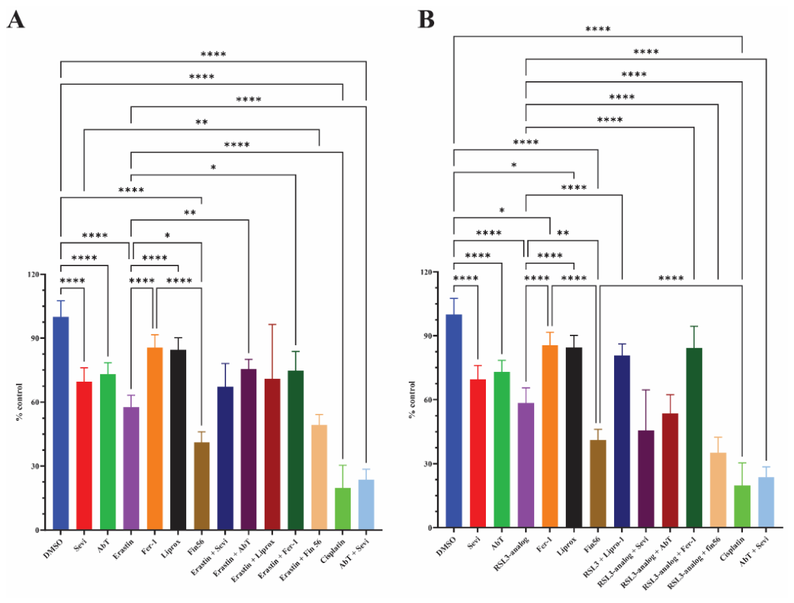
Figure 15.
Abiraterone and seviteronel do not detectably expand the intracellular labile Fe²⁺ pool. Representative confocal microscopy images of LNCaP prostate cancer cells stained with FerroOrange (red; Fe2+) and Hoechst 33342 (blue; nuclei) following treatment with abiraterone or seviteronel. Untreated cells (Control) and cells treated with FerroOrange alone serve as baseline indicators of the labile iron pool. For validation of probe specificity using exogenous iron loading and chelation. Cells were treated with 100 µmol/L ammonium iron (II) sulfate to induce iron overload or co-treated with the iron chelator 2,2-bipyridyl (50 µmol/L) to demonstrate signal quenching. Merged images are provided to show cell morphology. Zoom panel, Representative line-scan analysis (Zoom) depicting the relative fluorescence intensity of FerroOrange (red) and Hoechst (blue) across a single-cell diameter, illustrating the cytosolic distribution of the labile iron pool. Scale bars, 20 µM. Images are representative of n = 3 independent experiments.
Figure 15.
Abiraterone and seviteronel do not detectably expand the intracellular labile Fe²⁺ pool. Representative confocal microscopy images of LNCaP prostate cancer cells stained with FerroOrange (red; Fe2+) and Hoechst 33342 (blue; nuclei) following treatment with abiraterone or seviteronel. Untreated cells (Control) and cells treated with FerroOrange alone serve as baseline indicators of the labile iron pool. For validation of probe specificity using exogenous iron loading and chelation. Cells were treated with 100 µmol/L ammonium iron (II) sulfate to induce iron overload or co-treated with the iron chelator 2,2-bipyridyl (50 µmol/L) to demonstrate signal quenching. Merged images are provided to show cell morphology. Zoom panel, Representative line-scan analysis (Zoom) depicting the relative fluorescence intensity of FerroOrange (red) and Hoechst (blue) across a single-cell diameter, illustrating the cytosolic distribution of the labile iron pool. Scale bars, 20 µM. Images are representative of n = 3 independent experiments.
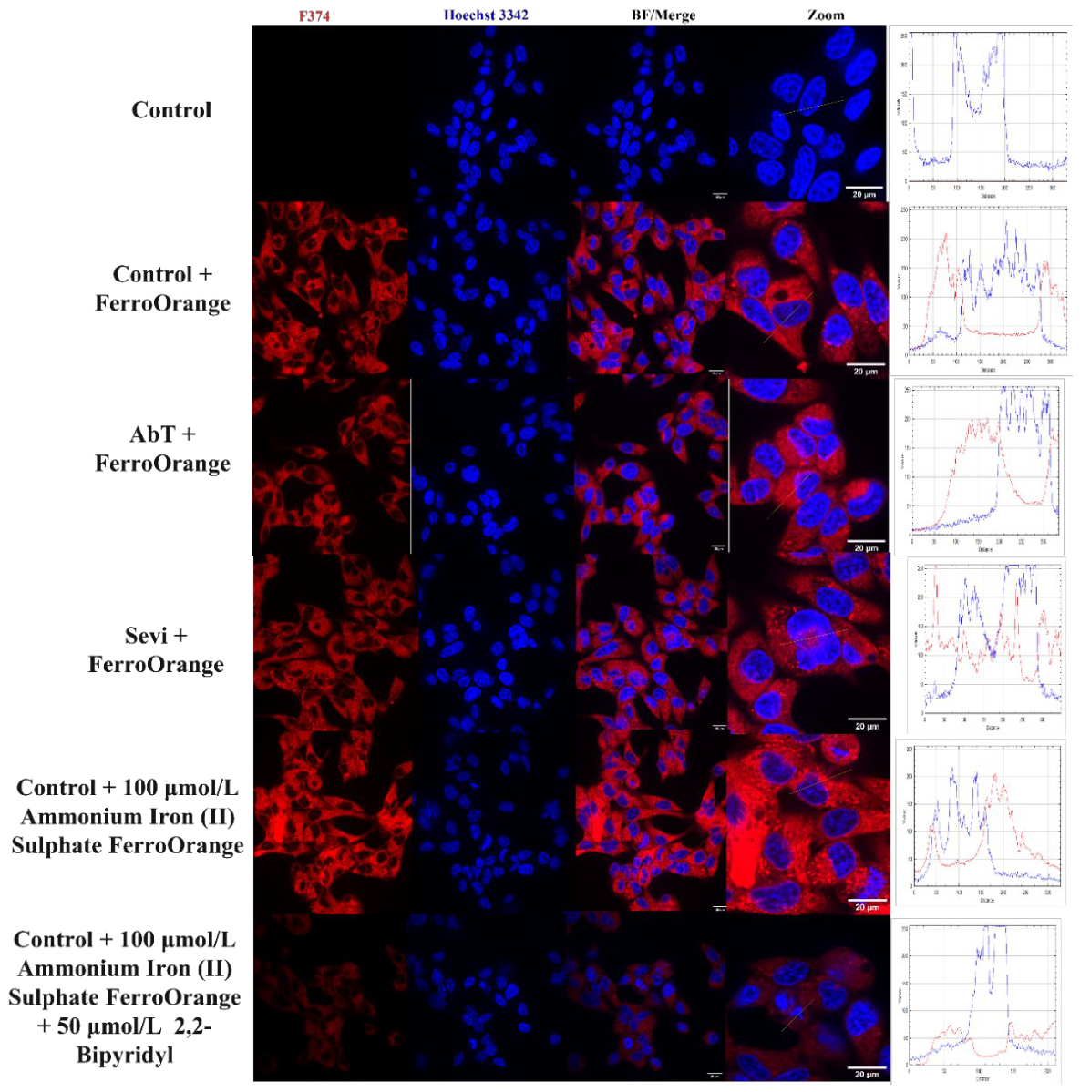
Figure 16.
Transcriptional responses to seviteronel and abiraterone. Seviteronel and abiraterone (Abiraterone) induce divergent transcriptional signatures in steroidogenic and ferroptotic pathways. (A, B) Normalized mRNA expression of (A) steroidogenic enzymes and cofactors and (B) ferroptosis-related markers following treatment with seviteronel. Note the significant attenuation of the essential cofactors POR and CYB5A (Panel A) alongside a robust, ~15-fold compensatory induction of AKR1C3 and the ferroptotic marker CHAC1 (Panel B). (C, D) Normalized mRNA expression of (C) steroidogenic enzymes and (D) ferroptosis markers following treatment with abiraterone. In contrast to seviteronel, abiraterone maintains a relatively stable transcriptional profile across the primary steroidogenic machinery (Panel C; ns = non-significant), while inducing a more moderate stress-response signature (Panel D). Data for steroidogenic genes (A, C) are presented as mean ± SD from two (n = 2) independent experiments. Data for ferroptotic genes (B, D) are presented as mean ± SEM from two (n = 2) technical replicates. Statistical significance was determined using a one-sample t-test against the normalized control value (dashed line at 1.0). Significance is indicated by asterisks (*p < 0.05, **p < 0.01); non-significant changes are denoted as ‘ns’.
Figure 16.
Transcriptional responses to seviteronel and abiraterone. Seviteronel and abiraterone (Abiraterone) induce divergent transcriptional signatures in steroidogenic and ferroptotic pathways. (A, B) Normalized mRNA expression of (A) steroidogenic enzymes and cofactors and (B) ferroptosis-related markers following treatment with seviteronel. Note the significant attenuation of the essential cofactors POR and CYB5A (Panel A) alongside a robust, ~15-fold compensatory induction of AKR1C3 and the ferroptotic marker CHAC1 (Panel B). (C, D) Normalized mRNA expression of (C) steroidogenic enzymes and (D) ferroptosis markers following treatment with abiraterone. In contrast to seviteronel, abiraterone maintains a relatively stable transcriptional profile across the primary steroidogenic machinery (Panel C; ns = non-significant), while inducing a more moderate stress-response signature (Panel D). Data for steroidogenic genes (A, C) are presented as mean ± SD from two (n = 2) independent experiments. Data for ferroptotic genes (B, D) are presented as mean ± SEM from two (n = 2) technical replicates. Statistical significance was determined using a one-sample t-test against the normalized control value (dashed line at 1.0). Significance is indicated by asterisks (*p < 0.05, **p < 0.01); non-significant changes are denoted as ‘ns’.
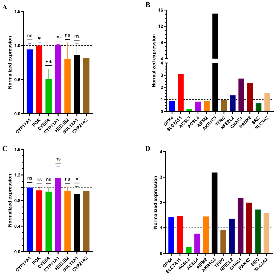
Figure 17.
ACTH-mediated signaling pathway architecture and transcriptional suppression by seviteronel and abiraterone. A) Schematic model of the canonical adrenocorticotropic hormone (ACTH) signaling cascade. The binding of ACTH to the melanocortin 2 receptor (MC2R) induces dissociation of the heterotrimeric G-protein complex, leading to Gα-mediated activation of adenylyl cyclase and generation of cyclic AMP (cAMP) from ATP. Downstream activation of protein kinase A (PKA) triggers the phosphorylation of steroidogenic acute regulatory protein (StAR) and hormone-sensitive lipase (HSL) to drive steroidogenesis, alongside concurrent phosphorylation of cAMP response element-binding protein (CREB) to stimulate gene transcription. B), Relative luciferase reporter activity in cells subjected to ACTH stimulation, DMSO vehicle control, seviteronel treatment, or abiraterone treatment (n = 9), independent biological replicates per group. Bars represent mean values ± s.d.; individual data points are plotted as dots. Statistical significance was determined using a two-tailed Student’s t-test (or one-way ANOVA with post hoc multiple comparisons); **P < 0.01, ****P < 0.0001, ns, not significant P > 0.05. RLU, relative luciferase units.
Figure 17.
ACTH-mediated signaling pathway architecture and transcriptional suppression by seviteronel and abiraterone. A) Schematic model of the canonical adrenocorticotropic hormone (ACTH) signaling cascade. The binding of ACTH to the melanocortin 2 receptor (MC2R) induces dissociation of the heterotrimeric G-protein complex, leading to Gα-mediated activation of adenylyl cyclase and generation of cyclic AMP (cAMP) from ATP. Downstream activation of protein kinase A (PKA) triggers the phosphorylation of steroidogenic acute regulatory protein (StAR) and hormone-sensitive lipase (HSL) to drive steroidogenesis, alongside concurrent phosphorylation of cAMP response element-binding protein (CREB) to stimulate gene transcription. B), Relative luciferase reporter activity in cells subjected to ACTH stimulation, DMSO vehicle control, seviteronel treatment, or abiraterone treatment (n = 9), independent biological replicates per group. Bars represent mean values ± s.d.; individual data points are plotted as dots. Statistical significance was determined using a two-tailed Student’s t-test (or one-way ANOVA with post hoc multiple comparisons); **P < 0.01, ****P < 0.0001, ns, not significant P > 0.05. RLU, relative luciferase units.
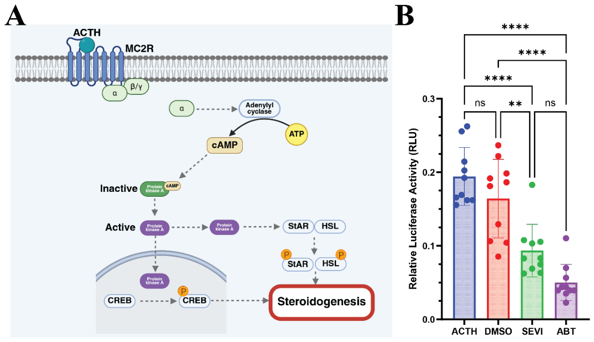

Table 1.
IC₅₀ values for abiraterone and seviteronel across key steroidogenic enzymes (CYP17A1 Hydroxylase and Lyase, CYP21A2). Selectivity index = IC50 hydroxylase / IC50 lyase; values >1 indicate lyase-biased inhibition relative to hydroxylase inhibition. All IC₅₀.values in µM.
Table 1.
IC₅₀ values for abiraterone and seviteronel across key steroidogenic enzymes (CYP17A1 Hydroxylase and Lyase, CYP21A2). Selectivity index = IC50 hydroxylase / IC50 lyase; values >1 indicate lyase-biased inhibition relative to hydroxylase inhibition. All IC₅₀.values in µM.
| Drugs | CYP17A1-Hydroxylase | CYP17A1-Lyase | CYP21A2 | Selectivity Index (SI) |
| Abiraterone | 0.05 ± 0.006 | 0.67 ± 0.098 | 4.6 ± 1.51 | 0.07 |
| Seviteronel | 8.6 ± 1.32 | 1.2 ± 0.14 | 9.8 ± 2.81 | 7.17 |
Table 2.
Impact of seviteronel and abiraterone on key steroid metabolites in NCI-H295R cells. All values are Mean nmol/L, normalized to protein levels.
Table 2.
Impact of seviteronel and abiraterone on key steroid metabolites in NCI-H295R cells. All values are Mean nmol/L, normalized to protein levels.
| Pathway | Steroid | DMSO control | Abiraterone | Seviteronel |
| CYP17A1 Substrates | Pregnenolone | 13.3 | 222.6 (↑↑↑) | 82.3 (↑↑) |
| Progesterone | 2.1 | 138.0 (↑↑↑) | 65.1 (↑↑) | |
| Androgens | Androstenedione | 119.2 | 11.0 (↓↓↓) | 11.4 (↓↓↓) |
| Glucocorticoid Pathway | 17α-Hydroxyprogesterone | 12.0 | 1.7 (↓↓) | 28.1 (↑↑) |
| 11-Deoxycortisol | 386.6 | 32.3 (↓↓↓) | 69.2 (↓↓↓) | |
| Mineralocorticoid Pathway | 11-Deoxycorticosterone (DOC) | 151.7 | 5.6 (↓↓↓) | 271.4 (↑) |
| Final Products | Cortisol | 16.5 | 0.002 (↓↓↓) | 2.8 (↓↓) |
Table 3.
Pearson correlation analysis of key steroidogenic metabolome.
| Group | Interaction | vs | Pearson’s r | p-value | Interpretation |
| Seviteronel | 11-Deoxycorticosterone | Androstenedione | -0.752 | 0.012* | Selective Shunt |
| Seviteronel | 17α-Hydroxyprogesterone | Androstenedione | -0.610 | 0.045* | Lyase Blockade |
| Abiraterone | Pregnenolone | Progesterone | 0.969 | < 0.001*** | Metabolic Blockade |
| Abiraterone | Progesterone | 17α-Hydroxyprogesterone | 0.112 | 0.724 | Hydroxylase Blockade |
Disclaimer/Publisher’s Note: The statements, opinions and data contained in all publications are solely those of the individual author(s) and contributor(s) and not of MDPI and/or the editor(s). MDPI and/or the editor(s) disclaim responsibility for any injury to people or property resulting from any ideas, methods, instructions or products referred to in the content. |
© 2026 by the authors. Licensee MDPI, Basel, Switzerland. This article is an open access article distributed under the terms and conditions of the Creative Commons Attribution (CC BY) license (http://creativecommons.org/licenses/by/4.0/).
Copyright: This open access article is published under a Creative Commons CC BY 4.0 license, which permit the free download, distribution, and reuse, provided that the author and preprint are cited in any reuse.