

Submitted:
20 August 2026
Posted:
21 August 2026
You are already at the latest version
Abstract
Background: Pharmacological neuroprotection and adjuvant physical therapies, including piracetam and hyperbaric oxygen therapy, have emerged as potential strategies to enhance nerve regeneration. We aimed to assess the effects of piracetam and hyperbaric oxygen on peripheral nerve regeneration. Methods: Forty male Wistar Albino rats were randomly allocated to four groups following experimental sciatic nerve injury: standard care (Group K), hyperbaric oxygen therapy (Group HBO), intraperitoneal piracetam (Group P), and combined hyperbaric oxygen plus piracetam (Group C). After 14 days, the rats were sacrificed, and sciatic nerve tissues and blood samples were collected for histopathological and biochemical analyses. Results: No statistically significant differences were observed among the four groups with respect to axonal continuity, capillary formation, or myelin sheath regeneration. Fibroblast density was increased in Group P compared with Group K (p = 0.025), while no significant differences were detected among the remaining groups. Inflammatory cell counts were higher in Group P than in Group K (p = 0.037) and elevated in Group C compared with Group K (p = 0.014). Hypoxia-inducible factor-1 alpha (HIF-1α) expression was highest in Group K. Additionally, neuron-specific enolase levels were significantly increased in Group P compared with Group K (p = 0.006). Conclusion: In this experimental model, neither piracetam nor hyperbaric oxygen therapy resulted in significant improvements in structural nerve regeneration parameters. However, distinct alterations in inflammatory and biochemical markers were observed, indicating that these interventions may modulate cellular and metabolic responses following sciatic nerve injury rather than directly enhancing axonal repair.
Keywords:
piracetam
; hyperbaric oxygen
; nerve injury
; animal model
; regeneration
Introduction
Peripheral nerve injuries remain a significant clinical challenge due to their high incidence and the often-incomplete functional recovery that follows despite advances in microsurgical repair techniques [1]. Sciatic nerve injury, one of the most commonly studied experimental models of peripheral nerve damage, closely mimics human traumatic neuropathies and provides a reliable platform for evaluating therapeutic interventions aimed at enhancing nerve regeneration and functional recovery [2,3]. Although spontaneous regeneration can occur in peripheral nerves, the process is slow and frequently insufficient, resulting in persistent motor and sensory deficits [4]. Consequently, there is an ongoing need to identify adjuvant therapies that can accelerate nerve repair and improve functional outcomes [5].
The pathophysiology of peripheral nerve injury involves a complex cascade of events, including axonal degeneration, Schwann cell dysfunction, inflammatory responses, oxidative stress, and impaired microcirculation at the injury site [6,7]. These mechanisms collectively hinder axonal regrowth and remyelination [7]. Pharmacological agents with neuroprotective and neurotrophic properties, as well as physical modalities that enhance tissue oxygenation and cellular metabolism, have therefore attracted increasing interest as potential strategies to support nerve regeneration [8].
Piracetam, a cyclic derivative of gamma-aminobutyric acid (GABA), is widely recognized for its neuroprotective, nootropic, and rheological effects [9]. Experimental studies have demonstrated that piracetam improves neuronal metabolism, enhances microcirculatory blood flow, reduces oxidative stress, and stabilizes neuronal membranes [10,11]. In the context of nerve injury, these properties suggest a potential role for piracetam in promoting axonal regeneration and preserving neural integrity, yet evidence regarding its efficacy in peripheral nerve repair remains limited and warrants further investigation [12].
Hyperbaric oxygen (HBO) therapy is another therapeutic modality that has gained attention for its beneficial effects in neural tissue repair [13]. By increasing the partial pressure of oxygen in plasma, HBO enhances oxygen delivery to hypoxic tissues, reduces edema, modulates inflammatory responses, and stimulates angiogenesis [14]. Several experimental and clinical studies have reported that HBO therapy can improve nerve regeneration and functional recovery following peripheral nerve injury [15,16]; however, the optimal application protocols and its potential synergistic effects with pharmacological agents have not been fully elucidated.
Given the distinct yet potentially complementary mechanisms of piracetam and hyperbaric oxygen therapy, their combined or comparative effects on peripheral nerve regeneration merit systematic evaluation. Therefore, the present study aimed to investigate the effects of piracetam and hyperbaric oxygen application on the reversal of nerve damage in a rat model of induced sciatic nerve injury. By assessing functional, histopathological, and biochemical outcomes, this study seeks to contribute to the growing body of evidence on adjuvant therapies that may enhance peripheral nerve repair and inform future translational approaches.
Materials and Methods
Study Plan
This study used 40 male Wistar-Albino rats (Düzce University, Düzce Faculty of Medicine, Experimental Animal Application and Research Center). The study was conducted in accordance with the Declaration of Helsinki, and the protocol was approved by Düzce University Düzce Faculty of Medicine Experimental Animal Local Ethics Committee (2020/132) on 2020/10/1. Laboratory animal care principles were followed in this study. The average age of the rats included in the study was 2.5 months (2-3 months) and their average weight was 250 grams (200-300 grams). The animals were randomly divided into 4 groups, with 10 rats in each cage, and monitored in the laboratory environment for 1 week preoperatively. During the study, the rats were given unlimited tap water (ad libitum) and standard rodent feed. The animals were monitored in cages in a room with controlled temperature (23-25°C), a 12/12 hour light/dark cycle, and 50-60% humidity. The animals used in the study were monitored in their cages for approximately one week without any procedures after being brought to the laboratory center. On the day of the study, the rats were brought to the research laboratory and weighed. Anesthesia was achieved by administering intramuscular ketamine hydrochloride 90 mg/kg (Ketalar®, Eczacıbaşı İlaç Sanayi ve Ticaret A.Ş., Istanbul) and intramuscular 6 mg/kg xylazine (Rompun®, Bayer Türk Kimya Sanayi, Istanbul) to the rats.
Sciatic Nerve Injury Model
Anesthesia was administered by intramuscular injection of 90 mg/kg ketamine hydrochloride (Alfasan, Netherlands) and 6 mg/kg xylazine hydrochloride (Alfasan, Netherlands). Booster injections were planned as needed. The animal's right hip was shaved and washed with an antiseptic solution (povidone iodine). An oblique gluteal incision was made in the right lower extremity, following the hip joint crease, to access the biceps femoris muscle. The biceps femoris muscle was opened by blunt dissection along the posterior border of the femur and knee joint, exposing the sciatic nerve. The nerve was freed from surrounding tissues from the sciatic notch to the branching region in the popliteal area. It was planned to subject the approximately 2 cm long nerve to blunt trauma (three times for at least 30 seconds with a bulldog clip) at its midpoint (Figure 1).
Study Groups
Prophylactic antibiotics were administered to all groups after surgery. No rats died in any group after the operation. No wound infection was observed in any of the rats during follow-up. The control group (n=10) was named Group K, the hyperbaric oxygen group (n=10) was named Group HBO, the piracetam group (n=10) was named Group P, and the group given both HBO and piracetam (n=10) was named Group C. Group K (Control group): After sciatic nerve damage was induced, no treatment was given; standard care conditions were applied. At the end of the 14th day, euthanasia was performed by cervical dislocation under anesthesia/tranquilizer, and macroscopic and histological evaluation was performed. Group HBO (HBO treated group): HBO treatment was given for 1 hour a day for 14 days under a pressure of 2.5 ATA. At the end of the 14th day, euthanasia was performed by cervical dislocation under anesthesia/tranquilizers, and macroscopic and histological evaluations were carried out. Group P (Piracetam-treated group): 250 mg/kg piracetam was administered intraperitoneally in two equal doses twice daily. At the end of the 14th day, euthanasia was performed by cervical dislocation under anesthesia/tranquilizers, and macroscopic and histological evaluations were carried out. Group C (Piracetam and HBO-treated group): 250 mg/kg piracetam was administered intraperitoneally in two equal doses twice daily. HBO treatment was given for 1 hour a day at 2.5 ATA pressure for 14 days. At the end of the 14th day, euthanasia was performed by cervical dislocation under anesthesia/tranquilizers, and macroscopic and histological evaluations were carried out. Blood samples were taken from the hearts of all groups before sacrifice. Blood samples were centrifuged to separate the plasma. The resulting samples were stored at -80°C. Biochemical parameters such as NSE, S100B, and HIF1 markers were analyzed.
Hyperbaric Oxygen Therapy
Treatment was started on the same postoperative day. Ten rats each in Group HBO and Group C, which had sciatic nerve damage, received a total of 14 sessions of HBO treatment, once a day. The treatment was performed in a hyperbaric oxygen chamber, specially designed for experimental animals, with dimensions of 100 × 55 cm2, capable of accommodating 20 rats simultaneously in their cages. The treatment was terminated after 14 sessions. Treatments were applied at a pressure of 2.5 ATA and in sessions lasting a total of 60 minutes. Before treatment, the chamber was flushed with 100% oxygen for 5 minutes, then the pressure inside the chamber was gradually increased until the desired treatment depth was reached in 5 minutes. Treatment was applied for 60 minutes at a pressure of 2.5 ATA, decompression was achieved in 10 minutes, and the treatment was terminated in a total of 80 minutes.
Piracetam Treatment
Ten rats in Group P and Group C were administered Nootropil (containing the active ingredient piracetam, Nootropil) in two equal doses of 250 mg/kg/day at the same time via standard intraperitoneal injection. No complications were observed.
Clinical Follow-up and Evaluation
Clinically, right foot drop was observed in all rats in Group K, Group HBO, Group P, and Group C in which sciatic nerve damage was induced. Treatment with HBO and piracetam was administered for 14 days under standard care conditions. All rats in Groups K, HBO, P, and C were sacrificed at the end of the 14th day, and their sciatic nerves were removed for histopathological examination.
Histopathological Examination
Histopathological examination was performed in the laboratory of the Department of Pathology, Düzce University Faculty of Medicine. At the end of the study, all rats in all groups were sacrificed, and their sciatic nerves were removed. The sciatic nerve was excised to the branching region, with an additional 0.5 cm removed distally and proximally. The removed sciatic nerve tissues were fixed in 10% formaldehyde solution for 24 hours for histopathological examination. The formaldehyde-fixed samples were embedded in paraffin after routine follow-up procedures. 5µm sections were obtained from the samples and stained with H&E, as well as histochemically with toluidine blue and Masson trichrome, and immunohistochemically with neurofilament and CD34. Subsequently, the preparations were evaluated by a pathologist according to the semi-quantitative scoring system described by Klopfleisch et al (17) (Figure 2).
Biochemical Analysis
All measurements were performed in the Medical Biochemistry Department Laboratory of Düzce University Faculty of Medicine by the same Biochemistry specialist. Blood samples taken from rats were centrifuged at 3000 rpm for 10 minutes. Serum samples were collected using a micropipette and stored in ependroff tubes at -80°C. HIF1alpha, S100B, and NSE levels were analyzed from the samples. The numbers of the commercial kits (USCN, Wuhan) were recorded.
HIF1ALFA: L210213034 LOT
S100B: L20405260 LOT
NSE: L210407794 LOT
Rat calorimetric ELISA kits were used according to the steps specified in the kit protocol. All stages were followed and completed. After preparing the standard solution, the samples were appropriately placed for PCR and incubated at 37°C for 1 hour. Next, Detection reagent A solution was added and incubated at 37°C for 1 hour. After 1 hour, the samples were washed using the washing solution. After washing, Detection reagent B solution was added and incubated at 37°C for 30 minutes. After 30 minutes, the samples were washed 5 times with the washing solution. After washing, the substrate solution was added and incubated at 37°C for 20 minutes. After incubation, the samples, to which the stop solution was added, were placed in the instrument for analysis. A Biotek-Epoch brand PCR instrument was used for the measurement. The samples were analyzed at a wavelength of 450 nm. Calculations were performed using the Genera 5.2.1 program with the curve export program. The results were recorded in nanomol/milliliter.
Statistical Analysis
The distribution of data was examined using the Shapiro-Wilk test. For group comparisons, One-Way ANOVA and post hoc Tukey HSD tests were used for variables showing normal distribution, and the Kruskal-Wallis test was used for variables not showing normal distribution. For multiple comparisons, the post hoc Dunn test was used to compare the mean rank scores of the groups. Descriptive statistics were summarized as mean ± standard deviation or median, interquartile range, and minimum-maximum values, depending on the distribution of the data. Statistical analyses were performed using the SPSS v.22 software package, and the statistical significance level was considered to be 0.05.
Results
Body Weight Comparison
All rats were weighed before surgery and at regular intervals thereafter. There was no statistically significant difference in body weight among the groups throughout the study period (p = 0.956), indicating that the groups were comparable with respect to baseline and follow-up weights (mean weight: group K: 250.5±14.8gr, group HBO: 247.7±14.5gr, group P: 247.9±11.8gr, group C: 247.4±14.4gr, p-value = 0.956).
Histopathological Score Distribution
Histopathological evaluation was performed using five parameters: axonal continuity, fibroblast count, myelin sheath thickness, inflammatory cell count, and capillary density. The distribution of scores across the groups is summarized in Table 1. In the control group (Group K), axonal continuity scores were predominantly 2 (70%) and 3 (30%). Similar distributions were observed for fibroblast count, whereas myelin sheath scores ranged from 1 to 3. Inflammatory cell scores were mostly high, with 60% of rats scoring 3. Capillary density showed a wide distribution, with 50% of rats scoring 3. In the HBO-treated group (Group HBO), most rats received a score of 2 for axonal continuity (80%) and fibroblast count (90%). Myelin sheath scores were predominantly 2 (90%). Inflammatory cell scores were more evenly distributed across scores 1–3. Capillary density scores were mainly low, with 70% of rats scoring 1. In the piracetam-treated group (Group P), axonal continuity was mostly scored as 2 (90%). Fibroblast count was predominantly score 2 (80%), while myelin sheath scores were mainly 2 (90%). Inflammatory cell scores were lower compared with the control group, with only 10% of rats scoring 3. Capillary density was mostly scored as 2 (80%) (Table 1).
In the combined treatment group (Group C), all rats received a score of 2 for axonal continuity, fibroblast count, and myelin sheath thickness. Inflammatory cell scores were limited to scores 1 and 2. Capillary density scores were evenly distributed across scores 1 to 3.
Statistical Comparison of Histopathological Parameters
Median values, interquartile ranges, and minimum–maximum values for histopathological parameters are presented in Table 1. No statistically significant differences were observed among the groups with respect to axonal continuity (p = 0.281), myelin sheath thickness (p = 0.931), or capillary density (p = 0.418). A statistically significant difference was found in fibroblast counts among the groups (p = 0.036). Post hoc analysis revealed that this difference was primarily attributable to a significant difference between Group P and Group K. Inflammatory cell counts also differed significantly among the groups (p = 0.011). Pairwise comparisons demonstrated that inflammatory cell scores were significantly higher in Group P compared with Group K (p = 0.037), and in Group C compared with Group K (p = 0.014). No other pairwise comparisons reached statistical significance (Figure 2 and Figure 3).
Biochemical Findings
Biochemical parameters, including hypoxia-inducible factor 1-alpha (HIF-1α), neuron-specific enolase (NSE), and S100β, are summarized in Table 2. There were statistically significant differences among the groups in HIF-1α levels (p = 0.001) and NSE levels (p = 0.011). No significant difference was observed for S100β levels (p = 0.070). Post hoc analysis revealed that HIF-1α levels were significantly higher in the control group compared with Group HBO (p = 0.028), Group P (p = 0.001), and Group C (p = 0.012). No significant differences were observed among Group HBO, Group P, and Group C (Table 3). For NSE, a statistically significant difference was observed only between Group P and Group K, with higher NSE levels in Group P (p = 0.006). No other intergroup comparisons showed significant differences (Table 3).
Discussion
Peripheral nerve injury initiates a cascade of pathological events, including Wallerian degeneration, ischemia, oxidative stress, inflammatory infiltration, and impaired Schwann cell function, all of which hinder axonal regrowth and remyelination. Although peripheral nerves retain a limited capacity for spontaneous regeneration, the rate and extent of recovery are often insufficient to restore normal function. Therefore, adjunctive therapeutic strategies aimed at modifying the post-injury microenvironment are of particular interest. The present study evaluated the effects of piracetam and hyperbaric oxygen (HBO) therapy, individually and in combination, on peripheral nerve regeneration in a rat model of sciatic nerve injury. The findings demonstrate that while structural indicators of nerve regeneration were comparable across groups, distinct biochemical and cellular responses were evident. Group P demonstrated heightened inflammatory activity and neuronal injury markers, whereas Group K showed a pronounced hypoxic response, suggesting differential mechanisms of tissue response rather than differences in gross regenerative outcomes. In other words, neither piracetam nor hyperbaric oxygen therapy resulted in significant improvements in structural nerve regeneration parameters. However, distinct alterations in inflammatory and biochemical markers were observed, indicating that these interventions may modulate cellular and metabolic responses following sciatic nerve injury rather than directly enhancing axonal repair.
Piracetam (2-oxo-1-pyrrolidine-acetamine) is a gamma-aminobutyric acid derivative. It has been used in the treatment of epilepsy, cerebrovascular events, motor aphasia, amnesia, and hair loss, and beneficial results have been observed [17]. Piracetam has entered clinical practice as a nootropic agent. Its cytoprotective, anti-inflammatory, anti-apoptotic, antihypoxic, antioxidant and microcirculation-protective effects have been demonstrated in studies. It also has both central and peripheral effects, reducing platelet aggregation, correcting morphological erythrocyte abnormalities and helping to regulate peripheral microcirculation [18]. In our study, piracetam could not significantly improve structural nerve regeneration parameters. In a similar animal study, Mehta et al investigated the effect of piracetam on a neuropathic pain model induced in the sciatic nerves of rats. They used piracetam at doses of 50 mg/kg/day, 100 mg/kg/day, and 200 mg/kg/day. They reported that the 200 mg/kg piracetam dose was beneficial in neuropathic pain, but no dose-dependent significant changes were recorded in nerve damage in histopathological samples [19]. The reason for choosing the 250 mg/kg/day dose in our study was to determine whether the functional improvement observed with the 200 mg/kg/day dose in this study would provoke any histopathological changes with the 250 mg/kg/day dose. Piracetam plays a role in regulating the membrane fluidity of reorganizing cells. Its effect is not cell or organ specific. Studies have shown that piracetam increases microcirculation [20]. It reduces platelet adhesion and aggregation, and also inhibits vasospasm. In Group P (the group treated only with piracetam), which was the piracetam-only component of our study, we investigated the effectiveness of piracetam's anti-inflammatory, anti-apoptotic, and microcirculation-improving effects in healing peripheral (sciatic) nerve injury. In the histopathological results, we observed an increase in the number of inflammatory cells and fibroblasts in the samples of rats that received piracetam. We observed that both of these parameters were highest in the piracetam group. We observed that piracetam was not as effective in suppressing inflammation as in Group K, Group HBO, and Group C (HBO+P), which constituted the other parts of our study.
In terms of response to oxidative damage, HIF1-alpha levels were determined on day 14 and were found to be statistically significantly lower in Group P compared to the control group (p=0.01). In the study conducted by Kandieser et al, it was observed that the levels of HIF1-alpha, which were examined histochemically in nerve tissue after mechanical peripheral nerve damage, started to decrease immediately after the damage, showed a decrease within 10-21 days, and started to rise again after the 21st day. In our study, samples were taken from the blood of rats on the 14th day, which corresponds to the time period when regeneration is most active [21]. HIF1 proteins are key regulators of the transcriptional activation responses to hypoxia of many genes related to events such as adaptation to low oxygen pressure in cells and tissues, cell survival and proliferation, angiogenesis, erythropoiesis, glucose uptake, and iron metabolism [22,23,24]. It has been stated in the literature that high HIF1-alpha levels attempt to limit apoptosis by stimulating adaptation mechanisms, and the low HIF1-alpha levels we observed in the piracetam group support the conclusion that neuronal damage continues or regeneration is not fully developed in this group. NSE levels were found to be statistically significantly higher in the piracetam group compared to the control group. This finding supports the fact that sufficient regeneration after peripheral nerve damage is not achieved in the piracetam group.
HBO promotes the survival of the marginal tissue (penumbra), has anti-edema properties, improves microcirculation, reduces the edema-hypoxia-edema vicious cycle, increases growth factors, and enhances neovascularization. At the cellular level (ATP deficiency is observed in neuronal damage), it protects tissue levels of ATP and prevents mitochondrial dysfunction. It has significant antioxidant and antapoptotic effects. Recently, HBO has been shown to improve nerve regeneration following peripheral nerve injury [25,26]. HBO treatment has been shown to have neuroprotective effects through mechanisms such as inhibition of inflammation. It reduces inflammation, lipid peroxidation, and improves the antioxidant status after sciatic nerve transection. In our study, anti-inflammatory effects in rats treated only with HBO were similar to the control group; no statistically significant difference was found. While the amount of HBO administered in studies with HBO varies, it is generally used between 0.1 ATA/min and 3.0 ATA/min. In our study, 2.5 ATA/min was applied. In studies of spinal cord injury induced in rats, HBO treatment has been shown to prevent apoptosis by reducing hypoxia-inducible factor-1α (HIF1-alpha) [27]. However, due to the location and structure of the nerve, it exhibits different regeneration characteristics than the central nerve. In a study in which Akın and his colleagues caused damage to the sciatic nerve of rabbits, they examined the parameters of edema, axonal continuity, myelin sheath formation, and vascular damage in their histopathological examinations and found no statistically significant histopathological difference between the group treated with HBO and the control group [28]. In our study, we also examined the parameters of axonal continuity, myelin sheath formation, capillary number, inflammatory cell number, and fibroblast number histopathologically [29]. We did not see any statistically significant histopathological difference between the group treated with HBO and the control group. When we examined myelin sheath formation, although there was no statistically significant difference between the groups, myelin sheath formation was numerically superior in the HBO-treated group compared to the control group. In the examination of the number of inflammatory cells, although there was no statistically significant difference between the control group and the hyperbaric group, the number of inflammatory cells was found to be lower in the HBO-treated group compared to the control group. In a study by Kato et al, they examined enolase isoenzyme levels in a sciatic nerve injury model and observed low enolase levels in the distal segment after injury, especially in the treated group. In our study, neuron-specific levels were found to be within the normal range except for Group P, and a statistically significant difference was observed in Group P compared to Group K [30].
S-100beta is a member of the S-100 family and is an acidic protein with approximately 21 kDa of homodimeric 2 beta subunits. It differs in that it is found in astrocytes and Schwann cells. S-100beta is an important protein structure that plays a role in events such as brain damage repair, neural development, and differentiation [31,32]. In peripheral nerve damage, S100 beta dimers regulate axonal development and have neurotrophic effects. Deleon et al showed that S100 beta levels decreased in the first 1-3 days (three times the normal level), then increased, and returned to control levels on the 14th day. In our study, the reason for analyzing blood samples taken on the 14th day was to determine the effectiveness of regeneration. We did not observe a statistically significant difference in the S-100 beta samples taken. Although there was no statistically significant difference in S-100 beta levels in our study, the values in the untreated control group were found to be higher than in the other groups (p=0.070). S-100beta levels, which can increase more in primary brain injuries, did not increase significantly in our experimental nerve injury model, and did not decrease in the treated groups. In parallel with this, we believe that the usability of S-100beta levels in monitoring peripheral nerve injury treatment requires controlled studies enriched with longer measurement periods.
Despite these encouraging results, several limitations should be acknowledged. First, this study was conducted in an experimental animal model, and the findings may not be directly generalizable to human peripheral nerve injuries. Second, the follow-up period was limited to the early and intermediate phases of regeneration; longer observation periods would be valuable to assess the durability of functional recovery. Third, molecular analyses of inflammatory mediators, oxidative stress markers, and neurotrophic factors were not performed, which limits mechanistic interpretation. Future studies incorporating biochemical and immunohistochemical analyses may help clarify the precise pathways through which piracetam and HBO exert their effects.
Conclusion
In this sciatic nerve injury model, piracetam and hyperbaric oxygen therapy did not produce measurable improvements in histological indices of axonal regeneration. Instead, the observed changes in inflammatory cell infiltration, hypoxia-related signaling, and neuronal injury markers suggest that these interventions primarily influence the local inflammatory milieu and cellular metabolic pathways rather than directly promoting structural nerve repair.
Ethical approval
The study was conducted in accordance with the Declaration of Helsinki, and the protocol was approved by Düzce University Düzce Faculty of Medicine Experimental Animal Local Ethics Committee (2020/132) on 2020/10/1.
Consent to participate
Not applicable.
Consent for publication
Not applicable.
Declaration of conflicting interest
All authors declared no potential conflicts of interest with respect to the research, authorship, and/or publication of this article.
Funding statement
The study was financially supported by Düzce University.
Authors’ contribution
“Conceptualization: [Umut Caner Canoğlu]; Methodology: [Ilknur Suidiye Yorulmaz]; Formal analysis and investigation: [Mehmet Gamsızkan], [Abdulkadir İskender]; Writing - original draft preparation: [Merve Alpay]; Writing – review and editing: [Merve Alpay]; Funding acquisition: [Mehmet Ali Sungur]; Resources: [Mehmet Ali Sungur]; Supervision: [Umut Caner Canoğlu].
Data availability statement
The datasets generated during and/or analyzed during the current study are available from the corresponding author on reasonable request.
References
- Grosu-Bularda, A.; Vancea, C.V.; Hodea, F.V.; Cretu, A.; Bordeanu-Diaconescu, E.M.; Dumitru, C.S.; et al. Optimizing Peripheral Nerve Regeneration: Surgical Techniques, Biomolecular and Regenerative Strategies-A Narrative Review. Int. J. Mol. Sci. 2025, 26(8), 3895. [Google Scholar] [CrossRef]
- Stocco, E.; Barbon, S.; Zamuner, A.; Confalonieri, M.; Tiengo, C.; De Caro, R.; et al. Self-assembling peptides for sciatic nerve regeneration: a review of conduit microenvironment modeling strategies in preclinical studies. Front Cell Dev. Biol. 2025, 13, 1637189. [Google Scholar] [CrossRef]
- Garg, S.P.; Weissman, J.P.; Shah, K.V.; Patel, A.; Hassan, A.M.; Ko, J.H. Characteristics of Sciatic Nerve Repair With Neurolysis, End-to-end Repair, and Nerve Grafting: A Narrative Review. Plast. Reconstr. Surg. Glob. Open 2025, 13(7), e6946. [Google Scholar] [CrossRef]
- Tabatabai, T.S.; Alizadeh, M.; Farahani, M.K.; Ehterami, A.; Kloucheh, S.G.; Salehi, M. Peripheral Nerve Repair: Historical Perspectives, Current Advances, and Future Directions in Natural and Synthetic Neural Conduits. J. Neurosci. Res. 2025, 103(7), e70060. [Google Scholar] [CrossRef]
- Ali, S.; Sun, M.; Khan, M.N.; Qiang, F. Advances in sciatic nerve regeneration: A review of contemporary techniques. Regen. Ther. 2025, 29, 563–74. [Google Scholar] [CrossRef]
- Voldřich, R.; Vondra, P.; Skalková, I.; Netuka, D. Management of peripheral nerve injuries. Rozhl. Chir. 2025, 104(10), 434–40. [Google Scholar] [CrossRef]
- Kitano, D.; Katana, D.; Madanat, A.R.; Bazell, A.E.; Smith, J.M.; Marra, K.G. Crush nerve injury model in the rat sciatic nerve: A comprehensive review and validation of various methods. J. Neurosci. Methods 2025, 423, 110556. [Google Scholar] [CrossRef]
- Kong, Y.; Pan, T.; Kuss, M.; Yang, K.; John, J.V.; Duan, B. Enhancing peripheral nerve regeneration with rehabilitation and biomaterial-driven drug delivery strategies. Prog. BioMed Eng. 2025, 7(4), 322–36. [Google Scholar] [CrossRef]
- Jafarisavari, Z.; Ai, J.; Abbas Mirzaei, S.; Soleimannejad, M.; Asadpour, S. Development of new nanofibrous nerve conduits by PCL-Chitosan-Hyaluronic acid containing Piracetam-Vitamin B12 for sciatic nerve: A rat model. Int. J. Pharm. 2024, 655, 123978. [Google Scholar] [CrossRef]
- Chelyshev, IuA; Khafiz'ianova, RKh; Raginov, I.S.; Vafin, AIu. Drug stimulation of the peripheral nerve regeneration. Eksp. Klin. Farmakol. 2000, 63(4), 17–19. [Google Scholar]
- Abomosallam, M.; Hendam, B.M.; Abdallah, A.A.; Refaat, R.; Elshatory, A.; Gad El Hak, H.N. Neuroprotective effect of piracetam-loaded magnetic chitosan nanoparticles against thiacloprid-induced neurotoxicity in albino rats. Inflammopharmacology 2023, 31(2), 943–65. [Google Scholar] [CrossRef]
- Mehta, A.K.; Bhati, Y.; Tripathi, C.D.; Sharma, K.K. Analgesic effect of piracetam on peripheral neuropathic pain induced by chronic constriction injury of sciatic nerve in rats. Neurochem Res. 2014, 39(8), 1433–9. [Google Scholar] [CrossRef]
- Wang, B.; Yu, K.; Wang, J.; Li, Y.; Li, M. Efficacy of hyperbaric oxygen combined with dual antiplatelet therapy in elderly patients with acute cerebral infarction and its impact on nerve factors. Thromb. J. 2025, 23(1), 93. [Google Scholar] [CrossRef]
- Kedar, D.J.; Shani, N.; Fliss, E.; Bracha, G.; Zvi, E.; Rosen, R.; et al. Long-term Hyperbaric Oxygen Treatment Enhances Nerve Regeneration and Remyelination in a Rat Sciatic Nerve Graft Model. Plast. Reconstr. Surg. Glob. Open 2025, 13(8), e7039. [Google Scholar] [CrossRef]
- Amiri, F.T.; Jafari, A.; Ahmadi, F.; Mokhtari, H.; Raoofi, A.; Moharrami Kasmaie, F.; et al. Exosomes derived from human placental mesenchymal stem cells in combination with hyperbaric oxygen therapy enhance neuroregeneration in a rat model of sciatic nerve crush injury. Regen. Ther. 2024, 28, 30–40. [Google Scholar] [CrossRef]
- Li, D.; He, X.; Li, Y.; Wu, S.; Liu, J. The effects of hyperbaric oxygen therapy on neuroprotection and recovery after brain resuscitation. Int. J. Neurosci. 2025, 135(10), 1097–1103. [Google Scholar] [CrossRef]
- Altas, E.; Ertekin, M.V.; Kuduban, O.; Gundogdu, C.; Demirci, E.; Sutbeyaz, Y. Effects of piracetam supplementation on cochlear damage occuring in guinea pigs exposed to irradiation. Bio Pharm. Bull. 2006, 29(7), 1460–5. [Google Scholar] [CrossRef]
- Akdur, O.; Küçük, C.; Durukan, P. The effect of piracetam on brain damage and serum nitric oxide levels in dogs submitted to hemorrhagic shock. Turk. J. Trauma Emerg. Surg. 2008, 14(4), 277–83. [Google Scholar]
- Ro, L.S.; Chen, S.T.; Tang, L.M.; Jacobs, J.M. Effect of NGF and anti-NGF on neuropathic pain in rats following chronic constriction injury of the sciatic nerve. Pain 1999, 79(2-3), 265–74. [Google Scholar] [CrossRef]
- Winblad, B. Piracetam: a review of pharmacological properties and clinical uses. CNS Drug Rev. 2005, 11(2), 169–82. [Google Scholar] [CrossRef]
- Kanngiesser, M.; Mair, N.; Lim, H.-Y.; Zschiebsch, K.; Blees, J.; Häussler, A. Hypoxia-inducible factor 1 regulates heat and cold pain sensitivity and persistence. Antioxid. Redox Signal. 2014, 20(16), 2555–71. [Google Scholar] [CrossRef]
- Ivan, M.; Kondo, K.; Yang, H.; Kim, W.; Valiando, J.; Ohh, M.; et al. HIFα targeted for VHL-mediated destruction by proline hydroxylation: implications for O2 sensing. Science 2001, 292(5516), 464–8. [Google Scholar] [CrossRef]
- Jaakkola, P.; Mole, D.R.; Tian, Y.-M.; Wilson, M.I.; Gielbert, J.; Gaskell, S.J.; et al. Targeting of HIF-α to the von Hippel-Lindau ubiquitylation complex by O2-regulated prolyl hydroxylation. Science 2001, 292(5516), 468–72. [Google Scholar] [CrossRef]
- Jain, S.; Maltepe, E.; Lu, M.M.; Simon, C.; Bradfield, C.A. Expression of ARNT, ARNT2, HIF1α, HIF2α and Ah receptor mRNAs in the developing mouse. Mech. Dev. 1998, 73(1), 117–23. [Google Scholar] [CrossRef]
- Nazario, J. Hyperbaric oxygen therapy and promoting neurological recovery following nerve trauma. Undersea Hyperb. Med. 2011, 38(5), 345. [Google Scholar]
- Oroglu, B.; Turker, T.; PROF, S.A.; Alp, M. Effect of hyperbaric oxygen therapy on tense repair of the peripheral nerves. Undersea Hyperb. Med. 2011, 38(5), 367. [Google Scholar]
- Yi, Z.; Liu, X.H.; Qu, S.D.; Jing, Y.; Wang, Z.W.; Gao, C.J.; et al. Hyperbaric oxygen intervention on expression of hypoxia-inducible factor-1α and vascular endothelial growth factor in spinal cord injury models in rats. Chin. Med. J. 2013, 126(20), 3897–903. [Google Scholar] [CrossRef]
- Akın, O.N.; Kutlay, M.; Demirel, D.; Kibici, K.; Elbüken, E. Tavşan siyatik sinirinde ezilme travması sonrasında akut dönemde oluşan histopatolojik değişiklikler üzerine hiperbarik oksijen tedavisinin etkileri. Atatürk Üniversitesi Tıp Derg. 1997, 29(3), 500–6. [Google Scholar]
- Klopfleisch, R. Multiparametric and semiquantitative scoring systems for the evaluation of mouse model histopathology-a systematic review. BMC Vet. Res. 2013, 9(1), 1–15. [Google Scholar] [CrossRef]
- And, K.K.; Satoh, T. Changes in the concentration of enolase isozymes and S-100 protein in degenerating and regenerating rat sciatic nerve. J. Neurochem. 1983, 40(4), 1076–81. [Google Scholar] [CrossRef]
- Kahn, H.; Baumal, R.; Van Eldik, L.; Dunn, R.; Marks, A. Immunoreactivity of S100 beta in heart, skeletal muscle, and kidney in chronic lung disease: possible induction by cAMP. Mod. Pathol. 1991, 4(6), 698–701. [Google Scholar]
- Rothermundt, M.; Peters, M.; Prehn, J.H.; Arolt, V. S100B in brain damage and neurodegeneration. Microsc. Res. Tech 2003, 60(6), 614–32. [Google Scholar] [CrossRef]
Figure 1.
The anesthetized rat is placed in the surgical position (A), Cleaning the skin with an antiseptic solution (B), Making a skin incision (C), Dissection of subcutaneous and muscle tissue (D), Location of the sciatic nerve (E), Sciatic nerve compression with clipping (F), Covering the skin (G).
Figure 1.
The anesthetized rat is placed in the surgical position (A), Cleaning the skin with an antiseptic solution (B), Making a skin incision (C), Dissection of subcutaneous and muscle tissue (D), Location of the sciatic nerve (E), Sciatic nerve compression with clipping (F), Covering the skin (G).
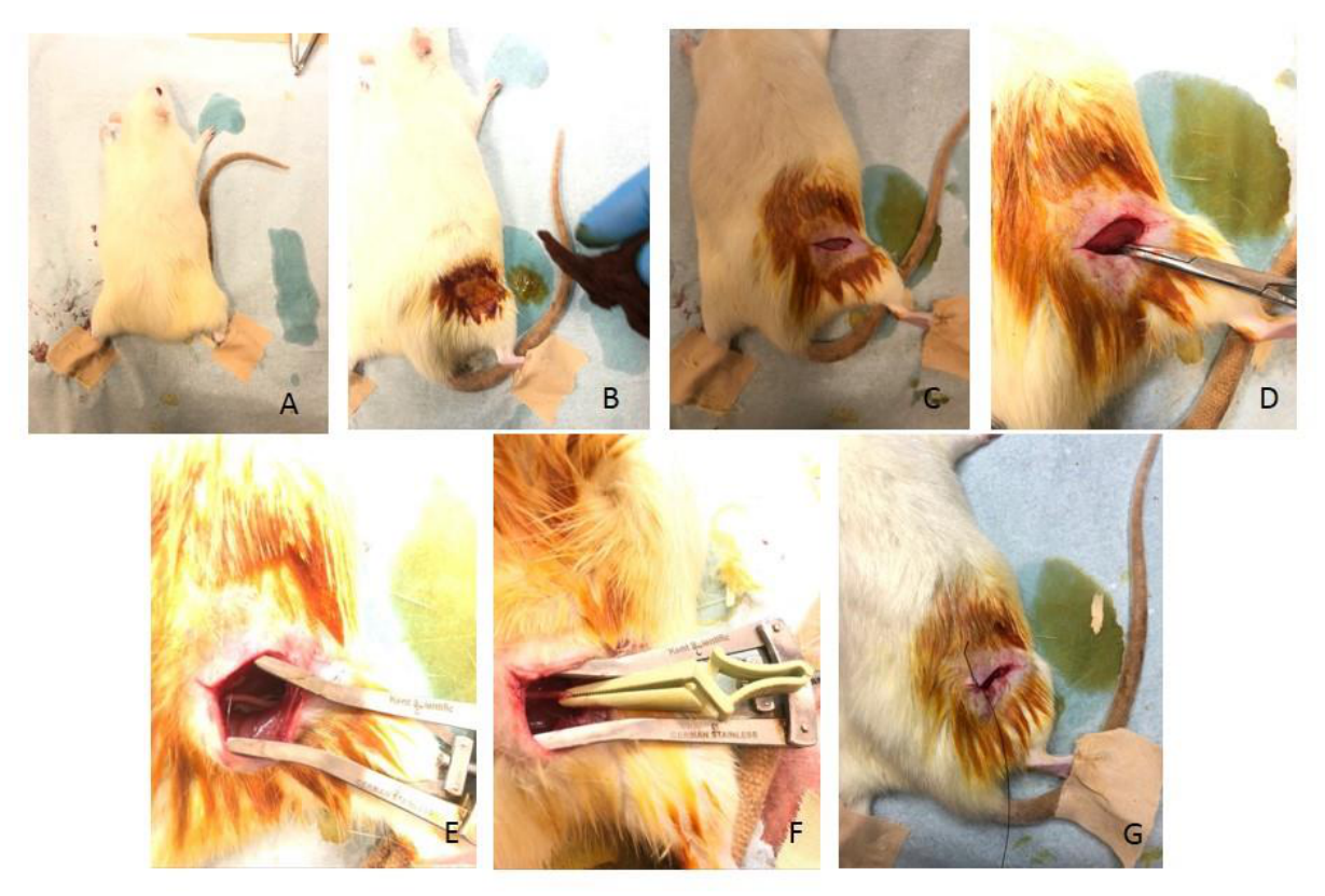
Figure 2.
Multiple comparison rank plot for fibroblast counts among groups (Rank-based graphical representation of fibroblast count scores in the control group (Group K), hyperbaric oxygen group (Group HBO), piracetam group (Group P), and combined treatment group (Group C). A statistically significant difference was observed between Group P and Group K).
Figure 2.
Multiple comparison rank plot for fibroblast counts among groups (Rank-based graphical representation of fibroblast count scores in the control group (Group K), hyperbaric oxygen group (Group HBO), piracetam group (Group P), and combined treatment group (Group C). A statistically significant difference was observed between Group P and Group K).
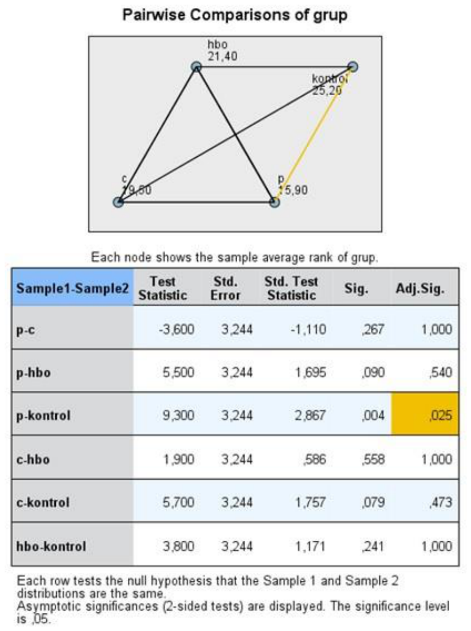
Figure 3.
Mean rank plot of inflammatory cell counts among groups (Rank-based comparison of inflammatory cell count scores among the four study groups. Statistically significant differences were observed between Group P and Group K, and between Group C and Group K).
Figure 3.
Mean rank plot of inflammatory cell counts among groups (Rank-based comparison of inflammatory cell count scores among the four study groups. Statistically significant differences were observed between Group P and Group K, and between Group C and Group K).
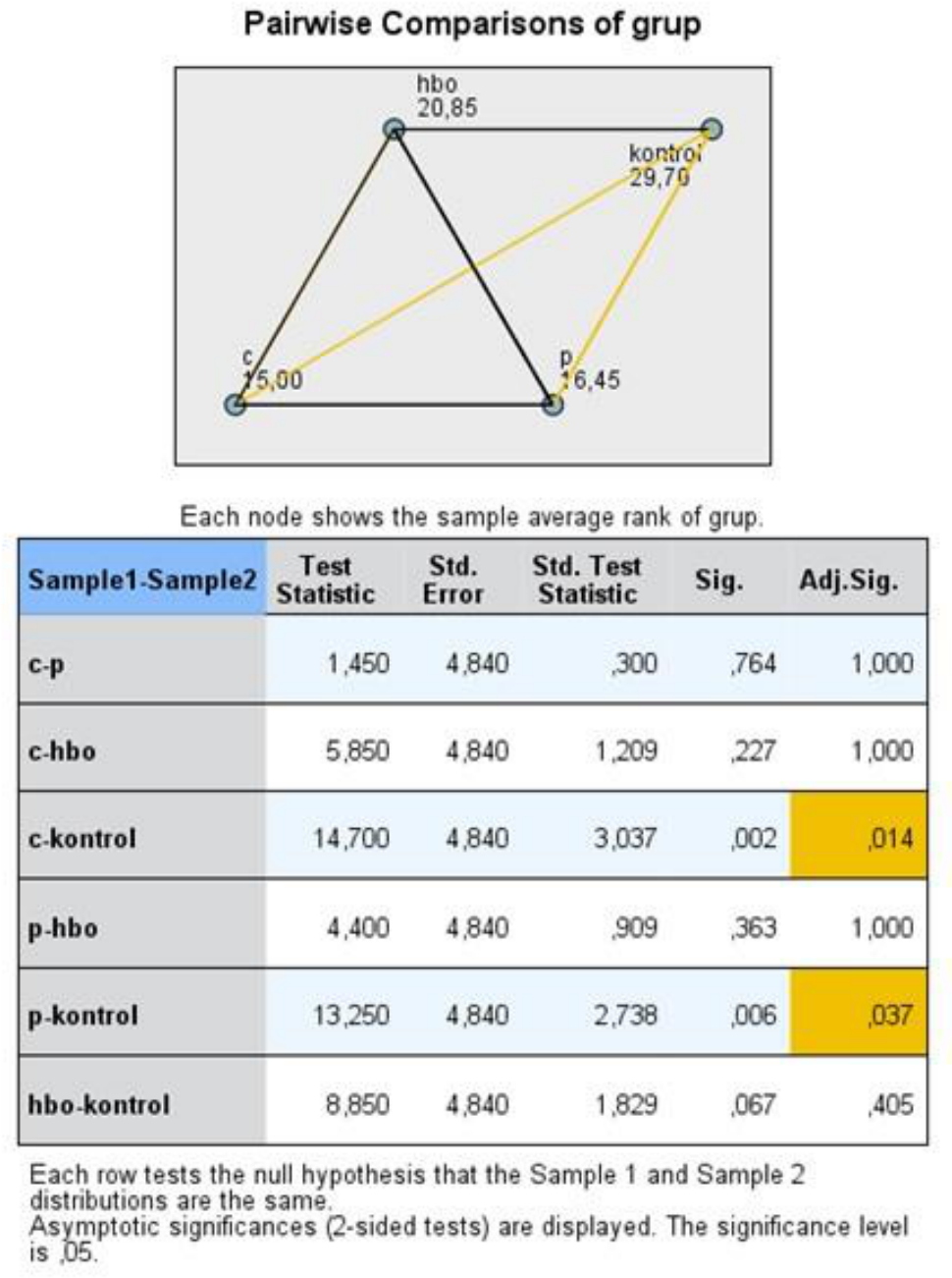

Table 1.
Distribution of histopathological scores among study groups.
| Parameters | Group K | Group HBO | Group P | Group C | P-value |
|---|---|---|---|---|---|
| Axonal continuity 1 2 3 |
0 (%0,0) 7 (%70,0) 3 (%30,0) |
0 (%0,0) 8 (%80,0) 2 (%20,0) |
0 (%0,0) 9 (%90,0) 1 (%10,0) |
0 (%0,0) 10 (%100) 0 (%0,0) |
0.281 |
| Fibroblast count 1 2 3 |
0 (%0,0) 7 (%70,0) 3 (%30,0) |
0 (%0,0) 9 (%90,0) 1 (%10,0) |
2 (%20,0) 8 (%80,0) 0 (%0,0) |
0 (%0,0) 10 (%100) 0 (%0,0) |
0.036 |
| Myelin sheath 1 2 3 |
3 (%30,0) 4 (%40,0) 3 (%30,0) |
0 (%0,0) 9 (%90,0) 1 (%10,0) |
0 (%0,0) 9 (%90,0) 1 (%10,0) |
0 (%0,0) 10 (%100) 0 (%0,0) |
0.931 |
| Inflammatory cells 1 2 3 |
0 (%0,0) 4 (%40,0) 6 (%60,0) |
3 (%30,0) 4 (%40,0) 3 (%30,0) |
4 (%40,0) 5 (%50,0) 1 (%10,0) |
4 (%40,0) 6 (%60,0) 0 (%0,0) |
0.011 |
| Capillary 1 2 3 |
4 (%40,0) 1 (%10,0) 5 (%50,0) |
7 (%70,0) 1 (%10,0) 2 (%20,0) |
2 (%20,0) 8 (%80,0) 0 (%0,0) |
4 (%40,0) 3 (%30,0) 3 (%30,0) |
0.418 |
Distribution of semi-quantitative scores for axonal continuity, fibroblast count, myelin sheath thickness, inflammatory cell count, and capillary density in the control group (Group K), hyperbaric oxygen group (Group HBO), piracetam group (Group P), and combined treatment group (Group C). Data are presented as number of animals and percentage for each score category.
Table 2.
Comparison of biochemical parameters among groups.
| Parameters | Group K | Group HBO | Group P | Group C | P-value |
|---|---|---|---|---|---|
| Nanomolar/mL | 0,838±0,181a | 0,571±0,254b | 0,445±0,199b | 0,540±0,165b | 0,001 |
| HIF1-alpha | 0,609±0,117a | 0,742±0,237ab | 0,967±0,344b | 0,773±0,124ab | 0,011 |
| NSE | 1,499±0,450 | 1,388±0,474 | 1,053±0,297 | 1,135±0,332 | 0,070 |
a,b: Different letters indicate a significant difference between groups according to multiple comparison results (Different superscript letters indicate statistically significant differences based on post hoc analysis). NSE: Neuron-specific enolase. Serum levels of hypoxia-inducible factor 1-alpha (HIF-1α), neuron-specific enolase (NSE), and S100β among study groups. Data are presented as mean ± standard deviation (nanomolar/mL).
Table 3.
Post hoc pairwise comparison of HIF-1α and Neuron-specific enolase (NSE) levels among groups.
Table 3.
Post hoc pairwise comparison of HIF-1α and Neuron-specific enolase (NSE) levels among groups.
| HIF1-alfa | Post-hoc p-value | NSE Nanomolar/mL |
Post-hoc p-value |
|---|---|---|---|
| Grup K vs Grup HBO | 0,028 | Grup K vs Grup HBO | 0,554 |
| Grup K vs Grup P | 0,001 | Grup K vs Grup P | 0,006 |
| Grup K vs Grup C | 0,012 | Grup K vs Grup C | 0,379 |
| Grup HBO vs Grup P | 0,510 | Grup HBO vs Grup P | 0,135 |
| Grup HBO vs Grup C | 0,986 | Grup HBO vs Grup C | 0,991 |
| Grup P vs Grup C | 0,721 | Grup P vs Grup C | 0,234 |
Disclaimer/Publisher’s Note: The statements, opinions and data contained in all publications are solely those of the individual author(s) and contributor(s) and not of MDPI and/or the editor(s). MDPI and/or the editor(s) disclaim responsibility for any injury to people or property resulting from any ideas, methods, instructions or products referred to in the content. |
© 2026 by the authors. Licensee MDPI, Basel, Switzerland. This article is an open access article distributed under the terms and conditions of the Creative Commons Attribution (CC BY) license (http://creativecommons.org/licenses/by/4.0/).
Copyright: This open access article is published under a Creative Commons CC BY 4.0 license, which permit the free download, distribution, and reuse, provided that the author and preprint are cited in any reuse.