

Submitted:
20 August 2026
Posted:
21 August 2026
You are already at the latest version
Abstract
Chronic pain imposes a significant burden on global healthcare systems, and existing pharmacological interventions often provide inadequate pain management. Long-term opioid therapy for chronic pain has contributed to the ongoing global opioid crisis, and its utilization is restricted by severe adverse effects including tolerance, addiction risks, and fatal respiratory suppression. However, recent preclinical studies have shown that peroxisome proliferator-activated receptor gamma coactivator-1α (PGC-1α) has the potential to provide effective chronic pain management and to suppress adverse outcomes of chronic opioid therapy. PGC-1α acts by coordinating mitochondrial renewal, redox balance, and neuroinflammation within neural pathways. Its suppression drives mitochondrial dysfunction and pain chronification. Available evidence highlights the role of oxidative damage and impaired mitochondrial function in chronic pain across musculoskeletal, neuropathic, cancer, and opioid-induced pain models. Therefore, advancing research into the role of PGC-1α in chronic pain management could uncover effective therapeutic interventions with safer clinical outcomes. However, comprehensive reviews that integrate mechanistic evidence on PGC-1α from multiple chronic pain models with drug-repurposing implications remain limited in the literature. This narrative review synthesizes evidence on PGC-1α involvement in chronic pain and examines potential therapeutic interventions through the modulation of PGC-1α as a novel neuroprotective target. Activation of PGC-1α has been shown to attenuate mitochondrial abnormalities and pain hypersensitivity in several preclinical models. Natural compounds, repurposed drugs, and synthetic small-molecule compounds targeting PGC-1α have shown promise as bioactive agents for modulating mitochondrial dysfunction, oxidative stress, and neuroinflammation. However, most available data are preclinical and highlight a translational gap between experimental findings and clinical application across various nociceptive models. Therefore, additional investigation is required to clarify tissue-specific pathways, optimize PGC-1α pharmacological activators, and establish the safety and translational relevance of modulating PGC-1α in chronic pain.
Keywords:
chronic pain
; PGC-1α
; mitochondrial dysfunction
; oxidative stress
; neuroinflammation
; analgesic mechanisms
1. Introduction
Pain encompasses a distressing neurosensory and psychological state characterized by established or possible tissue impairment [1]. Previous studies have shown that clinical control of severe chronic pain has relied on mu-opioid receptor agonists such as morphine [2]. However, extended opioid therapy is associated with hyperalgesia, addiction liability, and lethal respiratory hypoventilation, which poses risks to many patients [3]. These therapeutic setbacks have encouraged researchers to investigate alternative opioid strategies, including preclinical testing of delta and kappa receptor agonists [4,5]. However, studies have demonstrated some shortcomings related to kappa- and delta-opioid receptors [6,7]. For example, kappa receptor agonists have been limited by dysphoric and psychotomimetic effects, whereas certain delta-opioid receptor agonists are known to provoke convulsant behavior [6,7]. Thus, the development of non-opioid therapeutic alternatives for chronic pain management remains essential [8].
Non-opioid adjuncts, notably gabapentinoids, tricyclic antidepressants, and serotonin-norepinephrine reuptake inhibitors, offer alternative options to overcome the limitations of conventional opioids [8]. However, these options remain suboptimal and are limited by adverse effects [9,10]. Specifically, duloxetine carries a risk of cardiovascular adverse events, whereas gabapentinoids can cause fatigue, ataxia, and have misuse potential [9,10]. Furthermore, minocycline, which has been explored for managing neuroinflammatory responses in persistent lower back pain by inhibiting glial activation, can cause tooth discoloration with prolonged use [11,12].
Chronic pain arises from peripheral and central sensitization, abnormal ion channel and neurotransmitter signaling, glial activation, and neuroimmune remodeling that modifies synaptic function [13,14]. Recent evidence suggests that management of chronic pain could target mitochondrial dysfunction and neuroimmune responses [13,15]. Persistent oxidative damage and disruption of cellular bioenergetics are hallmark features of chronic mitochondrial dysfunction, thereby contributing to neuronal hyperexcitability as well as central pain sensitization [15]. Many currently available analgesics relieve pain by attenuating nociceptive signaling [16]. However, they do not address the underlying mitochondrial and redox abnormalities implicated in chronic pain. At high doses or extended use, analgesics such as non-steroidal anti-inflammatory agents and opioids tend to impair mitochondrial function and increase oxidative damage [2,17,18]. Given the central role of mitochondrial dysfunction and oxidative stress in chronic pain, there is a growing need to identify upstream regulators of mitochondrial biogenesis as potential therapeutic targets. However, a major research gap remains, as standard analgesics are limited in their ability to restore these upstream pathways in chronic pain [19].
Peroxisome proliferator-activated receptor gamma coactivator 1-alpha (PGC-1α) orchestrates mitochondrial generation and antioxidant pathways that modulate redox and inflammatory signaling in persistent pain conditions [20,21]. PGC-1α also regulates neuronal excitability and glial activation during nerve-injury-induced pain [22,23,24]. However, despite growing evidence implicating PGC-1α in various pain-related mitochondrial dysfunctions, its therapeutic exploration as a non-opioid target has not been documented for translational potential. This review synthesizes information from the recent literature on how PGC-1α modulates mitochondrial dysfunction and cellular oxidative damage in neuropathic, musculoskeletal, cancer-related, and opioid-induced hyperalgesia models [21,25,26]. It highlights the biological functions of PGC-1α and its impairment in the pathogenesis of persistent nociception [24]. It also explores targeting this coactivator using natural compounds and novel small-molecule activators or stabilizers as therapeutic approaches aimed at restoring mitochondrial integrity and mitigating oxidative burden [27,28]. This evidence positions PGC-1α as a promising druggable regulator of mitochondrial and redox homeostasis in chronic pain, with considerable potential to drive the development of mechanism-based, non-opioid analgesics. It also highlights the translational gaps that need to be addressed for clinical applications.
2. How Mitochondrial Dysfunction and Oxidative Stress Drive Chronic Pain
Chronic pain arises from complex cellular and molecular alterations within both central and peripheral neuronal networks [29]. Neuronal mitochondrial dysfunction reduces cellular respiration and increases reactive oxygen species (ROS) production [30]. This eventually contributes to sensory hyperexcitability and neuroinflammation [30]. Oxidative stress intensifies peripheral nociceptor sensitization and glial activation that sustain pain chronification [29]. ROS also disrupt ion channels, disturb inhibitory synaptic transmission, and alter monoaminergic signaling [31,32].
Within peripheral tissues, mitochondrial bioenergetic failure and oxidative stress activate sensory nerves and increase nociceptive signaling [33]. In the CNS, mitochondrial dysfunction also contributes to central sensitization and neuroinflammation [34,35]. Bioenergetic deficits within the spinal dorsal horn disrupt glutamate and calcium homeostasis, promoting excitotoxic nociceptive transmission [36,37]. Mitochondrial oxidative stress disrupts blood-brain barrier integrity by altering junctional protein complexes and increasing permeability, which facilitates neuroinflammation and contributes to central sensitization in persistent pain [35,38,39,40]. A reduction of PGC-1α expression correlates with mitochondrial ROS generation within chronic pain in experimental models [19,21,41]. Figure 1 illustrates the cellular mechanisms by which altered PGC-1α signaling contributes to mitochondrial dysfunction and persistent pain.
3. Induction of Mitochondrial Toxicity by Analgesics and Adjuvants
Some commonly used analgesics and adjuvants increase mitochondrial injury and ROS production, further reinforcing the need for mitochondria-targeted interventions [42,43,44]. For instance, NSAIDs such as diclofenac and indomethacin disrupt mitochondrial membrane potential and induce bioenergetic stress [45,46,47]. These mechanisms additionally elicit mitochondrial permeability and oxidative stress, which may culminate in apoptotic degradation [48,49]. In addition, indomethacin impairs PGC-1α signaling and triggers mitochondrial dysfunction within gastric cancer cells [50].
Chronic morphine exposure causes mitochondrial dysfunction, ROS accumulation, and morphine tolerance [51,52]. Opioids also increase mitochondrial DNA (mtDNA) release in immune cells to promote NF-κB activation [53,54,55]. Fentanyl and methadone disrupt mitochondrial morphology and reduce neuronal viability in neuronal models [56]. Consistently, PGC-1α prevents opioid-driven mitochondrial impairment [26].
Ketamine as an adjuvant for refractory neuropathic and cancer-related pain disrupts mitochondrial homeostasis in human-derived neurons by depleting ATP and compromising mitochondrial integrity [57,58,59]. These mitochondrial abnormalities may be associated with decreased hippocampal PGC-1α protein expression in neurobehavioral deficits [60]. Similarly, impairment of mitochondrial function could play a role in the side effects associated with adjunctive antidepressants prescribed for chronic pain [61]. Fluoxetine, for instance, downregulates mitochondrial ATP-production pathways in prefrontal cortex interneurons, suggesting an alteration in cellular bioenergetics [62]. Figure 2 summarizes the mitochondrial toxicity associated with commonly used analgesics and adjuvants.
4. Regulatory Signaling and Biological Functions of PGC-1α in Chronic Pain
PGC-1α is a central co-activator of transcription that governs mitochondrial formation, oxidative processes, and neuroinflammation [63]. It responds to epigenetic changes by recruiting histone acetyltransferases and the mediator complex to transcriptional complexes [64]. PGC-1α is highly enriched in tissues with elevated energy demands, specifically cardiac muscle, skeletal fibers, and brown adipose tissue [63]. In the CNS, PGC-1α supports neuronal resilience by promoting oxidative phosphorylation and endogenous antioxidant defenses [63]. Studies link disrupted PGC-1α pathway activity in neurodegenerative disorders and persistent pain conditions [23,65]. This evidence suggests that PGC-1α may help maintain protective metabolic plasticity in nociceptive circuits [41,66]. Impaired regulation of PGC-1α contributes to both neuropathic pain and neuroinflammation [67,68].
PGC-1α function is controlled via AMP-activated protein kinase (AMPK) alongside SIRT1 as energy-sensing pathways [63,69]. When cells experience sudden energy deficits such as during fasting, physical exertion, or oxygen deprivation, AMPK phosphorylates PGC-1α [63,70]. This phosphorylation event primes PGC-1α for subsequent deacetylation and activation by SIRT1 [63,69,70]. These modifications stimulate PGC-1α, promoting its transcriptional coactivation in the nucleus and establishing the AMPK/SIRT1/PGC-1α pathway as a core regulator of cellular energy balance [63,69,70].
Following post-translational activation, PGC-1α engages nuclear respiratory factors (NRF-1/NRF-2) to drive the expression of mitochondrial transcription factor A (TFAM) [63,70]. TFAM then enters the mitochondrial matrix, where it regulates mtDNA transcription and maintains oxidative phosphorylation [63,70]. PGC-1α also interacts with PPARs and estrogen-related receptors (ERRs) to regulate respiratory chain assembly alongside fatty acid oxidation pathways [20]. This cascade helps replace damaged mitochondria in neural tissue, protects cellular metabolism, and maintains mitochondrial energy demand [20]. This suggests that PGC-1α supports synaptic function in response to stress and persistent nociception [66,71].
Furthermore, PGC-1α drives CNS defenses against oxidative stress by upregulating catalase, superoxide dismutase 2, and glutathione peroxidase 1, thereby restricting ROS accumulation [63,72]. It also attenuates neuroinflammatory responses in astrocytes and microglia by suppressing the NF-κB axis and diminishing key cytokines [72]. In CNS models, PGC-1α preserves neuronal survival by limiting oxidative stress and inflammation, while its downregulation promotes neuronal damage [66]. The regulatory signaling pathways and protective biological functions of PGC-1α in chronic pain are summarized in Figure 3.
5. Experimental Evidence for PGC-1α in Chronic Pain
Experimental studies using rodent pain models, cultured cells, and human tissue samples indicate that PGC-1α promotes mitochondrial biogenesis alongside cellular redox balance and neuroimmune homeostasis in chronic pain [70,73,74,75]. Conversely, PGC-1α downregulation triggers mitochondrial dysfunction, increased ROS, glial activation, and pain chronification [70,74]. Restoration of PGC-1α signaling reverses mitochondrial quality control deficits while mitigating nociceptive behaviors [74,76,77]. However, PGC-1α pain-modulating mechanisms vary across chronic pain conditions, depending on tissue, cell type, and pathology. For example, PGC-1α limits cortical hyperexcitability [78], preserves cartilage integrity [79,80], and prevents muscle wasting in osteoarthritis [79]. In peripheral nerve injury, PGC-1α supports mitochondrial resilience in spinal sensory neurons and regulates microglial polarization [21,75]. In chemotherapy and diabetic-induced neuropathic pain, PGC-1α protects neural and peripheral nerves against mitochondrial dysfunction and oxidative injury [80,81,82,83,84]. PGC-1α reduces neuroinflammation and microglial polarization, while preserving GABAergic interneurons in cancer bone pain, [85,86]. In fibromyalgia, PGC-1α restores mitochondrial redox balance in muscle and hippocampal tissues [87,88], while in chronic headaches, it protects mitochondrial function and reduces trigeminal neuroinflammation [73,89,90]. Similarly, PGC-1α agonists counteract opioid-induced mitochondrial dysfunction and spinal apoptosis [26,91]. Thus, restoration of PGC-1α signaling could alleviate chronic pain through tissue- and mechanism-specific biological effects. Given that these biological effects vary across different pain conditions, evidence for PGC-1α in chronic pain subtypes is synthesized in the following sections.
5.1. Musculoskeletal Pain
Ageing-associated pain chronification reflects both peripheral tissue degeneration and altered pain processing within the primary somatosensory cortex [78]. Reduced PGC-1α in the cortex leads to insufficient control of excitatory neuronal signaling [78]. Research by Wu et al. (2024) showed that wild-type adult mice recovered from nerve-injury-induced mechanical and thermal hypersensitivity, whereas PGC-1α+/- mice failed to resolve these nociceptive behaviors [78]. Both genotypes showed increased calcium activity at day 7 post-injury, but persistent hyperactivation was observed only in PGC-1α+/- haploinsufficient mice [78]. In the cortex, reduced PGC-1α is associated with persistent hyperactivity and pain chronification, while in peripheral pain models, downregulation of PGC-1α directly influences mitochondrial dysfunction [67,78]. Such findings demonstrate PGC-1α supports pain resolution by maintaining mitochondrial homeostasis and limiting oxidative stress across central as well as peripheral pathways [67,78].
Osteoarthritis pain, characterized by joint damage involves spinal mitochondrial dysfunction and central sensitization [92]. Earlier work from Sun et al. (2022) indicated that upregulating spinal Sestrin2 (Sesn2) alleviates chronic pain via induction of AMPK/PGC-1α signaling to stimulate mitochondrial generation in spinal neurons [92]. Zhao et al. (2025) showcased that stromal cell-derived factor 1 (SDF-1) relieved osteoarthritis by activating PGC-1α in chondrocytes [93]. Consistently, Li and his colleagues (2024) showed that prenatal prednisone exposure (PPE) programs long-term osteoarthritis susceptibility in female offspring by suppressing PGC-1α in fetal cartilage [79]. PPE induced PGC-1α promoter hypermethylation, impaired mitochondrial biogenesis and glutamine metabolism, leading to chondrodysplasia [79]. Interestingly, restoration of PGC-1α expression via lentiviral transduction or ZLN005 reversed these mitochondrial defects and rescued cartilage matrix synthesis in osteoarthritis [79].
Post-traumatic osteoarthritis is accompanied by quadriceps wasting, which worsens joint instability and accelerates cartilage breakdown. A previous study demonstrated joint distraction paired with treadmill exercise reduced inflammatory responses within the joint space, protected cartilage, thereby delaying post-traumatic osteoarthritis development [75]. A combined human and rabbit study by Liu et al. (2024) found that lower skeletal muscle PGC-1α levels were associated with more severe osteoarthritis [75]. The quadriceps biopsies from patients with osteoarthritis also showed reduced PGC-1α mRNA and protein expression, supporting a role for muscular PGC-1α deficiency [75]. In the rabbit PTOA model, joint instability downregulated muscle PGC-1α while increasing Atrogin-1 and MuRF1 [75]. Exercise and joint distraction restored PGC-1α and suppressed atrophy markers, whereas ZLN005, a PGC-1α agonist, produced similar protective effects [75]. Table 1 summarizes PGC-1α dysregulation in musculoskeletal pain.
5.2. Peripheral Nerve Injury
Peripheral nerve injury models including chronic constriction injury (CCI), spinal nerve ligation (SNL), and spared nerve injury (SNI), consistently highlight the ability of PGC-1α to stimulate mitochondrial synthesis and abolish nerve injury-induced hypersensitivity [74,92]. However, the biological consequences of PGC-1α dysregulation differ across spinal neurons, microglia, dorsal root ganglion neurons, and immune cells within the injured peripheral nerve [21,23,41,71,74,77,95,96]. Therefore, mechanistic evidence for PGC-1α’s causal role in chronic pain should be considered as a strength of evidence.
In the spinal cord, PGC-1α appears to support mitochondrial biogenesis and redox homeostasis in nociception [92]. Sun et al. (2022) showed that mitochondrial biogenesis within spinal tissue is altered in association with CCI-driven neuropathic hypersensitivity [92]. Under this experimental design, application of the Nrf2 activator RTA-408 upregulated PGC-1α and mitigated mechanical hypersensitivity and thermal nociceptive responses [92]. The PGC-1α activator ZLN005 also produced significant analgesia. This effect, and that of RTA-408, was lost when PGC-1α was pharmacologically inhibited, supporting a causal role of PGC-1α in RTA-408-induced mitochondrial antinociception [92].
Heat shock protein 22 (HSP22) is involved in driving mitochondrial renewal and protects cells against oxidative damage and neuronal loss in brain disorders [97]. Notably, a marked reduction of HSP22 in spinal neurons has been linked to the progression of neuropathic pain behaviors [98]. Intrathecal recombinant HSP22 (rhHSP22) attenuated both mechanical hypersensitivity and thermal pain responses in SNI rat models [98]. In line with this evidence, rhHSP22 lowered ROS levels via the stimulation of spinal AMPK/PGC-1α signaling [98]. In addition, knocking down PGC-1α with siRNA eliminated the therapeutic pain relief conferred by rhHSP22 [98]. The findings provide causal evidence that PGC-1α is required for HSP22-mediated protection in neuropathic nociception [98].
PGC-1α also influences neuroimmune signaling in the spinal dorsal horn [21]. Chen et al. (2025) revealed that spinal PGC-1α activation alleviates CCI-induced neuropathic pain by interrupting a reciprocal cycle involving mitochondrial malfunction and NLRP3 inflammasome activation [21]. In the spinal dorsal horn, CCI increased pro-inflammatory CD68/IBA1 and decreased ARG1/IBA1 microglia while altering inflammatory cytokines [21]. PGC-1α activation restored mitochondrial homeostasis and suppressed NLRP3 inflammasome signaling to restrain microglial neuroinflammation in CCI [21]. Consistently, Zhou and colleagues showed that metformin increased PGC-1α signaling and reduced CD86, iNOS, TNF-α, and IL-1β as inflammatory macrophage markers [95]. Additionally, metformin enhanced nerve regeneration and remyelination by increasing NF200 and myelin basic protein [95].
Additional evidence supports a role for PGC-1α in spinal mitochondrial adaptation and neuroprotection [77]. The selective 5-HT1F receptor agonist lasmiditan alleviated SNI-induced pain through PGC-1α-dependent induction of spinal mitochondrial biogenesis [77]. These observations are consistent with research by Simmons and co-workers, who demonstrated that administration of the selective 5-HT1F receptor agonist LY344864 upregulated spinal cord PGC-1α mRNA levels and enhanced functional recovery following spinal cord injury [99]. This bioenergetic recovery suggests that 5-HT1F receptor agonists exert neuroprotective effects by activating PGC-1α and attenuating nerve injury-induced pain [77,99]. ChREBP was also reported to promote PGC-1α transcription in spinal microglia, thereby suppressing neuroinflammation and neuronal hyperexcitability in neuropathic pain [96].
In the CNS, mitochondrial outer membrane translocator protein (TSPO) shows promise in alleviating neuropathic pain through regulating mitochondrial activity [100]. Ro5-4864, a TSPO agonist, relieved mechanical hypersensitivity by upregulating PGC-1α pathways, which optimized mitochondrial activity and reinforced antioxidant protection [100]. Consistent with these findings, Li et al. (2025) established that boosting TSPO levels mitigated SNI-driven neuropathic pain through restored mitochondrial function and reduced pyroptosis in dorsal horn neurons [101]. The pharmacological upregulation of TSPO activated PGC-1α and conferred antinociceptive actions [101]. However, blocking PGC-1α abolished the beneficial effects of TSPO on mitochondrial balance and pyroptosis, supporting a causal contribution of PGC-1α to TSPO-mediated antinociception [101].
Epigenetic mechanisms also contribute to neuropathic pain by repressing antinociceptive genes [23]. PGC-1α can be suppressed transcriptionally during neuropathic pain through DNA methylation, histone modification, and chromatin remodeling [102]. RNA-seq analysis identified Sp1 as related to CCI-provoked neuropathic pain and predicted its binding to the PGC-1α promoter [23]. Spinal Sp1 upregulation repressed PGC-1α expression by recruiting histone deacetylase 2 (HDAC2) to the PGC-1α promoter [23]. Silencing Sp1 reduced inflammation, mitigated mitochondrial dysfunction in the spinal cord, and improved pain sensitivity [23]. Likewise, overexpressing PGC-1α ameliorated CCI-provoked neuropathic pain and mitochondrial dysfunction [23]. This points to epigenetic regulators such as Sp1 or HDAC2 as potential targets to prevent PGC-1α silencing in chronic neuropathic pain [23].
Additional evidence indicates that stimulating G protein-coupled receptor 39 (GPR39) recruits PGC-1α to drive mitochondrial biogenesis and mitigate mechanical allodynia [74]. Dietary ketosis boosted spinal dorsal horn PGC-1α levels, thereby mitigating CCI-triggered mechanical and thermal hypersensitivity [103]. Melatonin inhibited SNL-induced neuropathic pain by enhancing PGC-1α expression and improving mitochondrial membrane potential in dorsal root ganglia [104]. Bioactive molecules in turmeric alleviate mechanical hypersensitivity by inhibiting glial activation and restoring mitochondrial function across both the spinal cord and amygdala [105].
In essence, across CCI, SNI, and SNL models, reduced PGC-1α is consistently associated with mitochondrial malfunction, oxidative damage, and persistent neuropathic pain behaviors [98,103,104]. The Sp1/HDAC2 provide epigenetic repression of PGC-1α, thereby promoting a pro-nociceptive state [23]. Activation of PGC-1α by Nrf2, TSPO, AMPK, 5-HT1F, GPR39, HSP22, ChREBP, metformin, melatonin, and turmeric restores mitochondrial biogenesis in pain models [77,92,96,98,100,104]. However, these interventions produce biological effects through various molecular targets [106,107,108]. Therefore, increased PGC-1α expression alone could not establish that PGC-1α mediates the antinociceptive effects. The stronger evidence supporting PGC-1α as a causal regulator of neuropathic pain comes from studies in which its manipulation changes mitochondrial function and pain behaviour using PGC-1α inhibition, knockdown, overexpression, or rescue approaches [23,98,101,109]. A summary of PGC-1α dysregulation in peripheral nerve injury is provided in Table 2.
5.3. Chemotherapy-Induced Neuropathic Pain
Paclitaxel-induced neuropathic pain (PINP) impaired mitochondrial biogenesis in the nociceptive system, with reduced levels of PGC-1α in the CNS [110]. In a rat model of PINP, formoterol enhanced PGC-1α expression and blocked mechanical allodynia [110]. This is an indication that the β2-adrenergic system can relieve chemotherapy-induced neuropathic pain by inducing PGC-1α-dependent mitochondrial renewal [110]. Recently, Mo and colleagues (2026) documented that pituitary adenylate cyclase-activating polypeptide (PACAP) relieved PINP by activating neuronal PGC-1α [80]. PACAP upregulated PGC-1α and reduced oxidative damage, accompanied by a reduction in mechanical allodynia and hyperalgesia [80]. Pharmacological inhibition of PGC-1α abolished these effects, suggesting that PACAP alleviates neuropathic pain through regulation of mitochondrial health [80].
5.4. Cancer-Induced Bone Pain
In a rodent model of cancer-provoked bone pain, Ge et al. (2022) indicated polarization of spinal microglia alongside suppressed PGC-1α and reduced glutathione peroxidase 4 (GPx4) [85]. Intraperitoneal naringenin administration relieved bone cancer-induced mechanical allodynia and promoted spinal GPx4 expression via the AMPK/PGC-1α system [85]. Chen et al. (2025) support this mechanism by showing that spinal PGC-1α activation abolished neuropathic pain by reprogramming microglia [21]. Impressively, reduced PGC-1α expression was observed in spinal GABAergic interneurons [86]. Treatment with the intrathecally administered PGC-1α agonist ZLN005 preserved GABAergic interneuron populations by alleviating mechanical allodynia and restoring mitochondrial biogenesis [86]. These biological effects were reversed by the PGC-1α antagonist SR-18292, indicating that PGC-1α-dependent mitochondrial protection in inhibitory interneurons plays a role in limiting cancer-induced bone pain [86].
5.5. Diabetic-Induced Neuropathic Pain
Diabetic nerve injury is marked by oxidative stress and impairment in PGC-1α-dependent mitochondrial biogenesis. In painful diabetic neuropathy, suppression of this pathway is linked to mitochondrial abnormality and peripheral nerve damage, particularly in Schwann cells and sciatic nerve tissue [81,82,83,84]. Notably, neuroprotective compounds such as quercetin, honokiol, mitochondria-derived peptide, and Tang Bi formula have been shown to counteract these pathological changes by reactivating AMPK/PGC-1α-dependent mitochondrial biogenesis [81,82,83,84]. These studies support PGC-1α as a metabolic target through which potential neuroprotective agents could preserve mitochondrial integrity, improve redox balance, and attenuate nerve injury in diabetic neuropathy [81,82,83,84]. Table 3 summarizes evidence of PGC-1α dysregulation in chemotherapy-induced neuropathic pain, cancer-induced bone pain, and diabetic-induced neuropathic pain.
5.6. Fibromyalgia
Mitochondrial dysfunction in skeletal muscle and hippocampus is coupled to suppression of PGC-1α signaling in fibromyalgia models [76,87,88]. This suppression promotes redox imbalance and mitochondrial abnormalities, which contribute to nociplastic pain and cognitive impairment [76,87,88]. Belviranlı and colleagues reported that exercise training and coenzyme Q10 improve fibromyalgia symptoms [87,88]. These benefits resulted from the stimulation of hippocampal PGC-1α, which improved redox homeostasis and mitochondrial renewal in skeletal muscle fibers [87,88]. Similarly, melatonin restored PGC-1α signaling, mitigated neuronal sensitization, and suppressed neuroinflammation [76,111]. Such evidence highlights that PGC-1α serves as a neuroprotective target mediating the beneficial effects of both pharmacological and non-pharmacological measures, including exercise, coenzyme Q10, and melatonin, in fibromyalgia [76,87,88,111]. A summary of PGC-1α dysregulation in fibromyalgia is provided in Table 4.
5.7. Chronic Headache Disorders
Chronic headache disorders, particularly migraine are associated with mitochondrial malfunction and alteration in PGC-1α-related signaling in trigeminal pathways [73,89]. Prior findings using nitroglycerin-triggered migraine models show that lower PGC-1α levels correlate with central sensitization and inflammatory pathways in chronic migraine progression [73]. Guan and collaborators demonstrated that mitochondrial dysregulation in the trigeminal ganglion promotes nociception in nitroglycerin-triggered migraine by impairing PGC-1α and mitochondrial health [89]. Similarly, administration of the mitochondrial-targeted peptide SS-31 alleviated pain responses and revived mitochondrial activity by boosting PGC-1α in an inflammatory soup model [90]. Table 5 summarizes PGC-1α dysregulation in chronic headache disorders.
5.8. Opioid-Induced Hyperalgesia
Chronic opioid exposure is associated with opioid-induced hyperalgesia and tolerance through downregulation of PGC-1α-dependent mitochondrial signaling [26,91]. Jie et al. (2025) showed that morphine tolerance is correlated to enhanced CCL2 and inhibition of Nrf2/PGC-1α signaling [91]. These changes promote oxidative stress and neuronal apoptosis, thereby reducing analgesic efficacy [91]. In this study, pharmacological manipulation with the CCL2 inhibitor Bindarit or the PGC-1α agonist ZLN005 abolished spinal apoptosis and reversed tolerance from morphine administration [91]. Feng et al. (2025) reported a similar pattern in remifentanil-induced hyperalgesia, where activation of spinal p38 MAPK suppressed PGC-1α, promoted neuronal ROS release, thereby contributing to pain hypersensitivity [112]. In line with these results, Kashiwagi et al. (2021) showed that recombinant PGC-1α reduced mitochondrial superoxide and attenuated spinal morphine tolerance [26]. Interestingly, NeuroAid improved morphine-induced cognitive deficits, potentially by upregulating PGC-1α [113]. The basolateral amygdala SIRT1/PGC-1α pathway and mitochondrial dysregulation were also implicated in morphine withdrawal symptoms [114]. Table 6 summarizes PGC-1α dysregulation in opioid-induced hyperalgesia.
6. Opportunities in Targeting PGC-1α for Chronic Pain
PGC-1α represents a potential neurotherapeutic target in chronic pain due to its involvement in regulating mitochondrial health, antioxidant defense, and neuroinflammation [95,97]. In both chronic pain models and neurodegenerative or neurological conditions, declining expression of PGC-1α plays a key role in neuropathological alterations [63,97]. However, most available evidence is derived from preclinical brain and spinal cord samples, dorsal root ganglia, hippocampus or injured peripheral nerves, which are not readily accessible for assessment in patients [26,84,91,110,115,116]. Skeletal muscle could provide an accessible tissue source for evaluating PGC-1 in conditions involving muscle dysfunction such as osteoarthritis and fibromyalgia [75,111,117]. However, it remains unclear whether skeletal-muscle PGC-1α signaling reflects PGC-1α activity within central pain pathways.
Bioactive molecules, including resveratrol, berberine, quercetin, evodiamine, and salvianolic acid A, have been reported to confer neuroprotection by increasing PGC-1α levels and improving mitochondrial resilience in preclinical pain models [84,118,119,120,121]. Other phytochemicals, including magnolol and polyphenols, were also explored for PGC-1α-targeting activity [39,122]. These compounds have been linked to mitochondrial protection and reduced oxidative stress, supporting their potential relevance for PGC-1α-mediated modulation of pain pathways [105,119,121,122]. In addition, compounds such as mitoquinone, pyrroloquinoline quinone, alpha-lipoic acid, acetyl-L-carnitine, and nicotinamide riboside are promising mitochondrial protectants and antioxidants for further study of PGC-1α in chronic nociception [93,123,124,125].
Small-molecule PGC-1α activators and stabilizers such as ZLN005 and other high-throughput screening hits may offer an additional strategy for chronic pain by enhancing PGC-1α expression and mitochondrial biogenesis [27,126]. Neuroprotective agents such as N-acetylcysteine and idebenone may also act as antioxidants, mitochondrial protectants, or neuroinflammation modulators [127,128]. These compounds warrant further investigation for their potential roles in modulating PGC-1α in pain mechanisms.
An additional translational opportunity is that PGC-1α-targeted approaches could serve as an opioid-sparing add-on to opioid analgesics. This is evident by the fact that spinal PGC-1α was downregulated during morphine tolerance, whereas restoration of PGC-1α attenuated mechanical and thermal hypersensitivity in morphine-tolerant rodents [26]. Recent studies implicate PGC-1α/SIRT3 and Nrf2/PGC-1α impairment in remifentanil-induced hyperalgesia and morphine tolerance, respectively, both of which are reversed by ZLN005 intervention [91,112]. These identify PGC-1α activation as a candidate strategy to abolish opioid-induced tolerance and hyperalgesia [91,112]. Chronic pain management sometimes requires combination therapy to improve efficacy and limit opioid adverse effects [129]. Future studies should test whether PGC-1α activators can reduce opioid dose requirements or enhance the therapeutic efficacy of opioids. Direct comparisons of PGC-1α activators with opioid analgesics are also required to evaluate their potential as standalone non-opioid options.
7. Challenges and Limitations in Targeting PGC-1α for Chronic Pain
Despite its compelling role in mitochondrial function and pain modulation, targeting PGC-1α for chronic pain presents several biological, pharmacological, and translational limitations. A major translational challenge in targeting PGC-1α includes the need for tissue-specific modulation to avoid systemic effects, given its highly inducible regulation of mitochondrial and inflammatory pathways [63,65,130,131,132]. For instance, PGC-1α overexpression improves cardiac function in young wild-type rodents but accelerates ageing in older mice [133]. Similarly, PGC-1α supports metabolic homeostasis at physiological levels, but excessive expression may promote insulin resistance [134,135].
Sex differences can complicate PGC-1α targeting because males and females respond differently [136]. For example, exercise-induced PGC-1α expression appears to be sex-dependent. Besides, cardiac studies suggest that PGC-1α contributes to sex-specific variation in cardiac function and cardiovascular risk [136]. Recent studies also show that PGC-1α-dependent thermogenesis in brown adipose tissue is influenced by sex [137]. This is concerning because chronic pain, including fibromyalgia and migraine, is more common in females [138,139]. Specifically, sex representation varied across fibromyalgia models, with several reserpine-induced studies using female Wistar rats [76,87,115], while another study used male Sprague-Dawley rats [111]. In contrast, studies of nitroglycerin-induced migraine used male mice [73,89], whereas the inflammatory-soup headache study was predominantly male, with limited female inclusion [90]. Therefore, Such data reinforce the need to consider sex as a biological variable when interpreting PGC-1α findings from experimental models [136,137]. Given that PGC-1α is sex-dependent, this gap warrants direct attention.
Natural molecules such as curcumin and berberine have shown neuroprotective effects in preclinical studies by targeting PGC-1α [28,140,141]. However, these molecules influence other signaling pathways, including AMPK and Nrf2 [63]. Therefore, their effects may not be attributable solely to direct PGC-1α activation [142]. Clinically used compounds such as melatonin and metformin appear promising for PGC-1α-centered therapy [76,143]. However, more studies are required to define the optimal dosing and translational potential of metformin-melatonin combinations in pain models [144]. In addition, the small-molecule PGC-1α agonist ZLN005 has shown activity in mouse models, whereas SR-18292 has been documented to inhibit PGC-1α activity in animal studies [68]. However, variations in chronic pain models and the species gap between rodents and humans remain major barriers to clinical translation [145]. Finally, PGC-1α serves as a regulator of gene expression, coactivating transcription factors rather than a classic receptor, making direct pharmacological targeting particularly challenging [131]. Future research should consider modulating PGC-1α through post-translational protein modifications alongside interactions with target proteins to influence its stability and activity [20,27,131]. Furthermore, structural biology could potentially identify functional domains of PGC-1α for future structure-based drug design. These limitations highlight that substantial future research is required to translate PGC-1α modulation into safe, effective, and sex-specific clinical therapies for chronic pain.
8. Conclusions and Future Directions
PGC-1α appears to be a promising disease-modifying neuroprotective target in chronic pain by modulating mitochondrial dysfunction, oxidative stress, and neuroinflammation. However, its effects may vary across tissues and pain conditions. Natural compounds including resveratrol, berberine, quercetin, evodiamine, and salvianolic acid A, some clinically used drugs, and small-molecule modulators show potential as PGC-1α-targeting analgesic agents. However, most findings remain preclinical, with challenges related to sex specificity, tissue specificity, and limited translation to humans. Future scientific work should define the role of PGC-1α in sex-specific pain model and optimize dosing to develop more selective, effective, and tolerable analgesics.
The development of lipid nanoparticles or blood-brain-barrier penetrating formulations will be important for directing PGC-1α activators to the CNS. This is because targeted delivery systems could help reduce systemic side effects by limiting off-target exposure and improving CNS delivery. Future preclinical work should determine which types of chronic pain benefit most from PGC-1α modulation. Combining PGC-1α modulators with low-dose opioids may reduce tolerance, as PGC-1α stimulation has been demonstrated to mitigate both morphine tolerance and chronic pain. In addition, future studies should investigate whether PGC-1α upregulation can also mitigate other major opioid-related adverse effects, such as respiratory depression, gastrointestinal dysmotility, and physical dependence or withdrawal syndromes.
Authors’ Contribution: MHA: Conceptualization, Investigation, Writing - original draft, Writing – review & editing, Formal analysis, Data curation. KGM: Writing – review & editing, Methodology, Formal analysis, Visualization. YA: Writing – review & editing, Methodology, Visualization. MM: Writing – review & editing, Formal analysis, Visualization. AIJ: Writing – review & editing, Supervision, Project administration, Data curation. IY: Writing – review & editing, Formal analysis, Supervision, Project administration, Validation. All the authors read and approved the final manuscript.
Data Availability Statement
No data was generated or analyzed in this manuscript. Data sharing is not applicable to this article.
Conflicts of Interest
The authors declare no conflict of interest.
Abbreviations
The following abbreviations are used in this manuscript:
AMPK: AMP-activated protein kinase; CAT: catalase; CCI: chronic constriction injury; CNS: central nervous system; ERRs: estrogen-related receptors; GPX1: glutathione peroxidase 1; GPR39: G protein-coupled receptor 39; GPx4: glutathione peroxidase 4; HDAC2: histone deacetylase 2; HSP22: heat shock protein 22; mtDNA: mitochondrial DNA; mtROS: mitochondrial reactive oxygen species; NF-κB: nuclear factor kappa-light-chain-enhancer of activated B cells; NSAIDs: non-steroidal anti-inflammatory drugs; NRF: nuclear respiratory factor; PACAP: adenylate cyclase-activating polypeptide; PGC-1α: PPAR gamma coactivator-1α; PINP: paclitaxel-induced neuropathic pain; PPAR: peroxisome proliferator-activated receptor; PPE: prenatal prednisone exposure; PTOA: post-traumatic osteoarthritis; rhHSP22: recombinant heat shock protein 22; ROS: reactive oxygen species; SDF-1: stromal cell-derived factor 1; SNI: spared nerve injury; SNL: spinal nerve ligation; SOD2: superoxide dismutase 2; SS-31: Szeto-Schiller peptide 31; TSPO: translocator protein.
References
- Raja, S.N.; Carr, D.B.; Cohen, M.; Finnerup, N.B.; Flor, H.; Gibson, S.; Keefe, F.J.; Mogil, J.S.; Ringkamp, M.; Sluka, K.A.; et al. The Revised International Association for the Study of Pain Definition of Pain: Concepts, Challenges, and Compromises. Pain 2020, 161, 1976–1982. [Google Scholar] [CrossRef] [PubMed]
- Amiri, R.; Fallah, F.; Ghorbanzadeh, B.; Oroojan, A.A.; Behmanesh, M.A.; Alboghobeish, S. Mitigating Morphine Dependence and Withdrawal: The Role of Venlafaxine and Calcium Channel Blockers in Mitochondrial Damage and Oxidative Stress in the Brain. Brain Res. Bull. 2025, 226, 111364. [Google Scholar] [CrossRef] [PubMed]
- Badshah, I.; Anwar, M.; Murtaza, B.; Khan, M.I. Molecular Mechanisms of Morphine Tolerance and Dependence; Novel Insights and Future Perspectives. Mol. Cell. Biochem. 2024, 479, 1457–1485. [Google Scholar] [CrossRef] [PubMed]
- Atigari, D.V.; Paton, K.F.; Uprety, R.; Váradi, A.; Alder, A.F.; Scouller, B.; Miller, J.H.; Majumdar, S.; Kivell, B.M. The Mixed Kappa and Delta Opioid Receptor Agonist, MP1104, Attenuates Chemotherapy-Induced Neuropathic Pain. Neuropharmacology 2021, 185, 108445. [Google Scholar] [CrossRef] [PubMed]
- Quirion, B.; Bergeron, F.; Blais, V.; Gendron, L. The Delta-Opioid Receptor; a Target for the Treatment of Pain. Front. Mol. Neurosci. 2020, 13, 52. [Google Scholar] [CrossRef] [PubMed]
- Jutkiewicz, E.M.; Palleiko, B.A.; Dripps, I.J.; Livingston, K.E.; Roques, B.P.; Clements, B.M.; Traynor, J.R. Positive Allosteric Modulation of the Delta Opioid Receptor Attenuates Depression-like and Pain-like Behaviors but Does Not Promote Convulsions. J. Pharmacol. Exp. Ther. 2026, 393, 104942. [Google Scholar] [CrossRef] [PubMed]
- Ona, G.; Sampedro, F.; Romero, S.; Valle, M.; Camacho, V.; Migliorelli, C.; Mañanas, M.Á.; Antonijoan, R.M.; Puntes, M.; Coimbra, J.; et al. The Kappa Opioid Receptor and the Sleep of Reason: Cortico-Subcortical Imbalance Following Salvinorin-A. Int. J. Neuropsychopharmacol. 2022, 25, 54–63. [Google Scholar] [CrossRef] [PubMed]
- Nwosu, A.D.G.; Chukwu, L.C.; Onwuasoigwe, O.; Nweze, S.O.; Nwadike, K. Redefining the Role of Analgesic Adjuvants in Pain Management: A Narrative Review. Indian J. Pain 2023, 37, 65–73. [Google Scholar] [CrossRef] [PubMed]
- Evoy, K.E.; Covvey, J.R.; Peckham, A.M.; Ochs, L.; Hultgren, K.E. Reports of Gabapentin and Pregabalin Abuse, Misuse, Dependence, or Overdose: An Analysis of the Food And Drug Administration Adverse Events Reporting System (FAERS). Res. Soc. Adm. Pharm. 2019, 15, 953–958. [Google Scholar] [CrossRef] [PubMed]
- Park, K.; Kim, S.; Ko, Y.-J.; Park, B.-J. Duloxetine and Cardiovascular Adverse Events: A Systematic Review and Meta-Analysis. J. Psychiatr. Res. 2020, 124, 109–114. [Google Scholar] [CrossRef] [PubMed]
- Martins, A.M.; Marto, J.M.; Johnson, J.L.; Graber, E.M. A Review of Systemic Minocycline Side Effects and Topical Minocycline as a Safer Alternative for Treating Acne and Rosacea. Antibiotics 2021, 10, 757. [Google Scholar] [CrossRef] [PubMed]
- Mohammadian, M.; Morrissey, E.J.; Knight, P.C.; Brusaferri, L.; Kim, M.; Efthimiou, N.; Murphy, J.P.; Alshelh, Z.; Grmek, G.; Schnieders, J.H.; et al. Investigating the Potential of Minocycline in Reducing Brain Inflammation in Chronic Low Back Pain: A Randomized, Placebo-Controlled Mechanistic Clinical Trial. Pain 2025, 166, 2044–2053. [Google Scholar] [CrossRef] [PubMed]
- McKenzie, A.; Dombrower, R.; Theeraphapphong, N.; McKenzie, S.; Hijazin, M.A. Glial Activation, Neuroinflammation, and Loss of Neuroprotection in Chronic Pain: Cellular Mechanisms and Emerging Therapeutic Strategies. Biomedicines 2025, 14, 58. [Google Scholar] [CrossRef] [PubMed]
- Yang, J.; Jung, M.; Picco, L.; Grist, E.; Lloyd-Jones, M.; Giummarra, M.; Nielsen, S. Pain in People Seeking and Receiving Opioid Agonist Treatment: A Systematic Review and Meta-analysis of Prevalence and Correlates. Addiction 2024, 119, 1879–1901. [Google Scholar] [CrossRef] [PubMed]
- Fan, Y.-L.; Dai, F.-Y.; Wang, J.-Y.; Dai, X.-Y.; Song, W.-L.; Wu, H.; Gao, S.-J.; Tao, M.; Zhang, L.-Q.; Liu, T.-Z.; et al. Mitochondrial Dysfunction as a Driver of Chronic Pain: New Insights and Therapeutic Prospects. Pharmacol. Res. 2026, 227, 108170. [Google Scholar] [CrossRef] [PubMed]
- Van Den Ameele, J.; Fuge, J.; Pitceathly, R.D.S.; Berry, S.; McIntyre, Z.; Hanna, M.G.; Lee, M.; Chinnery, P.F. Chronic Pain Is Common in Mitochondrial Disease. Neuromuscul. Disord. 2020, 30, 413–419. [Google Scholar] [CrossRef] [PubMed]
- Mazumder, S.; Bindu, S.; Debsharma, S.; Bandyopadhyay, U. Induction of Mitochondrial Toxicity by Non-Steroidal Anti-Inflammatory Drugs (NSAIDs): The Ultimate Trade-off Governing the Therapeutic Merits and Demerits of These Wonder Drugs. Biochem. Pharmacol. 2024, 228, 116283. [Google Scholar] [CrossRef] [PubMed]
- Ramachandran, A.; Jaeschke, H. Mitochondria in Acetaminophen-Induced Liver Injury and Recovery: A Concise Review. Livers 2023, 3, 219–231. [Google Scholar] [CrossRef] [PubMed]
- Li, Y.; Wu, P.; Wen, Q. Effect of Mitochondrial Dysfunction on Neuropathic Pain. Biomed. Pharmacother. 2025, 193, 118760. [Google Scholar] [CrossRef] [PubMed]
- Abu Shelbayeh, O.; Arroum, T.; Morris, S.; Busch, K.B. PGC-1α Is a Master Regulator of Mitochondrial Lifecycle and ROS Stress Response. Antioxidants 2023, 12, 1075. [Google Scholar] [CrossRef] [PubMed]
- Chen, Y.; Ling, G.; Xu, Q.; Zhu, Q.; Huang, T.; Su, T.; Xie, Y.; Zhong, Y. Activation of Spinal PGC-1α Regulates Microglial Polarization through a Feedback Loop between ROS-Mediated Mitochondrial Dysfunction and the NLRP3 Inflammasome in Neuropathic Pain. Brain Res. Bull. 2025, 227, 111365. [Google Scholar] [CrossRef] [PubMed]
- Miao, J.; Zhou, X.; Ding, W.; You, Z.; Doheny, J.; Mei, W.; Chen, Q.; Mao, J.; Shen, S. Proliferator-Activated Receptor-Gamma Coactivator-1α Haploinsufficiency Promotes Pain Chronification After Burn Injury. Anesth. Analg. 2020, 130, 240–247. [Google Scholar] [CrossRef] [PubMed]
- Miao, J.; Chen, Z.; Wu, Y.; Hu, Q.; Ji, T. Sp1 Inhibits PGC-1α via HDAC2-Catalyzed Histone Deacetylation in Chronic Constriction Injury-Induced Neuropathic Pain. ACS Chem. Neurosci. 2022, 13, 3438–3452. [Google Scholar] [CrossRef] [PubMed]
- Wu, X.; Yang, L.; Li, Z.; Gu, C.; Jin, K.; Luo, A.; Rasheed, N.F.; Fiutak, I.; Chao, K.; Chen, A.; et al. Aging-associated Decrease of PGC -1α Promotes Pain Chronification. Aging Cell 2024, 23, e14177. [Google Scholar] [CrossRef] [PubMed]
- Chen, N.; Zhan, R.-N.; Liu, D.-Q.; Zhang, Y.; Tian, Y.-K.; Zhou, Y.-Q. PGC-1α Activation Ameliorates Cancer-Induced Bone Pain via Inhibiting Apoptosis of GABAergic Interneurons. Biochem. Pharmacol. 2024, 222, 116053. [Google Scholar] [CrossRef] [PubMed]
- Kashiwagi, Y.; Yi, H.; Liu, S.; Takahashi, K.; Hayashi, K.; Ikegami, D.; Zhu, X.; Gu, J.; Hao, S. Mitochondrial Biogenesis Factor PGC-1α Suppresses Spinal Morphine Tolerance by Reducing Mitochondrial Superoxide. Exp. Neurol. 2021, 339, 113622. [Google Scholar] [CrossRef] [PubMed]
- Pettersson-Klein, A.T.; Izadi, M.; Ferreira, D.M.S.; Cervenka, I.; Correia, J.C.; Martinez-Redondo, V.; Southern, M.; Cameron, M.; Kamenecka, T.; Agudelo, L.Z.; et al. Small Molecule PGC-1α1 Protein Stabilizers Induce Adipocyte Ucp1 Expression and Uncoupled Mitochondrial Respiration. Mol. Metab. 2018, 9, 28–42. [Google Scholar] [CrossRef] [PubMed]
- Wu, Y.; Sun, F.; Guo, Y.; Zhang, Y.; Li, L.; Dang, R.; Jiang, P. Curcumin Relieves Chronic Unpredictable Mild Stress-Induced Depression-Like Behavior through the PGC-1α/FNDC5/BDNF Pathway. Behav. Neurol. 2021, 2021, 1–13. [Google Scholar] [CrossRef] [PubMed]
- Yang, J.-X.; Wang, H.-F.; Chen, J.-Z.; Li, H.-Y.; Hu, J.-C.; Yu, A.-A.; Wen, J.-J.; Chen, S.-J.; Lai, W.-D.; Wang, S.; et al. Potential Neuroimmune Interaction in Chronic Pain: A Review on Immune Cells in Peripheral and Central Sensitization. Front. Pain Res. 2022, 3, 946846. [Google Scholar] [CrossRef] [PubMed]
- Meng, X.; Song, Q.; Liu, Z.; Liu, X.; Wang, Y.; Liu, J. Neurotoxic β-Amyloid Oligomers Cause Mitochondrial Dysfunction—the Trigger for PANoptosis in Neurons. Front. Aging Neurosci. 2024, 16, 1400544. [Google Scholar] [CrossRef] [PubMed]
- Mahapatra, C.; Thakkar, R.; Kumar, R. Modulatory Impact of Oxidative Stress on Action Potentials in Pathophysiological States: A Comprehensive Review. Antioxidants 2024, 13, 1172. [Google Scholar] [CrossRef] [PubMed]
- Yowtak, J.; Lee, K.Y.; Kim, H.Y.; Wang, J.; Kim, H.K.; Chung, K.; Chung, J.M. Reactive Oxygen Species Contribute to Neuropathic Pain by Reducing Spinal GABA Release. Pain 2011, 152, 844–852. [Google Scholar] [CrossRef] [PubMed]
- Nesuashvili, L.; Hadley, S.H.; Bahia, P.K.; Taylor-Clark, T.E. Sensory Nerve Terminal Mitochondrial Dysfunction Activates Airway Sensory Nerves via Transient Receptor Potential (TRP) Channels. Mol. Pharmacol. 2013, 83, 1007–1019. [Google Scholar] [CrossRef] [PubMed]
- Joshi, A.U.; Minhas, P.S.; Liddelow, S.A.; Haileselassie, B.; Andreasson, K.I.; Dorn, G.W.; Mochly-Rosen, D. Fragmented Mitochondria Released from Microglia Trigger A1 Astrocytic Response and Propagate Inflammatory Neurodegeneration. Nat. Neurosci. 2019, 22, 1635–1648. [Google Scholar] [CrossRef] [PubMed]
- Silva Santos Ribeiro, P.; Willemen, H.L.D.M.; Eijkelkamp, N. Mitochondria and Sensory Processing in Inflammatory and Neuropathic Pain. Front. Pain Res. 2022, 3, 1013577. [Google Scholar] [CrossRef] [PubMed]
- Mancuso, M.; Mezzalira, F.; Vignoli, B.; Greotti, E. Mitochondrial Ca2+ Signaling at the Tripartite Synapse: A Unifying Framework for Glutamate Homeostasis, Metabolic Coupling, and Network Vulnerability. Biomolecules 2026, 16, 171. [Google Scholar] [CrossRef] [PubMed]
- Martami, F.; Holton, K.F. Targeting Glutamate Neurotoxicity through Dietary Manipulation: Potential Treatment for Migraine. Nutrients 2023, 15, 3952. [Google Scholar] [CrossRef] [PubMed]
- DosSantos, M.F.; Holanda-Afonso, R.C.; Lima, R.L.; DaSilva, A.F.; Moura-Neto, V. The Role of the Blood—brain Barrier in the Development and Treatment of Migraine and Other Pain Disorders. Front. Cell. Neurosci. 2014, 8. [Google Scholar] [CrossRef] [PubMed]
- Kim, Y.; Cho, A.Y.; Kim, H.C.; Ryu, D.; Jo, S.A.; Jung, Y.-S. Effects of Natural Polyphenols on Oxidative Stress-Mediated Blood-Brain Barrier Dysfunction. Antioxidants 2022, 11, 197. [Google Scholar] [CrossRef] [PubMed]
- Wang, Y.; Wu, J.; Wang, J.; He, L.; Lai, H.; Zhang, T.; Wang, X.; Li, W. Mitochondrial Oxidative Stress in Brain Microvascular Endothelial Cells: Triggering Blood-Brain Barrier Disruption. Mitochondrion 2023, 69, 71–82. [Google Scholar] [CrossRef] [PubMed]
- Sun, J.; Li, J.-Y.; Zhang, L.-Q.; Li, D.-Y.; Wu, J.-Y.; Gao, S.-J.; Liu, D.-Q.; Zhou, Y.-Q.; Mei, W. Nrf2 Activation Attenuates Chronic Constriction Injury-Induced Neuropathic Pain via Induction of PGC-1 α -Mediated Mitochondrial Biogenesis in the Spinal Cord. Oxid. Med. Cell. Longev. 2021, 2021, 9577874. [Google Scholar] [CrossRef] [PubMed]
- AlMallah, B.; Al Laham, S.; Alkhatib, R.; Al-Manadili, A. Evaluation of the Therapeutic Effect of Cardamom Extract on Nephropathy Induced by Aspirin in Rats Model. Jordan J. Pharm. Sci. 2025, 18, 146–159. [Google Scholar] [CrossRef]
- Halcrow, P.W.; Kumar, N.; Hao, E.; Khan, N.; Meucci, O.; Geiger, J.D. Mu Opioid Receptor-Mediated Release of Endolysosome Iron Increases Levels of Mitochondrial Iron, Reactive Oxygen Species, and Cell Death. NeuroImmune Pharmacol. Ther. 2022, 0. [Google Scholar] [CrossRef] [PubMed]
- Kasala, S.; Briyal, S.; Prazad, P.; Ranjan, A.K.; Stefanov, G.; Donovan, R.; Gulati, A. Exposure to Morphine and Caffeine Induces Apoptosis and Mitochondrial Dysfunction in a Neonatal Rat Brain. Front. Pediatr. 2020, 8, 593. [Google Scholar] [CrossRef] [PubMed]
- Kim, S.R.; Park, J.W.; Choi, Y.-J.; Sonn, S.K.; Oh, G.T.; Lee, B.-H.; Chang, T.-S. Mitochondrial H2O2 Is a Central Mediator of Diclofenac-Induced Hepatocellular Injury. Antioxidants 2023, 13, 17. [Google Scholar] [CrossRef] [PubMed]
- Pinheiro, J.; Pinheiro, E.; De Deus, G.R.; Saito, G.; Luz, W.L.; Assad, N.; Da Cunha Palheta, M.R.; De Jesus Oliveira Batista, E.; Morais, S.; Passos, A.; et al. Brain Oxidative Stress Mediates Anxiety-like Behavior Induced by Indomethacin in Zebrafish: Protective Effect of Alpha-Tocopherol. Naunyn. Schmiedebergs Arch. Pharmacol. 2024, 397, 1715–1725. [Google Scholar] [CrossRef] [PubMed]
- Thai, P.N.; Ren, L.; Xu, W.; Overton, J.; Timofeyev, V.; Nader, C.E.; Haddad, M.; Yang, J.; Gomes, A.V.; Hammock, B.D.; et al. Chronic Diclofenac Exposure Increases Mitochondrial Oxidative Stress, Inflammatory Mediators, and Cardiac Dysfunction. Cardiovasc. Drugs Ther. 2023, 37, 25–37. [Google Scholar] [CrossRef] [PubMed]
- Debsharma, S.; Pramanik, S.; Bindu, S.; Mazumder, S.; Das, T.; Saha, D.; De, R.; Nag, S.; Banerjee, C.; Siddiqui, A.A.; et al. Honokiol, an Inducer of Sirtuin-3, Protects against Non-steroidal Anti-inflammatory Drug-induced Gastric Mucosal Mitochondrial Pathology, Apoptosis and Inflammatory Tissue Injury. Br. J. Pharmacol. 2023, 180, 2317–2340. [Google Scholar] [CrossRef] [PubMed]
- Salimi, A.; Atashbar, S.; Shabani, M. Gallic Acid Inhibits Celecoxib-Induced Mitochondrial Permeability Transition and Reduces Its Toxicity in Isolated Cardiomyocytes and Mitochondria. Hum. Exp. Toxicol. 2021, 40, S530–S539. [Google Scholar] [CrossRef] [PubMed]
- Debsharma, S.; Pramanik, S.; Bindu, S.; Mazumder, S.; Das, T.; Pal, U.; Saha, D.; De, R.; Nag, S.; Banerjee, C.; et al. NSAID Targets SIRT3 to Trigger Mitochondrial Dysfunction and Gastric Cancer Cell Death. iScience 2024, 27, 109384. [Google Scholar] [CrossRef] [PubMed]
- Jia, J.; Xu, G.; Zeng, X. The Biology of Morphine and Oxidative Stress. In Handbook of Substance Misuse and Addictions; Patel, V.B., Preedy, V.R., Eds.; Springer International Publishing: Cham, 2022; pp. 1–21. ISBN 978-3-030-67928-6. [Google Scholar]
- Malik, J.A.; Agrewala, J.N. Assessing the Implications of Morphine-Induced Dysregulation of Autophagy on Brain Health. Mol. Neurobiol. 2025, 62, 14706–14716. [Google Scholar] [CrossRef] [PubMed]
- Wen, S.; Jiang, Y.; Liang, S.; Cheng, Z.; Zhu, X.; Guo, Q. Opioids Regulate the Immune System: Focusing on Macrophages and Their Organelles. Front. Pharmacol. 2022, 12, 814241. [Google Scholar] [CrossRef] [PubMed]
- Xie, F.; Kitagawa, Y.; Ogata, H.; Yasuhara, S.; You, Z.; Jeevendra Martyn, J.A. Morphine Induces Inflammatory Responses via Both TLR4 and cGAS-STING Signaling Pathways. Cytokine 2024, 183, 156737. [Google Scholar] [CrossRef] [PubMed]
- Xu, Y.; Chen, R.; Zhi, F.; Sheng, S.; Khiati, L.; Yang, Y.; Peng, Y.; Xia, Y. δ-Opioid Receptor, Microglia and Neuroinflammation. Aging Dis. 2023, 14, 778. [Google Scholar] [CrossRef] [PubMed]
- Nylander, E.; Zelleroth, S.; Nyberg, F.; Grönbladh, A.; Hallberg, M. The Effects of Morphine, Methadone, and Fentanyl on Mitochondria: A Live Cell Imaging Study. Brain Res. Bull. 2021, 171, 126–134. [Google Scholar] [CrossRef] [PubMed]
- Azari, L.; Hemati, H.; Tavasolian, R.; Shahdab, S.; Tomlinson, S.M.; Babilonia, M.B.; Huang, J.; Tometich, D.B.; Turner, K.; Anaraki, K.S.; et al. The Efficacy of Ketamine for Acute and Chronic Pain in Patients with Cancer: A Systematic Review of Randomized Controlled Trials. Healthcare 2024, 12, 1560. [Google Scholar] [CrossRef] [PubMed]
- Bruna-Mejias, A.; Baeza, V.; Gamboa, J.; Baez Flores, B.; San Martin, J.; Astorga, C.; Leyton, J.; Nova-Baeza, P.; Orellana-Donoso, M.; Suazo-Santibañez, A.; et al. Use of Ketamine in Patients with Multifactorial Neuropathic Pain: A Systematic Review and Meta-Analysis. Pharmaceuticals 2024, 17, 1165. [Google Scholar] [CrossRef] [PubMed]
- Ito, H.; Uchida, T.; Makita, K. Ketamine Causes Mitochondrial Dysfunction in Human Induced Pluripotent Stem Cell-Derived Neurons. PLoS ONE 2015, 10, e0128445. [Google Scholar] [CrossRef] [PubMed]
- Hosseini, L.; Abolhasanpour, N.; Seyedaghamiri, F.; Hassanzadeh, P.; Shahabi, P.; Mafikandi, V.; Kalejahi, P.; Rajabi, M.; Shokri, Z.; Fakhari, A. Cerebrolysin Ameliorates Ketamine-Mediated Anxiety and Cognitive Impairments via Modulation of Mitochondrial Function and CREB/PGC-1α Pathway. Mol. Brain 2025, 18, 84. [Google Scholar] [CrossRef] [PubMed]
- Ľupták, M.; Fišar, Z.; Hroudová, J. Different Effects of SSRIs, Bupropion, and Trazodone on Mitochondrial Functions and Monoamine Oxidase Isoform Activity. Antioxidants 2023, 12, 1208. [Google Scholar] [CrossRef] [PubMed]
- Jetsonen, E.; Didio, G.; Suleymanova, I.; Teino, I.; Castrén, E.; Umemori, J. Chronic Treatment with Fluoxetine Regulates Mitochondrial Features and Plasticity-Associated Transcriptomic Pathways in Parvalbumin-Positive Interneurons of Prefrontal Cortex. Neuropsychopharmacology 2025, 50, 1864–1874. [Google Scholar] [CrossRef] [PubMed]
- Qian, L.; Zhu, Y.; Deng, C.; Liang, Z.; Chen, J.; Chen, Y.; Wang, X.; Liu, Y.; Tian, Y.; Yang, Y. Peroxisome Proliferator-Activated Receptor Gamma Coactivator-1 (PGC-1) Family in Physiological and Pathophysiological Process and Diseases. Signal Transduct. Target. Ther. 2024, 9, 50. [Google Scholar] [CrossRef] [PubMed]
- Krämer, A.I.; Handschin, C. How Epigenetic Modifications Drive the Expression and Mediate the Action of PGC-1α in the Regulation of Metabolism. Int. J. Mol. Sci. 2019, 20, 5449. [Google Scholar] [CrossRef] [PubMed]
- Rius-Pérez, S.; Torres-Cuevas, I.; Millán, I.; Ortega, Á.L.; Pérez, S. PGC-1 α, Inflammation, and Oxidative Stress: An Integrative View in Metabolism. Oxid. Med. Cell. Longev. 2020, 2020, 1–20. [Google Scholar] [CrossRef] [PubMed]
- Kuczynska, Z.; Metin, E.; Liput, M.; Buzanska, L. Covering the Role of PGC-1α in the Nervous System. Cells 2021, 11, 111. [Google Scholar] [CrossRef] [PubMed]
- Ye, L.; Li, M.; Wang, Z.; Yang, Z.; Zhang, J.; Fang, H.; He, Z.; Wang, X. Depression of Mitochondrial Function in the Rat Skeletal Muscle Model of Myofascial Pain Syndrome Is Through Down-Regulation of the AMPK-PGC-1α-SIRT3 Axis. J. Pain Res. 2020, Volume 13, 1747–1756. [Google Scholar] [CrossRef] [PubMed]
- Zhai, M.; Hu, H.; Zheng, Y.; Wu, B.; Sun, W. PGC1α: An Emerging Therapeutic Target for Chemotherapy-Induced Peripheral Neuropathy. Ther. Adv. Neurol. Disord. 2023, 16, 17562864231163361. [Google Scholar] [CrossRef] [PubMed]
- Cantó, C.; Auwerx, J. PGC-1α, SIRT1 and AMPK, an Energy Sensing Network That Controls Energy Expenditure. Curr. Opin. Lipidol. 2009, 20, 98–105. [Google Scholar] [CrossRef] [PubMed]
- Chen, J.; Liu, B.; Yao, X.; Yang, X.; Sun, J.; Yi, J.; Xue, F.; Zhang, J.; Shen, Y.; Chen, B.; et al. AMPK/SIRT1/PGC-1α Signaling Pathway: Molecular Mechanisms and Targeted Strategies From Energy Homeostasis Regulation to Disease Therapy. CNS Neurosci. Ther. 2025, 31, e70657. [Google Scholar] [CrossRef] [PubMed]
- Hao, C.; Ma, B.; Gao, N.; Jin, T.; Liu, X. Translocator Protein (TSPO) Alleviates Neuropathic Pain by Activating Spinal Autophagy and Nuclear SIRT1/PGC-1α Signaling in a Rat L5 SNL Model. J. Pain Res. 2022, Volume 15, 767–778. [Google Scholar] [CrossRef] [PubMed]
- D’Egidio, F.; Qosja, E.; Ammannito, F.; Topi, S.; d’Angelo, M.; Cimini, A.; Castelli, V. Antioxidant and Anti-Inflammatory Defenses in Huntington’s Disease: Roles of NRF2 and PGC-1α, and Therapeutic Strategies. Life 2025, 15, 577. [Google Scholar] [CrossRef] [PubMed]
- Liu, W.; Liu, Z.; Dong, H.; Ni, Z.; Wang, Y.; Li, Y.; Qian, H.; Hu, Y.; Zuo, Y. Circadian Rhythm Cascade SIRT1/PGC-1α/BMAL1 Pathway Regulates Nitroglycerin-Induced Chronic Migraine. Neurochem. Res. 2025, 50, 262. [Google Scholar] [CrossRef] [PubMed]
- Zhang, L.; Tan, X.; Song, F.; Li, D.; Wu, J.; Gao, S.; Sun, J.; Liu, D.; Zhou, Y.; Mei, W. Activation of G-Protein-Coupled Receptor 39 Reduces Neuropathic Pain in a Rat Model. Neural Regen. Res. 2024, 19, 687–696. [Google Scholar] [CrossRef] [PubMed]
- Liu, X.; Chen, R.; Song, Z.; Sun, Z. Exercise Following Joint Distraction Inhibits Muscle Wasting and Delays the Progression of Post-Traumatic Osteoarthritis in Rabbits by Activating PGC-1α in Skeletal Muscle. J. Orthop. Surg. 2024, 19, 325. [Google Scholar] [CrossRef] [PubMed]
- Osama, J.; El-Gazar, A.A.; Ragab, G.M.; EL-Sayed, N.S.; Kamel, A.S. Melatonin Mitigates Central Sensitization and Nociplastic Pain in Spinal Cord and Dorsal Root Ganglia of FM Rat Model: Modulation of SIRT1/PGC-1α/MAPK/NF-κB Signaling. J. Neuroimmune Pharmacol. 2026, 21, 12. [Google Scholar] [CrossRef] [PubMed]
- Zhang, L.-Q.; Zhou, Y.-Q.; Li, J.-Y.; Sun, J.; Zhang, S.; Wu, J.-Y.; Gao, S.-J.; Tian, X.-B.; Mei, W. 5-HT1F Receptor Agonist Ameliorates Mechanical Allodynia in Neuropathic Pain via Induction of Mitochondrial Biogenesis and Suppression of Neuroinflammation. Front. Pharmacol. 2022, 13, 834570. [Google Scholar] [CrossRef] [PubMed]
- Wu, X.; Yang, L.; Li, Z.; Gu, C.; Jin, K.; Luo, A.; Rasheed, N.F.; Fiutak, I.; Chao, K.; Chen, A.; et al. Aging-associated Decrease of PGC -1α Promotes Pain Chronification. Aging Cell 2024, 23, e14177. [Google Scholar] [CrossRef] [PubMed]
- Li, Q.; Zhang, F.; Dai, Y.; Liu, L.; Chen, L.; Wang, H. Activation of the PGC-1α-Mediated Mitochondrial Glutamine Metabolism Pathway Attenuates Female Offspring Osteoarthritis Induced by Prenatal Excessive Prednisone. Sci. China Life Sci. 2024, 67, 2382–2397. [Google Scholar] [CrossRef] [PubMed]
- Mo, R.; Wang, C.; Hu, H.; Shi, S.; Cao, D.; Luo, Z.; Yang, H.; Zhai, M.; Sun, W. PACAP Alleviates Paclitaxel-Induced Peripheral Neuropathy by Targeting Oxidative Stress and Mitochondrial Damage via the PGC-1α Pathway. CNS Neurosci. Ther. 2026, 32, e70745. [Google Scholar] [CrossRef] [PubMed]
- Huang, C.-C.; Chiu, H.-Y.; Lee, P.-H.; Fang, S.-Y.; Lin, M.-W.; Chen, H.-F.; Lee, J.-S. Mitochondrial Transplantation Attenuates Traumatic Neuropathic Pain, Neuroinflammation, and Apoptosis in Rats with Nerve Root Ligation. Mol. Pain 2023, 19, 17448069231210423. [Google Scholar] [CrossRef] [PubMed]
- Xu, L.; Tang, X.; Yang, L.; Chang, M.; Xu, Y.; Chen, Q.; Lu, C.; Liu, S.; Jiang, J. Mitochondria-Derived Peptide Is an Effective Target for Treating Streptozotocin Induced Painful Diabetic Neuropathy through Induction of Activated Protein Kinase/Peroxisome Proliferator-Activated Receptor Gamma Coactivator 1alpha -Mediated Mitochondrial Biogenesis. Mol. Pain 2024, 20, 17448069241252654. [Google Scholar] [CrossRef] [PubMed]
- Yang, C.; Yu, H.; Zhou, L.; Su, H.; Li, X.; Qi, W.; Lian, F. Tang Bi Formula Alleviates Diabetic Sciatic Neuropathy via AMPK/PGC-1α/MFN2 Pathway Activation. Sci. Rep. 2025, 15, 25069. [Google Scholar] [CrossRef] [PubMed]
- Zhang, Q.; Song, W.; Zhao, B.; Xie, J.; Sun, Q.; Shi, X.; Yan, B.; Tian, G.; Liang, X. Quercetin Attenuates Diabetic Peripheral Neuropathy by Correcting Mitochondrial Abnormality via Activation of AMPK/PGC-1α Pathway in Vivo and in Vitro. Front. Neurosci. 2021, 15, 636172. [Google Scholar] [CrossRef] [PubMed]
- Ge, M.-M.; Li, D.-Y.; Wang, L.; Zhang, L.-Q.; Liu, D.-Q.; Tian, Y.-K.; Ye, D.-W.; Liu, Z.-H.; Zhou, Y.-Q.; Yang, H. Naringenin Promoted Spinal Microglia M2 Polarization in Rat Model of Cancer-Induced Bone Pain via Regulating AMPK/PGC-1α Signaling Axis. Biomed. Pharmacother. 2022, 149, 112912. [Google Scholar] [CrossRef] [PubMed]
- Chen, N.; Zhan, R.-N.; Liu, D.-Q.; Zhang, Y.; Tian, Y.-K.; Zhou, Y.-Q. PGC-1α Activation Ameliorates Cancer-Induced Bone Pain via Inhibiting Apoptosis of GABAergic Interneurons. Biochem. Pharmacol. 2024, 222, 116053. [Google Scholar] [CrossRef] [PubMed]
- Belviranlı, M.; Okudan, N.; Sezer, T. Exercise Training Alleviates Symptoms and Cognitive Decline in a Reserpine-Induced Fibromyalgia Model by Activating Hippocampal PGC-1α/FNDC5/BDNF Pathway. Neuroscience 2024, 549, 145–155. [Google Scholar] [CrossRef] [PubMed]
- Belviranlı, M.; Okudan, N.; Sezer, T. Coenzyme Q10 Improves Redox Homeostasis, Mitochondrial Biogenesis, and Irisin Signaling in Fast- and Slow-Twitch Muscle Fibers in a Reserpine-Induced Fibromyalgia-like Myalgia Model. Life Sci. 2026, 394, 124366. [Google Scholar] [CrossRef] [PubMed]
- Guan, X.-Y.; Dong, X.; Wang, Y.-X.; Xu, B.-C.; Wu, X.-B. Mitochondrial Dysfunction in Trigeminal Ganglion Contributes to Nociceptive Behavior in a Nitroglycerin-Induced Migraine Mouse Model. Mol. Pain 2025, 21, 17448069251332100. [Google Scholar] [CrossRef] [PubMed]
- Shan, Z.; Wang, Y.; Qiu, T.; Zhou, Y.; Zhang, Y.; Hu, L.; Zhang, L.; Liang, J.; Ding, M.; Fan, S.; et al. SS-31 Alleviated Nociceptive Responses and Restored Mitochondrial Function in a Headache Mouse Model via Sirt3/Pgc-1α Positive Feedback Loop. J. Headache Pain 2023, 24, 65. [Google Scholar] [CrossRef] [PubMed]
- Jie, J.; Jihao, R.; Zheng, L.; Jie, L.; Xiaoling, P.; Wei, Z.; Feng, G. Unraveling Morphine Tolerance: CCL2 Induces Spinal Cord Apoptosis via Inhibition of Nrf2 Signaling Pathway and PGC-1α-Mediated Mitochondrial Biogenesis. Brain. Behav. Immun. 2025, 124, 347–362. [Google Scholar] [CrossRef] [PubMed]
- Sun, J.; Song, F.-H.; Wu, J.-Y.; Zhang, L.-Q.; Li, D.-Y.; Gao, S.-J.; Liu, D.-Q.; Zhou, Y.-Q.; Mei, W. Sestrin2 Overexpression Attenuates Osteoarthritis Pain via Induction of AMPK/PGC-1α-Mediated Mitochondrial Biogenesis and Suppression of Neuroinflammation. Brain. Behav. Immun. 2022, 102, 53–70. [Google Scholar] [CrossRef] [PubMed]
- Zhao, Y.; Lin, D.; Zhu, X.; Yan, J.; Liang, Y.; Wang, Y.; Dai, T.; Zhang, Z.; Wang, S. SDF-1 Alleviates Osteoarthritis by Resolving Mitochondrial Dysfunction through the Activation of the Sirt3/PGC-1α Signalling Pathway. Arthritis Res. Ther. 2025, 27, 51. [Google Scholar] [CrossRef] [PubMed]
- Gao, S.-J.; Li, D.-Y.; Liu, D.-Q.; Sun, J.; Zhang, L.-Q.; Wu, J.-Y.; Song, F.-H.; Zhou, Y.-Q.; Mei, W. Dimethyl Fumarate Attenuates Pain Behaviors in Osteoarthritis Rats via Induction of Nrf2-Mediated Mitochondrial Biogenesis. Mol. Pain 2022, 18, 17448069221124920. [Google Scholar] [CrossRef] [PubMed]
- Zhou, Z.; Luo, G.; Li, C.; Zhang, P.; Chen, W.; Li, X.; Tang, J.; Qing, L. Metformin Induces M2 Polarization. [CrossRef] [PubMed]
- Hu, Z.; Pang, J.; Liu, X.; Zhao, Y.; Lu, Y.; Chen, H.; Zeng, H.; Yu, Y.; Zhao, Y.; Gao, L.; et al. PGC -1α Transcriptionally Regulated by ChREBP Mitigates Neuropathic Pain Through Promoting Microglial Fatty Acid Oxidation and Anti-Inflammatory Response. CNS Neurosci. Ther. 2026, 32, e70744. [Google Scholar] [CrossRef] [PubMed]
- Fan, H.; Ding, R.; Liu, W.; Zhang, X.; Li, R.; Wei, B.; Su, S.; Jin, F.; Wei, C.; He, X.; et al. Heat Shock Protein 22 Modulates NRF1/TFAM-Dependent Mitochondrial Biogenesis and DRP1-Sparked Mitochondrial Apoptosis through AMPK-PGC1α Signaling Pathway to Alleviate the Early Brain Injury of Subarachnoid Hemorrhage in Rats. Redox Biol. 2021, 40, 101856. [Google Scholar] [CrossRef] [PubMed]
- Zhang, L.; Liu, L.; Li, D.; Wu, J.; Gao, S.; Song, F.; Zhou, Y.; Liu, D.; Mei, W. Heat Shock Protein 22 Attenuates Nerve Injury-Induced Neuropathic Pain Via Improving Mitochondrial Biogenesis and Reducing Oxidative Stress Mediated By Spinal AMPK/PGC-1α Pathway in Male Rats. J. Neuroimmune Pharmacol. 2024, 19, 5. [Google Scholar] [CrossRef] [PubMed]
- Simmons, E.C.; Scholpa, N.E.; Cleveland, K.H.; Schnellmann, R.G. 5-Hydroxytryptamine 1F Receptor Agonist Induces Mitochondrial Biogenesis and Promotes Recovery from Spinal Cord Injury. J. Pharmacol. Exp. Ther. 2020, 372, 216–223. [Google Scholar] [CrossRef] [PubMed]
- Hao, C.; Ma, B.; Gao, N.; Jin, T.; Liu, X. Translocator Protein (TSPO) Alleviates Neuropathic Pain by Activating Spinal Autophagy and Nuclear SIRT1/PGC-1α Signaling in a Rat L5 SNL Model. J. Pain Res. 2022, Volume 15, 767–778. [Google Scholar] [CrossRef] [PubMed]
- Li, B.; Yu, K.; Zhou, X.; Sun, J.; Qi, L.; Li, W.; Yang, T.; Li, W.; Wang, N.; Gu, X.; et al. Increased TSPO Alleviates Neuropathic Pain by Preventing Pyroptosis via the AMPK-PGC-1α Pathway. J. Headache Pain 2025, 26, 16. [Google Scholar] [CrossRef] [PubMed]
- Pethő, G.; Kántás, B.; Horváth, Á.; Pintér, E. The Epigenetics of Neuropathic Pain: A Systematic Update. Int. J. Mol. Sci. 2023, 24, 17143. [Google Scholar] [CrossRef] [PubMed]
- Deng, M.; Fang, Y.; Chu, Y.; Ding, R.; Fan, X.; Wei, H.; Song, H.; Jiang, G.; Zhang, H.; Han, C.; et al. SIRT3 Is Required for the Protective Function of Ketogenic Diet on Neural Inflammation and Neuropathic Pain. Int. J. Biol. Sci. 2025, 21, 3011–3029. [Google Scholar] [CrossRef] [PubMed]
- Zeng, Y.; Fang, Q.; Chen, J.; Wang, Y.; Liu, X.; Zhang, X.; Shi, Y.; Zhan, H.; Zhong, X.; Yao, M.; et al. Melatonin Improves Mitochondrial Dysfunction and Attenuates Neuropathic Pain by Regulating SIRT1 in Dorsal Root Ganglions. Neuroscience 2023, 534, 29–40. [Google Scholar] [CrossRef] [PubMed]
- Santos, J.; Wang, R.; Bhakta, V.; Driver, Z.; Vadim, Y.; Kiritoshi, T.; Ji, G.; Neugebauer, V.; Shen, C.-L. Turmeric Bioactive Compounds Alleviate Spinal Nerve Ligation-Induced Neuropathic Pain by Suppressing Glial Activation and Improving Mitochondrial Function in Spinal Cord and Amygdala. Nutrients 2023, 15, 4403. [Google Scholar] [CrossRef] [PubMed]
- Paoli, A.; Cerullo, G. Investigating the Link between Ketogenic Diet, NAFLD, Mitochondria, and Oxidative Stress: A Narrative Review. Antioxidants 2023, 12, 1065. [Google Scholar] [CrossRef] [PubMed]
- Cao, B.; Wang, J.; Feng, J. Signaling Pathway Mechanisms of Neurological Diseases Induced by G Protein-coupled Receptor 39. CNS Neurosci. Ther. 2023, 29, 1470–1483. [Google Scholar] [CrossRef] [PubMed]
- Rehman, S.; Ikram, M.; Ullah, N.; Alam, S.; Park, H.; Badshah, H.; Choe, K.; Ok Kim, M. Neurological Enhancement Effects of Melatonin against Brain Injury-Induced Oxidative Stress, Neuroinflammation, and Neurodegeneration via AMPK/CREB Signaling. Cells 2019, 8, 760. [Google Scholar] [CrossRef] [PubMed]
- Sun, J.; Li, J.-Y.; Zhang, L.-Q.; Li, D.-Y.; Wu, J.-Y.; Gao, S.-J.; Liu, D.-Q.; Zhou, Y.-Q.; Mei, W. Nrf2 Activation Attenuates Chronic Constriction Injury-Induced Neuropathic Pain via Induction of PGC-1 α -Mediated Mitochondrial Biogenesis in the Spinal Cord. Oxid. Med. Cell. Longev. 2021, 2021, 9577874. [Google Scholar] [CrossRef] [PubMed]
- Chen, N.; Ge, M.-M.; Li, D.-Y.; Wang, X.-M.; Liu, D.-Q.; Ye, D.-W.; Tian, Y.-K.; Zhou, Y.-Q.; Chen, J.-P. Β2-Adrenoreceptor Agonist Ameliorates Mechanical Allodynia in Paclitaxel-Induced Neuropathic Pain via Induction of Mitochondrial Biogenesis. Biomed. Pharmacother. 2021, 144, 112331. [Google Scholar] [CrossRef] [PubMed]
- Favero, G.; Bonomini, F.; Franco, C.; Rezzani, R. Mitochondrial Dysfunction in Skeletal Muscle of a Fibromyalgia Model: The Potential Benefits of Melatonin. Int. J. Mol. Sci. 2019, 20, 765. [Google Scholar] [CrossRef] [PubMed]
- Feng, G.; Song, F.; Wang, A.; Wang, L.; Zhang, H.; Wang, S.; Hou, H.; Zhang, L.; Deng, L. Spinal P38 MAPK/PGC-1α/SIRT3 Signaling Pathway Mediates Remifentanil-Induced Hyperalgesia in Rats via ROS Release and NR2B Activation. Neurochem. Int. 2025, 190, 106061. [Google Scholar] [CrossRef] [PubMed]
- Malboosi, N.; Nasehi, M.; Hashemi, M.; Vaseghi, S.; Zarrindast, M.-R. The Neuroprotective Effect of NeuroAid on Morphine-Induced Amnesia with Respect to the Expression of TFAM, PGC-1α, ΔfosB and CART Genes in the Hippocampus of Male Wistar Rats. Gene 2020, 742, 144601. [Google Scholar] [CrossRef] [PubMed]
- Yin, F.; Zhang, J.; Liu, Y.; Zhai, Y.; Luo, D.; Yan, X.; Feng, Y.; Lai, J.; Zheng, H.; Wei, S.; et al. Basolateral Amygdala SIRT1/PGC-1α Mitochondrial Biogenesis Pathway Mediates Morphine Withdrawal-Associated Anxiety in Mice. Int. J. Neuropsychopharmacol. 2022, 25, 774–785. [Google Scholar] [CrossRef] [PubMed]
- Belviranlı, M.; Okudan, N.; Sezer, T. Coenzyme Q10 Ameliorates Fibromyalgia-like Symptoms and Cognitive Deficits by Enhancing Hippocampal PGC-1α/FNDC5/BDNF Pathway in Reserpine-Treated Rats. Mol. Neurobiol. 2026, 63, 248. [Google Scholar] [CrossRef] [PubMed]
- Mo, R.; Wang, C.; Hu, H.; Shi, S.; Cao, D.; Luo, Z.; Yang, H.; Zhai, M.; Sun, W. PACAP Alleviates Paclitaxel-Induced Peripheral Neuropathy by Targeting Oxidative Stress and Mitochondrial Damage via the PGC-1α Pathway. CNS Neurosci. Ther. 2026, 32, e70745. [Google Scholar] [CrossRef] [PubMed]
- Yang, S.; Loro, E.; Wada, S.; Kim, B.; Tseng, W.-J.; Li, K.; Khurana, T.S.; Arany, Z. Functional Effects of Muscle PGC-1alpha in Aged Animals. Skelet. Muscle 2020, 10, 14. [Google Scholar] [CrossRef] [PubMed]
- Li, X.; Yang, S.; Wang, L.; Liu, P.; Zhao, S.; Li, H.; Jiang, Y.; Guo, Y.; Wang, X. Resveratrol Inhibits Paclitaxel-Induced Neuropathic Pain by the Activation of PI3K/Akt and SIRT1/PGC1α Pathway. J. Pain Res. 2019, Volume 12, 879–890. [Google Scholar] [CrossRef] [PubMed]
- Wang, P.; Sun, C.; Shi, C.; Gao, Y.; Luan, C.; Yan, C. Evodiamine Alleviates IL-1β-Induced Chondrocyte Damage by Regulating Mitochondrial Dysfunction via the SIRT1/PGC-1α Pathway. J. Mol. Histol. 2025, 56, 363. [Google Scholar] [CrossRef] [PubMed]
- Yu, R.; Kong, D.-L.; Liao, C.; Yu, Y.-J.; He, Z.-W.; Wang, Y. Natural Products as the Therapeutic Strategies for Addiction. Biomed. Pharmacother. 2024, 175, 116687. [Google Scholar] [CrossRef] [PubMed]
- Yu, X.; Zhang, L.; Yang, X.; Huang, H.; Huang, Z.; Shi, L.; Zhang, H.; Du, G. Salvianolic Acid A Protects the Peripheral Nerve Function in Diabetic Rats through Regulation of the AMPK-PGC1α-Sirt3 Axis. Molecules 2012, 17, 11216–11228. [Google Scholar] [CrossRef] [PubMed]
- Liu, Z.; Zhang, H.; Wang, H.; Wei, L.; Niu, L. Magnolol Alleviates IL-1β-Induced Dysfunction of Chondrocytes Through Repression of SIRT1/AMPK/PGC-1α Signaling Pathway. J. Interferon Cytokine Res. 2020, 40, 145–151. [Google Scholar] [CrossRef] [PubMed]
- Broome, S.C.; Pham, T.; Braakhuis, A.J.; Narang, R.; Wang, H.W.; Hickey, A.J.R.; Mitchell, C.J.; Merry, T.L. MitoQ Supplementation Augments Acute Exercise-Induced Increases in Muscle PGC1α mRNA and Improves Training-Induced Increases in Peak Power Independent of Mitochondrial Content and Function in Untrained Middle-Aged Men. Redox Biol. 2022, 53, 102341. [Google Scholar] [CrossRef] [PubMed]
- Visalli, F.; Capobianco, M.; Cappellani, F.; Rapisarda, L.; Spinello, A.; Avitabile, A.; Cannizzaro, L.; Gagliano, C.; Zeppieri, M. Mitochondrial Health Through Nicotinamide Riboside and Berberine: Shared Pathways and Therapeutic Potential. Int. J. Mol. Sci. 2026, 27, 485. [Google Scholar] [CrossRef] [PubMed]
- Zahedi, E.; Sadr, S.S.; Sanaeierad, A.; Hosseini, M.; Roghani, M. Acetyl-l-Carnitine Alleviates Valproate-Induced Autism-like Behaviors through Attenuation of Hippocampal Mitochondrial Dysregulation. Neuroscience 2024, 558, 92–104. [Google Scholar] [CrossRef] [PubMed]
- Zhang, L.-N.; Zhou, H.-Y.; Fu, Y.-Y.; Li, Y.-Y.; Wu, F.; Gu, M.; Wu, L.-Y.; Xia, C.-M.; Dong, T.-C.; Li, J.-Y.; et al. Novel Small-Molecule PGC-1a Transcriptional Regulator With Beneficial Effects on Diabetic Db/Db Mice. [CrossRef] [PubMed]
- Amador-Martínez, I.; Aparicio-Trejo, O.E.; Aranda-Rivera, A.K.; Bernabe-Yepes, B.; Medina-Campos, O.N.; Tapia, E.; Cortés-González, C.C.; Silva-Palacios, A.; Roldán, F.J.; León-Contreras, J.C.; et al. Effect of N-Acetylcysteine in Mitochondrial Function, Redox Signaling, and Sirtuin 3 Levels in the Heart During Cardiorenal Syndrome Type 4 Development. Antioxidants 2025, 14, 367. [Google Scholar] [CrossRef] [PubMed]
- Clementi, M.E.; Pizzoferrato, M.; Bianchetti, G.; Brancato, A.; Sampaolese, B.; Maulucci, G.; Tringali, G. Cytoprotective Effect of Idebenone through Modulation of the Intrinsic Mitochondrial Pathway of Apoptosis in Human Retinal Pigment Epithelial Cells Exposed to Oxidative Stress Induced by Hydrogen Peroxide. Biomedicines 2022, 10, 503. [Google Scholar] [CrossRef] [PubMed]
- Boccella, S.; De Filippis, L.; Giorgio, C.; Brandolini, L.; Jones, M.; Novelli, R.; Amorizzo, E.; Leoni, M.L.G.; Terranova, G.; Maione, S.; et al. Combination Drug Therapy for the Management of Chronic Neuropathic Pain. Biomolecules 2023, 13, 1802. [Google Scholar] [CrossRef] [PubMed]
- McMeekin, L.J.; Bartley, A.F.; Bohannon, A.S.; Adlaf, E.W.; Van Groen, T.; Boas, S.M.; Fox, S.N.; Detloff, P.J.; Crossman, D.K.; Overstreet-Wadiche, L.S.; et al. A Role for PGC-1α in Transcription and Excitability of Neocortical and Hippocampal Excitatory Neurons. Neuroscience 2020, 435, 73–94. [Google Scholar] [CrossRef] [PubMed]
- McMeekin, L.J.; Fox, S.N.; Boas, S.M.; Cowell, R.M. Dysregulation of PGC-1α-Dependent Transcriptional Programs in Neurological and Developmental Disorders: Therapeutic Challenges and Opportunities. Cells 2021, 10, 352. [Google Scholar] [CrossRef] [PubMed]
- Rios, W.Q.; Silva, C.M.; Ferreira, R.; Gomes, J.R.B. Molecular Regulation of PGC-1α: From Protein-Protein Interactions and Post-Translational Modifications to Pharmacological Modulation. J. Mol. Med. 2026, 104, 87. [Google Scholar] [CrossRef] [PubMed]
- Zhu, X.; Shen, W.; Yao, K.; Wang, H.; Liu, B.; Li, T.; Song, L.; Diao, D.; Mao, G.; Huang, P.; et al. Fine-Tuning of PGC1α Expression Regulates Cardiac Function and Longevity. Circ. Res. 2019, 125, 707–719. [Google Scholar] [CrossRef] [PubMed]
- Liang, H.; Balas, B.; Tantiwong, P.; Dube, J.; Goodpaster, B.H.; O’Doherty, R.M.; DeFronzo, R.A.; Richardson, A.; Musi, N.; Ward, W.F. Whole Body Overexpression of PGC-1α Has Opposite Effects on Hepatic and Muscle Insulin Sensitivity. Am. J. Physiol.-Endocrinol. Metab. 2009, 296, E945–E954. [Google Scholar] [CrossRef] [PubMed]
- Pagel-Langenickel, I.; Bao, J.; Joseph, J.J.; Schwartz, D.R.; Mantell, B.S.; Xu, X.; Raghavachari, N.; Sack, M.N. PGC-1α Integrates Insulin Signaling, Mitochondrial Regulation, and Bioenergetic Function in Skeletal Muscle. J. Biol. Chem. 2008, 283, 22464–22472. [Google Scholar] [CrossRef] [PubMed]
- Naumenko, N.; Mutikainen, M.; Holappa, L.; Ruas, J.L.; Tuomainen, T.; Tavi, P. PGC-1α Deficiency Reveals Sex-Specific Links between Cardiac Energy Metabolism and EC-Coupling during Development of Heart Failure in Mice. Cardiovasc. Res. 2022, 118, 1520–1534. [Google Scholar] [CrossRef] [PubMed]
- Takeuchi, A.; Tsujimoto, K.; Aoki, J.; Ikeda, K.; Kono, N.; Kano, K.; Niitsu, Y.; Horino, M.; Hara, K.; Okazaki, R.; et al. Sex Difference in BAT Thermogenesis Depends on PGC-1α–Mediated Phospholipid Synthesis in Mice. Nat. Commun. 2025, 16, 6072. [Google Scholar] [CrossRef] [PubMed]
- Al-Hassany, L.; Haas, J.; Piccininni, M.; Kurth, T.; Maassen Van Den Brink, A.; Rohmann, J.L. Giving Researchers a Headache – Sex and Gender Differences in Migraine. Front. Neurol. 2020, 11, 549038. [Google Scholar] [CrossRef] [PubMed]
- Arout, C.A.; Sofuoglu, M.; Bastian, L.A.; Rosenheck, R.A. Gender Differences in the Prevalence of Fibromyalgia and in Concomitant Medical and Psychiatric Disorders: A National Veterans Health Administration Study. J. Womens Health 2018, 27, 1035–1044. [Google Scholar] [CrossRef] [PubMed]
- Kim, J.-S.; Cho, H.-J. The Neuroprotective Effects of Berberine in Alzheimer’s Disease Mouse Model via Improvement of Mitochondrial Dysfunction and Enhanced Amyloid-Beta Peptide Clearance. Biomed. Sci. Lett. 2025, 31, 236–246. [Google Scholar] [CrossRef]
- Su, L.-Y.; Liu, Q.; Jiao, L.; Yao, Y.-G. Molecular Mechanism of Neuroprotective Effect of Melatonin on Morphine Addiction and Analgesic Tolerance: An Update. Mol. Neurobiol. 2021, 58, 4628–4638. [Google Scholar] [CrossRef] [PubMed]
- Gureev, A.P.; Shaforostova, E.A.; Popov, V.N. Regulation of Mitochondrial Biogenesis as a Way for Active Longevity: Interaction Between the Nrf2 and PGC-1α Signaling Pathways. Front. Genet. 2019, 10, 435. [Google Scholar] [CrossRef] [PubMed]
- Zhou, Z.; Luo, G.; Li, C.; Zhang, P.; Chen, W.; Li, X.; Tang, J.; Qing, L. Metformin Induces M2 Polarization. [CrossRef] [PubMed]
- Gauthereau-Torres, M.Y.; Martínez-Guillen, J.S.; Cervantes-Durán, C.; Gutiérrez-García, C.J.; Godínez-Hernández, D.; Aguilera Méndez, A.; Ortega-Varela, L.F. Preclinical Assessment of a Metformin–Melatonin Combination: Antinociceptive Synergism. Pharmaceutics 2025, 17, 1057. [Google Scholar] [CrossRef] [PubMed]
- Mouraux, A.; Bannister, K.; Becker, S.; Finn, D.P.; Pickering, G.; Pogatzki-Zahn, E.; Graven-Nielsen, T. Challenges and Opportunities in Translational Pain Research – An Opinion Paper of the Working Group on Translational Pain Research of the European Pain Federation (EFIC). Eur. J. Pain 2021, 25, 731–756. [Google Scholar] [CrossRef] [PubMed]
Figure 1.
Pathophysiological pathways linking PGC-1α dysregulation and mitochondrial dysfunction to pain chronification. ATP, adenosine triphosphate; BBB, blood-brain barrier; CNS, central nervous system; PGC-1α, peroxisome proliferator-activated receptor-gamma coactivator 1-alpha; ROS, reactive oxygen species.
Figure 1.
Pathophysiological pathways linking PGC-1α dysregulation and mitochondrial dysfunction to pain chronification. ATP, adenosine triphosphate; BBB, blood-brain barrier; CNS, central nervous system; PGC-1α, peroxisome proliferator-activated receptor-gamma coactivator 1-alpha; ROS, reactive oxygen species.
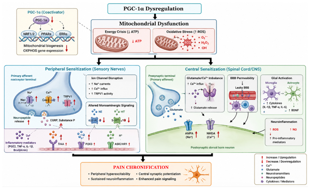
Figure 2.
Mitochondrial toxicity and oxidative stress induced by common analgesics and adjuvants. ATP, adenosine triphosphate; mtDNA, mitochondrial DNA; NF-κB, nuclear factor kappa B; NSAIDs, non-steroidal anti-inflammatory drugs; ROS, reactive oxygen species.
Figure 2.
Mitochondrial toxicity and oxidative stress induced by common analgesics and adjuvants. ATP, adenosine triphosphate; mtDNA, mitochondrial DNA; NF-κB, nuclear factor kappa B; NSAIDs, non-steroidal anti-inflammatory drugs; ROS, reactive oxygen species.
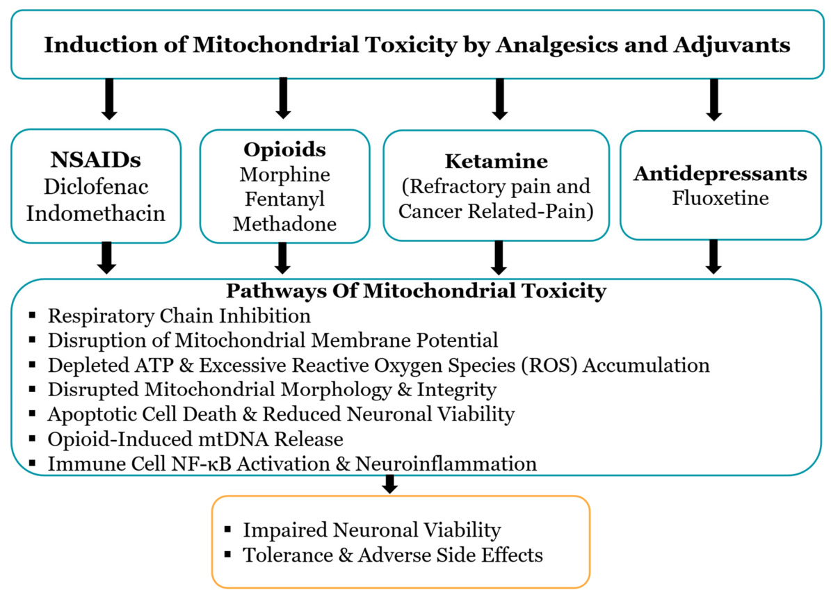
Figure 3.
The upstream activation and downstream neuroprotective pathways of PGC-1α in Chronic Pain. AMPK, AMP-activated protein kinase; CAT, catalase; ERRs, estrogen-related receptors; ETC, electron transport chain; GPX1, glutathione peroxidase 1; mtDNA, mitochondrial DNA; NF-κB, nuclear factor kappa B; NRF-1/2, nuclear respiratory factors 1 and 2; PPARs, peroxisome proliferator-activated receptors; ROS, reactive oxygen species; SIRT1, sirtuin 1; SOD2, superoxide dismutase 2; TFAM, mitochondrial transcription factor A.
Figure 3.
The upstream activation and downstream neuroprotective pathways of PGC-1α in Chronic Pain. AMPK, AMP-activated protein kinase; CAT, catalase; ERRs, estrogen-related receptors; ETC, electron transport chain; GPX1, glutathione peroxidase 1; mtDNA, mitochondrial DNA; NF-κB, nuclear factor kappa B; NRF-1/2, nuclear respiratory factors 1 and 2; PPARs, peroxisome proliferator-activated receptors; ROS, reactive oxygen species; SIRT1, sirtuin 1; SOD2, superoxide dismutase 2; TFAM, mitochondrial transcription factor A.
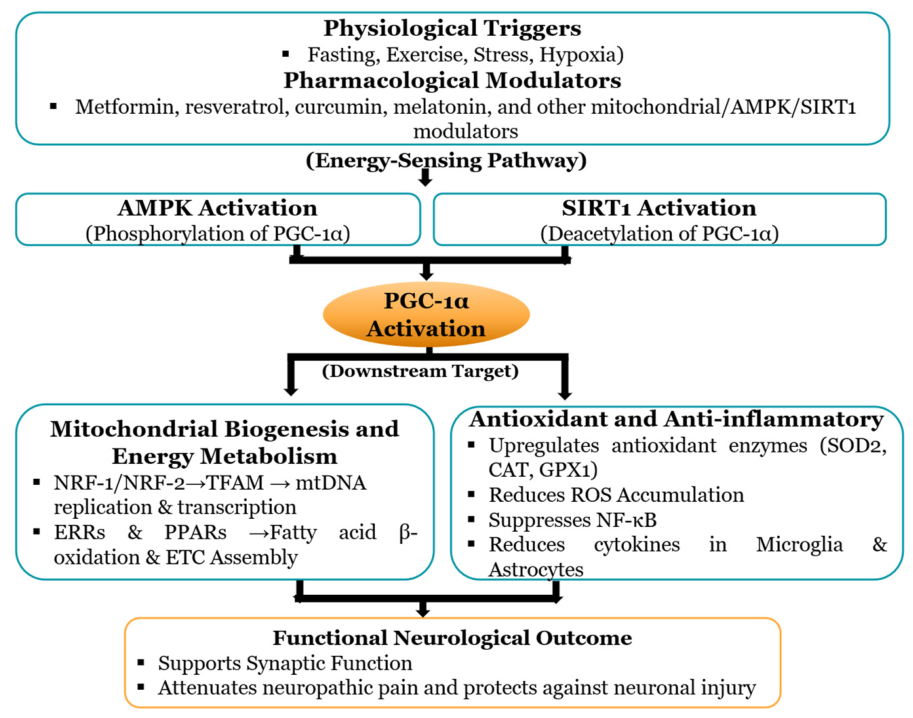

Table 1.
PGC-1α dysregulation in musculoskeletal pain.
| Pain Model/References | PGC-1α Expression | Tissue/Cell Affected | Effects of PGC-1α Deficiency | Biological Effects of PGC-1α Restoration |
| Ageing-associated pain after nerve injury [78] |
Age-related cortical PGC-1α decline | Somatosensory cortex (S1) GABAergic interneurons | Persistent S1 excitatory overactivity |
Restoration of cortical dynamics and reversal of chronification |
| Osteoarthritis [92] |
Downregulation of AMPK signaling | L4-L6 spinal dorsal horn neurons | Increased oxidative stress, reduced NRF1/TFAM/mtDNA, and spinal gliosis |
Sesn2 reactivated AMPK/PGC-1α signaling and alleviated mechanical allodynia. |
| Post-Traumatic Osteoarthritis & Muscle Wasting [75] |
PGC-1α downregulation in skeletal muscle | Quadriceps skeletal muscle | Muscle atrophy, inflammation, and cartilage loss |
Joint distraction and treadmill exercise restored PGC-1α and suppressed muscle wasting. |
| Myofascial pain syndrome [67] |
AMPK-PGC-1α-SIRT3 expression was downregulated in skeletal muscle. | Rat skeletal muscle. | Depressed mitochondrial function and myofascial pain. | Restoring AMPK/PGC-1α/SIRT3 axis could enhance mitochondrial health and alleviate myofascial pain. |
| Monosodium iodoacetate-induced osteoarthritis [94] |
PGC-1α level decreased in the lumbar spinal cord. | Lumbar spinal cord. | Mitochondrial dysfunction and pain behaviors. | Dimethyl fumarate alleviated osteoarthritis pain by increasing PGC-1α level |
| Collagen-induced osteoarthritis [93] |
Sirt3/PGC-1α expression was suppressed in osteoarthritic tissue. | Knee joint osteoarthritic tissue. | Mitochondrial dysfunction, oxidative stress, and cartilage degeneration. | SDF alleviated osteoarthritis via the Sirt3/PGC-1α pathway. |
Table 2.
PGC-1α Dysregulation in Peripheral Nerve Injury.
| Pain Model/Reference | PGC-1α expression | Tissue/Cell Affected | Effects of PGC-1α Deficiency | Biological Effect of PGC-1α Restoration |
| CCI-triggered neuropathic pain [41] |
Spinal PGC-1α, NRF1, TFAM, and mtDNA were reduced. |
L4-L6 spinal cord dorsal horn | Decrease in mitochondrial biogenesis and hyperalgesia. |
RTA-408 and ZLN005 increased PGC-1α and alleviated pain. |
| SNI-induced neuropathic pain [98] |
Spinal AMPK/PGC-1α signaling was impaired. | Spinal dorsal horn. | Reduced NRF1/TFAM-dependent mitochondrial biogenesis and increased ROS/oxidative stress. | rhHSP22 activated PGC-1α, restored mitochondrial biogenesis, and alleviated hypersensitivity. |
| CCI-induced neuropathic pain (Chen et al., 2025) |
Spinal PGC-1α activity was reduced. | Dorsal horn microglia. |
Mitochondrial dysfunction and NLRP3 activation |
PGC-1α activation restored mitochondrial function, shifted microglia toward an anti-inflammatory phenotype, and reduced pain. |
| SNI-induced neuropathic pain [77] | Spinal PGC-1α signaling was impaired. | Lumbar dorsal horn neurons. | Reduced mitochondrial biogenesis and persistent mechanical allodynia. | Lasmiditan activated PGC-1α, restored mitochondrial biogenesis, and reduced allodynia. |
| Peripheral nerve injury [95] |
AMPK/PGC-1α/PPAR-γ signaling was reduced in macrophages. | Bone-marrow-derived macrophages at the injury site. |
Increased iNOS/TNF-α/IL-1β, impaired axon regeneration and remyelination. | Metformin activated PGC-1α and improved regeneration and recovery. |
| CCI-induced neuropathic pain [23] |
PGC-1α transcription was repressed by Sp1/HDAC2. |
Spinal cord microglia and neurons. | Mitochondrial disruption, oxidative damage, and neuronal dysfunction. | PGC-1α overexpression reversed mitochondrial dysfunction, and alleviated pain. |
| SNL-induced neuropathic pain [100] |
Nuclear SIRT1/PGC-1α signaling was reduced. | Spinal dorsal horn | Abnormal mitochondrial biogenesis, high oxidative stress, and mechanical allodynia. |
Ro5-4864 restored PGC-1α, improved mitochondrial biogenesis, and reduced allodynia. |
| SNI-induced neuropathic pain [101] |
PGC-1α signaling was reduced. |
Spinal dorsal horn astrocytes. | Mitochondrial dysfunction, disrupted dynamics, pyroptosis, and inflammation. |
TSPO activation restored PGC-1α, improved mitochondrial health, and reduced pain. |
| SNI-induced neuropathic pain [96] |
ChREBP increased PGC-1α transcription. | Spinal dorsal horn microglia | Reduced PGC-1α lowered fatty acid oxidation and promoted pro-inflammatory effects. |
PGC-1α restored microglial fatty acid oxidation, reduced cytokines, and pain. |
| SNI-induced neuropathic pain [74] |
SIRT1/PGC-1α signaling and GPR39 expression were reduced. |
Spinal dorsal horn neurons and microglia | Mitochondrial dysfunction, neuroinflammation, and persistent allodynia |
TC-G 1008 activated PGC-1α, improved mitochondrial function, and reduced pain. |
| CCI-induced neuropathic pain [103] |
PGC-1α was reduced and restored by a ketogenic diet. |
Spinal dorsal horn microglia. | Neuroinflammation, ROS accumulation, and mitochondrial dysregulation. |
Ketogenic diet increased PGC-1α, improved mitochondrial function, and relieved pain. |
| SNL-induced neuropathic pain [104] |
SIRT1 and PGC-1α were reduced in DRG neurons. |
L5-L6 dorsal root ganglion neurons. |
Mitochondrial dysregulation and oxidative stress. | Melatonin restored PGC-1α expression, improved mitochondrial biogenesis, and reduced pain. |
Table 3.
PGC-1α Dysregulation in Chemotherapy-Induced Neuropathic Pain, Cancer-Induced Bone Pain, and Diabetic-Induced Neuropathic Pain.
Table 3.
PGC-1α Dysregulation in Chemotherapy-Induced Neuropathic Pain, Cancer-Induced Bone Pain, and Diabetic-Induced Neuropathic Pain.
| Pain Model/Reference | PGC-1α expression | Tissue/Cell | Effects of PGC-1α Deficiency | Biological Effect of PGC-1α Restoration |
| Paclitaxel-induced neuropathic pain [110] |
PGC-1α-mediated mitochondrial biogenesis was induced in the spinal cord. | Spinal cord. | Impaired mitochondrial biogenesis contributed to mechanical allodynia. | β2-adrenoreceptor activation restored PGC-1α and alleviated mechanical allodynia. |
| Paclitaxel-induced peripheral neuropathy [80] |
PGC-1α was expressed in dorsal root ganglion | Dorsal root ganglion | Oxidative stress and mitochondrial damage promoted peripheral neuropathy. | PACAP activated PGC-1α, enhanced mitochondrial health, reduced oxidative damage, and alleviated neuropathy. |
| Cancer-induced bone pain [85] |
PGC-1α signaling was suppressed. | Spinal microglia | M1 polarization, NF-κB activation, and reduced GPx4. | Naringenin activated PGC-1α, promoted M2 polarization, and alleviated pain. |
| Cancer-induced bone pain [86] |
PGC-1α was downregulated in the spinal cord. | Spinal GABAergic interneurons | Interneuron apoptosis, mitochondrial dysfunction, increased ROS, and allodynia. | ZLN005 restored PGC-1α, preserved interneurons, and reduced allodynia. |
| Diabetic peripheral neuropathy [84] |
PGC-1α was suppressed. | Sciatic nerves, dorsal root ganglia neurons, and Schwann cells. | Mitochondrial dysfunction and oxidative stress. |
Quercetin activated PGC-1α and improved neuropathy. |
| Streptozotocin-induced painful diabetic neuropathy [82] |
PGC-1α expression was induced by a mitochondria-derived peptide (MOTS-c) | Spinal dorsal horn. | Mitochondrial dysfunction with hyperalgesia and allodynia. |
MOTS-c activated PGC-1α, improved mitochondrial health, and alleviated pain |
| Streptozotocin-induced diabetic neuropathy [83] |
Suppression of PGC-1α signaling. | Sciatic nerves and Schwann cells. |
Mitochondrial imbalance, axonal degeneration, and oxidative stress. | Tang Bi formula activated PGC-1α and improved nerve injury |
Table 4.
PGC-1α Dysregulation in Fibromyalgia.
| Pain Model/Reference | PGC-1α expression | Tissue/Cell Affected | Effects of PGC-1α Deficiency | Biological Effect of PGC-1α Restoration |
| Reserpine-induced fibromyalgia [87] |
Alteration in hippocampal PGC-1α. | Hippocampus | Cognitive decline, reduced locomotion, increased depression-like behavior, and mechanical allodynia. | Exercise training activated hippocampal PGC-1α and improved fibromyalgia symptoms. |
| Reserpine-induced fibromyalgia [88] |
Reduction in PGC-1α expression. | Gastrocnemius and soleus muscle | Redox imbalance, reduced mitochondrial biogenesis, and fibromyalgia-like pain | CoQ10 restored redox homeostasis and increased PGC-1α |
| Reserpine-treated fibromyalgia [88] |
PGC-1α signaling was altered. | Hippocampus |
Fibromyalgia-like symptoms, cognitive deficits, and impaired mitochondrial and redox homeostasis. | CoQ10 enhanced hippocampal PGC-1α signaling and improved symptoms and cognition. |
| Fibromyalgia model [111] |
PGC-1α and CoQ10 were reduced. | Gastrocnemius muscle | Mitochondrial dysfunction and oxidative stress. | Melatonin preserved mitochondrial health and prevented oxidative stress via PGC-1α signaling |
| Reserpine-induced fibromyalgia [76] |
PGC-1α signaling was suppressed. | Spinal cord | Central sensitization and nociplastic pain, with MAPK/NF-κB activation | Melatonin restored PGC-1α signaling and reduced pain sensitization. |
Table 5.
PGC-1α Dysregulation in Chronic Headache Disorders.
| Pain Model/Reference | PGC-1α expression | Tissue/Cell Affected | Effects of PGC-1α Deficiency | Biological Effect of PGC-1α Restoration |
| Nitroglycerin-induced chronic migraine [73] |
Downregulation of PGC-1α. | Trigeminal nucleus | Inflammation and central sensitization. | SIRT1 overexpression or SRT1720 alleviated inflammation and pain sensitization. |
| Nitroglycerin-induced migraine mouse model [89] |
Decreased PGC-1α expression. | Trigeminal ganglion neurons. | Mitochondrial dysfunction, periorbital allodynia, and mechanical hyperalgesia. |
Schisandra chinensis and mitoquinone improved mitochondrial function and alleviated migraine pain. |
| Inflammatory-soup-induced headache model [90] |
Reduction in PGC-1α expression. | Trigeminal nucleus caudalis. | Nociceptive responses and mitochondrial dysfunction. | SS-31 abolished nociception and restored mitochondrial function. |
Table 6.
PGC-1α Dysregulation in Opioid-Induced Hyperalgesia.
| Pain Model/References | PGC-1α expression | Tissue/Cell Affected | Effects of PGC-1α Deficiency | Biological Effect of PGC-1α Restoration |
| Morphine tolerance [91] |
Reduced PGC-1α-expression | Spinal cord. | Nrf2 inhibition and mitochondrial dysfunction. |
PGC-1α activator ZLN005 protected against spinal cord apoptosis and morphine tolerance. |
| Remifentanil-induced hyperalgesia [112] |
p38 MAPK/PGC-1α/SIRT3 expression was suppressed. | Spinal cord. | ROS release and hyperalgesia. | Modulating p38 MAPK/PGC-1α signaling reduced hyperalgesia. |
| Morphine tolerance [26] |
PGC-1α suppressed spinal morphine tolerance. | Spinal cord. | Increased mitochondrial superoxide and tolerance. | Recombinant PGC-1α reduced mitochondrial superoxide and abolished morphine tolerance. |
Disclaimer/Publisher’s Note: The statements, opinions and data contained in all publications are solely those of the individual author(s) and contributor(s) and not of MDPI and/or the editor(s). MDPI and/or the editor(s) disclaim responsibility for any injury to people or property resulting from any ideas, methods, instructions or products referred to in the content. |
© 2026 by the authors. Licensee MDPI, Basel, Switzerland. This article is an open access article distributed under the terms and conditions of the Creative Commons Attribution (CC BY) license (http://creativecommons.org/licenses/by/4.0/).
Copyright: This open access article is published under a Creative Commons CC BY 4.0 license, which permit the free download, distribution, and reuse, provided that the author and preprint are cited in any reuse.