

Submitted:
20 August 2026
Posted:
21 August 2026
You are already at the latest version
Abstract
Equine asthma is a chronic neutrophilic airway disease that can be exacerbated by obesity, an immunometabolic condition increasingly linked to activation of the NLRP3 inflammasome and the release of interleukin-1 beta (IL-1β). Tamoxifen, a selective oestrogen receptor modulator with immunomodulatory properties independent of its hormonal action, has previously been shown to reduce neutrophilic airway inflammation in horses, although its effect on NLRP3 inflammasome activation remains unknown. This study aimed to determine whether tamoxifen modulates NLRP3, ASC and caspase-1 gene expression, and IL-1β production, in neutrophils isolated from obese and non-obese asthmatic horses in clinical remission (n = 3 per group), using an in vitro model of inflammasome activation induced with lipopolysaccharide (LPS) and nigericin. Gene expression was assessed by RT-qPCR and IL-1β secretion by ELISA. LPS–nigericin stimulation markedly increased NLRP3 and IL-1β expression in both groups; co-treatment with tamoxifen (5 µM) significantly attenuated the LPS–nigericin-induced increase in NLRP3 expression irrespective of body condition, without affecting ASC or caspase-1. By contrast, tamoxifen significantly reduced IL-1β gene expression and secretion specifically in neutrophils from obese horses, whereas non-obese horses showed little or no response. Obese horses also exhibited higher basal IL-1β gene expression and serum concentrations than non-obese horses. These findings indicate that tamoxifen modulates NLRP3 inflammasome activation and that its effect on IL-1β is body condition-dependent, supporting its potential as an immunomodulatory agent for neutrophilic, obesity-associated equine asthma.
Keywords:
tamoxifen
; equine
; pro-inflammatory cytokines
; NLRP3 inflammasome
1. Introduction
Equine asthma is a chronic respiratory disease affecting adult horses that shares important pathophysiological similarities with human asthma, particularly with regard to airway inflammation and the involvement of innate immune mediators [1]. The condition is characterised by reversible airflow obstruction, bronchial hyperresponsiveness, and marked neutrophilic infiltration, the persistence of which contributes to airway remodelling and the progressive deterioration of respiratory function [2]). Owing to these similarities, equine asthma has been proposed as a relevant natural model for studying chronic inflammatory respiratory diseases [3,4]
Obesity is defined as the abnormal or excessive accumulation of fat that can be harmful to the body, and is currently considered a global epidemic [5,6]. The worrying increase in overweight and obesity in today’s society is not limited to humans, with significant increases reported in companion and domestic animals [7,8]. In horses, numerous quantitative studies have also described a sustained growth in the prevalence of overweight and obesity [9,10,11], principally attributable to poor management practices, such as inappropriate feeding regimes and limited exercise [12]. Furthermore, obesity is often poorly recognized by owners, who tend to underestimate the body condition score (BCS) of their animals [13]. Evidence from several studies points to an increased risk of asthma among obese individuals relative to their lean counterparts, regardless of sex or age [14,15,16,17,18,19,20,21,22], an association that appears to be stronger in individuals with central adiposity [23,24]. Adipose tissue contributes to the modulation of inflammatory and immune processes through the secretion of adipokines, including leptin—a pro-inflammatory mediator—and adiponectin, which acts in an anti-inflammatory capacity [25,26]. In line with this, in asthmatic-obese subjects there is a rise in the serum concentrations of the pro-inflammatory adipokine leptin whereas adiponectin levels are decreased [27]. Furthermore, severe asthma is extremely common in patients with obesity [28]. Besides, human obese patients tend to have more severe asthma that does not respond as well to conventional therapy compared with lean asthmatics [29]. In horses, obesity, defined as a body condition score (BCS) of 7 or above [30], has been identified as a significant risk factor for the development of equine asthma [31]
On the other hand, the mechanisms that translate obesity into chronic inflammation also involve innate immunity sensors such as toll-like receptor 4 (TLR4) [32,33]; it has been shown that activation of this receptor leads to an increase in IL-1β expression; for this reason, this cytokine is elevated in the blood and airway of obese individuals [34,35]. The release of IL-β from cells depends on the activation of caspase-1, which cleaves it from its precursor protein. Caspase-1 is activated upon assembly of the nucleotide oligomerization domain-like receptor 3 (NLRP3, inflammasome) protein and the adaptor molecule, ASC (apoptosis-associated speck-like protein containing a caspase-recruitment domain) [36,37]. Initiation of expression and assembly of inflammasome components occurs through NF-ĸB activation [38,39,40,41]. Activation of the inflammasome in obese individuals is produced through TLR4 by fatty acids [28,42]. Recently, our research group demonstrated that obese asthmatic horses in remission exhibited a significantly higher expression of IL-1β compared with lean asthmatic horses in remission [43].
Corticosteroids remain the mainstay of treatment for equine asthma, but their long-term use is limited by the risk of laminitis, immunosuppression, and endocrine-metabolic disturbances [44]. Tamoxifen (TX), a selective oestrogen receptor modulator, is proposed as a therapeutic alternative, supported by several previous studies: TX preferentially induced apoptosis in granulocytes compared with mononuclear cells (in vitro and in vivo), activated the intrinsic apoptotic pathway in equine neutrophils, inhibited respiratory burst, chemotaxis, and chemokinesis of neutrophils in a dose-dependent manner, and induced efferocytosis by macrophages through an oestrogen-independent mechanismal. [45,46,47,48]. In vivo, oral TX (0.25 mg/kg, for five days) significantly reduced BALF neutrophil counts and improved the clinical status of horses with airway inflammation, with results similar to, or even better than, those observed with dexamethasone [49]; an independent group confirmed an improvement in airway resistance (although without a significant change in BALF neutrophil counts) in horses with severe exacerbated equine asthma [50].
Despite this evidence, it remains unknown whether TX is able to directly modulate NLRP3 inflammasome activation and IL-1β production in equine neutrophils, and whether this effect is influenced by the animal’s body condition. Given that obesity could amplify activation of this inflammatory pathway and reduce the response to conventional therapies, it is relevant to determine whether TX has a potential modulatory effect on this mechanism in the specific context of obesity-associated equine asthma. The present study aimed to evaluate the effect of TX on the expression of NLRP3 inflammasome components (NLRP3, ASC and caspase-1) and on IL-1β production in neutrophils from obese and non-obese asthmatic horses, using an in vitro model of inflammatory activation induced with lipopolysaccharide and nigericin
2. Materials and Methods
2.1. Animals
In this study, six horses with naturally occurring equine asthma in clinical remission were included and allocated into two experimental groups according to body condition: three obese and three non-obese horses. The initial diagnosis of severe equine asthma was established through a general clinical examination and bronchoalveolar lavage (BAL). At the time of diagnosis, all horses presented a clinical score greater than 15, according to the scoring system described by Lavoie et al. [51]. Cytological analysis of the BAL fluid confirmed a neutrophilic asthmatic phenotype, characterised by a neutrophil proportion exceeding 20%. During the clinical remission period, the horses showed no evident respiratory signs, such as coughing, dyspnoea, or nasal discharge. In addition, BAL cytology revealed a neutrophil proportion below 8%, consistent with disease remission.
The animals were between 5 and 12 years of age, were crossbred Chilean Criollo horses, and the experimental group comprised four mares and two geldings. None of the horses received treatment with corticosteroids or bronchodilators for at least two months prior to the study, in order to avoid potential pharmacological effects on the inflammatory response evaluated. The animals were classified according to their body condition using the nine-point Body Condition Score (BCS) system described by Henneke et al. [30]. Horses included in the obese group had a BCS ≥ 7/9, whereas non-obese horses had a BCS ≤ 6/9. This scale evaluates morphological parameters such as the difficulty in palpating the ribs, the presence of fat deposits around the tail head, fat accumulation at the withers and behind the scapula, thickening of the neck, and fat deposits in the lumbar region and inner thigh. Obese horses were maintained on pasture, and their diet consisted of a mixture of alfalfa, concentrate, oil, and hay. In contrast, non-obese horses were primarily fed daily hay. Both groups had ad libitum access to water throughout the entire experimental period at the Veterinary Teaching Hospital of the Universidad Austral de Chile. All experimental procedures were approved by the Bioethics Committee for the Use of Animals in Biomedical Research of the Universidad Austral de Chile (approval resolution No. 497/2023).
2.2. Leukocytes Isolation
Blood collection was performed via jugular venepuncture using 30 mL syringes connected to a 21 G scalp vein needle. A total of 20 mL of blood was obtained from each horse and transferred into sterile 50 mL conical tubes containing 2 mL of 3.8% (w/v) sodium citrate as an anticoagulant. Each animal was sampled once, and all samples were processed and analysed independently. Cell separation was conducted using a discontinuous Percoll® (GE Healthcare) gradient. In sterile 15 mL conical tubes, 4 mL of 70% Percoll was added to the bottom, followed by 4 mL of 80% Percoll layered above. Subsequently, 6 mL of whole blood (containing 3.8% sodium citrate) was carefully layered on top. Samples were centrifuged at 400 × g for 45 min to achieve separation of the cellular populations. Mononuclear and polymorphonuclear (PMN) cells were collected into sterile plastic tubes, washed with 1% citrate-phosphate-buffered saline (PBS), and resuspended in 2 mL of complete RPMI 1640 medium (Gibco) supplemented with penicillin–streptomycin–neomycin (PSN; Gibco) and 10% (v/v) foetal bovine serum (FBS; Gibco). Cell counts were determined using a Neubauer chamber and manual haemocytometer. All procedures were carried out in a Class II laminar flow cabinet to ensure sterility and prevent contamination.
2.3. Stimulation of Neutrophils for NLRP3 Inflammasome Expression
To induce in vitro activation of the NLRP3 inflammasome, purified neutrophils were stimulated with lipopolysaccharide (LPS) at a concentration of 100 ng/mL for 2.5 hours; subsequently, the cells were washed with PBS and centrifuged at 1200 × g for 7 minutes, after which nigericin was added at a final concentration of 5 μM and incubated for a further 1.5 hours, completing a total experimental duration of 4 hours. The effect of TX on inflammasome activation was assessed by incubation at a final concentration of 5 μM, and in all experiments a negative control with 0.1% DMSO was included as the vehicle of TX. The experimental model comprised the following conditions: a negative control (neutrophils incubated in culture medium without inflammatory stimulation), an inflammatory condition (neutrophils stimulated with LPS followed by nigericin to induce NLRP3 inflammasome activation), and TX conditions (cells subjected to the inflammatory protocol in the presence of the drug). The experimental model was applied to neutrophils obtained from obese and non-obese asthmatic horses. All incubations were performed in cell culture plates maintained at 37 °C in a humidified atmosphere containing 5% CO₂. At the end of the treatment, cells from each group were collected into microcentrifuge tubes and centrifuged at 15,000 × g for 1 min. The supernatant was carefully discarded, and 1 mL of TRIzol™ reagent was added. Samples were homogenised using a 1 mL syringe fitted with a 25 G needle and stored at −20 °C for 12 h prior to RNA extraction.
2.4. RNA Extraction and Real-Time PCR
Samples preserved at −20 °C in TRIzol™ were thawed, and 300 µL of absolute chloroform was added to each sample. The mixture was homogenised under refrigeration for 4 minutes, followed by centrifugation at 12,000 × g for 15 minutes. The supernatant was discarded, and 500 µL of isopropanol was added. Homogenisation was performed by immersion, and the samples were then incubated at −20 °C for 20 minutes. After incubation, the samples were centrifuged at 12,000 × g for 10 minutes. The supernatant was removed, and 1 mL of ethanol was added to wash the pellet. The mixture was vortexed and centrifuged at 8,000 × g for 5 minutes. Following supernatant removal, the pellet was resuspended in 30 µL of DEPC-treated water (autoclaved and filtered through a membrane filter), and incubated at 60 °C for 10 minutes. Subsequently, 30 µL of each sample was treated with 3.5 µL of DNase buffer and 1.5 µL of DNase enzyme. This mixture was incubated at 37 °C for 30 minutes. DNase inactivation was performed by adding 4 µL of DNase inactivator, followed by vortexing and centrifugation at 10,000 × g for 1.5 minutes. The resulting supernatant was collected and quantified using a NanoDrop™ spectrophotometer. Samples were then adjusted to a concentration of 1.5 µg and incubated at 65 °C for 10 minutes. For reverse transcription, 1 µL of M-MLVRT buffer, 1.25 µL of dNTP mix, 0.5 µL of RNaseOUT, 1 µL of M-MLVRT enzyme, and 2.25 µL of DEPC-treated water were added per sample. The reaction was incubated at 44 °C for 60 minutes. Finally, quantitative PCR (qPCR) was performed using a master mix containing SYBR Green®, the corresponding primers, cDNA, and nuclease-free water. Reactions were loaded into MicroAmp™ 8-tube strips, with eight wells allocated per gene, and amplification was conducted using an Applied Biosystems StepOne™ Real-Time PCR System. The primers used for real-time PCR amplification were as follows: NLRP3 (forward: 5′-AAA CTG GAA GAG CCG GAG TG-3′; reverse: 5′-TGT TGA GGT TGA CGC TCT CG-3′), IL-1β (forward: 5′-AGT ACC CGA CAC CAG TGA CA-3′; reverse: 5′-GCC ACA ATG ATT GAC ACG ACA-3′), ASC (Forward: 5′-TCTATCTGGAGGCGTACGGAG-3′, Reverse: 5′-ATCCACTTCTGTGACCCGTG-3′); Caspasa-1 (Forward: 5′-GGCTTGCGCTCATTATCTGC-3′, Reverse: 5′-AAAGTGCTGTCAGAGGACCG-3′).
2.5. Enzyme Linked Immunosorbent Assay (ELISA) Analysis
Secreted IL-1β in the supernatants was quantified in obese and non-obese asthmatic horses using a commercial equine-specific ELISA kit (MyBioSource, San Diego, CA, USA), following the manufacturer’s instructions. In the post-treatment protocol, inflammatory stimulation was initiated at time zero with LPS (100 ng/mL) for 2.5 hours; thereafter, the cells were washed and incubated with nigericin (5 μM) in the presence of TX (5 μM) for a further 1.5 hours. All incubations were performed in cell culture plates maintained at 37 °C in a humidified atmosphere containing 5% CO₂. The assay had a detection range of 31.25 to 2000 pg/mL and an inter-assay variability of 8%. Absorbance was measured at 450 nm using a Varioskan Flash multimode plate reader (Thermo Fisher Scientific, Waltham, MA, USA). A four-parameter logistic (4PL) model was applied to generate the standard curve and analyse the data using GraphPad Prism software (version 9.1.0; GraphPad Software Inc., Boston, MA, USA)
2.6. Statistics Analysis
Graph generation and statistical analyses were performed using GraphPad Prism (GraphPad Software Inc., version 10.2.2). Data normality was assessed using the Shapiro–Wilk test, and all datasets were found to follow a normal distribution. For each experiment, mean differences between conditions were analysed using Tukey’s multiple comparisons test. Results are presented as mean ± standard deviation (SD), and a p-value < 0.05 was considered statistically significant
3. Results
3.1. Expression of Inflammasome Components Following Tamoxifen Treatment
Figure 1 shows the relative gene expression of NLRP3 inflammasome components in neutrophils isolated from obese and non-obese horses with asthma. LPS–nigericin stimulation markedly increased nlrp3 expression in both groups compared with the control and TX-only conditions (Figure 1A). Treatment with TX significantly attenuated the LPS–nigericin-induced increase in nlrp3 expression, with no evident differences between obese and non-obese horses. In contrast, asc and casp1 expression remained relatively stable across the different experimental conditions (Figure 1B,C).
3.2. IL-1β Expression Following Tamoxifen Treatment After the Induction of Inflammation
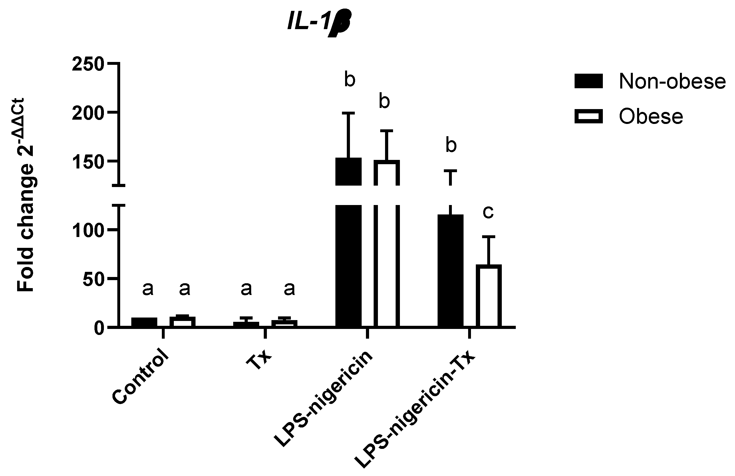
Figure 2 shows the relative gene expression of IL-1β in neutrophils isolated from obese and non-obese asthmatic horses. LPS–nigericin stimulation significantly increased IL1-β expression in both groups relative to the control and TX-only conditions. In neutrophils from non-obese horses, treatment with TX did not significantly attenuate LPS–nigericin-induced IL-1β expression. In obese horses, however, TX significantly reduced IL-1β expression compared with LPS–nigericin stimulation alone, although levels remained elevated relative to the control and TX-only conditions. Moreover, following co-treatment with LPS–nigericin and TX, IL-1β expression was significantly lower in obese than in non-obese horses. Taken together, these findings suggest that the modulatory effect of TX on IL-1β expression may be dependent on the body condition of asthmatic horses.
3.3. Effect of Tamoxifen on LPS–nigericin-induced IL-1β Secretion by Neutrophils from Obese and Non-Obese Asthmatic Horses in Vitro
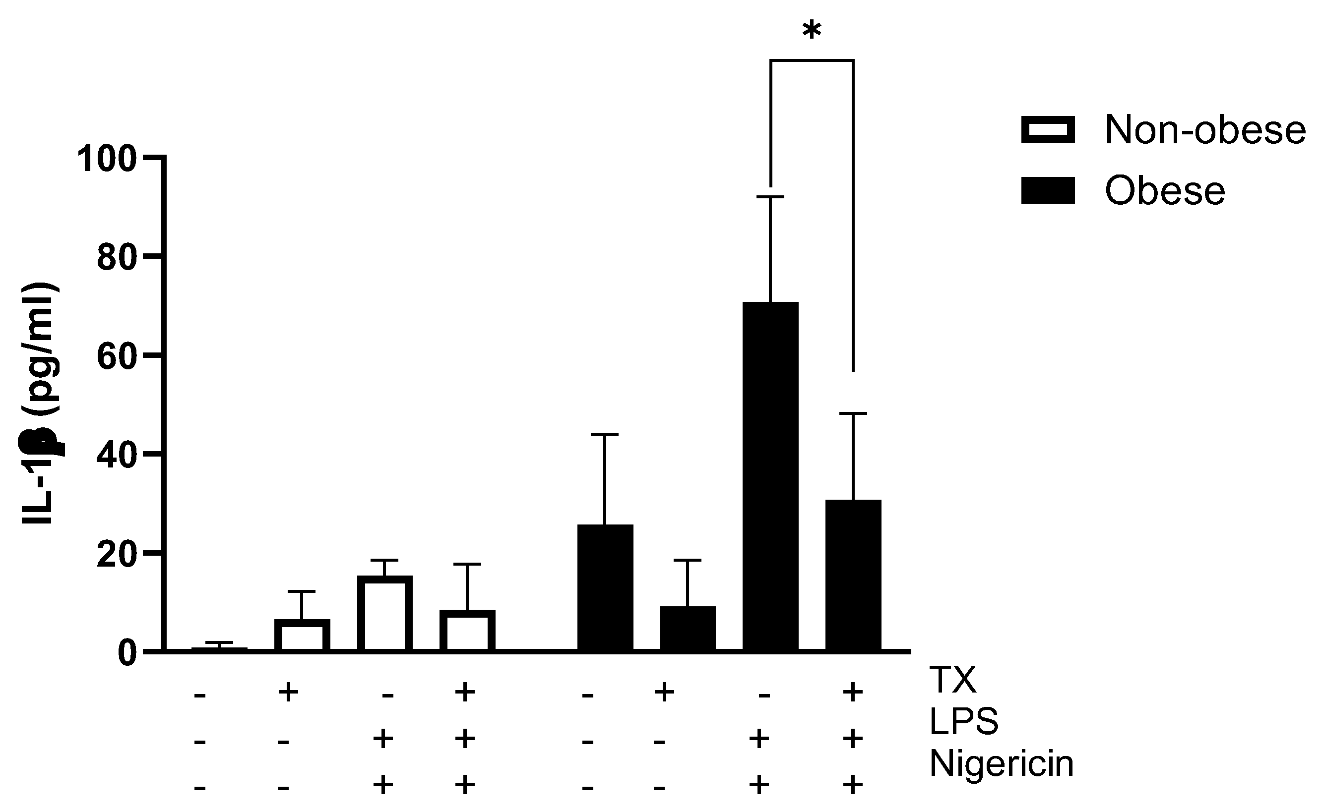
L-1β secretion, measured by ELISA, in neutrophils treated with tamoxifen following the induction of inflammation differed significantly between obese and non-obese animals. However, this difference was not directly attributable to TX treatment, but rather reflected a baseline difference between the two groups. Nevertheless, the observed trend suggests that obesity may enhance the inflammatory response in the context of equine asthma. When the experimental conditions were analysed within the obese group, significant differences were also detected among the inflammatory stimuli evaluated. In particular, IL-1β secretion induced by the complete inflammasome activation protocol (LPS + nigericin) was modulated by TX treatment, with a reduction in the extracellular levels of this cytokine observed in drug-treated samples. Taken together, these results indicate that TX exerts a modulatory effect on IL-1β secretion, particularly in neutrophils derived from obese horses, suggesting that this drug may contribute to regulating the exacerbated inflammatory response associated with the interaction between obesity and asthma.
Figure 3.
In vitro effect of tamoxifen on IL-1β secretion by neutrophils isolated from obese and non-obese horses with asthma following stimulation with LPS and nigericin. Neutrophils were incubated with tamoxifen (TX; 5 μM) and/or stimulated with LPS and nigericin. IL-1β concentrations were measured in the culture supernatants and expressed as pg/mL. Data are presented as the mean ± standard deviation of three independent experiments (n = 3 horses per group). *p < 0.05 indicates a significant difference between the LPS–nigericin condition and co-treatment with tamoxifen in neutrophils from obese horses.
Figure 3.
In vitro effect of tamoxifen on IL-1β secretion by neutrophils isolated from obese and non-obese horses with asthma following stimulation with LPS and nigericin. Neutrophils were incubated with tamoxifen (TX; 5 μM) and/or stimulated with LPS and nigericin. IL-1β concentrations were measured in the culture supernatants and expressed as pg/mL. Data are presented as the mean ± standard deviation of three independent experiments (n = 3 horses per group). *p < 0.05 indicates a significant difference between the LPS–nigericin condition and co-treatment with tamoxifen in neutrophils from obese horses.
4. Discussion
The present study aimed to determine whether TX modulates the activation of the NLRP3 inflammasome and the production of IL-1β in neutrophils from asthmatic horses, and whether this modulatory effect is influenced by body condition. Overall, our findings indicate that TX acts differentially at distinct levels of the inflammatory cascade: it markedly attenuated the transcriptional induction of NLRP3 irrespective of body condition, while its effect on IL-1β expression and secretion was more pronounced in neutrophils from obese horses, suggesting that obesity may render this inflammatory pathway both more active and more responsive to pharmacological modulation.
LPS–nigericin stimulation markedly increased nlrp3 transcription in neutrophils from both obese and non-obese horses, consistent with the two-signal model of canonical NLRP3 inflammasome activation, in which TLR4-mediated priming induces transcriptional upregulation of NLRP3 prior to the assembly of the inflammasome complex [52]. Treatment with TX significantly blunted this increase in both groups, indicating that the drug interferes with the priming phase itself rather than acting selectively in obese animals. This observation is consistent with previous reports showing that tamoxifen inhibits NF-κB activation more effectively than dexamethasone in vitro [53], since NF-κB is the principal transcription factor driving nlrp3 induction downstream of TLR4 [54]. In contrast, asc and caspase-1 expression remained essentially unchanged across all experimental conditions, in agreement with the notion that these components are constitutively expressed in innate immune cells and are therefore not subject to the same transcriptional regulation as the NLRP3 sensor itself [55]. Taken together, these results suggest that the transcriptional step most sensitive to tamoxifen is the priming-dependent induction of NLRP3, whereas the assembly components of the inflammasome complex are largely unaffected at the gene expression level.
A different pattern emerged for IL-1β gene expression. Although LPS–nigericin stimulation increased IL-1β transcription in both groups, TX significantly reduced this response only in neutrophils from obese horses, with no comparable effect observed in non-obese animals. This divergence indicates that the modulatory action of TX on IL-1β transcription is not uniform but is instead conditioned by the body condition of the animal. One plausible explanation is that neutrophils from obese horses operate under a higher basal inflammatory tone, driven by circulating adipokines such as leptin and by free fatty acid-mediated TLR4 signalling [56,57], which may render the IL-1β transcriptional programme more dependent on, and therefore more susceptible to, NF-κB inhibition. In non-obese horses, where baseline inflammatory activation is comparatively lower, additional or redundant signalling pathways may sustain IL-1β transcription independently of the NF-κB axis targeted by tamoxifen, thereby limiting the drug’s efficacy in this group.
Beyond gene expression, tamoxifen also modulated the extracellular secretion of IL-1β following LPS–nigericin stimulation, again with a clearer effect in neutrophils from obese horses. Since mature IL-1β release depends on caspase-1-mediated proteolytic cleavage of the pro-cytokine and on gasdermin D-dependent pore formation [58], this finding suggests that TX may additionally interfere with post-translational steps of inflammasome activation, beyond its transcriptional effect on NLRP3. Such an action would be consistent with previous work demonstrating that TX inhibits reactive oxygen species production and chemotaxis in equine neutrophils [47,48] and promotes their apoptotic clearance by alveolar macrophages [46], all of which point to a broader capacity of the drug to dampen neutrophil effector functions independently of, or in addition to, transcriptional regulation.
The observation that obese horses displayed a more pronounced TX-mediated reduction in both IL-1β expression and secretion is in line with the broader concept of obesity as an immunometabolic amplifier of NLRP3 inflammasome activity. In humans, obesity-associated free fatty acids and adipokines have been shown to potentiate NLRP3-dependent IL-1β production in the airway, contributing to a more severe, neutrophil-predominant and corticosteroid-resistant asthma phenotype [59,60]. Our results extend this concept to the equine species, suggesting that obese asthmatic horses may harbour neutrophils that are simultaneously more inflammatory at baseline and more responsive to NLRP3-targeted pharmacological modulation, a feature that could have direct therapeutic relevance given the limited efficacy of corticosteroids in this subgroup of patients [61].
This concept of obesity as an immunometabolic amplifier of NLRP3-driven inflamma-tion is further supported by a companion metabolomic study from our group using the same cohort of obese and non-obese asthmatic horses [43]. That analysis revealed that neutrophils from obese horses undergo a distinct metabolic re-programming, characterised by elevated tricarboxylic acid (TCA) cycle intermediates such as citrate, increased levels of the immunomodulatory metabolites itaconate and citraconate, and higher concentrations of branched-chain and aromatic amino acids and fatty acids, alongside an enhanced oxidative burst and a stronger IL-1β transcrip-tional response to LPS [43]. Itaconate is a well-established negative regulator of NLRP3 inflammasome assembly, so its concurrent accumulation together with an amplified IL-1β response in obese horses suggests that compensatory an-ti-inflammatory metabolic circuits are insufficient to restrain NLRP3-IL-1β signalling in this population, reinforcing the rationale for pharmacologically targeting this pathway and providing a broader metabolic context for the heightened TX re-sponsiveness observed in obese horses in the present study.
This study has several limitations that warrant consideration. First, the relatively small sample size (n = 3 horses per group) limits the statistical power to detect subtler differences and constrains the generalisability of these findings to the broader population of asthmatic horses. Second, although gene expression was assessed for NLRP3, ASC and caspase-1, functional readouts of inflammasome assembly and activity, such as caspase-1 enzymatic activity or ASC-speck formation, were not evaluated, and would help clarify whether the post-translational effects of TX on IL-1β secretion occur upstream or downstream of caspase-1 activation. Third, the use of isolated neutrophils stimulated in vitro, while allowing the direct effects of tamoxifen on these cells to be examined, does not fully reproduce the complexity of the pulmonary inflammatory microenvironment in vivo. Future studies incorporating larger cohorts, functional inflammasome assays, and in vivo validation will be necessary to confirm whether the body condition-dependent modulatory effect of TX observed here translates into a clinically meaningful advantage for the management of obesity-associated equine asthma
5. Conclusions
This study demonstrates thatTX modulates NLRP3 inflammasome-driven inflammation in equine neutrophils in a manner dependent on both the stage of the inflammatory cascade and the body condition of the animal: it consistently suppressed the LPS–nigericin-induced transcriptional upregulation of NLRP3 in neutrophils from both obese and non-obese horses, without affecting the constitutively expressed components ASC and caspase-1, indicating a selective action on the priming phase of inflammasome activation, whereas its effect on IL-1β—both at the gene expression and secretion levels—was body condition-dependent, being significantly attenuated only in neutrophils from obese horses, in line with the higher basal IL-1β production observed in this group and supporting the notion that obesity sensitises equine neutrophils to a more intense and more pharmacologically modulable NLRP3–IL-1β inflammatory response. These results position TX as a candidate immunomodulatory agent for neutrophilic, obesity-associated equine asthma, a phenotype in which conventional corticosteroid therapy is often less effective, although confirmation in larger cohorts, in vivo models, and functional inflammasome assays will be required to establish its clinical relevance as an adjunct or alternative therapeutic strategy
Author Contributions
NM, AA, and JQ performed the experiments. GM, CH and RB performed the data analysis. RB and GM conceived and designed the experiments. GM, wrote the manuscript.
Funding
This research was funded by Fondo Nacional de Desarrollo Científico y Tecnológico, FONDECYT grant number 1230101.
Institutional Review Board Statement
This study was authorized by the Institutional Animal Care and Use Committee, Universidad Austral de Chile #497/2023.
Informed Consent Statement
Not applicable.
Data Availability Statement
The data presented in this study are available on request from the corresponding author.
Conflicts of Interest
All authors declare that they have no conflicts of interest.
References
- Uberti, B.; Morán, G. Role of Neutrophils in Equine Asthma. Anim. Heal. Res. Rev. 2018, 19, 65–73. [Google Scholar] [CrossRef] [PubMed]
- Couetil, L.; Cardwell, J.M.; Leguillette, R.; Mazan, M.; Richard, E.; Bienzle, D.; Bullone, M.; Gerber, V.; Ivester, K.; Lavoie, J.P.; et al. Equine Asthma: Current Understanding and Future Directions. Front. Vet. Sci. 2020, 7, 450. [Google Scholar] [CrossRef] [PubMed]
- Bullone, M.; Lavoie, J.-P. Asthma “of Horses and Men”—How Can Equine Heaves Help Us Better Understand Human Asthma Immunopathology and Its Functional Consequences? Mol. Immunol. 2015, 66, 97–105. [Google Scholar] [CrossRef] [PubMed]
- Pirie, R.S.; Couëtil, L.L.; Robinson, N.E.; Lavoie, J.-P. Equine Asthma: An Appropriate, Translational and Comprehendible Terminology? Equine Vet. J. 2016, 48, 403–405. [Google Scholar] [CrossRef] [PubMed]
- Smith, U.; Kahn, B.B. Adipose Tissue Regulates Insulin Sensitivity: Role of Adipogenesis, de Novo Lipogenesis and Novel Lipids. J. Intern. Med. 2016, 280, 465–475. [Google Scholar] [CrossRef] [PubMed]
- Gomez-Llorente, M.A.; Romero, R.; Chueca, N.; Martinez-Cañavate, A.; Gomez-Llorente, C. Obesity and Asthma: A Missing Link. Int. J. Mol. Sci. 2017, 18. [Google Scholar] [CrossRef] [PubMed]
- Sandøe, P.; Palmer, C.; Corr, S.; Astrup, A.; Bjørnvad, C.R. Canine and Feline Obesity: A One Health Perspective. Vet. Rec. 2014, 175, 610–616. [Google Scholar] [CrossRef] [PubMed]
- Kosolofski, H.R.; Gow, S.P.; Robinson, K.A. Prevalence of Obesity in the Equine Population of Saskatoon and Surrounding Area. Can. Vet. J. 2017, 58, 967–970. [Google Scholar] [PubMed]
- Wyse, C.A.; McNie, K.A.; Tannahil, V.J.; Murray, J.K.; Love, S. Prevalence of Obesity in Riding Horses in Scotland. Vet. Rec. 2008, 162, 590–591. [Google Scholar] [CrossRef] [PubMed]
- Thatcher, C.D.; Pleasant, R.S.; Geor, R.J.; Elvinger, F. Prevalence of Overconditioning in Mature Horses in Southwest Virginia during the Summer. J. Vet. Intern. Med. 2012, 26, 1413–1418. [Google Scholar] [CrossRef] [PubMed]
- Jaqueth, A.L.; Iwaniuk, M.E.; Burk, A.O. Characterization of the Prevalence and Management of Over-Conditioned Ponies and Horses in Maryland. J. Equine Vet. Sci. 2018, 68, 26–32. [Google Scholar] [CrossRef] [PubMed]
- Durham, A.E. Endocrine Disease in Aged Horses. Vet. Clin. North Am.-Equine Pract. 2016, 32, 301–315. [Google Scholar] [CrossRef] [PubMed]
- Stephenson, H.M.; Green, M.J.; Freeman, S.L. Short Communications: Prevalence of Obesity in a Population of Horses in the UK. Vet. Rec. 2011, 168. [Google Scholar] [CrossRef] [PubMed]
- Camargo, C.A.; Weiss, S.T.; Zhang, S.; Willett, W.C.; Speizer, F.E. Prospective Study of Body Mass Index, Weight Change, and Risk of Adult- Onset Asthma in Women. Arch. Intern. Med. 1999, 159, 2582–2588. [Google Scholar] [CrossRef] [PubMed]
- Shaheen, S.O.; Sterne, J.A.C.; Montgomery, S.M.; Azima, H. Birth Weight, Body Mass Index and Asthma in Young Adults. Thorax 1999, 54, 396–402. [Google Scholar] [CrossRef] [PubMed]
- Beckett, W.S.; Jacobs, D.R.; Xinhua, Y.U.; Iribarren, C.; Dale Williams, O. Asthma Is Associated with Weight Gain in Females but Not Males, Independent of Physical Activity. Am. J. Respir. Crit. Care Med. 2001, 164, 2045–2050. [Google Scholar] [CrossRef] [PubMed]
- Chen, Y.; Dales, R.; Tang, M.; Krewski, D. Obesity May Increase the Incidence of Asthma in Women but Not in Men: Longitudinal Observations from the Canadian National Population Health Surveys. Am. J. Epidemiol. 2002, 155, 191–197. [Google Scholar] [CrossRef] [PubMed]
- Xu, B.; Pekkanen, J.; Laitinen, J.; Järvelin, M.R. Body Build from Birth to Adulthood and Risk of Asthma. Eur. J. Public Health 2002, 12, 166–170. [Google Scholar] [CrossRef] [PubMed]
- Guerra, S.; Sherrill, D.L.; Bobadilla, A.; Martinez, F.D.; Barbee, R.A. The Relation of Body Mass Index to Asthma, Chronic Bronchitis, and Emphysema. Chest 2002, 122, 1256–1263. [Google Scholar] [CrossRef] [PubMed]
- Huovinen, E.; Kaprio, J.; Koskenvuo, M. Factors Associated to Lifestyle and Risk of Adult Onset Asthma. Respir. Med. 2003, 97, 273–280. [Google Scholar] [CrossRef] [PubMed]
- Nystad, W.; Meyer, H.E.; Nafstad, P.; Tverdal, A.; Engeland, A. Body Mass Index in Relation to Adult Asthma among 135,000 Norwegian Men and Women. Am. J. Epidemiol. 2004, 160, 969–976. [Google Scholar] [CrossRef] [PubMed]
- Ford, E.S. The Epidemiology of Obesity and Asthma. J. Allergy Clin. Immunol. 2005, 115, 897–909. [Google Scholar] [CrossRef] [PubMed]
- Kronander, U.N.; Falkenberg, M.; Olle, Z. Prevalence and Incidence of Asthma Related to Waist Circumference and BMI in a Swedish Community Sample. Respir. Med. 2004, 98, 1108–1116. [Google Scholar] [CrossRef] [PubMed]
- Von Behren, J.; Lipsett, M.; Horn-Ross, P.L.; Delfino, R.J.; Gilliland, F.; McConnell, R.; Bernstein, L.; Clarke, C.A.; Reynolds, P. Obesity, Waist Size and Prevalence of Current Asthma in the California Teachers Study Cohort. Thorax 2009, 64, 889–893. [Google Scholar] [CrossRef] [PubMed]
- Apostolopoulos, V.; de Courten, M.P.J.; Stojanovska, L.; Blatch, G.L.; Tangalakis, K.; de Courten, B. The Complex Immunological and Inflammatory Network of Adipose Tissue in Obesity. Mol. Nutr. Food Res. 2016, 60, 43–57. [Google Scholar] [CrossRef] [PubMed]
- Sood, A. Obesity, Adipokines, and Lung Disease. J. Appl. Physiol. 2010, 108, 744–753. [Google Scholar] [CrossRef] [PubMed]
- Arteaga-Solis, E.; Zee, T.; Emala, C.W.; Vinson, C.; Wess, J.; Karsenty, G. Inhibition of Leptin Regulation of Parasympathetic Signaling as a Cause of Extreme Body Weight-Associated Asthma. Cell Metab. 2013, 17, 35–48. [Google Scholar] [CrossRef] [PubMed]
- Tashiro, H.; Shore, S.A. Obesity and Severe Asthma. Allergol. Int. 2019, 68, 135–142. [Google Scholar] [CrossRef] [PubMed]
- Dixon, A. The Treatment of Asthma in Obesity. Expert Rev. Respir. Med. 2012, 6, 331–340. [Google Scholar] [CrossRef] [PubMed]
- HENNEKE, D.R.; POTTER, G.D.; KREIDER, J.L.; YEATES, B.F. Relationship between Condition Score, Physical Measurements and Body Fat Percentage in Mares. Equine Vet. J. 1983, 15, 371–372. [Google Scholar] [CrossRef] [PubMed]
- Thomas, S.J.; de Solis, C.N.; Coleman, M.C. Case-Control Study of Risk Factors for Equine Asthma in Texas. J. Equine Vet. Sci. 2021, 103, 103644. [Google Scholar] [CrossRef] [PubMed]
- Umetsu, D.T. Mechanisms by Which Obesity Impacts upon Asthma. Thorax 2017, 72, 174–177. [Google Scholar] [CrossRef] [PubMed]
- Bantulà, M.; Roca-Ferrer, J.; Arismendi, E.; Picado, C. Asthma and Obesity: Two Diseases on the Rise and Bridged by Inflammation. J. Clin. Med. 2021, 10, 169. [Google Scholar] [CrossRef] [PubMed]
- Parisi, M.M.; Grun, L.K.; Lavandoski, P.; Alves, L.B.; Bristot, I.J.; Mattiello, R.; Mottin, C.C.; Klamt, F.; Jones, M.H.; Padoin, A.V.; et al. Immunosenescence Induced by Plasma from Individuals with Obesity Caused Cell Signaling Dysfunction and Inflammation. Obesity 2017, 25, 1523–1531. [Google Scholar] [CrossRef] [PubMed]
- Todoric, K.; Zhou, H.; Zhang, H.; Mills, K.; Peden, D.B.; Hernandez, M.L. Body Mass Index Correlates with Pollutant-Induced Interleukin-1β in Sputum and Blood. Ann. Allergy Asthma Immunol. 2015, 114, 251–253. [Google Scholar] [CrossRef] [PubMed]
- Meylan, E.; Tschopp, J.; Karin, M. Intracellular Pattern Recognition Receptors in the Host Response. Nature 2006, 442, 39–44. [Google Scholar] [CrossRef] [PubMed]
- Martinon, F.; Tschopp, J. Inflammatory Caspases and Inflammasomes: Master Switches of Inflammation. Cell Death Differ. 2007, 14, 10–22. [Google Scholar] [CrossRef] [PubMed]
- Lee, T.-H.; Song, H.J.; Park, C.-S. Role of Inflammasome Activation in Development and Exacerbation of Asthma. Asia Pac. Allergy 2014, 4, 187. [Google Scholar] [CrossRef] [PubMed]
- He, Y.; Franchi, L.; Núñez, G. TLR Agonists Stimulate Nlrp3-Dependent IL-1β Production Independently of the Purinergic P2X7 Receptor in Dendritic Cells and In Vivo. J. Immunol. 2013, 190, 334–339. [Google Scholar] [CrossRef] [PubMed]
- Martinon, F.; Tschopp, J. Inflammatory Caspases: Linking an Intracellular Innate Immune System to Autoinflammatory Diseases. Cell 2004, 117, 561–574. [Google Scholar] [PubMed]
- Martinon, F.; Mayor, A.; Tschopp, J. The Inflammasomes: Guardians of the Body. Annu. Rev. Immunol. 2009, 27, 229–265. [Google Scholar] [CrossRef] [PubMed]
- Masters, S.L.; Dunne, A.; Subramanian, S.L.; Hull, R.L.; Tannahill, G.M.; Sharp, F.A.; Becker, C.; Franchi, L.; Yoshihara, E.; Chen, Z.; et al. Activation of the NLRP3 Inflammasome by Islet Amyloid Polypeptide Provides a Mechanism for Enhanced IL-1β 2 in Type 2 Diabetes. Nat. Immunol. 2010, 11, 897–904. [Google Scholar] [CrossRef] [PubMed]
- Albornoz, A.; Morales, B.; Fernandez, V.B.; Henriquez, C.; Quiroga, J.; Alarcón, P.; Moran, G.; Burgos, R.A. Obesity-Associated Metabolomic and Functional Reprogramming in Neutrophils from Horses with Asthma. Anim. an open access J. from MDPI 2025, 15. [Google Scholar] [CrossRef] [PubMed]
- Mainguy-Seers, S.; Lavoie, J. Glucocorticoid Treatment in Horses with Asthma: A Narrative Review. J. Vet. Intern. Med. 2021, jvim.16189. [Google Scholar] [CrossRef] [PubMed]
- Albornoz, A.; Morales, N.; Uberti, B.; Henriquez, C.; Burgos, R.A.; Alarcon, P.; Moran, G. Tamoxifen and Its Metabolites Induce Mitochondrial Membrane Depolarization and Caspase-3 Activation in Equine Neutrophils. Vet. Med. Sci. 2020, 6, 673–678. [Google Scholar] [CrossRef] [PubMed]
- Olave, C.; Morales, N.; Uberti, B.; Henriquez, C.; Sarmiento, J.; Ortloff, A.; Folch, H.; Moran, G. Tamoxifen Induces Apoptotic Neutrophil Efferocytosis in Horses. Vet. Res. Commun. 2018, 42, 57–63. [Google Scholar] [CrossRef] [PubMed]
- Borlone, C.; Morales, N.; Henriquez, C.; Folch, H.; Olave, C.; Sarmiento, J.; Uberti, B.; Moran, G. In Vitro Effects of Tamoxifen on Equine Neutrophils. Res. Vet. Sci. 2017, 110, 60–64. [Google Scholar] [CrossRef] [PubMed]
- Morales, N.; Henriquez, C.; Sarmiento, J.; Uberti, B.; Moran, G. Tamoxifen Inhibits Chemokinesis in Equine Neutrophils. Ir. Vet. J. 2018, 71, 22. [Google Scholar] [CrossRef] [PubMed]
- Perez, B.; Henriquez, C.; Sarmiento, J.; Morales, N.; Folch, H.; Galesio, J.S.; Uberti, B.; Morán, G. Tamoxifen as a New Therapeutic Tool for Neutrophilic Lung Inflammation. Respirology 2016, 21, 112–118. [Google Scholar] [CrossRef] [PubMed]
- Mainguy-Seers, S.; Picotte, K.; Lavoie, J.P. Efficacy of Tamoxifen for the Treatment of Severe Equine Asthma. J. Vet. Intern. Med. 2018, 32, 1748–1753. [Google Scholar] [CrossRef] [PubMed]
- Lavoie, J.P.; Bullone, M.; Rodrigues, N.; Germim, P.; Albrecht, B.; von Salis-Soglio, M. Effect of Different Doses of Inhaled Ciclesonide on Lung Function, Clinical Signs Related to Airflow Limitation and Serum Cortisol Levels in Horses with Experimentally Induced Mild to Severe Airway Obstruction. Equine Vet. J. 2019, 51, 779–786. [Google Scholar] [CrossRef] [PubMed]
- Schroder, K.; Tschopp, J. The Inflammasomes. Cell 2010, 140, 821–832. [Google Scholar] [CrossRef] [PubMed]
- Takada, Y.; Bhardwaj, A.; Potdar, P.; Aggarwal, B.B. Nonsteroidal Anti-Inflammatory Agents Differ in Their Ability to Suppress NF-ΚB Activation, Inhibition of Expression of Cyclooxygenase-2 and Cyclin D1, and Abrogation of Tumor Cell Proliferation. Oncogene 2004, 23, 9247–9258. [Google Scholar] [CrossRef] [PubMed]
- Bharti, A.C.; Aggarwal, B.B. Nuclear Factor-Kappa B and Cancer: Its Role in Prevention and Therapy. Biochem. Pharmacol. 2002, 64, 883–888. [Google Scholar] [CrossRef] [PubMed]
- Chen, G.; Shaw, M.H.; Kim, Y.G.; Nuñez, G. NOD-like Receptors: Role in Innate Immunity and Inflammatory Disease. Annu. Rev. Pathol. 2009, 4, 365–398. [Google Scholar] [CrossRef] [PubMed]
- Otero, M.; Lago, R.; Gomez, R.; Dieguez, C.; Lago, F.; Gómez-Reino, J.; Gualillo, O. Towards a Pro-Inflammatory and Immunomodulatory Emerging Role of Leptin. Rheumatology 2006, 45, 944–950. [Google Scholar] [CrossRef] [PubMed]
- Wen, H.; Gris, D.; Lei, Y.; Jha, S.; Zhang, L.; Huang, M.T.H.; Brickey, W.J.; Ting, J.P.Y. Fatty Acid-Induced NLRP3-ASC Inflammasome Activation Interferes with Insulin Signaling. Nat. Immunol. 2011, 12, 408–415. [Google Scholar] [CrossRef] [PubMed]
- He, W.T.; Wan, H.; Hu, L.; Chen, P.; Wang, X.; Huang, Z.; Yang, Z.H.; Zhong, C.Q.; Han, J. Gasdermin D Is an Executor of Pyroptosis and Required for Interleukin-1β Secretion. Cell Res. 2015, 25, 1285–1298. [Google Scholar] [CrossRef] [PubMed]
- Kim, H.Y.; Lee, H.J.; Chang, Y.J.; Pichavant, M.; Shore, S.A.; Fitzgerald, K.A.; Iwakura, Y.; Israel, E.; Bolger, K.; Faul, J.; et al. Interleukin-17-Producing Innate Lymphoid Cells and the NLRP3 Inflammasome Facilitate Obesity-Associated Airway Hyperreactivity. Nat. Med. 2014, 20, 54–61. [Google Scholar] [CrossRef] [PubMed]
- Peters, U.; Dixon, A.E.; Forno, E. Obesity and Asthma. J. Allergy Clin. Immunol. 2018, 141, 1169–1179. [Google Scholar] [CrossRef] [PubMed]
- Sutherland, E.R.; Goleva, E.; Strand, M.; Beuther, D.A.; Leung, D.Y.M. Body Mass and Glucocorticoid Response in Asthma. Am. J. Respir. Crit. Care Med. 2008, 178, 682–687. [Google Scholar] [CrossRef] [PubMed]
Figure 1.
Relative gene expression of NLRP3 inflammasome components in neutrophils isolated from obese and non-obese horses with asthma and the effect of in vitro tamoxifen treatment (5 μM). Expression levels of (A) nlrp3, (B) asc, and (C) casp1. Data are presented as the mean ± standard deviation of three independent experiments (n = 3 horses per group). Differences letters were considered statistically significant at p < 0.05.
Figure 1.
Relative gene expression of NLRP3 inflammasome components in neutrophils isolated from obese and non-obese horses with asthma and the effect of in vitro tamoxifen treatment (5 μM). Expression levels of (A) nlrp3, (B) asc, and (C) casp1. Data are presented as the mean ± standard deviation of three independent experiments (n = 3 horses per group). Differences letters were considered statistically significant at p < 0.05.
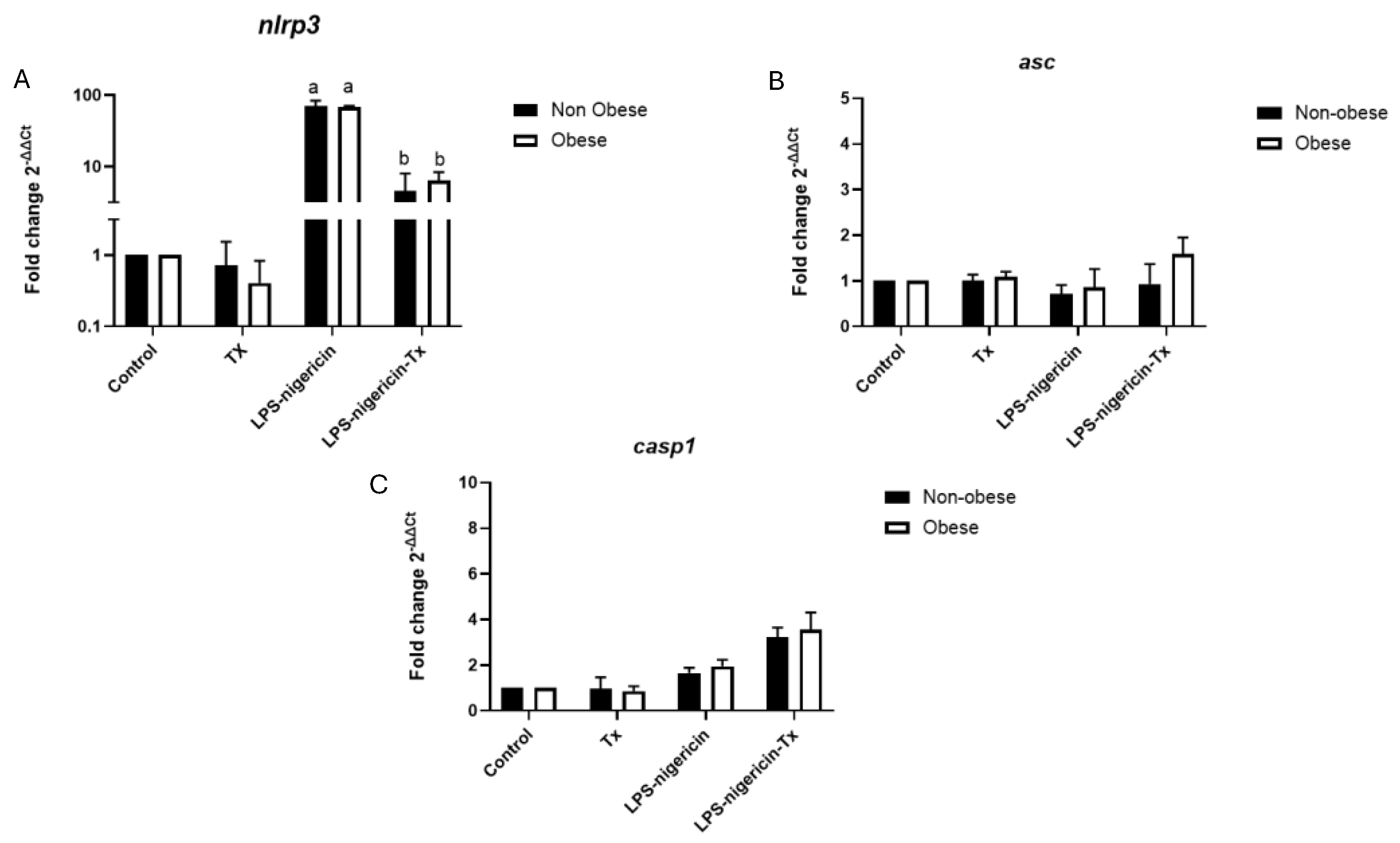
Figure 2.
Relative expression of IL-1β in neutrophils isolated from obese and non-obese horses with asthma and the effect of in vitro tamoxifen treatment. Neutrophils were stimulated with LPS–nigericin in the presence or absence of tamoxifen (5 μM). Data are presented as the mean ± standard deviation of three independent experiments (n = 3 horses per group). Differences letters were considered statistically significant at p < 0.05.
Figure 2.
Relative expression of IL-1β in neutrophils isolated from obese and non-obese horses with asthma and the effect of in vitro tamoxifen treatment. Neutrophils were stimulated with LPS–nigericin in the presence or absence of tamoxifen (5 μM). Data are presented as the mean ± standard deviation of three independent experiments (n = 3 horses per group). Differences letters were considered statistically significant at p < 0.05.
Disclaimer/Publisher’s Note: The statements, opinions and data contained in all publications are solely those of the individual author(s) and contributor(s) and not of MDPI and/or the editor(s). MDPI and/or the editor(s) disclaim responsibility for any injury to people or property resulting from any ideas, methods, instructions or products referred to in the content. |
© 2026 by the authors. Licensee MDPI, Basel, Switzerland. This article is an open access article distributed under the terms and conditions of the Creative Commons Attribution (CC BY) license (http://creativecommons.org/licenses/by/4.0/).
Copyright: This open access article is published under a Creative Commons CC BY 4.0 license, which permit the free download, distribution, and reuse, provided that the author and preprint are cited in any reuse.