

Submitted:
19 August 2026
Posted:
20 August 2026
You are already at the latest version
Abstract
Background: Facial pigmentary unevenness is frequently perceived as distressing and is associated with lower ratings of attractiveness, health, and youthfulness. Microdermabrasion together with selected cosmeceuticals can improve superficial discoloration, however, evaluation methods for objective color comparison differ considerably. Methods: We conducted an uncontrolled prospective interventional study in adults (n=5; ≥35 years) with pronounced skin pigmentation (all skin types) during four standardized institutional sessions every 14 days including a microdermabrasion and cosmetic protocol. Standardized facial photographs were rated in randomized, time-resolved order by expert estheticians (experts n=7) and lay assessors (novice n=13) using 5-point Likert scale questionnaire. Instrumental Colorimetry (VisioFace RD) quantified distinctive spot area (dL, %) and complexion evenness (dE) at baseline and over the following 10 weeks. Results: Pigment spot appearance and overall skin tone evenness improved significantly over time. Experts and novice rated most attributes equally. Even though analysis using traditional CIEDE color distance equations did not fully reflect hu-man perception, the triangulated, time-resolved assessment captured changes more comprehensively than colorimetry alone. Conclusions: This combined regimen was associated with improved facial pigmentation and tone uniformity. Instrumental colorimetry using CIEDE2000 calculation is recommended for future work.

Keywords:
pigmentation
; evaluation
; colorimetry
; questionnaire
; assessment
; microdermabrasion
; acid peel
1. Introduction
In many cultures, a uniform skin tone is regarded as a visible proxy for fitness and youthfulness. While an evenly tanned complexion may be preferred in many Western contexts and lighter facial skin is often valorized in parts of East Asia, both cultural frames converge in that facial pigmentary unevenness is commonly experienced as distressing and stigmatizing [1]. Objective image-analytic studies demonstrate that greater facial color homogeneity is associated with higher ratings of attractiveness, health, and with lower perceived age [2,3]. From the personal perspective, uneven facial pigmentation can negatively affect self-image and quality of life, particularly given the central role of facial cues in social interaction [4]. Therefore, affected individuals frequently seek evidence-based interventions to reduce hyperpigmentation.
From a clinical perspective, hyperpigmentation originates in melanogenesis: Melanin is an endogenous cutaneous pigment synthesized within epidermal melanosomes as part of an integrated photoprotective response to DNA photodamage and oxidative stress signals in skin. It is transferred to keratinocytes, where it functions as a broadband ultraviolet (UV) absorbent and radical scavenger, thereby contributing to photoprotection [5]. However, focal hyperpigmented macules can become clinically apparent when melanogenesis and/or melanin distribution is locally amplified by UV radiation, inflammatory mediators (e.g., post-inflammatory hyperpigmentation), or other metabolic and redox-related drivers of melanocyte activity, e.g. hormonal influences such as pregnancy or oral contraceptive use [6].
To address dyspigmentation, such as hyperpigmentation, different procedures for skin renewal are available in beauty salons or dermatology practices and skin clinics. While microneedling and microvibration treatments produce a stronger deep-tissue response, microdermabrasion excels at improving the skin’s surface, reducing superficial discoloration and irregularities, resulting in instantly even, clear, and smooth skin [7]. It mechanically removes the superficial epidermal layers and is known to promote skin renewal as well as to reduce scarring and pigmentary lesions, including hyperpigmented spots [8]. Across repeated regimens (e.g., 6 sessions to 12–14 weeks with typical session durations of ~10–30 min), clinical and histologic studies report improved skin texture and pigmentation, epidermal thickening/hyperplasia, partial acne-scar improvement, and altered melanosome distribution, alongside dermal remodeling with increased collagen density and variably reported elastin changes [9,10]. Mechanistically, abrasive injury activates inflammatory and wound-healing signaling (e.g., AP-1, IL-1β, TNF-α, MMPs) and can upregulate markers of neocollagenesis, with greater pathway activation after more aggressive abrasion [11,12]. Barrier perturbation appears transient: preclinical work indicates recovery of barrier function and stratum corneum within ~12–24 h, with increased permeability to topicals particularly during the first hours post-treatment, likely longer in humans [13,14]. Since evidence on enhanced active-ingredient delivery is context-dependent, penetration is known to increase for hydrophilic actives (e.g., niacinamide) [15]. This provides great potential for improving the absorption of active ingredients for hyperpigmentation and for enhancing their effectiveness.
Melanin-targeting actives have long attracted substantial interest in dermatology and the cosmetic industry, remaining the subject of intensive translational research. Established depigmenting agents include prescription-only drugs in many jurisdictions, such as hydroquinone, retinoids, topical corticosteroids, phenolic/resorcinol derivatives, and azelaic acid, which collectively address tyrosinase activity, epidermal turnover, and inflammation control [16]. Fixed triple-combination therapy (hydroquinone/tretinoin/fluocinolone acetonide) has demonstrated clinical efficacy in melasma in randomized trials, underscoring the benefit of multi-target intervention [17]. Beyond “classical” tyrosinase inhibition, contemporary concepts consider the full pigmentation cascade. Tranexamic acid is proposed to modulate upstream plasmin-driven inflammatory signaling and UV-induced melanogenic crosstalk – controlled trials support its role as monotherapy or adjunctive treatment in melasma [18]. Additional tyrosinase-directed compounds include kojic acid, arbutin, and licorice-derived flavonoids (e.g., glabridin), supported by mechanistic and preclinical evidence [19]. Ascorbates exert antioxidant and antimelanogenic effects, partly via modulation of tyrosinase-related pathways and inhibition of melanoidin oxidation [20]. Niacinamide reduces visible hyperpigmentation primarily by inhibiting melanosome transfer from melanocytes to keratinocytes, as clinical studies show [21]. Finally, chemical exfoliants (e.g. AHA, BHA, PHA) can accelerate pigment elimination via enhanced desquamation and have shown efficacy in melasma studies [22]. While the evidence supporting these ingredients varies in strength, in rational combinations, these modalities may enable comprehensive modulation of melanogenesis with acceptable tolerability making them particularly suitable for cosmetic approaches for prevention and care of dyspigmented skin.
Given this context, a holistic approach to addressing hyperpigmentation, combining device-based treatments and cosmetic products, is clearly indicated. To measure treatment effectiveness, some studies already pair objective methods (colorimeter) [23,24] with subjective assessments, such as self- and third-party rating questionnaires [25]. However, to the best of the authors’ knowledge, the literature has not yet described a triangulated framework that brings together (1) instrumental, image-based analysis, (2) investigator grading based on questionnaires, and (3) patient self-assessment. Therefore, the authors propose a double-blind, time-resolved approach to quantify pigmentation changes during long-term regimens that combine device-based treatment and cosmetic product application (serum, eye cream-, cream), integrating instrumental colorimetry with expert and lay evaluations of images, as well as self-perception and treatment acceptance/preference questionnaires.
2. Materials and Methods
2.1. Study Protocol and Participants
This work aimed to verify the reliability of a triangular image evaluation assessment. The data were collected from an uncontrolled prospective interventional study investigating the effects of microdermabrasion in combination with cosmetic treatment and aftercare in 5 adult volunteers. Participants were selected based on the following criteria: all skin types, pronounced pigmentation issues, irregular skin texture and uneven skin tone, enlarged pores, age: 35 to 60 years, without Vitamin C allergy, active acne, sensitive skin or relevant skin diseases.
Ethics committee approval was not obtained for this non-invasive study conducted in Germany. As this investigation involved the application of a complex cosmetic regimen and was not designed as a clinical trial of a medicinal product or medical device, formal review by an independent ethics committee was not sought. The procedures were performed on healthy adult volunteers after written informed consent. The study was conducted in accordance with the principles of the Declaration of Helsinki, and safety/tolerability were monitored throughout the study. Data protection was ensured through a double opt-in process on the testing platform. Consent regarding image rights was obtained using a standard form. No placebo control group was included, however, the documented state at the beginning of the study served as a baseline. Both expert and lay assessors were blinded to treatment group and time point during randomized image evaluation. All informational materials provided to the participants are included in the Appendix.
The treatment schedule and each treatment session followed a standardized, stepwise protocol. The participants attended treatment four times every 14 days. After initial skin cleansing and a preparatory peel, the skin was treated by microdermabrasion and subsequently exposed to an acid peel containing 14% AHA in combination with tranexamic acid and vitamin C. This was followed by application of a mask and a cooling massage over the mask, and a final leave-on finishing care product. The session was initiated and concluded with a company-specific ritual addressing breathing and relaxation. At-home care included a hyaluronic acid serum and an eye and face cream in the morning, as well as a vitamin C serum (weeks 1-7) or a longevity serum (week 8) and an eye and face cream in the evening. The volunteers were instructed to avoid UV exposure (vacation, tanning beds) during the study period. Full product specification related to the treatment and home-care step are presented in Table 2 (Appendix). The detailed step-by-step procedure are provided in the Appendix. Figure 1 shows the treatment schedule.
2.2. Data Collection
Standardized facial photographs were acquired before the first treatment, immediately after the first treatment, before each following treatment and 14 days after the last treatment (in sum 6 photographs) in the in-house photo studio with a fixed setup. For subjective assessment, images were anonymized, coded and presented in randomized, time-resolved order to 7 certified estheticians and 13 lay assessors on a stationary screen with consistent environmental conditions regarding light exposure and screen lighting. The questionnaire addressed the following characteristics: (1) Number of pigment spots, (2) Color intensity of pigment spots, (3) Intensity of color differences/contrast due to pigmentation disorders, (4) Size of pigment spots, and (5) Overall evenness of skin tone, using a 5-point Lickert scale and sample images as a visual reference, respectively. Colorimetric analysis was performed using the C+K VisioFace RD system (Courage + Khazaka electronic GmbH, Cologne, Germany). Calibrated facial scans were obtained at regular 14-day intervals as well, and processed using the manufacturer’s preconfigured analysis module, automatically segmenting conspicuous spot regions (displayed as red-circled areas) and reporting the proportion of conspicuous spots [%] and complexion evenness as a color-difference metric [dE] as well as distinctive spots [dL].
2.3. Statistics
We conducted a 2×2 mixed-design ANOVA with Group (novice vs. expert) as the between-subjects factor and Time (pre vs. post) and View (frontal vs. lateral) as within-subject factors, where each within-cell score was computed by averaging the five corresponding image ratings (frontal: images 1/3/5/7/9; lateral: images 2/4/6/8/10), and effects were tested using an OLS model with subjects nested within group (i.e., including a subject-within-group term) implemented in Python via statsmodels (type-III ANOVA). All pairwise within-subject comparisons were performed between weeks using paired-samples t-tests and then adjusted the resulting p-values for multiple testing with the Holm correction. The visualization was created using Microsoft® Excel® for Microsoft 365 MSO (Version 2604, Build 16.0.19929.20172), 64-bit.
3. Results
The objective of this analysis was to investigate how accurately the triangular assessment captures improvements in skin color. Across all participants, visual ratings of the time-resolved image-presentation showed significant changes in pigment-related parameters over time: the number (F (1, 54) = 62.05, p < .001, ηp² = .54), intensity (F (1, 54) = 62.05, p < .001, ηp² = .54), contrast (F (1, 54) = 83.54, p < .001, ηp² = .61), size of pigment spots (F (1, 54) = 14.70, p < .001, ηp² = .21) and skin tone evenness (F (1, 54) = 49.92, p < .001, ηp² = .48) improved between before and after treatment (see Figure 2).
No significant effects were found for group (novices vs. experts) on pigment number, intensity, contrast, or spot size, nor for any view effects (frontal vs. lateral) or interactions (group × time, group × view, time × view, or group × time × view) across the analyzed parameters (all p > .05). Only in the case of ratings for skin tone evenness significant differences were observed between expert and novice ratings (F (1, 54) = 5.24, p < .05, ηp² = .09). As Figure 3 shows, the variance was higher among novices, while the experts' ratings were more precise.
For instrumental analysis of distinctive spots (dL), the repeated-measures ANOVA showed a tendency for significance of week (F (5, 20) = 2.59, p < .10, ηp² = .39), however, none of the pairwise post-hoc comparisons remained significant after Holm correction (see Figure 4). In terms of changes in evenness (dE), neither ANOVA nor post-hoc tests revealed any significant changes.
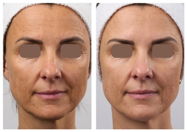
The photo in Figure 5 illustrates exemplary the change in skin appearance between the initial condition and after the treatment routine.
4. Discussion
Visual ratings showed clear pre–post improvements across all pigment-related endpoints (Figure 2), indicating that treatment-related changes were readily detectable in the time-resolved image presentation. The absence of group, view, and interaction effects for most parameters suggests that these improvements were largely robust to rater’s expertise and viewing angle. The only exception was skin tone evenness (Figure 3), where experts and novices differed, consistent with evenness being a more holistic and potentially criterion-dependent judgment. A key limitation is that the outcomes were based on ordinal visual ratings averaged across multiple images per view, and the modest, unbalanced novice/expert sample sizes may have limited power to detect small group, view, or interaction effects. Since there was a high variability in the evenness ratings provided by non-experts and the instrumental colorimetric analysis did not reveal any significant results, we recommend evaluating evenness only by experts, as this yields the most accurate results.
Although the instrumental analysis of weekly colorimetric means of spots and evenness showed small fluctuations, the repeated-measures analyses did not provide significant evidence for a reliable time-dependent change across the measurement period, suggesting that any observed differences are more likely due to within-subject variability and the limited sample size than to a systematic effect of time. However, the identification and quantification of skin pigmentation depend strongly on the color-difference formulas used. While the dE values frequently reported in the literature describe the mathematically precise color difference as a vector in the Lab color space—accounting for the light/dark, red/green, and yellow/blue components—the “spot fraction” considers areas that vary in their gray value (dL). Using the CIELAB2000 color-difference metric, which has so far been used only to a limited extent in dermatology but is well established in colorimetry, enables a determination of color differences that most closely matches human perception, as it incorporates correction factors for the orange, blue, and saturation regions. For a more accurate assessment of melasma, evaluating the difference in the yellow component b is recommended, whereas erythema is best quantified by analysis of the red component a [23,24,26].
A subjectively or objectively increased visibility of pigmentation after an active ingredient–supported microdermabrasion treatment may have several causes. First, desquamation of the most superficial skin layers, whether induced by microdermabrasion or acid-based exfoliation, can reveal deeper-lying pigment while removing superficial pigmentation, which can be measured objectively. In addition, a reduction in baseline pigmentation and an increase in non-pigmented areas may lead to a stronger subjective perception of the remaining pigmented areas, because the contrast is enhanced and the observer visually focuses on these regions. Second, transient inflammation and a reduced intrinsic photoprotective capacity of the skin can potentiate the effects of UV radiation, which itself may exacerbate hyperpigmentation. Therefore, diligent photoprotection is essential, including during darker seasons [27].
As already described in the introduction, UV radiation is the primary trigger for pigmentation. During the study period, the UV index in the geographic region (Dortmund, Germany for the period 17 October to 22 December 2025.) ranged between 0.4 and 1.8 [28]. Participants were instructed to avoid UV exposure and use photoprotection, however, compliance could not be systematically assessed. Nevertheless, performing the procedure under different UV-index conditions may yield different results. As described in numerous microdermabrasion guidelines, treatment during the summer months is generally not recommended [29].
In this study, a standardized treatment and skincare protocol was applied to ensure comparability between participants. However, since each individual’s skin may respond differently at the clinical, structural, and inflammatory levels [7], we recommend -aiming for optimal outcomes- the investigation of biomarker substances and indicators to identify key milestones during regeneration and to address them with appropriate active ingredients.
Beyond directly pigment-modulating actives, the tested regimen also incorporated additional functional ingredient classes intended to support skin condition during repeated treatment and home care. These included antioxidants, soothing and barrier-supportive agents, regeneration-associated peptides, vitamins, hydroxy acids, humectants, and botanical oils and extracts (see Table 2 Appendix). Although the efficacy of individual formulations depends on concentration, vehicle properties, ingredient interactions, and application regimen, this classification provides a useful framework for understanding the intended cosmetic functions of the products used in the present study setting.
Antioxidants such as epigallocatechin gallate, superoxide dismutase, resveratrol, and thiotaine may help counteract oxidative stress through reactive oxygen species scavenging and support of endogenous defense systems [30,31], while polyphenolic antioxidants such as epigallocatechin gallate and resveratrol have also been discussed in the context of topical photoprotection [32,33]. Soothing and barrier-supportive ingredients such as bisabolol, Lactobacillus ferment, niacinamide, and dexpanthenol may help reduce visible irritation, support epidermal barrier function, and improve skin resilience [34,35,36,37]. Humectants including hyaluronic acid, glycerin, pentylene glycol, butylene glycol, urea, and biosaccharide gum contribute to stratum corneum hydration and may improve skin texture and viscoelasticity [38,39]. In parallel, lipid-based emollients and botanical oils may reduce transepidermal water loss and support barrier homeostasis [40,41,42], whereas peptides are commonly used to support renewal-related processes and structure-associated skin properties [43]. Hydroxy acids and related acids, including salicylic acid, glycolic acid, and tranexamic acid, address complementary cosmetic targets such as desquamation, surface smoothing, pore appearance, and tone uniformity [44,45,46]. Additional vitamins and botanical extracts may further contribute antioxidative, soothing, balancing, and skin-conditioning effects [47,48,49,50]. In addition, antioxidant plant extracts with high antioxidant capacity are not only believed to reduce cutaneous oxidative stress and skin aging [51] but also known for extending the oxidative stability of cosmetic emulsions [52].
Overall, the ingredient profile suggests a multimodal cosmetic strategy aimed not only at uneven pigmentation, but also at hydration, barrier support, antioxidative protection, soothing, and overall improvement in skin appearance.
5. Conclusions
Although the multifactorial design does not allow causal attribution to any single procedure step or active ingredient, over the course of four biweekly treatments and standardized home care, the combined microdermabrasion–cosmetic regimen was associated with visible improvements in pigment spot appearance and overall facial skin tone evenness. These changes were consistently reflected across time-resolved, randomized evaluations of standardized photographs by both trained estheticians and lay assessors, supporting the robustness of the observed improvement. However, instrumental analysis with former CIEDE calculations could not fully reproduce the consistent results. Therefore, integrating objective image-based metrics with blinded novine vs. expert perception-based ratings provides a more comprehensive representation of treatment benefit than colorimetry alone. However, instrumental colorimetry using CIEDE2000 calculation is recommended for future work. Taken together, these findings support the continued need for assessment by a trained expert, or at minimum a human evaluator, in evaluating the effectiveness of combination regimens targeting hyperpigmentation in cosmetic and dermatologic practice.
Author Contributions
A. Springer led data curation, statistical analysis and visualization, interpretation and discussion of findings, and writing of the manuscript (abstract, introduction, methodology, results, discussion and conclusion) with focus on dermatology/ skin physiology, ingredients and colorimetry. E. Helfenbein was responsible for study conduct, including participant coordination, implementation of the intervention protocol, acquisition of study data and revisions and corrections to the manuscript. A. Spitz was responsible for the relevant product development and selection of corresponding active ingredients, additionally for writing and preparing related information and manuscript contributions with focus on test products. J. Blaak provided senior oversight, strategic direction, and overall scientific supervision. All authors contributed to the interpretation of the data, reviewed the final version of the manuscript and approved its submission.
Funding
This research received no external funding. Dr. Babor GmbH & Co. KG, Neuenhofstraße 180, 52078 Aachen, Germany covered the author’s working time and the Article Processing Charge (APC) and provided the test products. Participants received an honorarium/compensation for the study participation and publication of images.
Institutional Review Board Statement
All procedures were performed in healthy adult volunteers and in accordance with the Declaration of Helsinki. Ethical review and approval were waived for this study due to the use of commercially available cosmetic products and materials, the well-established nature of the treatments, and the exclusively non-invasive measurement methods. In addition, an internal ethics/risk assessment was documented, and safety and tolerability were monitored throughout the evaluation.
Informed Consent Statement
The study was performed between September and December of 2025. Informed consent was obtained from all subjects involved in the study (data protection, consent for facial photography).
Data Availability Statement
The data that support the findings of this study are available from the corresponding author upon reasonable request. Deidentified participant data and associated study materials will be shared where feasible and in accordance with applicable data protection regulations and the informed consent provided.
Acknowledgments
We thank all study participants for volunteering for the treatment regimen. We also thank the certified estheticians (expert raters) and novice assessors for their time and contribution to the randomized, time-resolved image evaluations. Many thanks to our partner Courage+Khazaka electronic GmbH for their collaboration on the colorimetric instrumental analysis. Our sincere appreciation belongs to Lisa Arndt for supporting product development, lab work and data presentation. We would like to acknowledge the collaborative efforts and mutual support that contributed to the completion of this manuscript.
Conflicts of Interest
The authors declare no conflicts of interest. Authors A. Springer, E. Helfenbein, A. Spitz and J. Blaak are affiliated with Dr. Babor GmbH & Co. KG, Neuenhofstraße 180, 52078 Aachen, Germany.
Appendix
Information Material for Participants
Participants received the following documents as PDF documents for their knowledge and signature before the start of the study:
- Image Release & Privacy Information
- Information on possible skin reactions following treatments with microdermabrasion and guidelines to follow
- Information on possible skin reactions following 14 – 30% AHA treatments and guidelines to follow
- Possible contraindications for the use of Microdermabrasion
- Possible contraindications for the use of 14–30% AHA peels
- Instructions for product use during the treatment study
Only after informed consent and expert examination of suitability the subject was admitted to participate.
Study Schedule
Table 1 summarizes the chronological schedule of the study from recruitment/screening through the end-of-study visit (Day 56), detailing visit settings, consent and safety procedures, standardized photo documentation, instrumental skin assessments, in-clinic interventions, and the prescribed homecare regimen with adherence monitoring.

Table 1.
Study schedule, in-clinic procedures, and homecare regimen.
| Day | Visit / setting | Consent & safety | Documentation (photos) | Assessments / analyses | In-clinic intervention | Homecare / adherence |
| Pre-Day 0 (recruitment/screening) | Screening | Selection by skin type & skin condition; eligibility confirmation; review of contraindications; participant counseling on required behaviors & potential adverse effects | – | – | – | – |
| Day 0 (baseline & treatment 1) | Visit 1: Baseline | Informed consent + data privacy; photo use rights; pre-treatment safety check | Pre-treatment: frontal + lateral views; Immediate post-treatment: frontal + lateral views | Visio Face facial skin analysis (baseline) | Treatment: MDA & 14% AHA | Provide homecare package + written instructions for Weeks 1–7: 1) HA serum 2) Vit. C serum 3) Skin tone eye cream 4) Skin tone cream |
| Days 1–13 | Home use | Tolerability monitoring; contact/withdrawal criteria per protocol if clinically relevant | – | – | – | Continue homecare as instructed (Weeks 1–7) |
| Day 14 (follow-up & treatment 2) | Visit 2 | Interval history since Day 0 (tolerability, events, adherence) | Standardized photos (frontal + lateral views) | Visio Face analysis pre-treatment | Treatment: MDA & 14% AHA | Continue homecare (Weeks 1–7) |
| Days 15–27 | Home use | Ongoing tolerability/adherence monitoring | – | – | – | Continue homecare |
| Day 28 (follow-up & treatment 3) | Visit 3 | Interval history since Day 14 | Standardized photos (frontal + lateral views) | Visio Face analysis pre-treatment | Treatment: 14% AHA | Continue homecare |
| Days 29–41 | Home use | Ongoing tolerability/adherence monitoring | – | – | – | Continue homecare |
| Day 42 (follow-up & treatment 4 + regimen update) | Visit 4 | Interval history since Day 28 | Standardized photos (frontal + lateral views) | Visio Face analysis pre-treatment | Treatment: MDA & 14% AHA | Update homecare from Week 8 + written instructions: add Antioxidative serum (as add-on or replacement per study plan) |
| Days 43–55 | Home use (Week 8) | Ongoing tolerability/adherence monitoring | – | – | – | Continue updated homecare (incl. Antioxidative serum) |
| Day 56 (end-of-study) | Final visit | End-of-study safety check; capture AEs through Day 56 | Final photos: frontal + lateral views | Visio Face analysis (end point) | – | Participant questionnaire/survey (e.g., perceived efficacy, tolerability, satisfaction; instrument/scales per protocol) |
Skin Care Products
Table 2 provides an overview of the cosmetic formulations used during the preparation stage, microdermabrasion, after care and home care of the treatment protocol.
Table 2.
Test product specification including formulation name, formulation type and internal formulation number (Formulation), ingredient list (INCI), physicochemical specification with pH and viscosity (Spec.). Key ingredients are highlighted in bold.
Table 2.
Test product specification including formulation name, formulation type and internal formulation number (Formulation), ingredient list (INCI), physicochemical specification with pH and viscosity (Spec.). Key ingredients are highlighted in bold.
| Formulation | INCI | Spec. |
| Cleanser (oil gel) 280677-11 |
CAPRYLIC/CAPRIC TRIGLYCERIDE, GLYCERIN, HELIANTHUS ANNUUS SEED OIL, AQUA, RICINUS COMMUNIS SEED OIL, SUCROSE PALMITATE, VITIS VINIFERA SEED OIL, BISABOLOL, LACTOBACILLUS FERMENT, SUPEROXIDE DISMUTASE, EPIGALLOCATECHIN GALLATYL GLUCOSIDE, ERGOTHIONEINE, MALTODEXTRIN, ETHYLHEXYLGLYCERIN, PHENOXYETHANOL | pH: / Viscosity: 28.300mPa*s |
| Tonic (watery) 280678-6 |
AQUA, GLYCERIN, BETAINE, PANTHENOL, SACCHARIDE ISOMERATE, SODIUM PCA, SODIUM LACTATE, ZINC GLUCONATE, PCA, GLYCINE, SERINE, ALANINE, BIOSACCHARIDE GUM-1, COPPER GLUCONATE, GLUTAMIC ACID, LYSINE HCL, THREONINE, FRUCTOSE, ARGININE, INOSITOL, LACTIC ACID, NIACINAMIDE, UREA, PROLINE, ETHYLHEXYLGLYCERIN, MAGNESIUM ASPARTATE, CITRIC ACID, SODIUM CITRATE, PHENOXYETHANOL, SODIUM BENZOATE | pH: 5.0 Viscosity: / |
| Peeling tonic (watery) 281874-1 |
AQUA, GLYCOLIC ACID, POLYGLYCERYL -10 LAURATE, PANTHENOL, GLYCERIN, SODIUM LACTATE, SODIUM PCA, LECITHIN, BIOSACCHARIDE GUM -1, FRUCTOSE, GLYCINE, INOSITOL, LACTIC ACID, NIACINAMIDE, UREA, HEXANOYL DIPEPTIDE -3 NORLEUCINE ACETATE, XANTHAN GUM, SODIUM HYDROXIDE, ETHYLHEXYLGLYCERIN, CITRIC ACID, PHENOXYETHANOL, SODIUM BENZOATE | pH: 4.0 Viscosity: 400mPA*s |
| Peeling mask (o/w emulsion) 281890-3 |
AQUA, DECYL OLEATE, KAOLIN, CETEARYL ALCOHOL, GLYCERIN, C12 -15 ALKYL BENZOATE, ALCOHOL DENAT., CI 77891, COCAMIDOPROPYL BETAINE, CETEARYL GLUCOSIDE, DIMETHYLIMIDAZOLIDINONE RICE STARCH, HELIANTHUS ANNUUS SEED OIL, PANTHENOL, SPHINGOMONAS FERMENT EXTRACT, LECITHIN, BISABOLOL, TOCOPHEROL, HEXANOYL DIPEPTIDE -3 NORLEUCINE ACETATE, MICROCRYSTALLINE CELLULOSE, XANTHAN GUM, SODIUM CETEARYL SULFATE, SODIUM CHLORIDE, CELLULOSE GUM, HYDROGENATED PALM GLYCERIDES CITRATE, ETHYLH EXYLGLYCERIN, CITRIC ACID, SODIUM HYDROXIDE, DISODIUM PHOSPHATE, POTASSIUM PHOSPHATE, PHENOXYETHANOL | pH: 5.8 Viscosity: 49.200 mPA*s |
| Tonic (alcoholic) 256240-1 |
AQUA, ALCOHOL DENAT., SALICYLIC ACID | pH: 3.1 Viscosity: / |
| AHA Peeling (watery) 280673-2 |
AQUA, PENTYLENE GLYCOL, ARGININE, GLYCOLIC ACID, 3 -O-ETHYL ASCORBIC ACID, BETAINE, TRANEXAMIC ACID, SALICYLIC ACID, ERGOTHIONEINE, XANTHAN GUM, SODIUM HYDROXIDE, PHENOXYETHANOL | pH: 4.0 Viscosity: 6.400mPA*s |
| Calming serum (o/w emulsion) 280679-9 |
AQUA, GLYCERIN, BETAINE, DECYL OLEATE, PRUNUS AMYGDALUS DULCIS OIL, HYPERICUM PERFORATUM EXTRACT, HELIANTHUS ANNUUS SEED OIL, SODIUM PCA, SODIUM LACTATE, PCA, SUPEROXIDE DISMUTASE, SERINE, ALANINE, BIOSACCHARIDE GUM-1, GLYCINE, TOCOPHEROL, GLUTAMIC ACID, LYSINE HCL, THREONINE, ARGININE, PROLINE, EPIGALLOCATECHIN GALLATYL GLUCOSIDE, ERGOTHIONEINE, XANTHAN GUM, CETEARYL ALCOHOL, GLYCERYL STEARATE, GELLAN GUM, SCLEROTIUM GUM, MALTODEXTRIN, ETHYLHEXYLGLYCERIN, SODIUM HYDROXI DE, PHENOXYETHANOL | pH: 5.5 Viscosity: 6.100mPA*s |
| Sheet mask (watery) 3SM3B503 |
AQUA, GLYCERIN, PANTHENOL, NIACINAMIDE, ASCOPHYLLUM NODOSUM EXTRACT, EPILOBIUM ANGUSTIFOLIUM FLOWER/LEAF/STEM EXTRACT, ZANTHOXYLUM PIPERITUM FRUIT EXTRACT, LONICERA JAPONICA FLOWER EXTRACT, CITRUS PARADISI FRUIT EXTRACT, HAMAMELIS VIRGINIANA EXTRACT, 1,2-HEXANEDIOL, SODIUM GLUCONATE, SODIUM HYALURONATE, XANTHAN GUM, BUTYLENE GLYCOL, CITRIC ACID, SODIUM CITRATE, PHENOXYETHANOL | pH: 4.5-6.5 Viscosity: / |
| B12 serum (o/w emulsion) 280680-1 |
AQUA, PRUNUS AMYGDALUS DULCIS OIL, POLYGLYCERYL-6 STEARATE, BUTYLENE GLYCOL, COCO-CAPRYLATE/CAPRATE, NIACINAMIDE, PANTHENOL, HELIANTHUS ANNUUS SEED OIL, BIOTIN, SUPEROXIDE DISMUTASE, SPHINGOMONAS FERMENT EXTRACT, TOCOPHEROL, MAGNESIUM CARBOXYMETHYL BETA -GLUCAN, BIOSACCHARIDE GUM-1, CYANOCOBALAMIN, EPIGALLOCATECHIN GALLATYL GLUCOSIDE, ERGOTHIONEINE, CETEARYL ALCOHOL, GLYCERYL STEARATE, POLYGLYCERYL -6 BEHENATE, MICROCRYSTALLINE CELLULOSE, SCLEROTIUM GUM, CELLULOSE GUM, MAL TODEXTRIN, ETHYLHEXYLGLYCERIN, CITRIC ACID, PHENOXYETHANOL | pH: 5.4 Viscosity: 1.300mPA*s |
| HA Serum (watery) 236036-7 |
AQUA, PENTYLENE GLYCOL, SODIUM ACETYLATED HYALURONATE, SODIUM HYALURONATE, SODIUM HYALURONATE CROSSPOLYMER, HYDROLYZED SODIUM HYALURONATE, CARBOMER, ETHYLHEXYLGLYCERIN, SODIUM HYDROXIDE, PHENOXYETHANOL | pH: 5.2 Viscosity: 1.500mPA*s |
| Vit. C serum (watery) 280695-6 |
AQUA, 3-O-ETHYL ASCORBIC ACID, BETAINE, DISODIUM PHOSPHATE, GLYCERIN, CITRIC ACID, SUPEROXIDE DISMUTASE, BIOSACCHARIDE GUM -1, PALMITOYL TRIPEPTIDE-5, EPIGALLOCATECHIN GALLATYL GLUCOSIDE, ERGOTHIONEINE, XANTHAN GUM, MALTODEXTRIN, SODIUM HYDROXIDE, ETHYLHEXYLGLYCERIN, PHENOXYETHANOL | pH: 5.2 Viscosity: 700mPA*s |
| Antioxidative serum (o/w emulsion) 280741-7 |
AQUA, CAPRYLIC/CAPRIC TRIGLYCERIDE, POLYGLYCERYL -6 STEARATE, PRUNUS AMYGDALUS DULCIS OIL, SQUALANE, PANTHENOL, ECHINACEA PURPUREA EXTRACT, CUCURBITA PEPO SEED EXTRACT, HELIANTHUS ANNUUS SEED OIL, GLYCERIN, LEC ITHIN, ALLANTOIN, SPHINGOMONAS FERMENT EXTRACT, SUPEROXIDE DISMUTASE, TOCOPHEROL, TEPRENONE, BIOSACCHARIDE GUM -1, GLYCERYL GLUCOSIDE, RESVERATROL, LACTIC ACID, EPIGALLOCATECHIN GALLATYL GLUCOSIDE, ERGOTHIONEINE, COCO-CAPRYLATE/CAPRATE, PROPANEDIOL, ALCOHOL , CETEARYL ALCOHOL, GLYCERYL STEARATE, SODIUM STEAROYL GLUTAMATE, MICROCRYSTALLINE CELLULOSE, POLYGLYCERYL-6 BEHENATE, SCLEROTIUM GUM, BEHENTRIMONIUM CHLORIDE, CELLULOSE GUM, MALTODEXTRIN, ETHYLHEXYLGLYCERIN, CITRIC ACID, POTASSIUM PHOSPHATE, PANTOLACTONE, PHENOXYETHANOL, SODIUM BENZOATE, POTASSIUM SORBATE, ISONIACINAMIDE | pH: 5.4 Viscosity: 1.600 mPA*s |
| Skin tone eye cream (o/w emulsion) 280696-4 |
AQUA, BUTYROSPERMUM PARKII BUTTER, HELIANTHUS ANNUUS SEED OIL, GLYCERIN, CETEARYL ALCOHOL, PRUNUS AMYGDALUS DULCIS OIL, SIMMONDSIA CHINENSIS SEED OIL, SQUALANE, NIACINAMIDE, CETEARYL GLUCOSIDE, PANTHENOL, TOCOPHERYL ACETATE, ALLANTOIN, SPHINGOMONAS FERMENT EXTRACT, SUPEROXIDE DISMUTASE, TOCOP HEROL, BIOSACCHARIDE GUM-1, LECITHIN, ACETYL GLUTAMINE, SODIUM HYALURONATE, BACILLUS/SOYBEAN FERMENT EXTRACT, 1,2 -HEXANEDIOL, CAPRYLYL GLYCOL, EPIGALLOCATECHIN GALLATYL GLUCOSIDE, LINOLENIC ACID, FOLIC ACID, ERGOTHIONEINE, OLIGOPEPTIDE -1, HEXAPEPTIDE-11, OLIGOPEPTIDE-3, BUTYLENE GLYCOL, MICROCRYSTALLINE CELLULOSE, XANTHAN GUM, SODIUM CETEARYL SULFATE, CELLULOSE GUM, SCLEROTIUM GUM, MALTODEXTRIN, ISOPROPYL ALCOHOL, ETHYLHEXYLGLYCERIN, CITRIC ACID, PANTOLACTONE, PHENOXYETHANOL, CI77288, OLIGOPEPTIDE | pH: 5.9 Viscosity: 21.900mPA*s |
| Skin tone cream (o/w emulsion) 280772-5 |
AQUA, BUTYROSPERMUM PARKII BUTTER, POLYGLYCERYL-3 METHYLGLUCOSE DISTEARATE, HYDROGENATED VEGETABLE GLYCERIDES, MACADAMIA INTEGRIFOLIA SEED OIL, SESAMUM INDICUM SEED OIL, COCO-CAPRYLATE/CAPRATE, GLYCERIN, NIACINAMIDE, CETEARYL ALCOHOL, PANTHENOL, HELIANTHUS ANNUUS SEED OIL, TOCOPHEROL, ALLANTOIN, SUPEROXIDE DISMUTASE, BIOSACCHARIDE GUM-1, LECITHIN, ACETYL GLUTAMINE, SODIUM HYALURONATE, BACILLUS/SOYBEAN FERMENT EXTRACT, 1,2-HEXANEDIOL, CAPRYLYL GLYCOL, LINOLENIC ACID, EPIGALLOCATECHIN GALLATYL GLUCOSIDE, FOLIC ACID, ERGOTHIONEINE, OLIGOPEPTIDE-1, HEXAPEPTIDE-11, OLIGOPEPTIDE-3, BUTYLENE GLYCOL, XANTHAN GUM, SCLEROTIUM GUM, SODIUM CETEARYL SULFATE, SODIUM STEAROYL GLUTAMATE, MALTODEXTRIN, ISOPROPYL ALCOHOL, ETHYLHEXYLGLYCERIN, CITRIC ACID, DISODIUM PHOSPHATE, POTASSIUM PHOSPHATE, PHENOXYETHANOL, OLIGOPEPTIDE-2 | pH: 5.6 Viscosity: 29.700mPA*s |
Treatment Protocol
The following Table 3 outlines the sequential procedure used in the in-clinic treatment protocol, including the objective of each step, generic materials/products, non-proprietary reproducible procedures, key time and treatment parameters, safety or stop criteria, and recommended aftercare. The workflow covers preparation, cleansing, double peeling, microdermabrasion, acid peel application, active infusion, mask treatment, finishing care, and patient guidance/follow-up. Outcomes and adverse events were documented throughout and at the end of the visit.
Table 3.
Standardized treatment workflow of the combined cosmetic intervention.
| Step | Objective | Materials / Products (generic) | Procedure (non-proprietary, reproducible) | Time / Parameters | Safety / Stop Criteria | Aftercare |
| 1 | Preparation | Warm compress water; cool compress water; cooling sticks; headband; gloves | Prepare warm water (38–42°C) and cool water (14–18°C) for compresses. Pre-chill cooling sticks in a cold cabinet ~15–20 min before mask step. | Temps as stated | Do not use extreme temperatures; avoid broken skin with heat/cold exposure | Document baseline skin status (erythema, sensitivity) |
| 2 | Cleansing | Cleanser; hydrating/soothing toner | Cleanse face/neck/décolleté with gentle technique. Tone to remove residue and normalize skin surface. | 2–5 min | Stop if significant burning/stinging occurs already during cleansing | Pat dry; avoid friction |
| 3 | Double Peeling | Peeling tonic; peeling mask; cotton pads; gauze; fan brush; damp sponges | Apply peeling tonic with cotton pad evenly; allow to act. Apply thin layer of peeling mask with brushes; let dry/settle. Allow the mask to dry; then rub off the dried mask using a cotton pad wrapped in gauze; remove any remaining residue with a damp sponge. | Tonic: per tolerance; Mask: ~10 min | Stop immediately if intense burning, rapidly increasing erythema, swelling, urticaria-like reaction | Rinse/neutralize as per product instructions; cool compress if needed |
| 4 | MDA (Microdermabrasion) – face/neck/décolleté | Tonic; eye protection pads; MDA device with ionic crystals (aluminium oxide); towels/sponges | Put on gloves. Pre-wipe with tonic. Apply protecting eye pads; protect hair/ears if needed. Perform MDA passes per area with conservative pressure; finish with thorough residue removal and tissue drying. Gentle cleansing with damp sponge. | Intensity 1–2 (low); passes: conservative | Stop, if pinpoint bleeding, marked pain, skin tearing, or excessive erythema | Reassess barrier; proceed only if skin response is mild/moderate and stable |
| 5 | Acid Peel | AHA Peeling + tranexamic acid + vitamin C, pH ~4; lip protectant (occlusive ointment); cool compresses; timer | Protect lips with occlusive ointment. Apply AHA peeling using a standardized technique to face/neck/décolleté, avoiding mucosa and high-risk zones. Strictly time exposure. Optional layering after 8 min only if reaction is mild and uniform. Remove peeling thoroughly with cool compresses; remove protecting eye pads. | 10 min – max 12 min | Continuous observation required. Immediate removal if severe burning, rapidly spreading intense erythema, marked edema, blistering, or patient distress | Cool compress; barrier-supportive care before next step if needed |
| 6 | Active infusion / concentrate | Calming serum | Apply evenly over treated areas with minimal friction. | 2 ml (approx. 4 dropper lengths) | Stop if new burning develops or erythema escalates | Continue with mask step for calming effect |
| 7 | Mask | Sheet mask | Apply mask; ensure full contact. Gentle cooling massage over the mask to enhance comfort. | 8 min | Stop, if itching, wheals or escalating discomfort | Remove mask; do not rinse unless required |
| 8 | Finishing care | B12 serum; barrier protectant/moisturizer | Apply calming and barrier supporting serum, to reduce transepidermal water loss and support recovery. | 1–3 min | If marked irritation persists, simplify regimen to bland emollient only | Counsel on strict photoprotection |
| 9 | Patient guidance / follow-up | Broad-spectrum sunscreen; gentle cleanser; bland moisturizer | Provide written aftercare: avoid heat, sauna, intense exercise same day; avoid acids/retinoids/scrubs until recovered; emphasize daily sunscreen. Arrange follow-up based on risk profile. | Follow-up typically 1–2 weeks (adapt) | Seek medical review if blistering, progressive swelling, severe pain, crusting, infection signs, or pigment change | Document outcomes and adverse events |
Parameters were adjusted according to skin type and tolerability. We documented erythema, burning/stinging, edema, and any adverse events at baseline, during exposure, and post-treatment.
References
- Lu, Y.; Yang, J.; Xiao, K.; Pointer, M.; Li, C.; Wuerger, S. Skin coloration is a culturally-specific cue for attractiveness, healthiness, and youthfulness in observers of Chinese and western European descent. PLoS ONE 2021, 16(10), e0259276. [Google Scholar] [CrossRef] [PubMed]
- Matts, P.J.; Fink, B.; Grammer, K.; Burquest, M. Color homogeneity and visual perception of age, health, and attractiveness of female facial skin. J. Am. Acad. Dermatol. 2007, 57(6), 977–984. [Google Scholar] [CrossRef] [PubMed]
- Fink, B.; Matts, P.J. The effects of skin colour distribution and topography cues on the perception of female facial age and health. J. Eur. Acad. Dermatol. Venereol. 2008, 22(4), 493–498. [Google Scholar] [CrossRef] [PubMed]
- Raveendra, L.; Sidappa, H.; Shree, S. A Study of Quality of Life in Patients with Facial Melanoses. Indian Dermatol. Online J. 2020, 11(2), 154–157. [Google Scholar] [CrossRef] [PubMed]
- Gilchrest, B.A.; Eller, M.S. DNA photodamage stimulates melanogenesis and other photoprotective responses. J. Investig. Dermatol. Symp. Proc. 1999, 4(1), 35–40. [Google Scholar] [CrossRef] [PubMed]
- Handel, A.C.; Miot, L.D.B.; Miot, H.A. Melasma: a clinical and epidemiological review. An. Bras. Dermatol. 2014, 89(5), 771–782. [Google Scholar] [CrossRef] [PubMed]
- Springer, A.; Helfenbein, E.; Blaak, J. Mechanically induced skin renewal: evidence-based insights into clinical, structural and inflammatory changes activated by microneedling, microdermabrasion and microvibration. J. Cosmet. Dermatol. 2026, 25(5), e70908. [Google Scholar] [CrossRef] [PubMed]
- AlKhawam, L.; Alam, M. Dermabrasion and microdermabrasion. Facial Plast. Surg. 2009, 25(5), 301–310. [Google Scholar] [CrossRef] [PubMed]
- Davari, P.; Gorouhi, F.; Jafarian, S.; Dowlati, Y.; Firooz, A. A randomized investigator-blind trial of different passes of microdermabrasion therapy and their effects on skin biophysical characteristics. Int. J. Dermatol. 2008, 47(5), 508–513. [Google Scholar] [CrossRef] [PubMed]
- El-Domyati, M.; Hosam, W.; Abdel-Azim, E.; Abdel-Wahab, H.; Mohamed, E. Microdermabrasion: a clinical, histometric, and histopathologic study. J. Cosmet. Dermatol. 2016, 15(4), 503–513. [Google Scholar] [CrossRef] [PubMed]
- Karimipour, D.J.; Karimipour, G.; Orringer, J.S. Microdermabrasion: an evidence-based review. Plast. Reconstr. Surg. 2010, 125(1), 372–377. [Google Scholar] [CrossRef] [PubMed]
- Karimipour, D.J.; Karimipour, G.; Orringer, J.S. Microdermabrasion: an evidence-based review. Plast. Reconstr. Surg. 2010, 125(1), 372–377. [Google Scholar] [CrossRef] [PubMed]
- Keck, C.M.; Chaiprateep, E.O.; Dietrich, H.; Sengupta, S. Influence of mechanical skin treatments on dermal penetration efficacy of active ingredients. Pharmaceutics 2022, 14(9), 1788. [Google Scholar] [CrossRef] [PubMed]
- Kaushik, V.; Keck, C.M. Influence of mechanical skin treatment (massage, ultrasound, microdermabrasion, tape stripping and microneedling) on dermal penetration efficacy of chemical compounds. Eur. J. Pharm. Biopharm. 2021, 169, 29–36. [Google Scholar] [CrossRef] [PubMed]
- Zhou, Y.; Banga, A.K. Enhanced delivery of cosmeceuticals by microdermabrasion. J. Cosmet. Dermatol. 2011, 10(3), 179–184. [Google Scholar] [CrossRef] [PubMed]
- Kim, H.; Choi, H.R.; Kim, D.S.; Park, K.C. Topical hypopigmenting agents for pigmentary disorders and their mechanisms of action. Ann. Dermatol. 2012, 24(1), 1–6. [Google Scholar] [CrossRef] [PubMed]
- Chan, R.; Park, K.C.; Lee, M.H.; Lee, E.S.; Chang, S.E.; Leow, Y.H.; et al. A randomized controlled trial of the efficacy and safety of a fixed triple combination (fluocinolone acetonide 0.01%, hydroquinone 4%, tretinoin 0.05%) compared with hydroquinone 4% cream in Asian patients with moderate to severe melasma. Br. J. Dermatol. 2008, 159(3), 697–703. [Google Scholar] [CrossRef] [PubMed]
- Kanechorn Na Ayuthaya, P.; Niumphradit, N.; Manosroi, A.; Nakakes, A. Topical 5% tranexamic acid for the treatment of melasma in Asians: a double-blind randomized controlled clinical trial. J. Cosmet. Laser Ther. 2012, 14(3), 150–154. [Google Scholar] [CrossRef] [PubMed]
- Sarkar, R.; Arora, P.; Garg, K.V. Cosmeceuticals for hyperpigmentation: What is available? J. Cutan. Aesthet. Surg. 2013, 6(1), 4–11. [Google Scholar] [CrossRef] [PubMed]
- Miao, F.; Su, M.Y.; Jiang, S.; Luo, L.F.; Shi, Y.; Lei, T.C. Intramelanocytic acidification plays a role in the antimelanogenic and antioxidative properties of vitamin C and its derivatives. Oxid. Med. Cell Longev. 2019, 2019, 2084805. [Google Scholar] [CrossRef] [PubMed]
- Hakozaki, T.; Minwalla, L.; Zhuang, J.; Chhoa, M.; Matsubara, A.; Miyamoto, K.; et al. The effect of niacinamide on reducing cutaneous pigmentation and suppression of melanosome transfer. Br. J. Dermatol. 2002, 147(1), 20–31. [Google Scholar] [CrossRef] [PubMed]
- Ilknur, T.; Unlü Biçak, M.; Demirtaşoğlu, M.; Ozkan, S. Glycolic acid peels versus amino fruit acid peels in the treatment of melasma. Dermatol. Surg. 2010, 36(4), 490–495. [Google Scholar] [CrossRef] [PubMed]
- Spencer, J.M.; Accioly, J.; Kitchen, N. Double blind, placebo controlled evaluation of a novel skin lightening agent. J. Drugs Dermatol. 2018, 17(1), 113–115. [Google Scholar] [PubMed]
- Alamgir, H.; Dhaliwal, S.; Choksi, A.; et al. An investigator-blinded, randomized trial of a broad-spectrum sunscreen containing sclareolide and niacinamide for the prevention of post-inflammatory hyperpigmentation in skin of color. Dermatol. Ther. (Heidelb) 2025. [Google Scholar] [CrossRef] [PubMed]
- Rodrigues, M.; Pandya, A.G. Self-applied topical interventions for melasma: a systematic review and meta-analysis of data from randomized, investigator-blinded clinical trials. Br. J. Dermatol. 2022, 187(3), 309. [Google Scholar] [CrossRef] [PubMed]
- Gómez-Polo, C.; Portillo Muñoz, M.; Lorenzo Luengo, M.C.; Vicente, P.; Galindo, P.; Martín Casado, A.M. Comparison of the CIELab and CIEDE2000 color difference formulas. J. Prosthet. Dent. Epub. 2016, 115(1), 65–70. [Google Scholar] [CrossRef] [PubMed]
- Solano, F. Photoprotection and Skin Pigmentation: Melanin-Related Molecules and Some Other New Agents Obtained from Natural Sources. Molecules 2020, 25(7), 1537. [Google Scholar] [CrossRef] [PubMed]
- TEMIS; KNMI. Daily UV index and UV dose data from satellite (UV archive; yearly netCDF files). Tropospheric Emission Monitoring Internet Service. Available online: https://www.temis.nl/uvradiation/UVarchive/uvncfiles.php (accessed on 2026-06-12).
- Ship, A.G.; Weiss, P.R. Pigmentation after dermabrasion: an avoidable complication. Plast. Reconstr. Surg. 1985, 75(4), 528–32. [Google Scholar] [CrossRef] [PubMed]
- Pillai, S.; Oresajo, C.; Hayward, J. Ultraviolet radiation and skin aging: roles of reactive oxygen species, inflammation and protease activation, and strategies for prevention of inflammation-induced matrix degradation-a review. Int. J. Cosmet. Sci. 2005, 27(1), 17–34. [Google Scholar] [CrossRef] [PubMed]
- Rinnerthaler, M.; Bischof, J.; Streubel, M.K.; Trost, A.; Richter, K. Oxidative stress in aging human skin. Biomolecules 2015, 5(2), 545–589. [Google Scholar] [CrossRef] [PubMed]
- Katiyar, S.K. Skin photoprotection by green tea: antioxidant and immunomodulatory effects. Curr. Drug Targets Immune Endocr. Metab. Disord. 2003, 3(3), 234–242. [Google Scholar] [CrossRef]
- Berman, A.Y.; Motechin, R.A.; Wiesenfeld, M.Y.; Holz, M.K. The therapeutic potential of resveratrol: a review of clinical trials. npj Precis Oncol. 2017, 1, 35. [Google Scholar] [CrossRef] [PubMed]
- Draelos, Z.D.; Matsubara, A.; Smiles, K. The effect of 2% niacinamide on facial sebum production. J. Cosmet. Laser Ther. 2006, 8(2), 96–101. [Google Scholar] [CrossRef] [PubMed]
- Bissett, D.L.; Oblong, J.E.; Berge, C.A.; Miyamoto, K. Topical niacinamide provides improvement in aging facial skin appearance. Dermatol. Surg. 2005, 31 7 Pt 2, 860–865. [Google Scholar] [CrossRef]
- Gehring, W. Nicotinamide-mechanisms of action and its topical use in dermatology. J. Cosmet. Dermatol. 2004, 3(2), 88–93. [Google Scholar] [CrossRef]
- Ebner, F.; Heller, A.; Rippke, F.; Tausch, I. Topical use of dexpanthenol in skin disorders. Am. J. Clin. Dermatol. 2002, 3(6), 427–433. [Google Scholar] [CrossRef] [PubMed]
- Fluhr, J.W.; Darlenski, R.; Surber, C. Glycerol and the skin: holistic approach to its origin and functions. Br. J. Dermatol. 2008, 159(1), 23–34. [Google Scholar] [CrossRef] [PubMed]
- Papakonstantinou, E.; Roth, M.; Karakiulakis, G. Hyaluronic acid: A key molecule in skin aging. Dermatoendocrinol 2012, 4(3), 253–258. [Google Scholar] [CrossRef] [PubMed]
- Blaak, J.; Staib, P. An updated review on efficacy and benefits of sweet almond, evening primrose and jojoba oils in skin care applications. Int. J. Cosmet. Sci. 2022, 44(1), 1–9. [Google Scholar] [CrossRef] [PubMed]
- Danby, S.G.; Al Enezi, T.; Sultan, A.; Lavender, T.; Chittock, J.; Brown, K.; Cork, M.J.; Danby, A. Effect of olive and sunflower seed oil on the adult skin barrier: implications for neonatal skin care. Pediatr. Dermatol. 2013, 30(1), 42–50. [Google Scholar] [CrossRef] [PubMed]
- Lodén, M. The clinical benefit of moisturizers. J. Eur. Acad. Dermatol. Venereol. 2005, 19(6), 672–688. [Google Scholar] [CrossRef] [PubMed]
- Blaak, J. Bioaktive Peptide in der Kosmetik: Wirkweisen und Applikationen. In Behr’s Jahrbuch Kosmetische Mittel 2026; Behr’s Verlag: Hamburg, 2026. [Google Scholar]
- Arif, T. Salicylic acid as a peeling agent: a comprehensive review. Clin. Cosmet. Investig. Dermatol. 2015, 8, 455–461. [Google Scholar] [CrossRef] [PubMed]
- Tang, S.C.; Yang, J.H. Dual effects of alpha-hydroxy acids on the skin. Molecules 2018, 23(4), 863. [Google Scholar] [CrossRef] [PubMed]
- Steiner, D.; Feola, C.; Bialeski, N.; da Silva, F.A.M. Tranexamic acid in melasma: A review. J. Cosmet. Dermatol. 2020, 19(11), 2829–2834. [Google Scholar] [CrossRef] [PubMed]
- Pullar, J.M.; Carr, A.C.; Vissers, M.C.M. The roles of vitamin C in skin health. Nutrients 2017, 9(8), 866. [Google Scholar] [CrossRef] [PubMed]
- Thiele, J.J.; Ekanayake-Mudiyanselage, S. Vitamin E in human skin: organ-specific physiology and considerations for its use in dermatology. Mol. Asp. Med. 2007, 28(5-6), 646–667. [Google Scholar] [CrossRef] [PubMed]
- Mukherjee, P.K.; Maity, N.; Nema, N.K.; Sarkar, B.K. Bioactive compounds from natural resources against skin aging. Phytomedicine 2012, 19(1), 64–73. [Google Scholar] [CrossRef] [PubMed]
- Draelos, Z.D. Botanicals as topical agents. Clin. Dermatol. 2001, 19(4), 474–477. [Google Scholar] [CrossRef]
- Palmer, D.M.; Kitchin, J.S. Oxidative damage, skin aging, antioxidants and a novel antioxidant rating system. J. Drugs Dermatol. 2010, 9(1), 11–5. [Google Scholar] [PubMed]
- Springer, A.; Ziegler, H.; Bach, K. The Influence of Antioxidant Plant Extracts on the Oxidation of O/W Emulsions. Cosmetics 2023, 10(2), 40. [Google Scholar] [CrossRef]
Figure 1.
Study design and timeline of the clinical trial. Six assessment visits were conducted over 56 days, including standardized photography (frontal and lateral views), Visio Face skin analysis, and repeated treatment sessions with microdermabrasion (MDA) and 14 % alpha hydroxy acid (AHA). A home-care kit was dispensed at Day 0 (Weeks 1–7) and supplemented with PRO Longevity Serum from Week 8 onwards. MDA = microdermabrasion; AHA = alpha hydroxy acid.
Figure 1.
Study design and timeline of the clinical trial. Six assessment visits were conducted over 56 days, including standardized photography (frontal and lateral views), Visio Face skin analysis, and repeated treatment sessions with microdermabrasion (MDA) and 14 % alpha hydroxy acid (AHA). A home-care kit was dispensed at Day 0 (Weeks 1–7) and supplemented with PRO Longevity Serum from Week 8 onwards. MDA = microdermabrasion; AHA = alpha hydroxy acid.
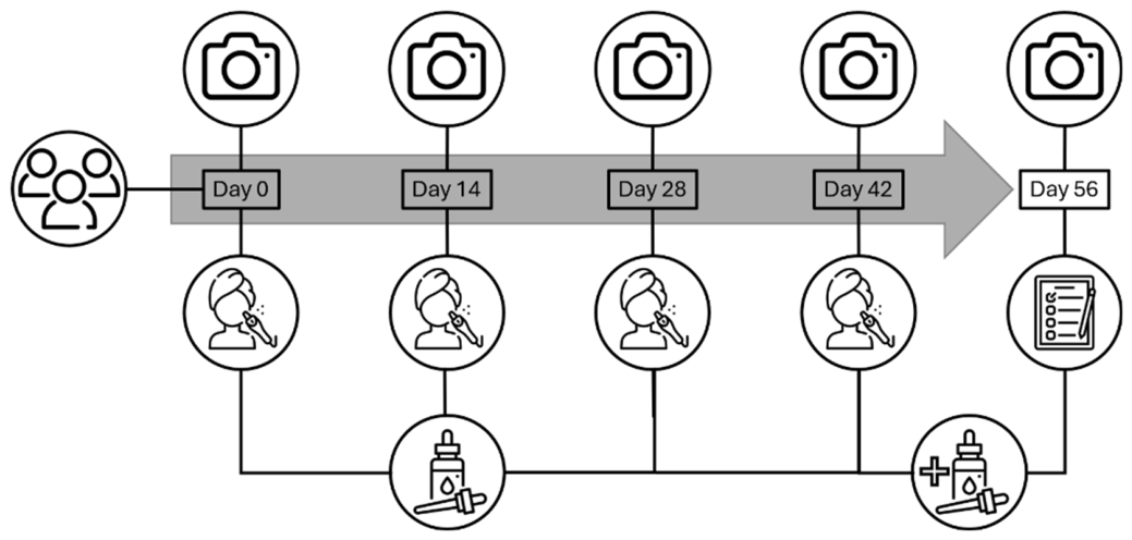
Figure 2.
Improvement of number, intensity, contrast, size of pigment spots, and skin tone evenness based on visual ratings of time-resolved image presentation. Significant improvements after 56 days in all pigment-related parameters marked with *** for p<.001.
Figure 2.
Improvement of number, intensity, contrast, size of pigment spots, and skin tone evenness based on visual ratings of time-resolved image presentation. Significant improvements after 56 days in all pigment-related parameters marked with *** for p<.001.
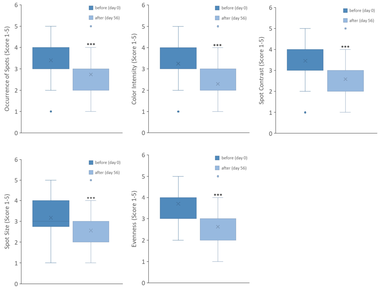
Figure 3.
Improvement of skin tone evenness based on visual ratings of time-resolved image presentation by experts and novices. Significantly different ratings marked with * for p<.05.
Figure 3.
Improvement of skin tone evenness based on visual ratings of time-resolved image presentation by experts and novices. Significantly different ratings marked with * for p<.05.
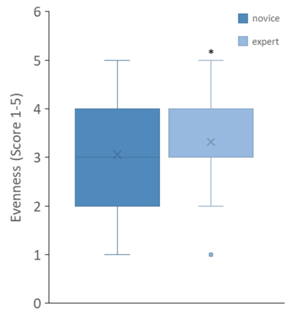
Figure 4.
Reduction of distinctive spots (dL) [%] based on time-resolved instrumental image analysis with tendency of significance p < .10 (*).
Figure 4.
Reduction of distinctive spots (dL) [%] based on time-resolved instrumental image analysis with tendency of significance p < .10 (*).
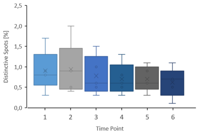
Figure 5.
Photographs of subject 2 before and after the MDA/AHA-TXA treatment routine: left, before the study; right, after four treatments and accompanying at-home product application.
Figure 5.
Photographs of subject 2 before and after the MDA/AHA-TXA treatment routine: left, before the study; right, after four treatments and accompanying at-home product application.
Disclaimer/Publisher’s Note: The statements, opinions and data contained in all publications are solely those of the individual author(s) and contributor(s) and not of MDPI and/or the editor(s). MDPI and/or the editor(s) disclaim responsibility for any injury to people or property resulting from any ideas, methods, instructions or products referred to in the content. |
© 2026 by the authors. Licensee MDPI, Basel, Switzerland. This article is an open access article distributed under the terms and conditions of the Creative Commons Attribution (CC BY) license.
Copyright: This open access article is published under a Creative Commons CC BY 4.0 license, which permit the free download, distribution, and reuse, provided that the author and preprint are cited in any reuse.