

Submitted:
19 August 2026
Posted:
20 August 2026
You are already at the latest version
Abstract
Mucormycosis is a rapidly progressive angioinvasive fungal infection predominantly affecting immunocompromised individuals and is associated with significant morbidity and mortality. Despite advances in standard care involving surgical debridement and systemic antifungal therapy, outcomes remain suboptimal due to recurrence risk, treatment-related toxicity (notably nephrotoxicity with amphotericin B), and substantial economic burden. These limitations justify continued investigation of adjunctive therapies and other novel approaches that may improve outcomes or reduce treatment burden, while established antifungal therapy and surgical management remain the cornerstone of care. Ayurveda, a traditional system of medicine in India, has been utilized by patients with mucormycosis; however, its evidence base remains limited and largely restricted to adjunctive or isolated case reports.
This study presents a case series of four mucormycosis patients managed predominantly with Ayurveda-based interventions. Three patients opted to receive stand-alone Ayurveda-based treatment after declining recommended standard biomedical treatment. significant radiological improvement as interpreted by the reporting radiologists with no evidence of recurrence. One case with poor adherence showed no radiological improvement, though survival was maintained for one year. All patients had complex comorbidities, including diabetes mellitus, hypertension, and renal impairment.
These observations advance hypothesis generation and provide conceptual and methodological insights for the development of future clinical research protocols. As an uncontrolled descriptive case series, they do not establish clinical efficacy, safety, non-inferiority, or suitability for clinical application, nor should they be interpreted as advocating replacement of the current standard-of-care management of mucormycosis.
Keywords:
mucormycosis
; Ayurveda
; case series
; emergency care
; complex disease management
1. Introduction
Mucormycosis is a rare, rapidly progressive, angioinvasive fungal infection caused by filamentous fungi belonging to the order Mucorales [1]. The disease predominantly affects individuals with impaired host immunity, particularly those with uncontrolled diabetes mellitus, haematological malignancies, recipients of solid-organ or hematopoietic stem cell transplantation, and patients receiving prolonged immunosuppressive therapy [2]. Following tissue invasion, the fungi exhibit a marked propensity for vascular infiltration, leading to thrombosis, tissue ischemia, and extensive necrosis [3]. Infection typically originates within the nasal cavity and paranasal sinuses and may rapidly extend to the orbit, cranial nerves, skull base, and intracranial structures [1]. Among the various clinical manifestations, rhino-orbito-cerebral mucormycosis (ROCM) is the most frequently encountered presentation and remains associated with mortality rates ranging from approximately 30% to 50% [4].
Beyond its substantial mortality, mucormycosis imposes considerable long-term morbidity. Survivors frequently experience irreversible visual impairment, cranial neuropathies, maxillofacial defects, facial disfigurement, and persistent functional limitations affecting mastication, speech, swallowing, and nasal breathing, all of which profoundly compromise health-related quality of life [5]. Consequently, mucormycosis constitutes a medical emergency that demands timely diagnosis and coordinated multidisciplinary management [1].
Contemporary management relies on a multimodal therapeutic strategy comprising early diagnosis, prompt reversal of underlying predisposing factors whenever feasible, initiation of systemic antifungal therapy with high-dose liposomal amphotericin B as the preferred first-line agent, aggressive surgical debridement of necrotic tissue, and step-down therapy with oral triazoles such as posaconazole or isavuconazole when clinically indicated [6]. The implementation of these evidence-based interventions through multidisciplinary care has substantially improved survival compared with historical outcomes and remains the cornerstone of mucormycosis management [7].
Nevertheless, important clinical challenges persist despite these therapeutic advances. Overall case-fatality rates remain approximately 40–50%, varying according to the anatomical site of infection, disease extent, underlying comorbidities, and timeliness of intervention [1]. Furthermore, radical surgical procedures frequently result in permanent anatomical and functional deficits, while prolonged antifungal therapy may lead to nephrotoxicity, hepatotoxicity, electrolyte disturbances, infusion-related adverse effects, and considerable financial burden [8,9]. Persistent or recurrent disease also continues to occur despite apparently appropriate medical and surgical management, particularly among patients with delayed presentation or persistent immunosuppression [10]. Collectively, these challenges underscore the need to investigate additional evidence-based therapeutic strategies aimed at preserving tissue integrity, improving functional recovery, reducing treatment-related morbidity, and enhancing patient-centred outcomes, while recognising that currently established systemic antifungal therapy and surgical management remain the standard of care for mucormycosis. Ayurveda, one of the major traditional medical systems practiced in India, continues to play an important role in healthcare delivery across the country [11]. During the COVID-19 pandemic, disruptions in healthcare delivery, restricted access to conventional medical services, and increased public interest in complementary healthcare were associated with greater utilisation of Ayurveda and other traditional medical systems. This pattern of healthcare-seeking extended to patients with COVID-19-associated complications, including mucormycosis who sought Ayurveda either as a primary therapeutic approach or as an adjunct to conventional management [12,13].
Despite its widespread utilization, robust clinical evidence supporting Ayurveda in the management of mucormycosis remains limited [14]. This evidence gap presents two important challenges. On one hand, potentially promising therapeutic approaches cannot be rigorously evaluated or appropriately integrated into evidence-informed clinical practice [15]. On the other hand, the absence of robust evidence creates uncertainty regarding efficacy and safety, thereby limiting informed clinical decision-making and may affect patient safety as well [16]. Accordingly, systematic evaluation of Ayurveda-based interventions is essential to establish an objective and scientifically robust evidence base [17].
Generating high-quality evidence in mucormycosis presents substantial methodological challenges. The rarity of the disease, rapid clinical progression, marked heterogeneity in patient characteristics, and ethical constraints surrounding randomization in life-threatening conditions limit the feasibility of adequately powered randomized controlled trials [18]. Consequently, well-conducted observational studies, rigorously documented case series, and other forms of real-world clinical evidence assume an important role in characterizing therapeutic responses, generating hypotheses, and informing future clinical research and guideline development [1].
Experimental investigations have demonstrated antifungal, anti-inflammatory, and immunomodulatory properties of several Ayurvedic medicinal plants and formulations against clinically relevant fungal pathogens, providing preliminary experimental rationale [19].However, the available clinical literature on Ayurveda in mucormycosis remains limited to non-randomized studies evaluating Ayurveda as an adjunct to conventional treatment, together with a single published case report describing successful stand-alone Ayurveda management [13,20]. Consequently, important evidence gaps persist regarding the evaluation of predominantly Ayurveda-based management, particularly with respect to objective clinical and radiological outcomes, safety, and long-term follow-up
To our knowledge, this is one of the first case series describing predominantly Ayurveda-based management of mucormycosis, with comprehensive documentation of therapeutic strategies, serial clinical and radiological outcomes, and long-term follow-up. As one of the first studies of its kind, this work contributes to the emerging evidence base by providing preliminary real-world clinical evidence to inform future hypothesis-driven research on the potential role of Ayurveda in mucormycosis management.
1.1. Case 1
A 65-year-old male from western India with a four-year history of diabetes mellitus and hypertension presented with a 15-day history of progressive orofacial symptoms, including a bulge over the hard palate, mobility of the right maxillary teeth, numbness of the upper palate, persistent bitter taste, and foul-smelling nasal discharge predominantly from the right nostril. The patient had no significant family history.
The patient initially consulted an otorhinolaryngologist. Microbiological examination of tissue obtained from the right nasal cavity by potassium hydroxide (KOH) preparation demonstrated septate branching fungal hyphae with numerous spores together with occasional broad aseptate hyphae. Subsequent fungal culture demonstrated growth of Rhizopus species and Aspergillus fumigatus according to the clinical microbiology laboratory report. Based on the clinical presentation and initial microbiological findings, invasive fungal rhino sinusitis was considered the leading diagnosis, with mucormycosis as the principal differential diagnosis and the possibility of concomitant aspergillosis also considered. Non contrast computed tomography (CT)of Paranasal Sinuses and orbits performed on 17 January 2025 demonstrated osteomyelitis involving the hard palate and bilateral maxillary alveolar processes, together with right ethmoidal and maxillary sinusitis and associated rhinitis (Figure 1A). These radiological findings were highly suggestive of an invasive rhino-maxillary fungal infection. Subsequent clinicopathological and microbiological evaluation established the diagnosis of mucormycosis.
The treating team recommended contemporary standard-of-care management comprising functional endoscopic sinus surgery (FESS) and systemic antifungal therapy. Following discussion of the available treatment options, the patient declined the proposed interventions because of concerns regarding surgical invasiveness and the anticipated financial burden, and subsequently sought consultation for Ayurveda-based management. The patient underwent stand-alone Ayurveda treatment while continuing biomedical follow-up and treatment for diabetes mellitus.
The patient demonstrated progressive clinical improvement throughout follow-up, with complete resolution of the palatal swelling after approximately 150 days of treatment [Table 1.1]. A follow-up 96 Slice CT scan (Thin 0.75-mm contiguous) of Paranasal Sinuses and orbits performed after approximately 11 months of Ayurveda treatment was interpreted by the reporting radiologist as demonstrating complete resolution of sinusitis, stabilization of the previously identified osseous changes, and radiological features suggestive of new bone formation and healing (Figure 1B).
1.2. Case 2
A 47-year-old male from western India presented with pain and heaviness over the left side of the head, a necrotic lesion involving the left hard palate (Figure 3A), restricted ocular movements, and blurred vision. The patient’s medical history was significant for uncontrolled diabetes mellitus, hypertension, chronic kidney disease, previous chikungunya infection, and a solitary functioning kidney. Baseline laboratory investigations demonstrated elevated serum creatinine and reduced estimated glomerular filtration rate.
Plain and contrast (Intravenous (IV) gadolinium contrast) Magnetic resonance imaging (MRI) of Brain with paranasal sinuses performed on 1 March 2024 demonstrated invasive fungal involvement of the left ethmoidal sinus with extension to the hard palate, skull-base osteomyelitis, surrounding inflammatory soft-tissue changes, the black turbinate sign, and perineural spread along the ophthalmic and maxillary divisions of the left trigeminal nerve (Figure 2A).
Potassium hydroxide examination performed on 2 March 2024 demonstrated zygomycete fungal hyphae, while subsequent fungal cultures of palatal and sinonasal specimens yielded Mucor species and Candida krusei, as reported by the clinical microbiology laboratory. Based on the clinical, microbiological, and radiological findings, the patient was diagnosed with rhino-orbital mucormycosis with skull-base involvement. The treating team recommended surgical debridement together with systemic antifungal therapy. However, after discussion of the proposed management and considering the anticipated surgical risks in the presence of multiple comorbidities, the patient elected to undergo stand-alone Ayurveda treatment. At the initial Ayurveda consultation, the patient’s serum creatinine was 1.48 mg/dL, with an estimated glomerular filtration rate of approximately 54 mL/min/1.73 m² [Supplementary Files 1.1]. The patient was advised to continue biomedical management and regular follow-up for diabetes mellitus, hypertension, and chronic kidney disease.
The patient demonstrated progressive clinical improvement within two weeks of treatment initiation [Table 1.1]. Complete symptomatic recovery together with complete clinical healing of the palatal lesion was achieved within approximately five months (Figure 3B).
Follow-up Non-contrast MRI(Multiplaner multi echo 1.5T) of Brain and Paranasal sinuses performed after approximately four months of Ayurveda treatment (8 July 2024) was interpreted by the reporting radiologist as demonstrating marked reduction of inflammatory changes involving the left pterygopalatine fossa and sphenopalatine foramen, near-complete resolution of abnormalities within the retro-maxillary fat space, and residual findings considered consistent with post-inflammatory sequelae rather than active invasive infection (Figure 2B).At the nine-month follow-up, the patient remained clinically stable without evidence of disease recurrence or treatment-related adverse events. The serum creatinine concentration had increased to 3.3 mg/dL, consistent with progression of the underlying chronic kidney disease.
Figure 2(A&B): MRI Image before(A) and after (B) Ayurveda treatment
1.3. Case 3
A 75-year-old male from western India with a 15-year history of diabetes mellitus presented with a 20-day history of nasal regurgitation of fluids during swallowing, persistent bitter taste, foul-smelling nasal discharge, and heaviness over the right side of the face.
Initial potassium hydroxide examination performed on 17 May 2022 did not demonstrate fungal elements. However, histopathological examination of maxillary sinus tissue performed on 25 May 2022 confirmed mucormycosis by demonstrating fungal invasion into bone.
Computed tomography Non-contrast Multidetector Computed Tomography on 14 May 2022 demonstrated extensive mucosal thickening involving the right maxillary and frontal sinuses together with bilateral ethmoidal and sphenoidal sinuses. Additional findings included inflammatory infiltration of the right retro-antral fat space and infratemporal region with early erosive changes involving the nasal septum, suggestive of osteomyelitis. Further radiological evaluation demonstrated extensive destruction involving the floor and walls of the right maxillary sinus, resulting in oro-antral fistula formation and displaced osseous fragments within the maxillary sinus cavity, indicative of advanced local tissue destruction [Supplementary File 1.0].
The patient initially underwent functional endoscopic sinus surgery (FESS) as recommended by the treating otorhinolaryngologist. Immediately following surgery, the patient sought Ayurveda treatment and declined systemic antifungal therapy. The patient subsequently underwent stand-alone Ayurveda management while continuing biomedical care for diabetes mellitus.
The patient demonstrated substantial clinical improvement during approximately 90 days of treatment [Table 1.1]. Long-term follow-up extending to three years demonstrated sustained symptom resolution without clinical evidence of recurrence. Follow-up non-contrast Multidetector Computed Tomography imaging performed three years after treatment (on 27 August 2025) demonstrated persistent residual mucosal thickening involving multiple paranasal sinuses but no radiological evidence of active invasive fungal disease or disease progression [Supplementary File 1.0].
1.4. Case 4
A 75-year-old female with a 20-year history of diabetes mellitus and hypertension, and a history of cholecystectomy one month before presentation, presented with headache, facial pain and heaviness, ocular pain, and fever.
Microbiological examination of tissue specimens obtained from the right nostril on 13 March 2025 demonstrated filamentous fungal elements. Although direct smear microscopy did not clearly distinguish septate from aseptate hyphae, the findings confirmed the presence of fungal structures within the tissue specimen.
96 Slice (0.5 mm high resolution) non-contrast Computed tomography of the paranasal sinuses performed on 8 March 2025 demonstrated bilateral chronic pan sinusitis, more pronounced on the right side, with complete obstruction of the right osteomeatal complex (Figure 4A). Additional CT findings included diffuse rarefaction and thinning of the adjacent osseous structures, moderate polypoidal mucosal thickening involving the right frontal and ethmoidal sinuses, severe mucosal thickening involving both maxillary and sphenoidal sinuses, and destruction of the right middle turbinate. Collectively, these radiological findings were consistent with invasive fungal rhino sinusitis, with mucormycosis considered the leading differential diagnosis
Histopathological examination of a biopsy specimen obtained from the right nasal cavity on 17 March 2025 demonstrated necrotic nasal mucosa containing broad aseptate fungal hyphae morphologically consistent with Mucorales, together with additional thin fungal hyphae and bacterial colonies. Fungal invasion of the soft tissue was evident, and no evidence of malignancy was identified. These histopathological findings established the diagnosis of mucormycosis, while also suggesting the presence of concomitant fungal elements consistent with a mixed fungal rhino sinusitis
Considering the histopathological evidence of tissue invasion, extensive Sino nasal involvement, and radiological evidence of bony destruction, the treating team recommended conventional management. Following discussion of the available treatment options, the patient elected to pursue Ayurveda-based management because of concerns regarding the perceived risks associated with standard therapy.
The patient underwent Ayurveda treatment; the patient had significant improvement with most of the symptoms, with 3 months of follow-up [Table 1.1]. However, adherence to the prescribed therapeutic regimen and dietary recommendations remained suboptimal because of personal constraints. Follow-up 96 Slice Non contrast Computed tomography of the paranasal sinuses performed after approximately three months (13 June 2025) demonstrated persistent destructive osseous changes involving the maxilla, hard palate, sphenoid bone, clivus, and right ethmoidal region together with residual inflammatory soft-tissue opacification involving the bilateral sphenoid, right ethmoidal, and right maxillary sinuses (Figure 4B). These findings were interpreted as post-treatment sequelae of invasive fungal rhino sinusitis without convincing radiological evidence of disease regression. The patient survived for approximately one year following diagnosis.
Figure 4(A&B): CT Image before(A) and after (B) Ayurveda treatment
2. Treatment and Diet
From an Ayurvedic perspective, the clinical presentation of the cases was interpreted in relation to the concepts of Raktaja Krimi (pathological organisms affecting blood and vascular tissues) and Asthi-Majjagata Krimi (pathological involvement of bone and marrow tissues), as described in the WHO International Standard Terminologies on Ayurveda (ITA 5.13.7; ITA 2.2.1; ITA 5.13). The treatment strategy was formulated through an integrative understanding of the contemporary clinical manifestations of mucormycosis and their Ayurvedic interpretation. A central therapeutic consideration was the regulation of kleda (moistness or pathological fluid accumulation; ITA-D.2534), which is understood to create conditions conducive to disease progression [Table 1.0]. Accordingly, both therapeutic interventions and supportive dietary measures were directed toward reducing excessive kleda and restoring physiological balance [21].
All patients were advised to follow a diet and lifestyle regimen designed to minimize factors considered to promote kleda accumulation. The dietary recommendations emphasized easily digestible foods, bitter vegetables, and moderate consumption of camel milk, while discouraging excessive intake of sweet, sour, heavy, and moisture-promoting foods. Lifestyle guidance included avoiding daytime sleep, prolonged exposure to cold environments, and other factors traditionally associated with increased kleda, while encouraging measures that promote warmth and metabolic regulation. Notably, Cases 1–3 demonstrated satisfactory adherence to these recommendations, whereas adherence was suboptimal in Case 4. Within the Ayurvedic framework, such variations in compliance may represent an important factor influencing therapeutic outcomes and warrant further consideration in future clinical investigations. Dietary and lifestyle recommendations based on the Ayurvedic concept of kleda reduction were incorporated as supportive components of the individualized management approach and should be interpreted separately from the primary therapeutic interventions. As this retrospective case series did not employ standardized methods to assess adherence or evaluate the independent contribution of these recommendations, no causal relationship between adherence and clinical outcomes can be inferred.
Given the emergency nature of mucormycosis, all patients underwent a comprehensive informed consent process that included discussion of the severity of the disease, anticipated prognosis, available management options, the potential benefits and risks of the proposed Ayurveda-based treatment, uncertainties regarding available evidence, and measures planned to mitigate foreseeable risks. Treatment decisions were reached through a shared decision-making process, and patients voluntarily opted for Ayurveda-based management after providing written informed consent.
3. Discussion
The present case series describes the clinical course, radiological evolution, and long-term outcomes of four cases of mucormycosis managed predominantly through Ayurveda-based interventions following diagnosis by conventional biomedical investigations [Table 1.2]. To our knowledge, this represents one of the first published case series documenting predominantly Ayurveda-based management of mucormycosis with comprehensive clinical, radiological, and long-term follow-up. The cohort represented a spectrum of advanced craniofacial mucormycosis, comprising three patients with rhino-maxillary disease (including cases with maxillary osteomyelitis and skull-base extension) and one patient with rhino-orbital disease complicated by skull-base involvement and perineural spread. Two patients also demonstrated concomitant mixed fungal infections involving Aspergillus spp. or Candida spp. Among the four patients, three achieved sustained clinical improvement accompanied by favourable radiological improvement following Ayurveda-based management, with follow-up ranging from nine months to four years. Notably, no clinical or radiological evidence of disease recurrence was documented during the respective follow-up periods in these three patients. An important observation was the sustained clinical and radiological improvement documented in the patient with skull-based mucormycosis and perineural spread (Case 2). Skull-base involvement represents one of the most aggressive manifestations of mucormycosis and is frequently associated with extensive tissue invasion, neurological complications, and poor clinical outcomes [22]. Such patients generally require aggressive multimodal management consisting of prolonged systemic antifungal therapy together with extensive surgical debridement and, in selected cases, repeated surgical procedures to achieve disease control [23]. Against this clinical background, the sustained clinical recovery and favourable radiological response observed over approximately nine months following predominantly Ayurveda-based management merit further investigation. Although causal inferences cannot be drawn from an uncontrolled case series, this clinical observation highlights the need for systematic evaluation of Ayurveda-based therapeutic strategies in advanced mucormycosis through appropriately designed clinical studies.
Case 4 demonstrated a comparatively different clinical trajectory. The patient experienced moderate symptomatic improvement during the initial three months of treatment; however, follow-up computed tomography did not demonstrate corresponding radiological resolution. Interpretation of this observation requires caution because treatment adherence became suboptimal after the initial treatment period, potentially influencing therapeutic response. Although convincing radiological improvement was not demonstrated, the patient’s survival for approximately one year remains a clinically relevant descriptive outcome, particularly as three-month survival is a widely reported endpoint in mucormycosis research [24].
Furthermore, persistent radiological abnormalities identified approximately three months after treatment initiation should not be interpreted as definitive evidence of treatment failure, since radiological resolution of invasive fungal rhino sinusitis may lag behind clinical recovery. A follow-up imaging assessment performed after five to six months under satisfactory treatment adherence may have provided a more reliable evaluation of disease evolution. In this case, clinical records documented substantially reduced medicine consumption followed by treatment discontinuation during the course of management. This observation is reported descriptively as one of several factors that may have influenced the overall clinical course, alongside disease severity, baseline clinical characteristics, concomitant management, and other unmeasured variables. Accordingly, no causal attribution is made between treatment adherence and the observed outcome. Collectively, these findings highlight the importance of standardized radiological follow-up protocols in future Ayurveda-based mucormycosis studies, while accounting for baseline disease severity, treatment adherence, and temporal changes in radiological response.
The diagnosis of mucormycosis in this case series was established through a comprehensive multidisciplinary assessment integrating clinical presentation, radiological findings, microbiological investigations, and histopathological examination where available. Although the EORTC/MSSGERC consensus definitions consider histopathological demonstration of tissue invasion a key criterion for the classification of proven mucormycosis, these definitions were primarily developed to standardize patient enrolment in epidemiological studies, clinical trials, and diagnostic research rather than to direct routine clinical decision-making [25]. As acknowledged by the consensus update, these research definitions are not intended to serve as the sole framework for day-to-day clinical diagnosis. Accordingly, this descriptive case series reflects real-world clinical practice, in which diagnostic decisions were based on multidisciplinary integration of compatible clinical features, characteristic radiological findings, microbiological evidence from tissue specimens, and histopathological confirmation where available. This pragmatic diagnostic approach is consistent with contemporary clinical management of mucormycosis while transparently presenting the diagnostic evidence supporting each individual case. Diabetes mellitus was present in all four patients, consistent with its well-established role as the predominant predisposing factor for mucormycosis, particularly in low- and middle-income countries [4]. One of the patient (case 2) had CKD which is considered to be emerging risk factor [26]. Additional comorbidities included hypertension, previous chikungunya infection, and solitary functioning kidney, thereby reflecting the complex clinical profile frequently encountered in routine practice. Consequently, this case series provides preliminary documentation regarding the clinical course, radiological outcomes, and long-term follow-up associated with predominantly Ayurveda-based management in patients with mucormycosis presenting with multiple high-risk clinical characteristics [Table 1.2].
The findings should also be interpreted within the broader healthcare context of low- and middle-income countries, where delayed diagnosis, challenges in achieving optimal glycaemic control, limited access to specialized centres, and financial constraints frequently contribute to advanced disease at presentation and poorer clinical outcomes [27,28]. Current evidence consistently identifies early initiation of systemic antifungal therapy together with prompt surgical debridement as the cornerstone of mucormycosis management, while delayed intervention has repeatedly been associated with increased mortality [29]. Boqaaiya et al. and other investigators have similarly emphasized that timely antifungal therapy and appropriate surgical intervention are critical determinants of survival [30]. Despite these established recommendations, mortality and substantial morbidity continue to be reported even in tertiary-care settings employing aggressive multimodal treatment strategies.
Against this background, the observations from the present series warrant careful consideration. All patients had already undergone comprehensive biomedical evaluation and diagnostic confirmation before initiating Ayurveda-based management, and most sought Ayurveda relatively late in the disease course after conventional treatment options had been discussed. Nevertheless, three patients demonstrated sustained clinical improvement together with favourable radiological findings on serial follow-up imaging, as interpreted in the respective radiology reports.
Although these observations should not be interpreted as evidence of therapeutic equivalence or superiority to contemporary standard-of-care management, they generate clinically relevant hypotheses for future research evaluating predominantly Ayurveda-based management in carefully selected clinical contexts.
These preliminary findings support the need for prospective observational studies and appropriately designed comparative clinical investigations to evaluate efficacy, safety, patient-centred outcomes, and to determine whether any role exists for integration alongside current standards of care.
An additional observation arising from the present series relates to the predominantly non-invasive therapeutic approach employed. Surgical management of mucormycosis, although considered an essential component of contemporary standard-of-care treatment, is frequently associated with substantial functional and cosmetic sequelae, including extensive tissue loss, craniofacial deformity, visual impairment, and neurological deficits, depending on disease extent [31]. Recent evidence has suggested that organ-preserving and minimally invasive endoscopic approaches may achieve satisfactory disease control in selected patients with rhino-orbito-cerebral mucormycosis, highlighting that successful management may depend not only on radical surgical excision but also on effective reduction of fungal burden [32]. Within this context, the present observations generate hypotheses for future research exploring whether predominantly Ayurveda-based interventions could be evaluated as adjuncts to contemporary management, including organ-preserving surgical approaches where clinically appropriate. Such hypotheses require rigorous clinical investigation and should not be interpreted as supporting modification of current standard-of-care management.
Safety remains a critical consideration in the evaluation of any therapeutic strategy for mucormycosis. Systemic antifungal agents, particularly amphotericin B, are well recognized for their association with nephrotoxicity, electrolyte disturbances, and infusion-related adverse reactions [8]. In the present series, no treatment-related adverse events were documented during routine clinical follow-up. Serial biochemical monitoring demonstrated stable hepatic and renal function throughout treatment in Cases 1 and 3. In contrast, Case 2 presented with substantial baseline renal risk, including a solitary functioning kidney, chronic kidney disease, treatment-resistant hypertension, and impaired renal function before initiation of Ayurveda-based management [Table 1.2]. Although serum creatinine increased from 1.48 mg/dL to 3.3 mg/dL during follow-up, the temporal pattern and underlying clinical context were more consistent with progression of pre-existing chronic kidney disease than with an acute treatment-related nephrotoxic event. However, within the limitations of an uncontrolled case series, a treatment-related contribution cannot be definitively excluded. These observations reinforce the importance of incorporating standardized longitudinal monitoring of renal and hepatic function into future clinical investigations evaluating Ayurveda-based management, particularly among patients with pre-existing renal impairment or other high-risk comorbidities.
The use of herbo-mineral formulations in Case 2 should be interpreted within the context of advanced skull-based mucormycosis, a rapidly progressive and potentially fatal infection associated with limited therapeutic windows. Within the Ayurveda emergency care treatment strategy adopted for this patient, herbo-mineral preparations constituted as an essential component of Ayurveda treatment. Their administration followed a structured risk-benefit assessment that considered both the severity of the underlying disease and the patient’s pre-existing renal risk factors. Risk mitigation strategies included continuation of conventional management for hypertension, regular biomedical follow-up, serial assessment of renal and hepatic function, clinical surveillance for adverse events, use of formulations manufactured by licensed Good Manufacturing Practice (GMP)-certified pharmaceutical facilities, and comprehensive informed consent addressing anticipated benefits, potential risks, therapeutic uncertainties, and predefined referral criteria in the event of clinical deterioration. Collectively, these measures reflect a structured patient-safety framework designed to minimize foreseeable risks while facilitating access to comprehensive treatment in a life-threatening clinical scenario.
Intranasal fumigation was administered at a distance of approximately 30–45 cm from the patient’s nostrils, allowing cooled and diluted fumes to reach the nasal passages while avoiding direct exposure to heat or concentrated smoke. The exposure was intermittent and delivered as a gentle, controlled flow rather than continuous or excessive inhalation. Patients were monitored throughout the procedure and specifically assessed for potential local and respiratory adverse effects, including nasal or throat irritation, burning sensation of the eyes or throat, breathlessness, worsening nasal symptoms, and other respiratory discomfort.
When transient symptoms such as mild throat or ocular irritation or mild breathlessness occurred following exposure, they were self-limiting and generally resolved spontaneously within approximately 5–10 minutes without requiring medical intervention. No clinically significant mucosal burns, persistent airway symptoms, secondary respiratory infections, or sustained worsening of local inflammatory symptoms were documented during the treatment period or subsequent clinical follow-up in the reported cases. Patients with severe underlying respiratory illness were not administered this intervention.
The available human evidence concerning the safety of Ayurvedic nasal fumigation remains limited. A recent pilot clinical study evaluated polyherbal Dhoopana in children with rhinitis, and a subsequently published clinical trial protocol describes structured administration and adverse-event monitoring; however, these studies concern a substantially different clinical population and indication and cannot be extrapolated to establish the safety of fumigation in patients with invasive fungal rhinosinusitis [33,34].
Accordingly, the present observations should not be interpreted as establishing the safety of intranasal fumigation. The safety observations reported here are limited to clinically documented adverse effects in this small retrospective case series. Larger prospective studies with predefined adverse-event criteria, standardised exposure parameters, and systematic assessment of nasal, ocular, and respiratory effects are required to characterise the safety profile of this intervention more reliably.
Although herbo-mineral formulations may pose safety concerns when manufactured without appropriate quality assurance or administered irrationally, the formulations employed in the present series were produced under established regulatory standards and prescribed according to recognized Ayurvedic clinical practice [Table 1.0]. Furthermore, published toxicological and clinical investigations have suggested that properly manufactured herbo-mineral formulations prepared using standardized purification and processing procedures described in classical Ayurveda may demonstrate acceptable safety profiles when used within recommended therapeutic dosages [35]. Similarly, the only previously published report describing stand-alone Ayurveda-based management of mucormycosis documented normal hepatic and renal function throughout treatment despite the use of comparable formulations [20]. However, such observations remain insufficient to draw definitive conclusions regarding safety, they support the need for rigorous prospective pharmacovigilance and standardized safety evaluation in future clinical research.
The financial burden of mucormycosis management represents an important consideration, particularly within resource-constrained healthcare systems. An earlier observational study from India observed that a considerable proportion of patients left standard biomedical treatment against medical advice, with economic hardship frequently cited as the reason for treatment discontinuation [36]. In this case series, the patients reflected similar economic constraints as one of the major factors impacting their preference for the treatment. All four patients reported that the anticipated cost of conventional treatment influenced their therapeutic decision-making. Although preferential pricing has reduced the acquisition cost of liposomal amphotericin B to approximately US$20 per 50-mg vial for low- and middle-income countries [35], the recommended induction regimen typically requires multiple vials daily for several weeks. Based on standard dosing recommendations (5 mg/kg/day for a 60-kg adult over 4–6 weeks), the estimated acquisition cost of liposomal amphotericin B alone may range from approximately US$3,360–5,040 (approximately ₹2.8–4.3 lakh). These estimates represent only the anticipated acquisition cost of induction antifungal therapy and do not include hospitalization, surgical procedures, diagnostic investigations, electrolyte replacement, intensive care support when required, management of treatment-related complications, or subsequent oral antifungal therapy. Consequently, the overall expenditure associated with contemporary standard-of-care management is expected to be substantially higher, particularly in resource-constrained healthcare settings.
In comparison, the direct expenditure associated with the predominantly Ayurveda-based management in the present case series was approximately ₹30,000–40,000 per patient. Although this expenditure was considerably lower than published estimates of medication acquisition costs and overall treatment expenditure reported for conventional mucormycosis management, these observations should be interpreted with considerable caution. The present study did not collect patient-level comparative economic data and was not designed to undertake a formal cost-effectiveness, cost-minimization, or cost-utility analysis. Furthermore, differences in disease severity, hospitalization requirements, surgical interventions, healthcare resource utilization, treatment pathways, and follow-up preclude direct economic comparisons between contemporary standard-of-care management and predominantly Ayurveda-based management. Nevertheless, the observations highlight the potential influence of treatment affordability on patient decision-making and identify an important area for future prospective health-economic research integrating clinical outcomes, patient-centred outcomes, quality of life, and healthcare resource utilization.
The biological plausibility of Ayurveda-based therapeutic approaches has been explored in experimental research evaluating the pharmacological properties of individual medicinal plants and formulations used in Ayurvedic practice. Laboratory investigations, including computational, in vitro, and animal studies, have reported antifungal, immunomodulatory, and anti-inflammatory activities for several Ayurvedic botanicals against selected fungal pathogens or inflammatory targets, although these findings largely derive from preclinical models rather than studies of invasive mucormycosis specifically [37,38].
Experimental evaluation of an Ayurvedic intranasal formulation (Anu Taila) demonstrated inhibitory activity against Mucor spores and modulation of TNF-α-associated inflammatory pathways in laboratory models [39]. Several medicinal plants incorporated into the individualized treatment protocols in the present case series, including Embelia ribes (Vidanga), Butea monosperma (Palash), Semecarpus anacardium (Bhallataka), Albizia lebbeck (Shirisha), and Azadirachta indica (Neem), were also components of the stand alone Ayurveda-based treatment previously reported for mucormycosis [20]. Experimental studies have demonstrated antifungal activity for extracts of E. ribes and A. lebbeck, while embelin, the principal constituent of E. ribes, has shown inhibition of TNF-α-converting enzyme and modulation of NF-κB signalling pathways under experimental conditions [40,41,42,43]. These findings identify biological activities of individual constituents but do not establish the therapeutic effects of the individualized formulations used in this case series.
Collectively, these experimental observations provide hypotheses that may justify further mechanistic investigation but should not be interpreted as evidence of clinical efficacy in invasive mucormycosis. Establishing biological activity requires dedicated in vitro studies against Mucorales, appropriate animal models, translational pharmacological investigations.
Clinical evidence supporting Ayurveda in mucormycosis remains limited. Madikonda et al. reported favourable outcomes following adjunctive Ayurveda treatment in a non-randomized study involving 92 patients, evaluating clinically meaningful endpoints including survival, recurrence, requirement for advanced surgical procedures, and overall clinical improvement [44]. Subsequently, Rajnikant et al. described successful predominantly Ayurveda-based management of COVID-19-associated rhino-orbital mucormycosis with documented radiological resolution [20] Building upon these reports, the present case series expands the available evidence by documenting multiple microbiologically confirmed cases representing diverse clinical presentations, mixed fungal infections, advanced skull-base disease, and prolonged follow-up extending to four years. Furthermore, the incorporation of serial radiological assessment provides additional objective outcome measures that have been infrequently reported in the existing literature.
The findings should also be interpreted within the broader methodological landscape of mucormycosis research. The rarity of the disease, rapid clinical progression, marked heterogeneity in patient characteristics, and ethical challenges associated with withholding established treatment substantially limit the feasibility of adequately powered randomized controlled trials. Consequently, well-documented observational studies and real-world clinical evidence remain important sources of information for hypothesis generation, refinement of outcome measures, and development of future clinical research. Within this context, the present series contributes preliminary evidence regarding clinical outcomes, radiological assessment, long-term follow-up, and safety monitoring associated with predominantly Ayurveda-based management.
Nevertheless, these findings should be interpreted cautiously and should not be construed as evidence supporting generalized treatment recommendations or therapeutic equivalence with contemporary standard-of-care management. Rather, this case series provides hypothesis-generating evidence that may inform future prospective observational studies and carefully designed comparative clinical investigations. Importantly, the study demonstrates the feasibility of applying conventional biomedical outcome measures, including survival, microbiological confirmation, serial radiological assessment, and longitudinal safety monitoring, to evaluate Ayurveda-based interventions. Collectively, these observations contribute methodological insights that may facilitate the development of scientifically rigorous, ethically appropriate, and contextually relevant research protocols for investigating the role of Ayurveda within the multidisciplinary management of mucormycosis.
Patient perspectives:
Case 1: The patient reported concerns regarding potential facial disfigurement associated with surgery and the anticipated financial burden of conventional treatment. During follow-up, the patient expressed satisfaction with the recovery and the non-surgical management received.
Case 2: The patient reported similar concerns regarding surgery and treatment-related costs and stated that a recommendation from a previously treated patient influenced the decision to seek Ayurveda care. During follow-up, the patient expressed satisfaction with the clinical outcome.
Case 3: The patient described anxiety regarding surgery and concerns about financial constraints before treatment. During follow-up, the patient reported satisfaction with the clinical improvement achieved.
Limitations
The findings of this case series should be interpreted in light of several limitations. Follow-up assessments were not conducted at standardized time points, resulting in variability in the timing of clinical, radiological, and laboratory evaluations across cases. This limitation restricted systematic comparison of treatment trajectories and highlights the need for standardized follow-up schedules, particularly for radiological assessment within clinically relevant time windows. Similarly, a predefined safety monitoring framework, including uniform kidney and liver function testing intervals, was not implemented, which is especially relevant for patients with baseline renal impairment or other high-risk comorbidities. Comprehensive ophthalmological outcome assessment was also limited, as formal visual field evaluation was not available for Case 2. The symptom assessment presented in this case series is based on patient reported outcome and not based on standardised assessment.
4. Conclusion
This case series describes four cases of mucormycosis managed predominantly through Ayurveda-based interventions, with comprehensive clinical, radiological, and long-term follow-up. Three patients demonstrated sustained clinical improvement together with favourable radiological findings on serial follow-up imaging over follow-up periods ranging from 9 months to 4 years, without documented disease recurrence. One patient demonstrated persistent radiological abnormalities at the three-month assessment in the context of suboptimal treatment adherence and subsequently discontinued treatment, although survival was maintained for approximately one year following diagnosis. No treatment-related adverse events were reported during follow-up, and serial safety monitoring did not reveal clinically significant treatment-related hepatic or renal toxicity. One patient with pre-existing chronic kidney disease exhibited progressive deterioration in renal function during follow-up; however, the clinical course was considered more consistent with progression of underlying renal disease than with a treatment-related adverse event.
Although the findings should not be interpreted as evidence of treatment effectiveness or as support for generalized clinical recommendations, they provide descriptive clinical observations that contribute to hypothesis generation regarding predominantly Ayurveda-based management of microbiologically proven mucormycosis. To our knowledge, this is one of the first case series to comprehensively document predominantly Ayurveda-based management with objective microbiological confirmation, serial radiological assessment, and prolonged follow-up across patients with diverse clinical presentations, including advanced skull-base disease.
Beyond its clinical observations, this paper provides methodological insights for future investigations by demonstrating the feasibility of incorporating conventional biomedical outcome measures including survival, radiological response, microbiological confirmation, and longitudinal safety monitoring into the evaluation of Ayurveda-based interventions. These observations support the need for well-designed clinical studies to evaluate clinical outcomes, safety, patient-centred outcomes, and to determine whether any role exists for Ayurveda-based interventions within multidisciplinary management strategies for mucormycosis while contemporary antifungal therapy and surgical management are considered as the current standard of care.
Supplementary Materials
The following supporting information can be downloaded at the website of this paper posted on Preprints.org.
Funding
No funding was received for this work.
Informed Consent Statement
Written informed consent was obtained from each patient (or their legally accepted representative, where applicable) for participation in the study and for the publication of the clinical details and accompanying images.
Data Availability Statement
This article presents a descriptive clinical case series derived from routine clinical care rather than a prospective protocol-based research study. The anonymized clinical data supporting the findings are presented within the article and its Supplementary Materials. Additional source data, derived from confidential patient medical records, are available to the Editorial Office or reviewers upon reasonable request for verification purposes. These records are not publicly available because unrestricted disclosure could compromise patient privacy and confidentiality and may be inconsistent with the informed consent provided by the patients and applicable legal and ethical requirements.
Conflicts of Interest
The authors declare that there are no conflicts of interest.
References
- Cornely, O.A.; Alastruey-Izquierdo, A.; Arenz, D.; Chen, S.C.A.; Dannaoui, E.; Hochhegger, B.; Hoenigl, M.; Jensen, H.E.; Lagrou, K.; Lewis, R.E.; et al. Global guideline for the diagnosis and management of mucormycosis: an initiative of the European Confederation of Medical Mycology in cooperation with the Mycoses Study Group Education and Research Consortium. Lancet Infect. Dis. 2019, 19, e405-e421. [CrossRef]
- Skiada, A.; Lass-Floerl, C.; Klimko, N.; Ibrahim, A.; Roilides, E.; Petrikkos, G. Challenges in the diagnosis and treatment of mucormycosis. Med. Mycol. 2018, 56, S93-S101. [CrossRef]
- Ibrahim, A.S.; Spellberg, B.; Walsh, T.J.; Kontoyiannis, D.P. Pathogenesis of Mucormycosis. Clin. Infect. Dis. 2012, 54, S16-S22. [CrossRef]
- Jeong, W.; Keighley, C.; Wolfe, R.; Lee, W.; Slavin, M.; Kong, D.; Chen, S.-A. The epidemiology and clinical manifestations of mucormycosis: a systematic review and meta-analysis of case reports. Clin. Microbiol. Infect. 2019, 25, 26-34. [CrossRef]
- Sen, M.; Honavar, S.G.; Bansal, R.; Sengupta, S.; Rao, R.; Kim, U.; Sharma, M.; Sachdev, M.; Grover, A.K.; Surve, A.; et al. Epidemiology, clinical profile, management, and outcome of COVID-19-associated rhino-orbital-cerebral mucormycosis in 2826 patients in India—Collaborative OPAI-IJO Study on Mucormycosis in COVID-19 (COSMIC), Report 1. Indian J. Ophthalmol. 2021, 69, 1670-1692. [CrossRef]
- Hoenigl, M.; Seidel, D.; Carvalho, A.; Rudramurthy, S.M.; Arastehfar, A.; Gangneux, J.-P.; Nasir, N.; Bonifaz, A.; Araiza, J.; Klimko, N.; et al. The emergence of COVID-19 associated mucormycosis: a review of cases from 18 countries. Lancet Microbe 2022, 3, e543-e552. [CrossRef]
- Patel, A.; Agarwal, R.; Rudramurthy, S.M.; Shevkani, M.; Xess, I.; Sharma, R.; Savio, J.; Sethuraman, N.; Madan, S.; Shastri, P.; et al. Multicenter Epidemiologic Study of Coronavirus Disease-Associated Mucormycosis, India. Emerg. Infect. Dis. 2021, 27, 2349-2359. [CrossRef]
- Tragiannidis, A.; Gkampeta, A.; Vousvouki, M.; Vasileiou, E.; Groll, A.H. Antifungal agents and the kidney: pharmacokinetics, clinical nephrotoxicity, and interactions. Expert Opin. Drug Saf. 2021, 20, 1061-1074. [CrossRef]
- Murthy, R.; Gote, Y.S.; Bagchi, A. Localized surgical debridement for the management of orbital mucormycosis. Indian J. Ophthalmol. 2022, 70, 649-652. [CrossRef]
- Rao, K.M.; Pooja, N. Management of Mucormycosis in Post COVID-19 Patients and Its Outcome in a Tertiary Care Center: Our Experience. Indian J. Otolaryngol. Head Neck Surg. 2022, 74, 3344-3351. [CrossRef]
- Chandra, S. Status of Indian medicine and folk healing: With a focus on integration of AYUSH medical systems in healthcare delivery. AYU 2012, 33, 461. [CrossRef]
- Samal, J.; Singh, H. A systematic review and meta-analysis on the utilization of ayush systems of medicine in India’s COVID response. Discov. Health Syst. 2026, 5, 16. [CrossRef]
- Adluri, U.S.P.; Perugu, S. Evaluation of efficacy and safety of adjuvant Ayurvedic therapy in patients with severe post-covid mucor-mycosis at a Government tertiary care hospital—A Case-Control study. J. Ayurveda Integr. Med. 2022, 13, 100585. [CrossRef]
- Rastogi, S.; Verma, A. Jalaneti as primary intervention in suspected rhino-orbito-cerebral mucormycosis helps improving the recovery: A case report. J. Ayurveda Integr. Med. 2022, 13, 100516. [CrossRef]
- Alice Wong, Y.M.; Ahn, S.; Bana, A.; Kumar Dua, P.; Eggers, R.; Kuruvilla, S.; Li, Y.; Liu, Q.; Shen, Y.; Kim, S. Policy implications of WHO’s global traditional medicine strategy 2025-2034. Bull. World Health Organ. 2025, 103, 715-721. [CrossRef]
- Patwardhan, B. Bridging Ayurveda with evidence-based scientific approaches in medicine. EPMA J. 2014, 5, 19. [CrossRef]
- Patwardhan, B. Time for evidence-based Ayurveda: A clarion call for action. J. Ayurveda Integr. Med. 2013, 4, 63. [CrossRef]
- Sipsas, N.V.; Gamaletsou, M.N.; Anastasopoulou, A.; Kontoyiannis, D.P. Therapy of Mucormycosis. J. Fungi 2018, 4, 90. [CrossRef]
- Karthik, K.P.; Dileep, A.; Rajagopala, S.; Mahapatra, A.K.; Dharmarajan, P. Ayurveda approach to mucormycosis and other fungal infections: A comprehensive review. J. Indian Syst. Med. 2021, 9, 216-225. [CrossRef]
- Patel, R.; Lakshmanan, S.; Padikkal, V.; Mandaliya, K. The stand-alone Ayurveda treatment for the management of COVID-associated mucormycosis (CAM): a case report. Front. Med. 2026, 13, 1848849. [CrossRef]
- World Health Organization. WHO International Standard Terminologies on Ayurveda; World Health Organization: Geneva, Switzerland, 2023.
- Marzi Manfroni, A.; Arcuri, F.; Spinzia, A.; Sako, M.; Bianchi, B.; Laganà, F. Aggressive Mucormycosis with Extensive Craniofacial Involvement: A Case Report of Radical Surgical Management and Prosthetic Rehabilitation. Reports 2025, 8, 187. [CrossRef]
- Chakrabarti, A.; Das, A.; Sharma, A.; Panda, N.; Das, S.; Gupta, K.; Sakhuja, V. Ten Years’ Experience in Zygomycosis at a Tertiary Care Centre in India. J. Infect. 2001, 42, 261-266. [CrossRef]
- Denis, B.; Resche-Rigon, M.; Raffoux, E.; Ronchetti, A.-M.; Dudoignon, E.; Verillaud, B.; Valade, S.; Lorillon, G.; Rabian, F.; Xhaard, A.; et al. Epidemiology, Clinical Manifestations, Treatment, and Outcome of Mucormycosis: A Review of 77 Cases From a Single Center in France. Open Forum Infect. Dis. 2024, 11, ofae426. [CrossRef]
- Pappas, P.G.; Chen, S.C.-A.; Donnelly, J.P. The evidence supporting the revised EORTC/MSGERC definitions for invasive fungal infections. Clin. Infect. Dis. 2021, 72 (Suppl. 2), S77–S78. [CrossRef]
- Prakash, H.; Ghosh, A.K.; Rudramurthy, S.M.; Singh, P.; Xess, I.; Savio, J.; Pamidimukkala, U.; Jillwin, J.; Varma, S.; Das, A.; et al. A prospective multicenter study on mucormycosis in India: Epidemiology, diagnosis, and treatment. Med. Mycol. 2019, 57, 395-402. [CrossRef]
- Prakash, H.; Chakrabarti, A. Global Epidemiology of Mucormycosis. J. Fungi 2019, 5, 26. [CrossRef]
- Gupta, N.; Srinivas, S.; Harikumar, A.; Devaraja, K.; Nallapati, V.T.; Saravu, K. Deoxycholate amphotericin for management of mucormycosis: a retrospective cohort study from South India. Infez. Med. 2022, 30. [CrossRef]
- Brunet, K.; Rammaert, B. Mucormycosis treatment: Recommendations, latest advances, and perspectives. J. Mycol. Med. 2020, 30, 101007. [CrossRef]
- Boqaaiya, S.; Cohen, Y.; Beiruti Wiegler, K.; Chassid, O. Treatment and Survival Outcomes in Rhino-Orbital Mucormycosis with and without Orbital Exenteration: A Retrospective Case Series and Literature Review. Int. Med. Case Rep. J. 2025, Volume 18, 747-762. [CrossRef]
- Barrera-López, F.J.; Noyola-García, M.E.; Barrera-López, N.G.; Albarrán-Sánchez, A. Surgical and medical complications associated with mucormycosis in a tertiary hospital. Gac. Med. Mex. 2025, 161, 17463. [CrossRef]
- Sweed, A.H.; Anany, A.M.; Hussein, A.; Nada, W.; Eesa, M.; Elnashar, I.; Mobashir, M.; Ibrahim, E.M.; Elmaghawry, M.E. Endoscopic Orbital Clearance/Debridement: A Potential Substitute for Orbital Exenteration in Rhino-orbital Mucormycosis. Int. Arch. Otorhinolaryngol. 2025, 29, 001-007. [CrossRef]
- Monika; Rathi, R.B.; Rathi, B.; Balakrishnana, D. Evaluation of efficacy of Dhoopana (polyherbal nasal fumigation) versus polyherbal steam inhalation in children with Pratishyaya (rhinitis): A pilot study. Int. J. Ayurvedic Med. 2025, 16, 130–135. [CrossRef]
- Kakar, M.; Rathi, R.; Balakrishnan, D.; Rathi, B. Evaluation of comparative efficacy of polyherbal steam inhalation versus polyherbal nasal fumigation (Dhoopana) in children with rhinitis (Pratishyaya): Protocol for an open-label randomized controlled trial. JMIR Res. Protoc. 2025, 14, e58197. [CrossRef]
- Kamath, S.U.; Rajendran, K.; Pemiah, B.; Rajan, K.S.; Krishnaswamy, S.; Sethuraman, S.; Krishnan, U.M. Pharmacological and toxicological insights into the ayurvedic formulation Rasasindura. Sci. Rep. 2025, 15, 43591. [CrossRef]
- Priya, P.; Ganesan, V.; Rajendran, T.; Geni, V. Mucormycosis in a tertiary care center in South India: A 4-year experience. Indian J. Crit. Care Med. 2020, 24, 168–171. [CrossRef]
- Berman, J. Liposomal Amphotericin B Treatment and the Leishmaniases. Am. J. Trop. Med. Hyg. 2019, 101, 727-728. [CrossRef]
- Esmaeili, A.; Saleh, I.; Abu-Dieyeh, M.H. Antifungal potential of plant-based extracts against Candida species: values, safety concerns, and possible applications. Phytochem. Rev. 2025, 24, 5801-5844. [CrossRef]
- Shah, M.; Zia, M.; Ahmad, I.; Umer Khan, M.; Ejaz, H.; Alam, M.; Aziz, S.; Nishan, U.; Dib, H.; Ullah, R.; et al. Computational analysis of Ayurvedic metabolites for potential treatment of drug-resistant Candida auris. Front. Cell. Infect. Microbiol. 2025, 15, 1537872. [CrossRef]
- Balkrishna, A.; Rastogi, S.; Kharayat, B.; Tomer, M.; Varshney, Y.; Singh, K.; Kumari, P.; Dev, R.; Srivastava, J.; Haldar, S.; et al. Anu Taila, an herbal nasal drop, suppresses mucormycosis by regulating host TNF-α response and fungal ergosterol biosynthesis. J. Appl. Microbiol. 2022, 132, 3355-3374. [CrossRef]
- Rani, A.; Nagamani, V.; Saritha, K.; Sulakshana, G. In vitro evaluation of antifungal activity of the seed extract of embelia ribes. Indian J. Pharm. Sci. 2011, 73, 247. [CrossRef]
- Dhanjal, J.K.; Nigam, N.; Sharma, S.; Chaudhary, A.; Kaul, S.C.; Grover, A.; Wadhwa, R. Embelin inhibits TNF-α converting enzyme and cancer cell metastasis: molecular dynamics and experimental evidence. BMC Cancer 2014, 14, 775. [CrossRef]
- Lam, S.K.; Ng, T.B. First report of an anti-tumor, anti-fungal, anti-yeast and anti-bacterial hemolysin from Albizia lebbeck seeds. Phytomedicine 2011, 18, 601-608. [CrossRef]
- Alzohairy, M.A. Therapeutics Role of Azadirachta indica (Neem) and Their Active Constituents in Diseases Prevention and Treatment. Evid.-Based Complement. Altern. Med. 2016, 2016, 7382506. [CrossRef]
- Madikonda, P.K.; Perugu, S.B.; Ramadevi, C.H. Effect of Ayurvedic Intervention as an adjunct therapy in Post COVID-19 Mucormycosis (PCM): A non-randomized parallel group study. J. Ayurveda Integr. Med. 2022, 13, 100672. [CrossRef]
Figure 1.
A—(Black arrow) Significant osseous erosion and destruction of bilateral posterior hard palate and adjacent aspect of posterior maxillary alveolus process, with associated paraosseous soft tissue component. B- (White arrow) Interval significant increase in sclerosis & regression in paraosseous soft tissue at bilateral posterior hard palate region, and increase in changes of osteitis.
Figure 1.
A—(Black arrow) Significant osseous erosion and destruction of bilateral posterior hard palate and adjacent aspect of posterior maxillary alveolus process, with associated paraosseous soft tissue component. B- (White arrow) Interval significant increase in sclerosis & regression in paraosseous soft tissue at bilateral posterior hard palate region, and increase in changes of osteitis.
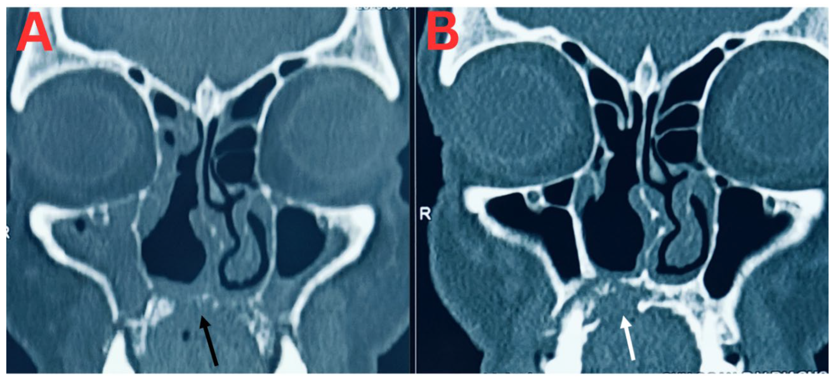
Figure 2.
A- White arrow: Showing left middle turbinate “Black Turbinate” Sign. White Star: Showing diffuse oedematous changes and soft tissue thickening involving medial intraconal compartment of left orbit. White arrow head: Diffuse marrow oedema and adjacent soft tissue at left posterior hard palate region. B- Yellow arrow: Showing autologous extirpatation of prior involved left middle turbinate. Yellow Star: Showing of resolution of prior noted soft tissue involving medial intraconal compartment of left orbit. Yellow arrow head: Interval near complete resolution of prior noted marrow oedema & adjacent soft tissue at left posterior hard palate region.
Figure 2.
A- White arrow: Showing left middle turbinate “Black Turbinate” Sign. White Star: Showing diffuse oedematous changes and soft tissue thickening involving medial intraconal compartment of left orbit. White arrow head: Diffuse marrow oedema and adjacent soft tissue at left posterior hard palate region. B- Yellow arrow: Showing autologous extirpatation of prior involved left middle turbinate. Yellow Star: Showing of resolution of prior noted soft tissue involving medial intraconal compartment of left orbit. Yellow arrow head: Interval near complete resolution of prior noted marrow oedema & adjacent soft tissue at left posterior hard palate region.
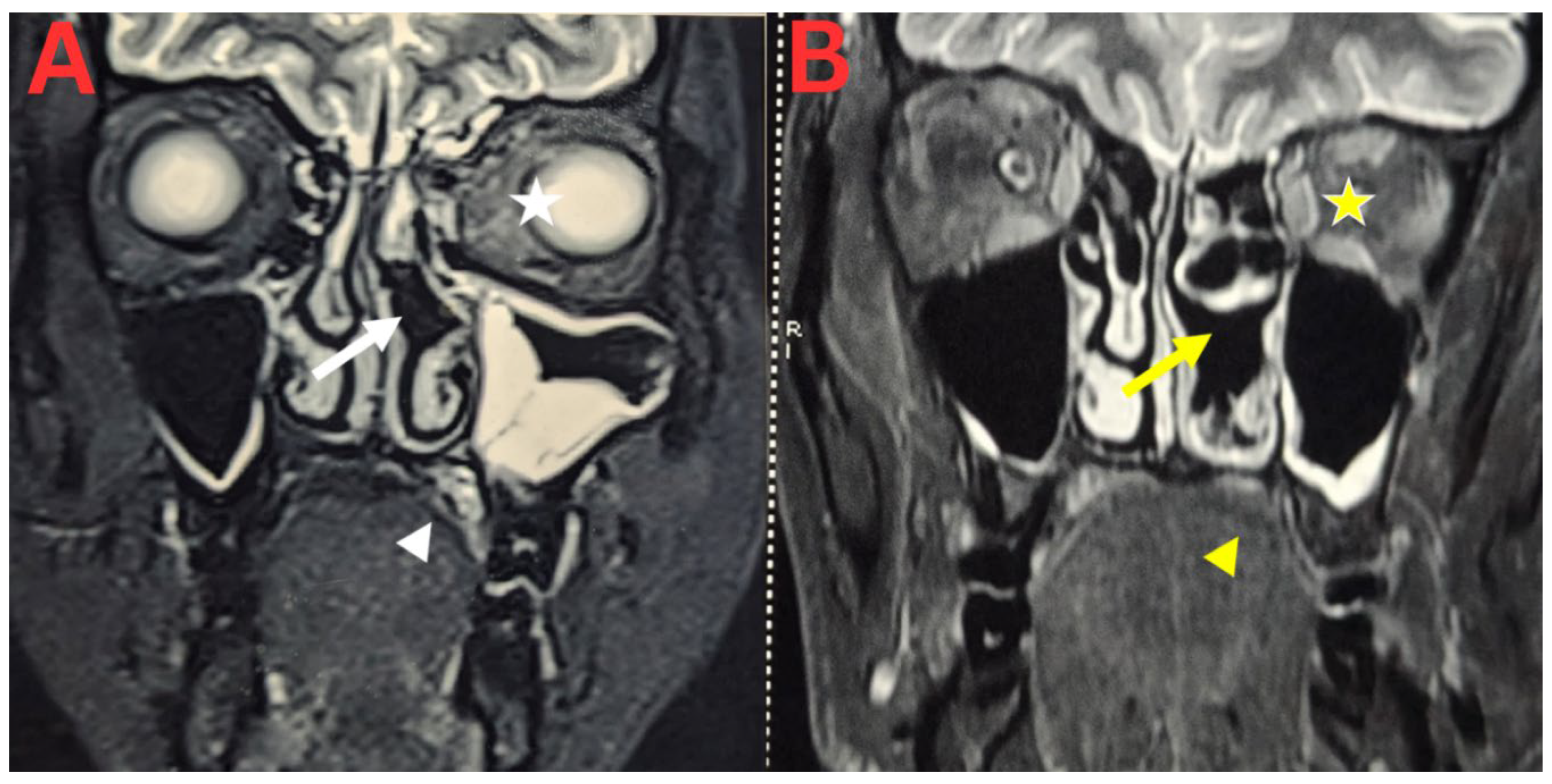
Figure 3.
A- Necrotic lesion in the hard palate. B- complete healing of the hard palate lesion.
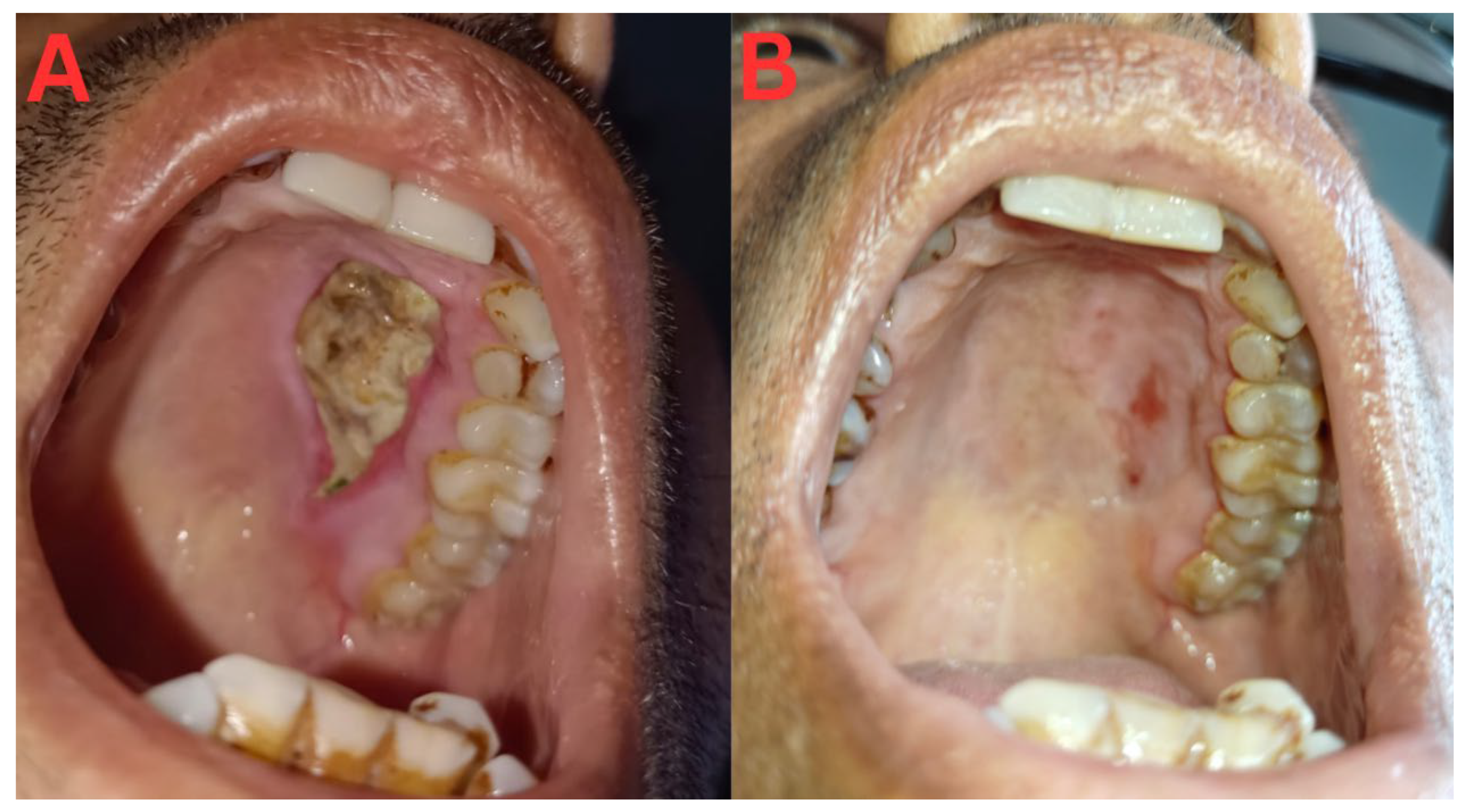
Figure 4.
A- white star: Subtle rarefaction of right middle turbinate, with polypoidal mucosal thickening at right middle meatus. White arrow): No evidence of osseous destruction or reflection of right hard palate. B. Yellow star: Interval increase in osseous destruction of right middle meatus, with mucosal irregularities. yellow arrow Interval significant increase in osseous destruction of right posterior hard palate and adjacent aspect of right maxillary bone.
Figure 4.
A- white star: Subtle rarefaction of right middle turbinate, with polypoidal mucosal thickening at right middle meatus. White arrow): No evidence of osseous destruction or reflection of right hard palate. B. Yellow star: Interval increase in osseous destruction of right middle meatus, with mucosal irregularities. yellow arrow Interval significant increase in osseous destruction of right posterior hard palate and adjacent aspect of right maxillary bone.
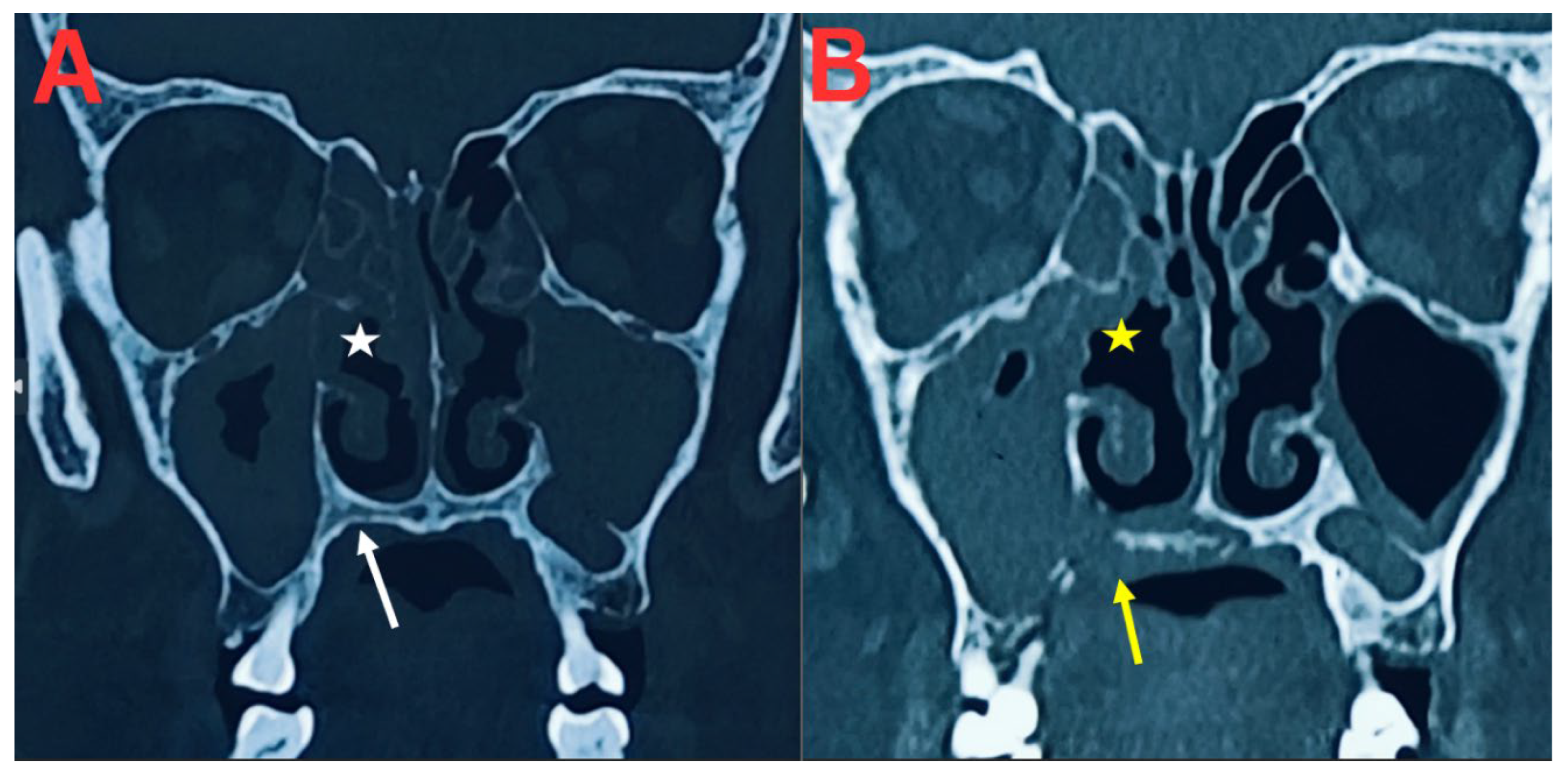

Table 1. 0.
Treatment Table.
| Intervention(Manufacture name with manufacturing licence number | Particulars | Case 1 (Duration in Days) And Dose |
Case 2 (Duration in Days) And dose |
Case 3 (Duration in Days) And Dose | Case 4 (Duration In days) And Dose |
Reference and Rational of use |
|---|---|---|---|---|---|---|
| Brihat Vata Chintamani Rasa (Oral)(Shree Dhootpapeshwar Ltd., AYU-150) |
Herbo mineral formulation containing purified Rasa (Purified Mercury), Gandhaka (Purified Sulfur), Abhraka Bhasma (Calcinated Mica) | 150 (125 mg) 1 tab twice a day |
120 (125 mg) 1 tab thrice a day |
105 (125 mg) 1 tab twice a day |
120 (125 mg) 1 tab twice a day |
Book-Bhaishajya Ratnavali, 26/145-148, InFdicated in disease of Vata dosha (ITA-2.1.1.1 Vata, dosha regulating movement and cognition) and Rasayana (ITA-9.1.35 Rejuvenative therapy) |
| Krumi Kuthar Rasa (Oral) (Dhanvantari Guj.Herb, GA-428) |
Herbo mineral Formulation containing Kajjali (Black Sulfide of Mercury), Tankana Bhasma (Borax) | 120 (125 mg) 2 tab thrice a day |
120 (125 mg) 2 tab thrice a day |
105 (125 mg) 2 tab thrice a day |
120 (125 mg) 2 tab thrice a day |
Book—Ayurved sara Sangrah (Page no 323), Indicated in Krimi roga (Disease of Krimi, ITA-5.13.4 Internal worm Infestation) |
| Krumighna Vatika (Oral) (Nagarjuna herbal concentrates pvt. Ltd., 69/25-D/88) |
Herbal Formulation containing Vidanga (Embelia ribes), Ajamoda (Apium graveolens), Haritaki (Terminalia chebula) | 150 (250 mg) 2 tabs thrice a day |
120 (250 mg) 2 tabs thrice a day |
105 (250 mg) 2 tabs thrice a day |
120 (250 mg) 2 tabs thrice a day |
Book- Sahasrayogam Indicated in Krimi roga (Disease of Krimi, ITA-5.13.4 Internal worm Infestation) |
| Serankottai Nei Capsule (Intra nasal) (SKM siddha & Ayurveda company pvt. Ltd., 739) |
Herbal combination containing Shuddha Bhallataka (Semecarpus anacardium), Milk, Ghee | 120 1 capsule (10-15 ghee drops) twice a day as a nasal drop (Half a capsule has to be instilled in both nostrils) |
120 1 capsule (10-15 ghee drops) thrice a day as a nasal drop (Half a capsule has to be instilled in both nostrils) |
105 1 capsule (10-15 ghee drops) twice a day as a nasal drop (Half a capsule has to be instilled in both nostrils) |
120 1 capsule (10-15 ghee drops) twice a day as a nasal drop (Half a capsule has to be instilled in both nostrils) |
Book- Siddha Vaithya Thirattu Indicated in Kapha vyadhi (disease of Kapha, ITA-2.1.3.1 Kapha, dosha responsible for cohesiveness) |
| Boniheal Tablet (Oral) (AIMIL pharmaceuticals (INDIA) pvt. Ltd.—HP-15/Ay) |
Contains mainly Godanti Bhasam (Classical Ayurvedic Preparation, Prcd.) –, Jahar Mohra Pishti (Classical Ayurvedic Preparation, Prcd.) –, Shalaki (Boswellia serrata, Exd.) –, Mukta Shukti Bhasam (Classical Ayurvedic Preparation, Prcd.) –, Sudh Laksha (Laccifera lacca purified, Secretion. Prcd.) –, Raal (Shorea robusta, Exd.) –, Vanshlochan (Bambusa sps., Sili. Concre.) –, Yashad Bhasam (Classical Ayurvedic Preparation, Prcd.) –, Shudh Shilajeet (Black bitumen purified, Prcd.)—,etc. | 150 250 mg 1 tab twice a day |
120 250 mg 1 tab twice a day |
105 250 mg 1 tab twice a day |
120 250 mg 1 tab twice a day |
Ayurveda Proprietary Medicine. Indicated to strengthen the bony tissue |
| Vidanga Choorna (Oral) Single herb powder |
Herbal Powder contains Vidanga (Embelia ribes) | 90 (5–10 gm) To drink and eat food with vidang choorna added into it |
120 (5–10 gm) To drink and eat food with vidang choorna added into it |
105 (5–10 gm) To drink and eat food with vidang choorna added into it |
120 (5–10 gm) To drink and eat food with vidang choorna added into it |
Book- Bhava Prakash Indicated as the Best herb for Krimi (ITA-5.13.4 Internal worm Infestation) |
| Palasha Choorna + Bhallataka Choorna + Vidanga Choorna (Intra nasal fumigation) Single herb Powder |
Herbal Formulations contains powder of Palash (Butea monosperma), Bhallathaka (Semecarpus anacardium), Vidanga (Embelia ribes) | 90 Mix all together and the patient has to inhale and exhale the smoke generated in it morning and in the evening |
120 Mix all together and the patient has to inhale and exhale the smoke generated in it morning, afternoon and in the evening |
105 Mix all together and the patient has to inhale and exhale the smoke generated in it morning and in the evening |
120 Mix all together and the patient has to inhale and exhale the smoke generated in it morning and in the evening |
Palash choorna Book- Bhava Prakash Indicated as the Best herb for Krimi (ITA-5.13.4 Internal worm Infestation), Bhallatak choorna (Indicated in Kapha vyadhi disease of Kapha, ITA-2.1.3.1 Kapha, dosha responsible for cohesiveness) |
| Vyadhi Harana Rasa (Oral) (Krushan gopal Ayurved Bhavan—539-D) |
Herbo mineral formulation containing purified Rasa (Purified Mercury), Gandhaka (Purified Sulfur), Hartal (Purified Arsenic trisulfide, manshila (Purified Arsenic disulphide) | 90 75 mg twice a day |
120 75 mg thrice a day |
105 75 mg twice a day |
0 | Rasa tantrasar and siddha prayoga-1-indicated in Nadivrana (ITA-5.56.1- Deep ulcer with fistula formation) |
| Panchtiktaka Kwatha (Oral) (Panchamrit herbals- GA/1762) |
Herbal formulation containing mainly Guduchi (Tinospora cordifolia), Nimba (Azadirachta indica), Vasa (Adhatoda vasica), Kantakari (Solanum xanthocarpum), Patola (Trichosanthes dioica) | 90 15 ml twice a day |
0 | 0 | 0 | Book- Ras Tantra Saar & Siddha Prayog Sangrah Volume 1, Indicated to strengthen Asthi dhatu (ITA-2.2.10 Bone, The fifth of the seven fundamental structural components. Its function is to provide bodily stability) |
| Netra Sudarshana Arka (Eye Drops) (Topical (Ophthalmic) (Krushan gopal Ayurved Bhavan—539-D) |
Eye drop containing Palash (Butea monosperma) | 150 2 eye drops in affected eye 3 times a day |
120 2 eye drops in affected eye 3 times a day |
0 | 120 2 eye drops in affected eye 3 times a day |
Book- Ras Tantra Saar & Siddha Prayog Sangrah Volume 1, Indicated in netra roga (ITA-5.66.1 Eye disorders) |
| Panchtiktaka Guggulu Ghrita tablet (Oral) (Jamna herbal research Ltd- MP/25D/11/139) |
Herbal formulation containing mainly Guduchi (Tinospora cordifolia), Nimba (Azadirachta indica), Vasa (Adhatoda vasica), Kantakari (Solanum xanthocarpum), Patola (Trichosanthes dioica) | 60 250 mg, 2 tab twice a day |
0 | 0 | 120 250 mg, 2 tab twice a day |
Book- Ras Tantra Saar & Siddha Prayog Sangrah Volume 1, Indicated to strengthen Asthi dhatu (ITA-2.2.10 Bone, The fifth of the seven fundamental structural components. Its function is to provide bodily stability) |
| Vranaphari Rasa (Oral) (Shri Jwala Ayurved Bhavan—15/1977) |
Herbo mineral formulation containing purified Rasa (Purified Mercury), Gandhaka (Purified Sulfur), Hartal (Purified Arsenic trisulfide, manshila (Purified Arsenic disulphide) and guggulu (Commiphora wightii) | 60 (125 mg) 1 tab twice a day |
120 (125 mg) 2 tab twice a day |
0 | 120 (125 mg) 1 tab twice a day |
Rasa tantrasar and siddha prayoga-2 indicated in Drusht vrana (ITA-5.54.7 Septic non healing ulcer) |
This table reflects the comprehensive information regarding Ayurveda therapeutic employed in this case
Table 1. 1.
Follow-up table.
| Follow up | Case 1 | Case2 | Case 3 | Case 4 |
|---|---|---|---|---|
| 1st consultation | Mobile teeth in upper right side numbness in upper palate and teeth feeling of bitter taste in mouth foul smell from nostrils (right>left) bulging of hard palate—15 days (17/01/2025) |
Pain in left side of nose, lips and eye Severe headache Numbness in left side of nose, palate, lips, eye region No perception of smell No movement of left eyeball Blurred vision in left eye o/e Large lesion at upper palate Since 8 days (10/03/2024) |
nasal regurgitation of fluids during oral intake feeling of bitter taste in mouth foul smell from nostrils heaviness of right side of face Since 20 days (02/06/2022) |
headache, facial pain and heaviness, ocular pain and heaviness, and fever. Since 10 days (13/03/2025) |
| 2nd visit |
Mobile teeth in upper right side—still present numbness in upper palate and teeth—40% improvement feeling of bitter taste in mouth—50% improvement foul smell from nostrils (right>left)—50% improvement bulging of hard palate—15 days—slightly reduced (16/02/2025) |
Improvement in pain—20% Improvement in numbness- 20% (18/03/2024) |
nasal regurgitation of fluids during oral intake—as it is feeling of bitter taste in mouth—30% improvement foul smell from nostrils 50% improvement heaviness of right side of face—100% improvement (29/06/2022) |
Headache 30% improvement facial pain and heaviness—30% improvement ocular pain- as it is Fever—Not there (25/03/2025) |
| 3rd Visit | Mobile teeth in upper right side—30% improvement numbness in upper palate and teeth—60% improvement feeling of bitter taste in mouth—70% improvement foul smell from nostrils (right>left)—70% improvement bulging of hard palate—15 days—40% improvement (23/03/2025) |
Improvement in pain -40% Improvement in numbness -40% Improvement in smell perception -40% Movement of left eyeball -30% Improvement in vision -10% (01/04/2024) |
nasal regurgitation of fluids during oral intake—as it is feeling of bitter taste in mouth—70% improvement foul smell from nostrils 80% improvement (20/07/2022) |
Headache 70% improvement facial pain and heaviness—50% improvement ocular pain- 20% improvement (21/04/2025) |
| 4th Visit | Mobile teeth in upper right side—60% improvement numbness in upper palate and teeth— 100% improvement feeling of bitter taste in mouth—100% improvement foul smell from nostrils (right>left)—100% improvement bulging of hard palate—15 days—70% improvement (17/04/2025) |
Improvement in pain -70% Improvement in numbness- 70% Improvement in smell perception- 80% Movement of left eyeball- 60% Improvement in vision- 40% o/e improvement in lesion at upper palate—40% (04/05/2024) |
nasal regurgitation of fluids during oral intake—as it is feeling of bitter taste in mouth—100% improvement foul smell from nostrils 100% improvement (19/08/2022) |
Headache 70% improvement facial pain and heaviness—50% improvement ocular pain- 20% improvement (06/05/2025) |
| 5th Visit | Mobile teeth in upper right side—90% improvement bulging of hard palate—15 days—90% improvement (26/05/2025) |
Improvement in pain- 90% Improvement in numbness 90% Improvement in smell perception 90% Movement of left eyeball- 75% Improvement in vision- 60% (04/06/2024) |
nasal regurgitation of fluids during oral intake—as it is (15/09/2022) |
Headache 80% improvement facial pain and heaviness—70% improvement ocular pain- 50% improvement (13/06/2025) |
| 6th Visit | Mobile teeth in upper right side—100% improvement bulging of hard palate—– 100% improvement (16/06/2025) |
Improvement in pain—100% Improvement in numbness 100% Improvement in smell perception 100% Movement of left eyeball 80% Improvement in vision 70% o/e lesion almost healed (08/07/2024) |
Nasal regurgitation of fluids during oral intake reduced up to 50-60% (28/08/2025) |
|
| 7th Visit | Movement of left eyeball- 100% Improvement in vision—80% (08/08/2024) |
|||
| 8th Visit | Improvement in vision 90-95% (07/09/2024) |
|||
| 9th Visit | Improvement in vision 100% (05/10/2024) |
This table presents follow-up information on clinical status based on non-standardised, patient-reported outcomes
Table 1. 2.
Clinical summary.
| Particulars | Case 1 | Case 2 | Case 3 | Case 4 |
|---|---|---|---|---|
| Age/Sex | 65/M | 47/M | 75/M | 75/F |
| Comorbidities | Diabetes mellitus, Hypertension | Diabetes mellitus, Hypertension, Chronic kidney disease, Solitary functioning kidney | Diabetes mellitus | Diabetes mellitus, Hypertension |
| Site and extent of disease | Rhino-maxillary mucormycosis involving hard palate, bilateral posterior maxillary alveolar process, right ethmoid and maxillary sinus with osteomyelitis | Rhino-orbital mucormycosis with skull-base osteomyelitis, ethmoid sinus, hard palate, pterygopalatine fossa, orbit and perineural spread | Rhino-maxillary mucormycosis with extensive maxillary sinus destruction, oro-antral fistula and osteomyelitis | Invasive fungal rhinosinusitis involving bilateral paranasal sinuses with destruction of the right middle turbinate and subsequent involvement of the maxilla, hard palate, sphenoid bone, clivus and right ethmoidal region. |
| Diagnostic evidence | Positive KOH microscopy, fungal culture, contrast-enhanced CT findings consistent with invasive fungal infection | Positive KOH microscopy, fungal culture, MRI findings characteristic of ROCM | Negative KOH microscopy, histopathology demonstrating fungal invasion into bone, and CT findings consistent with invasive fungal disease. | Positive tissue microscopy showing fungal structures, histopathology demonstrating broad aseptate hyphae with tissue invasion, and CT findings consistent with invasive fungal rhinosinusitis. |
| Organism identified | Rhizopus spp. and Aspergillus fumigatus | Mucor spp. and Candida krusei | Histopathology consistent with Mucorales (species not identified) | Broad aseptate hyphae consistent with Mucorales with additional thin fungal hyphae (mixed fungal infection suspected) |
| Standard treatment recommended | Functional endoscopic sinus surgery (FESS) with systemic liposomal amphotericin B | Surgical debridement with systemic liposomal amphotericin B | FESS followed by systemic liposomal amphotericin B | Surgical debridement with systemic liposomal amphotericin B |
| Treatment received before Ayurveda | None | None | FESS (declined antifungal therapy) | None |
| Ayurveda intervention | Predominantly Ayurveda-based treatment (oral formulations, intranasal therapy, fumigation, dietary and lifestyle advice) | Predominantly Ayurveda-based treatment (oral formulations, intranasal therapy, fumigation, dietary and lifestyle advice) | Predominantly Ayurveda-based treatment after FESS (oral formulations, intranasal therapy, fumigation, dietary and lifestyle advice | Predominantly Ayurveda-based treatment (oral formulations, intranasal therapy, fumigation, dietary and lifestyle advice) |
| Follow-up duration | 17 months | 9 months | 4 years | 1 year |
| Radiological outcome | Resolution of sinusitis with sclerosis and healing suggestive of new bone formation | Marked reduction in inflammatory changes with residual post-inflammatory sequelae; no radiological evidence of active invasive disease | Persistent residual mucosal thickening without radiological evidence of active invasive fungal disease or progression | Persistent destructive osseous changes without convincing radiological regression |
| Clinical outcome | Complete resolution of symptoms and palatal lesion | Complete healing of palatal lesion with sustained clinical improvement | Sustained symptom resolution without clinical recurrence | Partial symptomatic improvement despite persistent radiological disease |
| Adverse events | No treatment-related adverse events reported | Progressive renal dysfunction during follow-up; contribution of treatment could not be excluded, although clinically considered more consistent with progression of underlying CKD | No treatment-related adverse events reported | No treatment-related adverse events reported |
| Baseline Serum creatinine (Standard value -0.5-1.4 mg/dl) |
13/01/2025 1.05mg/dl |
01/03/2024 1.48 mg/dl |
16/05/2022 1.74 mg/dl |
12/03/2025 1.4 mg/dl |
| Follow up Serum creatinine (Standard value -0.5-1.4 mg/dl) |
13/06/2026 1.17 mg/dl |
03/12/2024 3.3 mg/dl |
14/06/2026 1.54 mg/dl |
Not Applicable |
| Baseline Liver function SGPT(Serum Glutamate Pyruvate Transaminase,standar value -0-45 U/L),SGOT(Serum Glutamic-Oxaloacetic Transaminase- Standard value- 0-35 U/L), Total Bilirubin (Standard value—0.0-1.2 mg/dl) |
Not available | Not available | Not available | Not available |
| Follow up Liver function SGPT(Serum Glutamate Pyruvate Transaminase,standar value -0-45 U/L),SGOT(Serum Glutamic-Oxaloacetic Transaminase- Standard value- 0-35 U/L), Total Bilirubin (Standard value—0.0-1.2 mg/dl) |
13/06/2026 SGPT- 41 U/L SGOT- 30 U/L Total Bilirubin—1.56 mg/dl |
03/12/2024 SGPT-22.75 U/L SGOT- 27 U/L Total bilirubin- 0.23 mg/dl |
14/06/2026 SGPT- 17.7 U/L SGOT- 19.1 U/L Total Bilirubin—0.33 mg/dl |
Not Applicable |
| Final status | Alive, clinically well, no recurrence during follow-up | Alive, clinically well, no recurrence during follow-up | Alive, disease-free at 4-year follow-up | Survived approximately 1 year after diagnosis; persistent radiological disease |
This table reflects the overall clinical course of the patients
Disclaimer/Publisher’s Note: The statements, opinions and data contained in all publications are solely those of the individual author(s) and contributor(s) and not of MDPI and/or the editor(s). MDPI and/or the editor(s) disclaim responsibility for any injury to people or property resulting from any ideas, methods, instructions or products referred to in the content. |
© 2026 by the authors. Licensee MDPI, Basel, Switzerland. This article is an open access article distributed under the terms and conditions of the Creative Commons Attribution (CC BY) license.
Copyright: This open access article is published under a Creative Commons CC BY 4.0 license, which permit the free download, distribution, and reuse, provided that the author and preprint are cited in any reuse.