

Submitted:
19 August 2026
Posted:
20 August 2026
You are already at the latest version
Abstract
Background/Objectives: Cognitive impairment in neuromyelitis optica spectrum disorder (NMOSD) is increasingly recognized, but its profile and clinical correlates remain uncertain. This study sought to describe cognitive performance in individuals with NMOSD and to explore its associations with disease activity, disability level, and immunosuppressive treatment. Methods: This exploratory cross-sectional study characterized cognitive performance in 36 AQP4-IgG–positive individuals with NMOSD and 18 healthy volunteers using domain-level cognitive scores adjusted for age, education, and visual acuity. Results: Cognitive impairment was present in at least one domain in 27 participants, most frequently affecting memory (36%), praxis (33%), and information processing speed (IPS; 31%). IPS was the only domain significantly impaired in NMOSD compared with controls in unadjusted analyses (p = 0.007), but no domain remained significantly different after adjustment, despite a medium-to-large effect for IPS. Higher disability burden was associated with worse IPS (p = 0.011) and with lower scores on a clinically sensitive cognitive composite integrating memory, IPS, and executive function (β = –0.125, p = 0.040). Among clinical activity measures, annualized relapse rate showed a positive association only with executive function (β = 0.282, p = 0.007), whereas neither relapse count nor immunosuppressive treatment was associated with cognitive performance. Conclusions: These findings indicate a heterogeneous cognitive profile in NMOSD strongly influenced by demographic and sensory factors, with disability burden emerging as the main clinical correlate of cognitive performance.
Keywords:
cognition
; disabilities
; NMOSD
; AQP4-IgG
1. Introduction
Neuromyelitis optica spectrum disorder (NMOSD) is an autoimmune inflammatory disease of the central nervous system characterized by AQP4-IgG antibodies and demyelinating attacks that predominantly affect the optic nerves, spinal cord, and area postrema [1]. It affects individuals across all ages, with a marked female predominance [2], and typically follows a relapsing course that leads to cumulative disability and reduced quality of life [3,4].
Beyond the classical motor and sensory manifestations, growing evidence indicates that NMOSD may also involve cognitive dysfunction. Its neurobiological basis is heterogeneous, with studies reporting hippocampal and thalamic abnormalities linked to cognitive impairment [5,6,7], while others describe preserved microstructural integrity even among cognitively impaired individuals [8].
Cognitive impairment has been reported in 29–70% of people with NMOSD (pwNMOSD) [5,9] and appears to be influenced by age, education, and disability level [10]. It most frequently affects attention, information processing speed (IPS), and memory [9,11]. Although this resembles patterns observed in multiple sclerosis [12], the frequency and characteristics of cognitive impairment in NMOSD may be related to multiple factors such as heterogeneity in samples, differences in disease duration, relapse history, and the neuropsychological instruments used across studies [9,10,11].
Given these inconsistencies, a clearer characterization of cognitive performance in NMOSD—particularly in relation to demographic factors, visual function, disability burden, and clinical activity—is needed. However, no study to date has systematically evaluated domain-level cognitive performance in AQP4-IgG–positive NMOSD using models that simultaneously account for demographic and sensory confounders while examining disability burden and relapse-based activity. Therefore, this study aimed to characterize domain-level cognitive performance in individuals with NMOSD and compare it with healthy volunteers. A secondary aim was to examine associations between cognition, disease activity, disability level, and immunosuppressive treatment.
2. Materials and Methods
2.1. Study Design and Participants
This cross-sectional study included 36 consecutive participants recruited from the Neuroimmunology Outpatient Clinic of the Neurology Division, Hospital das Clínicas, Faculdade de Medicina da Universidade de São Paulo, Brazil. Written informed consent was obtained from all participants, and the study protocol was approved by the Universidade de São Paulo Review Board (#2046215), in accordance with the Declaration of Helsinki (1975), as revised in 2024. Additional details regarding the overarching research project and ethical procedures are available in the group’s publication [13].
Participants were aged 18 years or older (mean age 44 ± 13 years) and met the 2015 International Panel for NMO Diagnosis (IPND) criteria for NMOSD [1], with AQP4-IgG seropositivity required for inclusion. Individuals with other neurological or psychiatric conditions were excluded.
Eighteen healthy volunteers (HV), who were accompanying patients awaiting care at the neurology outpatient clinic, were invited to participate. They were selected to match the sample of people with NMOSD (pwNMOSD) according to age and years of education and had no sensory or motor impairments that could interfere with the administration of the cognitive tests.
2.2. Clinical and Cognitive Assessment
Participants were first evaluated by a medical team specialized in neuroimmunology and subsequently by a neuropsychologist (K.R.C.). Demographic information for pwNMOSD and healthy volunteers, along with disease-specific clinical data, was collected during structured interviews. All procedures, including clinical and cognitive assessments, were completed on the same day.
Clinical assessment included the Kurtzke Expanded Disability Status Scale (EDSS) [14] to quantify disability and the Mini-Mental State Examination for cognitive screening [15]. Distance visual acuity was assessed using a standardized Snellen chart at 20 feet, with best optical correction [1]. Visual acuity values were recorded in Snellen notation [16] and subsequently converted to logMAR [17] for statistical analysis.
The cognitive battery included the Rey Auditory Verbal Learning Test [18], the Rey–Osterrieth Complex Figure Test (copy and delayed recall), administered without time constraints, the Trail Making Test, the Stroop Test [19], the Modified Wisconsin Card Sorting Test, and phonemic (F-A-S) and semantic (animals) verbal fluency tasks [20]. All instruments used in the battery have standardized normative data and validated procedures for the Brazilian population, ensuring culturally and linguistically appropriate interpretation of test performance.
Patients whose motor or visual disability prevented completion of specific tasks were assigned missing values, which were retained in subsequent analyses. Cognitive test scores were converted to z-scores based on normative data, and cognitive domains were derived from specific test scores or domain-level means (Supplementary Table S01). Cognitive impairment was defined as a z-score ≤ –1.5.
2.3. Statistical Analysis
Continuous variables were summarized as median and interquartile range, and categorical variables as absolute and relative frequencies. Between-group comparisons were performed using the Mann–Whitney test for continuous variables, with rank-biserial correlations reported as effect sizes, and Pearson’s chi-square or Fisher’s exact tests for categorical variables.
Group differences in cognitive domains were then assessed using ANCOVA models adjusted for age, years of education, and bilateral visual acuity. Estimated marginal means and partial eta-squared effect sizes were reported. Model assumptions were evaluated using residual diagnostics, Shapiro-Wilk tests, and Breusch-Pagan tests (Supplementary Table S02 and Figure S01).
To investigate clinical correlates of cognition in NMOSD, multivariable linear regression models were fitted using visual-acuity-adjusted cognitive residuals as dependent variables, with age and education included as covariates. Clinical predictors were tested in separate models. Because memory was the most frequently impaired domain, and information processing speed and executive function showed the strongest associations with clinical disease measures, these three domains were also combined in an exploratory clinically sensitive cognitive composite (CSCC), calculated as their arithmetic mean. The CSCC was subsequently evaluated in additional exploratory regression models adjusted for age and education, with each clinical predictor tested separately.
Robustness of regression estimates was assessed using HC3 heteroscedasticity-consistent standard errors and 5,000-repetition bias-corrected and accelerated (BCa) bootstrap confidence intervals and empirical two-tailed p-values. Multiple comparisons were controlled using the Benjamini-Hochberg false discovery rate procedure. Multicollinearity and model specification were assessed using variance inflation factors and the Regression Specification Error Test, when applicable.
Statistical analyses were performed in RStudio version 4.5.1. Statistical significance was set at p < 0.05.
3. Results
3.1. Sample Characteristics
Table 1 summarizes the sociodemographic matching between groups and the clinical characterization of the pwNMOSD sample. The groups did not differ in age, sex, education and skin color, indicating no significant sociodemographic differences between groups.
Among pwNMOSD, disease duration had a median of 6 years, with 4 relapses and an annualized relapse rate (ARR) of 0.82. The EDSS score was 3.25. Regarding treatment, 92% of pwNMOSD were receiving immunosuppressive therapy (IST), most commonly azathioprine. Prednisone was used by 95.5% of participants.
3.2. Cognitive Profile in NMOSD
When cognitive domains were examined, similar patterns of impairment were observed across the cohort. Delayed recall showed the highest frequency of impairment (n = 13; 33%), followed by praxis (n = 12; 33%) and IPS (n = 11; 31%). Overall, 27 pwNMOSD presented impairment in at least one cognitive domain, including one participant who exhibited impairment across all domains and nine participants who showed no impairment in any domain (Table 2). The full set of impairment frequencies is detailed in Supplementary Table S03.
Comparative cognitive performance, without covariate adjustment, between healthy volunteers and pwNMOSD across domains is illustrated in Figure 1. Group comparisons revealed statistically significant impairment in IPS performance in pwNMOSD compared with controls (p = 0.001, pFDR = 0.007, rank-biserial r = 0.47). Although memory showed an initial significant difference (p = 0.039, pFDR = 0.115, r = 0.28), and marginal trends were observed for praxis (p = 0.058, pFDR = 0.115, r = 0.27) and learning (p = 0.099, pFDR = 0.148, r = 0.23), only IPS remained statistically significant after correction for multiple comparisons. Executive functions and language showed no significant differences between the groups.
To account for potential confounding factors, adjusted models by age, years of education and visual acuity were employed, as detailed in the Supplementary Material (S04). No statistically significant differences were observed between pwNMOSD and controls across any of the assessed cognitive domains after covariate adjustment and multiple-comparison correction (pFDR > 0.05 for all comparisons).
Specifically, performance in learning, memory, praxis, executive function, and language was comparable between groups, with minimal effect sizes ( ranging from 0.001 to 0.070). The inclusion of covariates, particularly age and visual acuity, which exerted strong independent effects on performance, accounted for the initial group disparities observed in unadjusted comparisons, including those in memory and IPS. These findings indicate that such confounding factors strongly modulate cognitive efficiency in this cohort.
Although IPS showed a marginal group difference (p = 0.065) with a medium-to-large effect size ( = 0.204), indicating lower adjusted scores in pwNMOSD (-0.16 ±0.26) relative to controls (-0.23 ±0.34), this difference did not remain statistically significant after final adjustment (pFDR = 0.391). Complete parameter estimates for all individual model terms, including intercepts, group coefficients, and the specific contributions of age, education, and visual acuity across all six domains, are presented in Table 3.
3.3. Clinical Predictors of Cognitive Performance in NMOSD
Visual-acuity-adjusted cognitive residuals were used as outcomes. Multivariable analyses evaluating associations between clinical predictors and cognitive residuals revealed domain-specific vulnerabilities (Figure 2; see Supplementary Table S05 for details). Higher EDSS scores showed a significant negative association exclusively with IPS (β = -0.309, 95% BCa CI [-0.517, -0.163], p = 0.002), and this association remained significant after false discovery rate correction (pFDR = 0.011).
Longer disease duration showed a significant negative association only with executive functions (β = -0.071, 95% BCa CI [-0.142, -0.008], p = 0.031), but this association did not remain significant after multiple comparison correction (pFDR = 0.184). The total number of relapses showed no significant associations with any cognitive domain (all p ≥ 0.05). However, the annualized relapse rate exhibited a significant positive association with executive function (β = 0.282, 95% BCa CI [0.116, 0.479], p = 0.007, pFDR = 0.041), while remaining unrelated to the other cognitive domains. Regarding the different ISTs used, no statistically significant predictive effects on cognition were observed.
To further synthesize these domain-specific findings, an exploratory CSCC was derived from the cognitive domains that were either most frequently impaired or most closely associated with clinical disease measures. This composite was subsequently analyzed as an integrated marker of clinically relevant cognitive performance in NMOSD.
Among the clinical variables tested, EDSS showed the strongest association with the CSCC and provided the best model fit, with the highest explained variance and the lowest information criteria (R2 = 0.516, adjusted R2 = 0.460, AIC = 69.68, BIC = 76.68). Higher EDSS scores were significantly associated with lower CSCC scores (β = -0.125, 95% BCa CI [-0.244, -0.033], p = 0.026, pFDR = 0.040) (Figure 3). A summary of the results is provided in the Supplementary Material (Table S06).
4. Discussion
This study characterized the cognitive profile of individuals with NMOSD and its associations with clinical disease activity, disability level, and IST. Importantly, the NMOSD and control groups were broadly comparable in demographic characteristics and global cognitive screening, reducing the likelihood that group-level cognitive differences were primarily driven by baseline demographic imbalance. Although age and years of education were satisfactorily matched between groups, both variables showed substantial variability within the sample and are well-established determinants of cognitive performance [9], supporting their inclusion as covariates in all adjusted cognitive models. The clinical profile of the NMOSD cohort, including moderate disability burden and frequent immunosuppressive treatment [4], should also be considered when interpreting the generalizability of the findings.
In this cohort, the distribution of cognitive impairment shows memory, praxis, and IPS as the most frequently affected domains in NMOSD.Memory impairment frequently appears in NMOSD [21,22], but its expression should be interpreted alongside deficits in IPS. In demyelinating disorders, the IPS is the main cognitive disfunction [8,11,21,23], this slowing may constrain working memory resources, compromise the encoding of information into episodic memory, and disrupt associative processes involved in recall [23,24]. Therefore, memory deficits in NMOSD may partly reflect broader inefficiency in information processing rather than isolated mnemonic dysfunction.
Praxis impairment has been less consistently reported, although it may be present in pwNMOSD [23,24]. However, the interpretation of praxis performance is challenging because visual and motor impairments are frequent in this disease and may directly interfere with tasks requiring visuomotor integration [11,25]. Although the present analyses accounted for reduced visual acuity, no specific psychomotor performance measure was available to formally adjust for motor-related contributions, which should be considered when interpreting praxis-related findings. In the absence of NMOSD-specific batteries, cognitive assessment often relies on Multiple Sclerosis-based tools, which may not fully separate cognitive deficits from visual and motor contributions [11,26].
When these domains were directly compared with those of healthy volunteers, only IPS showed a significant unadjusted difference. However, this finding should be interpreted considering the substantial influence of demographic and sensory factors on cognitive performance. After adjustment for age, years of education, and visual acuity, the IPS difference was markedly attenuated and no longer survived correction for multiple comparisons, although it retained a medium-to-large effect size. This discrepancy between a sizeable effect and a non-significant adjusted result may reflect limited statistical power in this relatively small cohort, even though statistical prerequisites were met.
These findings should be interpreted with caution, as the expression of cognitive impairment in NMOSD appears to be influenced by demographic and sensory factors, as well as by disease-related network disruption and reduced neurological reserve associated with accumulated disability. Because IPS tasks include a psychomotor component, part of the EDSS–IPS association may reflect motor slowing not fully captured by visual-acuity adjustment. Thus, cognitive dysfunction in NMOSD is likely multifactorial, reflecting not only disease-specific neurobiological mechanisms but also visual, motor, and demographic contributions that may affect cognitive test performance [8].
This attenuation at the group level contrasts with the substantial heterogeneity observed within the NMOSD sample itself. While nine participants showed no impairment in any domain, twenty-seven were impaired in at least one domain, including one individual who showed impairment across all six domains. This distribution suggests that cognitive vulnerability in NMOSD may be concentrated in a subgroup of patients rather than uniformly distributed across the population.
This interpretation is consistent with previous proposals of distinct cognitive phenotypes in NMOSD [10] and may help explain why clinically meaningful impairment in part of the sample can be obscured in group-level comparisons. This more circumscribed cognitive profile also contrasts with the broader multidomain phenotypes described in multiple sclerosis [12], raising the possibility that NMOSD may involve a distinct and less pervasive pattern of cognitive vulnerability.
Clinical correlates of cognition in this cohort did not converge on a single measure of disease severity, and the observed pattern was, in some respects, counterintuitive. EDSS is weighted heavily toward ambulation, particularly at the intermediate scores observed in this cohort, and was not, a priori, the clinical variable most likely to track cognitive performance [9]. By contrast, annualized relapse rate, which more directly reflects inflammatory activity over time, might have been expected to show stronger cognitive associations. Nevertheless, EDSS emerged as the strongest predictor of both IPS and the CSCC, whereas ARR was associated only with executive function and in an unexpected positive direction. These findings suggest that the relationship between EDSS and cognition may not be explained solely by shared lesion burden, but may instead reflect a broader construct captured by EDSS beyond ambulation, potentially indexing global neurological reserve, neuroinflammation, diffuse network disruption, or cumulative disease burden not fully captured by relapse-based metrics [1].
The ARR finding is more difficult to reconcile with a simple activity-burden model. In NMOSD, the relationship between relapses and disability is not uniform: relapse-associated residual disability varies by attack phenotype, and some NMOSD-specific correlates of disease burden may not be captured by the EDSS [25]. The positive association with executive function may therefore reflect, at least in part, a temporal mismatch between relapse occurrence and subsequent disability accumulation. Because most participants were receiving immunosuppressive therapy (92%), one possible explanation is that relapses occurring after treatment initiation may have had less impact on residual disability, whereas earlier, pre-treatment relapses may already have contributed substantially to EDSS accumulation before study entry [3,4]. Under this interpretation, a higher ARR could reflect more recent disease activity without necessarily indicating a higher cumulative inflammatory burden. However, this treatment- and time-dependent explanation was not directly tested and should therefore be regarded as hypothesis-generating, particularly given the modest sample size. Consistent with this caution, the total number of relapses was not associated with any cognitive domain in the present study, arguing against interpreting ARR as a simple proxy for activity-related cognitive risk. Immunosuppressive treatment was also unrelated to cognitive performance, although this finding is difficult to interpret given the small number of treatment-naïve participants.
This study has several limitations. First, the sample size was relatively small, particularly for the healthy volunteer group (n = 18), which may have reduced statistical power to detect moderate effects. Second, the cross-sectional design precludes conclusions about temporal or causal relationships between clinical disease measures and cognitive performance. Longitudinal studies are needed to determine whether cognitive decline follows, precedes, or develops in parallel with disability accumulation. Third, this was a single-center study, which may limit generalizability to populations with different demographic, socioeconomic, or healthcare access profiles. Fourth, healthy volunteers were recruited among patient companions at the outpatient clinic; although the groups were adequately matched, this strategy may have introduced selection bias related to caregiving burden or other unmeasured characteristics. Fifth, because several clinical predictors were tested and the CSCC was exploratory, the findings should be interpreted cautiously and require replication in larger cohorts. Additionally, high-dose corticosteroid therapy—used by approximately 95% of participants—has been associated with acute and chronic executive dysfunction, raising the possibility that part of the cognitive impairment observed may be iatrogenic rather than disease-related; however, because corticosteroid exposure (current dose, cumulative dose, or dose strata) was not quantified, its independent impact on cognition could not be evaluated, representing an important limitation and a potential target for future analyses.
Despite these limitations, the findings have practical implications for the clinical management of pwNMOSD. The observation that IPS was the domain most consistently associated with disease-related clinical measures suggests that brief, targeted screening tools focused on processing speed may be useful in routine clinical settings, particularly where access to comprehensive neuropsychological assessment is limited. In addition, the association between EDSS and cognitive performance supports the use of physical disability status as a practical marker to identify patients who may benefit from closer cognitive monitoring [9] or referral for formal neuropsychological assessment. Future studies should prioritize larger, multicenter, longitudinal cohorts to clarify the temporal relationship between disease activity, disability accumulation, and cognitive trajectories. Incorporating structural and functional neuroimaging may also help elucidate the neurobiological substrates underlying cognitive changes in NMOSD.
5. Conclusions
In this cohort, cognitive impairment in NMOSD appeared more circumscribed than suggested by unadjusted comparisons alone. Information processing speed emerged as the cognitive domain most robustly related to disease-specific clinical measures, particularly disability level as measured by EDSS. These findings support a model in which cognitive dysfunction in NMOSD is more closely linked to accumulated neurological disability than to relapse-based inflammatory activity per se. They also highlight the importance of accounting for demographic and sensory confounders when characterizing the cognitive profile of this population.
Supplementary Materials
The following supporting information can be downloaded at the website of this paper posted on Preprints.org. Supplementary Material Supplementary Table S03–S06 provide extended results for cognitive impairment frequencies, covariate-adjusted group comparisons, clinical predictor analyses, and CSCC model parameters.
Author Contributions
Forma analysis, data curation, writing—original draft preparation, conceptualization, methodology K.R.C.. Writing—original draft preparation, L.A.S.M.. Conceptualization and writing—review and editing, M.F. M.. Conceptualization and writing—review, editing supervision, S.L.A.P. Writing—review, editing supervision, D.A., D.C.; T.A. and G.D.
Funding
This research received no external funding.
Institutional Review Board Statement
The study was conducted in accordance with the Declaration of Helsinki (1975), as revised in 2024 and, approved by the Hospital das Clínicas da Faculdade de Medicina da Universidade de São Paulo Review Board (#2046215).
Informed Consent Statement
Informed consent was obtained from all subjects involved in the study.
Data Availability Statement
The data supporting the findings of this study include sensitive clinical and cognitive information collected during routine hospital care. In accordance with institutional policies and national regulations on confidentiality and data protection, the dataset is not publicly available. Any request for access would require prior evaluation and approval by the relevant institutional ethics and governance bodies.
Conflicts of Interest
Some authors declare conflicts of interest, as well: S.L.A-P- Speaker, Board advisory e patrovinio de Congresso da Astra Zeneca, Roche e Amgen.
Abbreviations
The following abbreviations are used in this manuscript:
| ARR | Annualized relapse rate |
| CSCC | Clinically sensitive cognitive composite |
| EDSS | Expanded Disability Status Scale |
| EF | Executive function |
| IPS | Information processing speed |
| IST | Immunosuppressive therapy |
| NMOSD | Neuromyelitis optica spectrum disorder |
| pwNMOSD | People with neuromyelitis optica spectrum disorder |
References
- Wingerchuk, D.M.; Banwell, B.; Bennett, J.L.; Cabre, P.; Carroll, W.; Chitnis, T.; De Seze, J.; Fujihara, K.; Greenberg, B.; Jacob, A.; et al. International Consensus Diagnostic Criteria for Neuromyelitis Optica Spectrum Disorders. Neurology 2015, 85, 177–189. [Google Scholar] [CrossRef] [PubMed]
- Papp, V.; Magyari, M.; Aktas, O.; Berger, T.; Broadley, S.A.; Cabre, P.; Jacob, A.; Kira, J.; Leite, M.I.; Marignier, R.; et al. Worldwide Incidence and Prevalence of Neuromyelitis Optica: A Systematic Review. Neurology 2021, 96, 59–77. [Google Scholar] [CrossRef] [PubMed]
- Chen, B.; Francis, A.; Cooper, S.A.; Dobson, R.; Hacohen, Y.; Halfpenny, C.; Hemingway, C.; Hobart, J.C.; O’Sullivan, E.; Rashid, W.; et al. Quantitative Contribution of Clinical Attacks to Residual Disability in Patients With AQP4-Antibody Neuromyelitis Optica Spectrum Disorder. Neurology 2025, 104, e210137. [Google Scholar] [CrossRef] [PubMed]
- Berthele, A.; Levy, M.; Wingerchuk, D.M.; Pittock, S.J.; Shang, S.; Kielhorn, A.; Royston, M.; Sabatella, G.; Palace, J. A Single Relapse Induces Worsening of Disability and Health-Related Quality of Life in Patients with Neuromyelitis Optica Spectrum Disorder. Front. Neurol. 2023, 14, 1099376. [Google Scholar] [CrossRef] [PubMed]
- Wan, X.; Tang, Y.; Wu, Y.; Xu, Z.; Chen, W.; Chen, F.; Luo, C.; Wang, F. Abnormal Functional Connectivity of White-Matter Networks and Gray-White Matter Functional Networks in Patients with NMOSD. Brain Res. Bull. 2024, 211, 110949. [Google Scholar] [CrossRef] [PubMed]
- Kim, S.-H.; Park, E.Y.; Park, B.; Hyun, J.-W.; Park, N.Y.; Joung, A.; Lee, S.H.; Kim, H.J. Multimodal Magnetic Resonance Imaging in Relation to Cognitive Impairment in Neuromyelitis Optica Spectrum Disorder. Sci. Rep. 2017, 7, 9180. [Google Scholar] [CrossRef] [PubMed]
- Liu, Y.; Fu, Y.; Schoonheim, M.M.; Zhang, N.; Fan, M.; Su, L.; Shen, Y.; Yan, Y.; Yang, L.; Wang, Q.; et al. Structural MRI Substrates of Cognitive Impairment in Neuromyelitis Optica. Neurology 2015, 85, 1491–1499. [Google Scholar] [CrossRef] [PubMed]
- Boaventura, M.; Fragoso, D.C.; Avolio, I.; Martins, P.N.; Matos, A.D.M.B.; Pereira, S.A.; Callegaro, D.; Sato, D.K.; Leite, C.D.C.; Rovira, À.; et al. Cognitive Impairment and Multiparametric Brain MRI in Aquaporin-4-IgG Positive Neuromyelitis Optica Spectrum Disorder. Mult. Scler. Relat. Disord. 2026, 105, 106875. [Google Scholar] [CrossRef] [PubMed]
- Vlahovic, L.; McDonald, J.; Hinman, J.; Tomczak, A.; Lock, C.; Palmer, C.A.; Cook, L.J.; Yeaman, M.R.; Burnett, M.S.; Deutsch, G.K.; et al. Prevalence, Demographic, and Clinical Factors Associated With Cognitive Dysfunction in Patients With Neuromyelitis Optica Spectrum Disorder. Neurology 2024, 102, e207965. [Google Scholar] [CrossRef] [PubMed]
- Kong, L.; Lang, Y.; Wang, X.; Wang, J.; Chen, H.; Shi, Z.; Zhou, H. Identifying Different Cognitive Phenotypes and Their Relationship with Disability in Neuromyelitis Optica Spectrum Disorder. Front. Neurol. 2022, 13, 958441. [Google Scholar] [CrossRef] [PubMed]
- Hümmert, M.W.; Stern, C.; Paul, F.; Duchow, A.; Bellmann-Strobl, J.; Ayzenberg, I.; Schwake, C.; Kleiter, I.; Hellwig, K.; Jarius, S.; et al. Cognition in Patients with Neuromyelitis Optica Spectrum Disorders: A Prospective Multicentre Study of 217 Patients (CogniNMO-Study). Mult. Scler. J. 2023, 29, 819–831. [Google Scholar] [CrossRef] [PubMed]
- Hancock, L.M.; Galioto, R.; Samsonov, A.; Busch, R.M.; Hermann, B.; Matias-Guiu, J.A. A Proposed New Taxonomy of Cognitive Phenotypes in Multiple Sclerosis: The International Classification of Cognitive Disorders in MS (IC-CoDiMS). Mult. Scler. J. 2023, 29, 615–627. [Google Scholar] [CrossRef] [PubMed]
- Rosa, R.F.; Ugolini-Lopes, M.R.; Gandara, A.P.R.; Vendramini, M.B.G.; Campanholo, K.R.; Dutra, L.; De Andrade, D.C.O. Cognitive Dysfunction and Serum Levels of Brain-Derived Neurotrophic Factor (BDNF) in Primary Anti-Phospholipid Syndrome (PAPS). Rheumatology 2021, 60, 179–187. [Google Scholar] [CrossRef] [PubMed]
- Kurtzke, J.F. Rating Neurologic Impairment in Multiple Sclerosis: An Expanded Disability Status Scale (EDSS). Neurology 1983, 33, 1444–1444. [Google Scholar] [CrossRef] [PubMed]
- Brucki, S.M.D.; Nitrini, R.; Caramelli, P.; Bertolucci, P.H.F.; Okamoto, I.H. Sugestões Para o Uso Do Mini-Exame Do Estado Mental No Brasil. Arq. Neuropsiquiatr. 2003, 61, 777–781. [Google Scholar] [CrossRef] [PubMed]
- Snellen, Herman. Probebuchstaben zur Bestimmung der Sehschärfe; Van de Weijer: Utrecht, 1862. [Google Scholar]
- Holladay, J.T. Proper Method for Calculating Average Visual Acuity. J. Refract. Surg. 1997, 13, 388–391. [Google Scholar] [CrossRef] [PubMed]
- De Paula, J.J.; Malloy-Diniz, L.R. RAVLT—Teste de Aprendizagem Auditivo-Verbal de Rey: Livro de Intruções; Editora Vetor: São Paulo, 2018. [Google Scholar]
- Campanholo, K.R.; Romão, M.A.; Machado, M.D.A.R.; Serrao, V.T.; Coutinho, D.G.C.; Benute, G.R.G.; Lucia, M.C.S.D. Performance of an Adult Brazilian Sample on the Trail Making Test and Stroop Test. Dement. Neuropsychol. 2014, 8, 26–31. [Google Scholar] [CrossRef] [PubMed]
- Campanholo, K.R.; Boa, I.N.F.; Hodroj, F.C.D.S.A.; Guerra, G.R.B.; Miotto, E.C.; Lucia, M.C.S.D. Impact of Sociodemographic Variables on Executive Functions. Dement. Neuropsychol. 2017, 11, 62–68. [Google Scholar] [CrossRef] [PubMed]
- Czarnecka, D.; Oset, M.; Karlińska, I.; Stasiołek, M. Cognitive Impairment in NMOSD—More Questions than Answers. Brain Behav. 2020, 10, e01842. [Google Scholar] [CrossRef] [PubMed]
- Kazzi, C.; Alpitsis, R.; O’Brien, T.J.; Malpas, C.B.; Monif, M. Cognitive and Psychopathological Features of Neuromyelitis Optica Spectrum Disorder and Myelin Oligodendrocyte Glycoprotein Antibody-Associated Disease: A Narrative Review. Mult. Scler. Relat. Disord. 2024, 85, 105596. [Google Scholar] [CrossRef] [PubMed]
- Löffler, C.; Frischkorn, G.T.; Hagemann, D.; Sadus, K.; Schubert, A.-L. The Common Factor of Executive Functions Measures Nothing but Speed of Information Uptake. Psychol. Res. 2024, 88, 1092–1114. [Google Scholar] [CrossRef] [PubMed]
- McCabe, D.P.; Roediger, H.L.; McDaniel, M.A.; Balota, D.A.; Hambrick, D.Z. The Relationship between Working Memory Capacity and Executive Functioning: Evidence for a Common Executive Attention Construct. Neuropsychology 2010, 24, 222–243. [Google Scholar] [CrossRef] [PubMed]
- Gholizadeh, S.; Exuzides, A.; Sinnott, J.; Palmer, C.; Waltz, M.; Rose, J.W.; Jolley, A.M.; Behne, J.M.; Behne, M.K.; Blaschke, T.F.; et al. Assessment of Disability and Disease Burden in Neuromyelitis Optica Spectrum Disorders in the CIRCLES Cohort. Sci. Rep. 2024, 14, 26150. [Google Scholar] [CrossRef] [PubMed]
- Kim, S.-H.; Kwak, K.; Jeong, I.H.; Hyun, J.-W.; Jo, H.-J.; Joung, A.; Yu, E.-S.; Kim, J.-H.; Lee, S.H.; Yun, S.; et al. Cognitive Impairment Differs between Neuromyelitis Optica Spectrum Disorder and Multiple Sclerosis. Mult. Scler. J. 2016, 22, 1850–1858. [Google Scholar] [CrossRef] [PubMed]
Figure 1.
Cognitive comparison between groups. Note. Mann-Whitney Test & Rank-Biserial Correlation. Acronyms. EF- Executive Function; HV- Healthy Volunteers; IPS- Information Processing Speed and NMOSD- neuromyelitis optica syndrome disorder.
Figure 1.
Cognitive comparison between groups. Note. Mann-Whitney Test & Rank-Biserial Correlation. Acronyms. EF- Executive Function; HV- Healthy Volunteers; IPS- Information Processing Speed and NMOSD- neuromyelitis optica syndrome disorder.
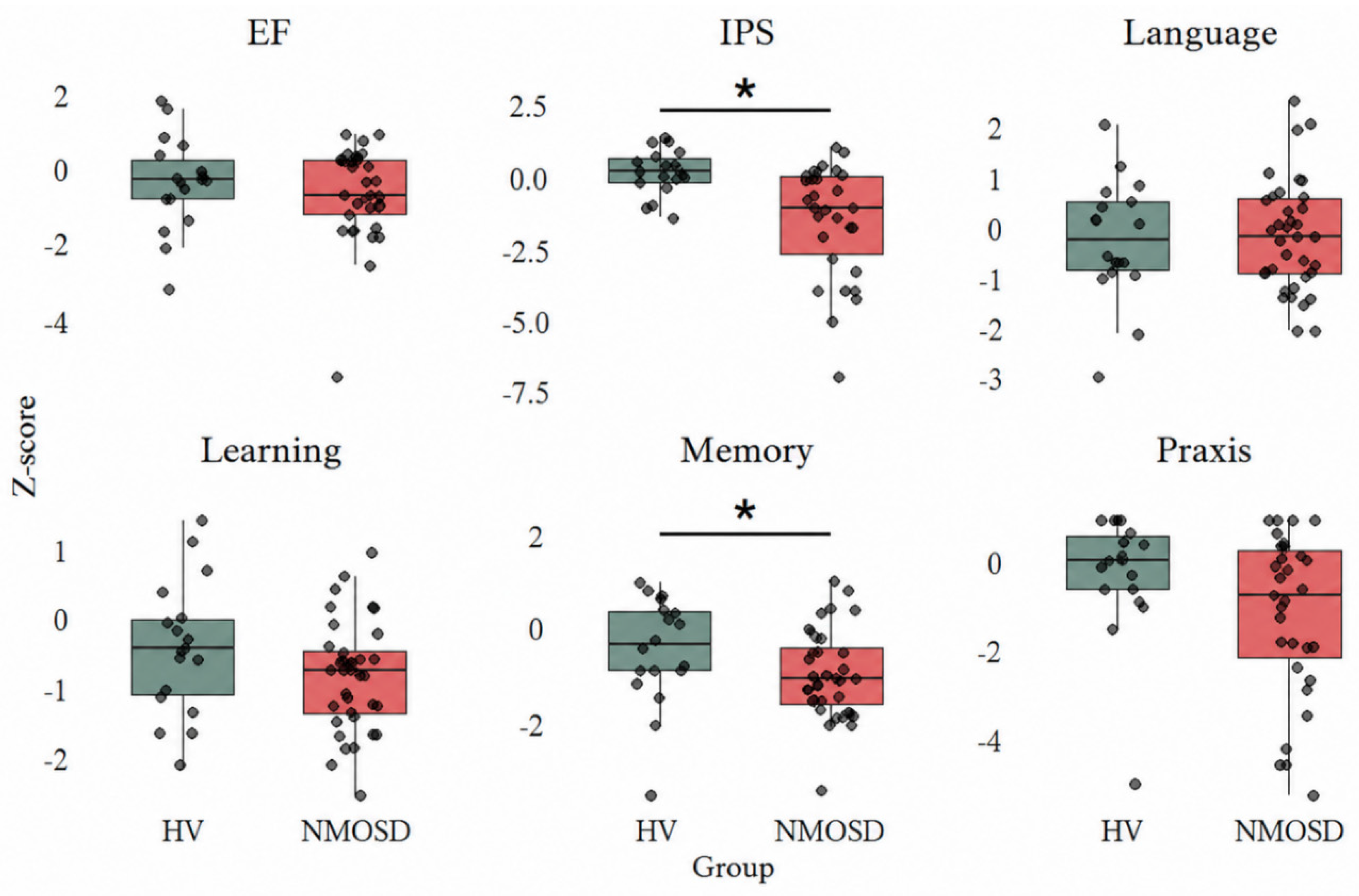
Figure 2.
Predictive effect of clinical variables on cognitive domains. Note. All models are adjusted for Age and Education. Robust standard errors (HC3) and bias-corrected and accelerated (BCa) bootstrap confidence intervals (5,000 resamples) are reported. The p-values were obtained by Bootstrap method and the (*) indicate the significance after Benjamini-Hochberg method. Acronyms. EDSS- Expanded Disability Status Scale.
Figure 2.
Predictive effect of clinical variables on cognitive domains. Note. All models are adjusted for Age and Education. Robust standard errors (HC3) and bias-corrected and accelerated (BCa) bootstrap confidence intervals (5,000 resamples) are reported. The p-values were obtained by Bootstrap method and the (*) indicate the significance after Benjamini-Hochberg method. Acronyms. EDSS- Expanded Disability Status Scale.
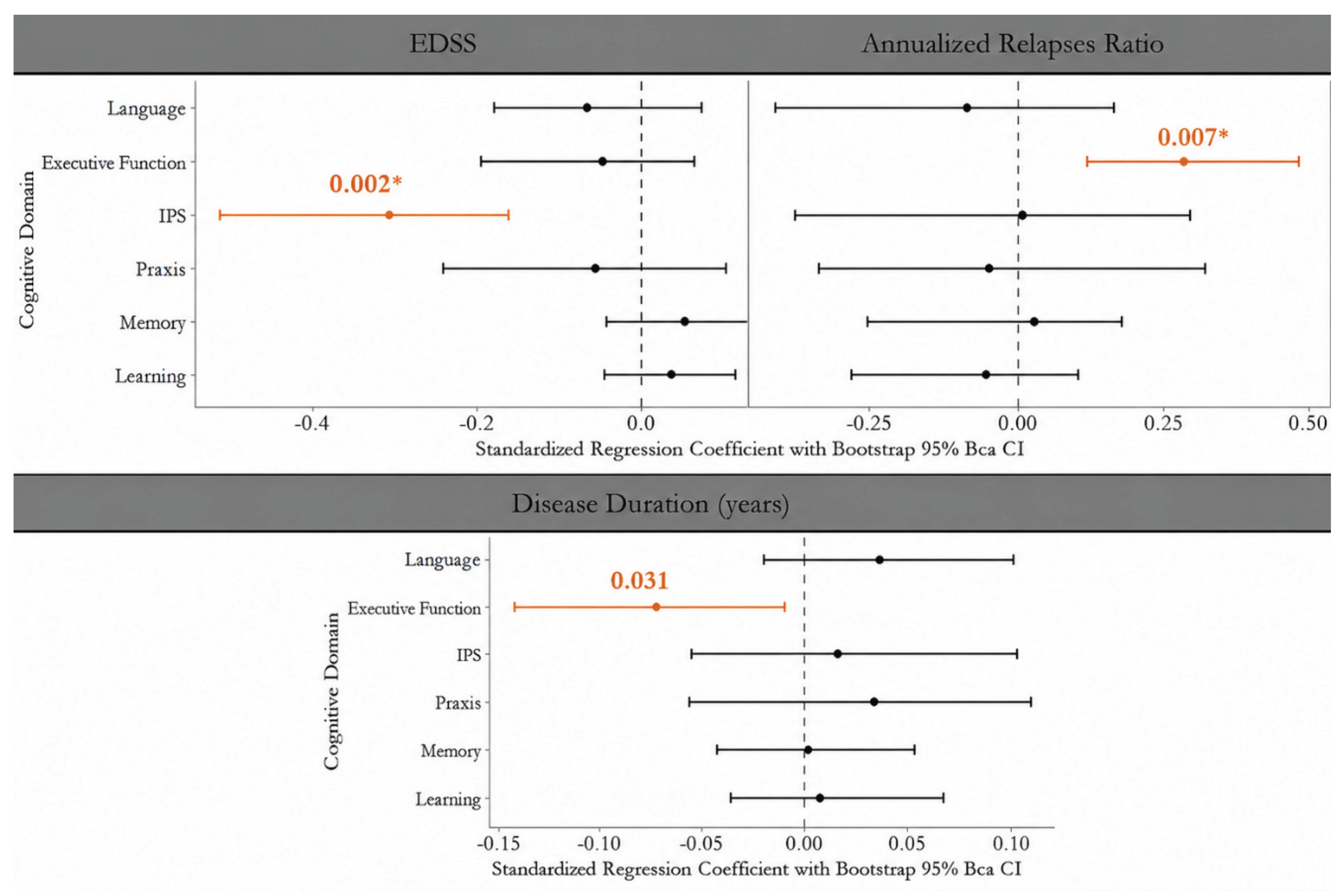
Figure 3.
Association Between EDSS and the Clinically Sensitive Cognitive Composite in NMOSD. Note. Scatterplot shows the association between Expanded Disability Status Scale (EDSS) and the clinically sensitive cognitive composite (CSCC) in patients with NMOSD. Each point represents an individual participant’s observed CSCC value. The solid line represents the model-predicted CSCC from a multivariable linear regression adjusted for age and years of education. The shaded area indicates the 95% BCa bootstrap confidence interval for the adjusted prediction. Higher EDSS values were associated with lower CSCC scores, indicating worse performance across clinically relevant cognitive domains. The CSCC was calculated as the arithmetic mean of visual-acuity-adjusted residual scores for memory, information processing speed, and executive function.
Figure 3.
Association Between EDSS and the Clinically Sensitive Cognitive Composite in NMOSD. Note. Scatterplot shows the association between Expanded Disability Status Scale (EDSS) and the clinically sensitive cognitive composite (CSCC) in patients with NMOSD. Each point represents an individual participant’s observed CSCC value. The solid line represents the model-predicted CSCC from a multivariable linear regression adjusted for age and years of education. The shaded area indicates the 95% BCa bootstrap confidence interval for the adjusted prediction. Higher EDSS values were associated with lower CSCC scores, indicating worse performance across clinically relevant cognitive domains. The CSCC was calculated as the arithmetic mean of visual-acuity-adjusted residual scores for memory, information processing speed, and executive function.
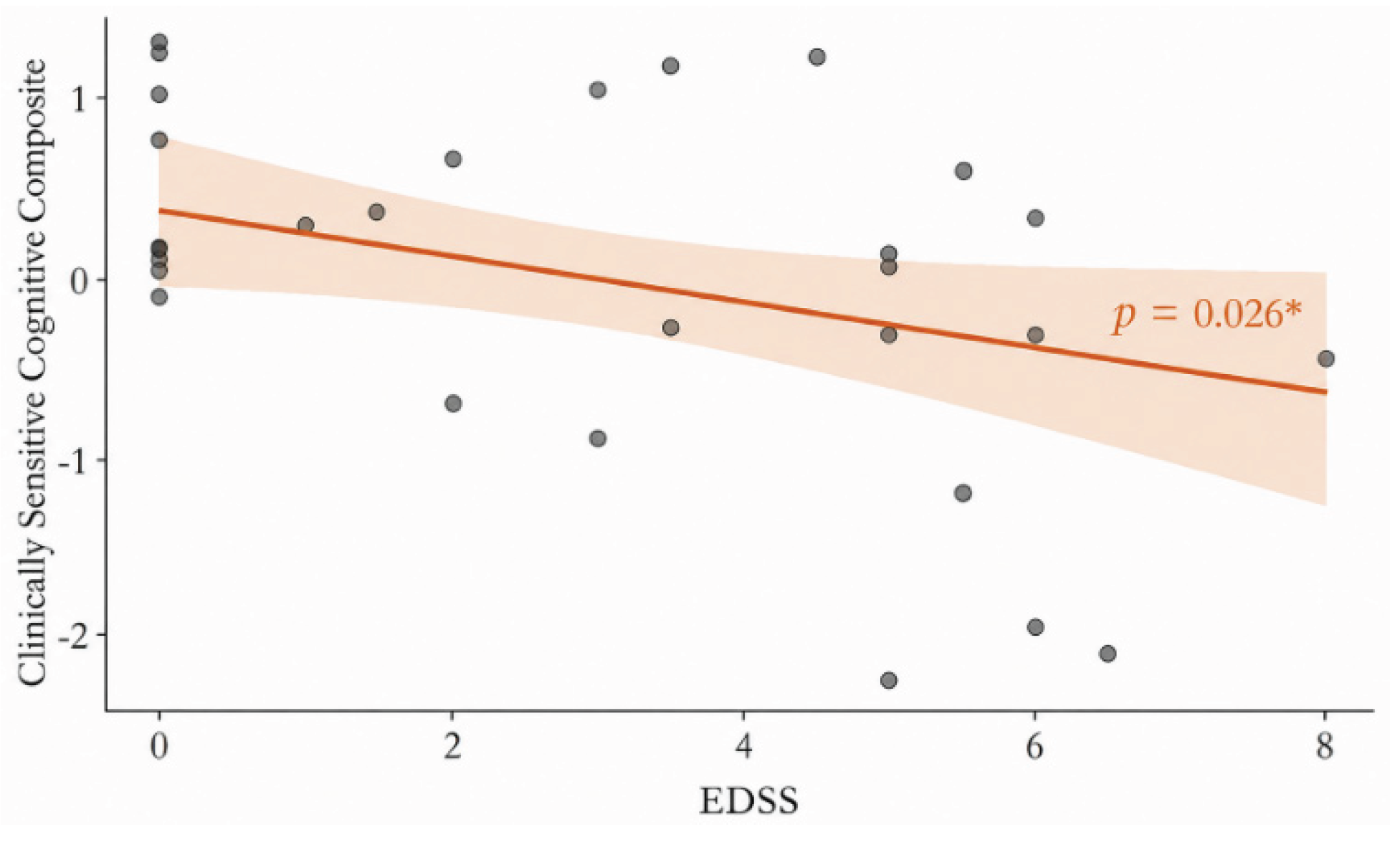

Table 1.
Sample characterization.
| Clinical variables | Healthy Volunteers (n = 18) |
People with NMOSD (n = 36) |
p | |
|---|---|---|---|---|
| Age | Median (IQR) | 38 (31.25–49.75) | 43.5 (34.75–55) | 0.236* |
| Years of Education | Median (IQR) | 14 (11–15) | 11 (9–12.75) | 0.071* |
| Sex (female) | N (%) | 13(72.2%) | 32 (88.9%) | 0.142** |
| Skin color | ||||
| Asian | N (%) | 0 | 1 (2.8%) | 1.000** |
| Non-White | N (%) | 13 (72.2%) | 25 (69.4%) | |
| White | N (%) | 5 (27.8%) | 10 (27.8%) | |
| MMSE | Median (IQR) | 29 (28–29) | 28 (26–29) | 0.212* |
| EDSS | Median (IQR) | - | 3.25 (0–5.5) | |
| Treatment | ||||
| IST | ||||
| Azathioprine | N (%) | - | 28 (84.8%) | |
| Methotrexate | N (%) | - | 3 (9.1%) | |
| Rituximab | N (%) | - | 2 (6.1%) | |
| Prednisone | N (%) | - | 21(95.5%) |
Note. (*) Mann-Whitney Test; (**) Fisher’s exact test. Acronyms. EDSS- Expanded Disability Status Scale; IST- Immunosuppressive Therapy; MMSE- Mini-Mental State Examination.
Table 2.
Cognitive impairment frequencies.
|
Cognitive Impairment ≤ -1.5 |
||
| Learning | 9 | (25%) |
| Memory | 13 | (36%) |
| Praxis | 12 | (33%) |
| Information Processing Speed | 11 | (31%) |
| Executive function | 8 | (22%) |
| Language | 6 | (17%) |
Table 3.
Cognitive comparisons across groups adjusted by age, years of education and visual acuity.
| Cognitive Domain | N | Controls | NMOSD | p | pFDR | Partial η2 |
| Adjusted Mean (SE) | Adjusted Mean (SE) | |||||
| Learning | 54 | -0.58 (0.23) | -0.79 (0.15) | 0.495 | 0.742 | 0.048 |
| Memory | 54 | -0.59 (0.27) | -0.90 (0.18) | 0.422 | 0.742 | 0.059 |
| Praxis | 49 | -0.84 (0.38) | -0.82 (0.28) | 0.980 | 0.980 | 0.070 |
| Information Processing Speed | 48 | -0.23 (0.34) | -1.16 (0.26) | 0.065 | 0.391 | 0.204 |
| Executive Function | 51 | -0.65 (0.31) | -0.45 (0.22) | 0.633 | 0.760 | 0.012 |
Note. Values represent estimated marginal means and standard errors (SE) adjusted for age, years of education, and bilateral visual acuity (LogMAR scores). Group comparisons were performed using an analysis of covariance (ANCOVA) with heteroscedasticity-consistent standard errors (HC3), followed by Benjamini-Hochberg false discovery rate (FDR) correction for multiple comparisons. Partial values represent effect sizes.
Disclaimer/Publisher’s Note: The statements, opinions and data contained in all publications are solely those of the individual author(s) and contributor(s) and not of MDPI and/or the editor(s). MDPI and/or the editor(s) disclaim responsibility for any injury to people or property resulting from any ideas, methods, instructions or products referred to in the content. |
© 2026 by the authors. Licensee MDPI, Basel, Switzerland. This article is an open access article distributed under the terms and conditions of the Creative Commons Attribution (CC BY) license (http://creativecommons.org/licenses/by/4.0/).
Copyright: This open access article is published under a Creative Commons CC BY 4.0 license, which permit the free download, distribution, and reuse, provided that the author and preprint are cited in any reuse.