

Submitted:
18 August 2026
Posted:
19 August 2026
You are already at the latest version
Abstract
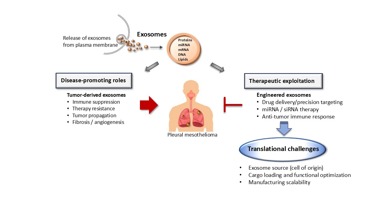
Pleural mesothelioma (PM) is an aggressive malignancy with limited therapeutic options, underscoring the need for novel therapeutic alternatives. Exosomes, a subset of extracellular vesicles mediating intercellular communication, have emerged as potential therapeutic platforms due to their ability to transfer functional biomolecules and modulate cellular responses. In PM, however, exosomes exert context-dependent effects: tumor-derived exosomes actively contribute to tumor progression, immune modulation, and therapy resistance, while exosomes of non-tumor origin or those subjected to engineering approaches may be exploited for targeted therapeutic delivery.
This review discusses the mechanistic foundations of exosome biology relevant to PM and current preclinical efforts to harness exosomes as carriers of nucleic acids, small molecules, and immune-modulatory agents. It further evaluates the influence of exosome cell-of-origin and the intrinsic therapeutic paradox arising from the dual biological roles of exosomes. Finally, key translational challenges, including optimization of exosome source, cargo loading efficiency and manufacturing scalability are examined as major obstacles to clinical implementation. Addressing these challenges is essential for translating exosome-based strategies into more effective therapeutic interventions in this malignancy.
Keywords:
pleural mesothelioma
; lung malignancy
; exosomes
; vesicles
1. Introduction
Pleural mesothelioma is a rare and highly aggressive malignancy arising from the mesothelial cells of the pleura and is etiologically linked primarily to occupational or environmental asbestos exposure [1]. This malignancy is characterized by a prolonged latency period, often exceeding two to three decades and a complex pathogenesis driven by chronic inflammation, oxidative stress, and recurrent genetic alterations. These include BRCA-1-associated protein 1 (BAP1) [2], cyclin-dependent kinase inhibitor 2A gene (CDKN2A), neurofibromin (NF2), [3], and tumor protein p53 (TP53) [4], creating a pro-tumorigenic environment that ultimately promotes malignant transformation [5]. Histologically, PM is categorized into epithelioid, sarcomatoid, and biphasic subtypes, with the epithelioid variant comprising 50–80% of cases and carrying a marginally better prognosis. The sarcomatoid subtype, the rarest, accounting for less than 10% of cases and the biphasic subtype both exhibit significantly more aggressive behavior and poorer clinical outcomes [6].
Treatment has conventionally been restricted to platinum-pemetrexed chemotherapy, surgery, and radiotherapy, and therapeutic outcomes remain poor [7]. Aggressive multimodal strategies have failed to yield survival benefits, as confirmed by the phase 3 MARS 2 trial, which demonstrated increased morbidity and no improvement in overall survival with extended pleurectomy/decortication compared to chemotherapy alone [8]. Despite advances in multimodal management, including anti-angiogenic agents like bevacizumab and dual immune checkpoint inhibitors, nivolumab and ipilimumab, median overall survival remains limited to 9–18 months [9,10]. These clinical limitations underscore the need for novel, mechanism-based therapeutic approaches.
Exosomes, a subtype of extracellular vesicles ranging from 30 to 150 nm in diameter, have emerged as important mediators of intercellular communication through the transfer of proteins, lipids, and nucleic acids. In accordance with the Minimal Information for Studies of Extracelllular Vesicles (MISEV2023) nomenclature recommendations [11], "vesicle" is used interchangeably with "exosome" in this manuscript, including in the title. For clarity, both terms refer specifically to exosomes unless otherwise indicated. In oncology, increasing attention has been directed toward their roles in tumor progression, immune modulation, and drug resistance, as well as their potential exploitation as therapeutic delivery vehicles [12,13].
In the context of PM, studies have demonstrated that tumor-derived exosomes actively contribute to disease biology. Proteomic analyses have shown that mesothelioma-derived exosomes are enriched in proteins associated with angiogenesis, including the angiogenic factor DEL-1, providing early evidence of their potential to shape a tumor-supportive microenvironment [14]. Additional mechanistic work has revealed that PM cells preferentially export tumor-suppressive microRNAs, including miR-16-5p, via exosomes, resulting in intracellular depletion of anti-oncogenic signals; notably, pharmacological inhibition of this same export mechanism restored intracellular miR-16-5p and suppressed tumor growth in preclinical models, pointing to the dual biological role of exosomes explored in this review[15]. Further, exosomes derived from malignant mesothelioma cell lines were found enriched in cancer-associated proteins linked to angiogenesis, metastasis, and immunoregulation, suggesting they help transmit tumor-promoting signals to surrounding and distant cells [16] .
Nevertheless, the available literature remains largely confined to preclinical investigations, focusing on mechanistic insights or biomarker discovery rather than systematic evaluation of exosome-based therapeutic strategies. Accordingly, this review evaluates the therapeutic potentials of exosomes in PM, focusing on the biological opportunities, inherent limitations and critical translational challenges that must be addressed to enable clinical application.
2. Exosome Biology
2.1. Classification of Extracellular Vesicles
Extracellular vesicles (EVs) represent a highly heterogeneous population of membrane-bound particles actively secreted by virtually all cell types, including cancer cells [17,18]. These vesicles serve as vehicles for transfer of molecular cargos such as proteins, lipids, and nucleic acids that can dynamically modulate recipient cell behavior and reprogram cellular phenotypes [13,19]. To date, the principal classes of EVs include exosomes, microvesicles and apoptotic bodies, which are distinguished primarily by their biogenesis pathways and physical size distributions [20]. Additional specialized subpopulations, such as large oncosomes, are distinctly associated with malignant transformation, high-grade tumors, and metastatic progression [21,22].
2.2. Fundamental Properties of Exosomes
Among these diverse EV subtypes, exosomes are uniquely distinguished by their endosomal origin and a relatively uniform, nanoscale diameter typically constrained between 30 and 150 nm [18,20]. Under native conditions, they exhibit a characteristic buoyant density of 1.13 to 1.19 g/mL in sucrose gradients and display a classic cup-shaped morphology under standard electron microscopy [23]. Exosomes are actively secreted into the extracellular environment by normal parenchymal cells, immune cells and stem cells as well as malignant populations [18,24].
Structurally, exosomes possess an inherent phospholipid bilayer membrane heavily enriched with specialized lipid structures, notably ceramide and cholesterol. This rigid lipid architecture protects the internal molecular cargo from extracellular enzymatic degradation while simultaneously enabling cargo sorting, vesicle secretion, and the establishment of functional cellular networks between the host cell and the extracellular environment [25]. Exosomes encapsulate a variety of functional genetic materials, including messenger RNA (mRNA), fragments of genomic DNA, and small non-coding regulatory RNAs like microRNAs (miRNAs) capable of mediating post-transcriptional gene exchange [19,26].
Through the safe transport of these diverse cellular elements, exosomes act as crucial mediators capable of regulating a wide array of biological processes, including inflammatory cascades, systemic immune responses, cellular communication within the nervous system, and various types of complex tissue repair [20].
2.3. Exosome Biogenesis
Exosome biogenesis is initiated through endocytic invagination of the plasma membrane to form early endosomes, followed by inward budding events that generate intraluminal vesicles within multivesicular bodies (MVBs) [27]. The fate of MVBs is tightly regulated, with subsets undergoing lysosomal degradation while others fuse with the plasma membrane to release exosomes into the extracellular space [28]. These processes are governed by two systems: the endosomal sorting complexes required for transport (ESCRT)-dependent and ESCRT-independent pathways. The ESCRT-dependent process utilizes core protein complexes, such as TSG101 [29] and the accessory protein ALIX [30], to sort cargo and drive membrane budding, a process finalized by the ATPase Vps4. Alternatively, ESCRT-independent pathways rely on lipid microdomains and the conversion of sphingomyelin to ceramide to induce membrane curvature, while tetraspanins (CD9, CD63, and CD81) act as scaffolds for cargo organization. Following formation within multivesicular bodies (MVBs), release is mediated by Rab GTPases specifically Rab27a and Rab27b, which regulate MVB trafficking and docking at the plasma membrane [31]. This process culminates in membrane fusion driven by SNARE proteins such as VAMP7 and SNAP-23, which triggers the external secretion of exosomes into the extracellular space [28]. These pathways are dynamically regulated by oncogenic signaling, linking exosome production to cellular transformation and stress responses [32,33].
2.4. Cargo Incorporation
Cargo incorporation into exosomes is a selective and regulated process rather than a passive reflection of cytosolic content. Proteins, messenger RNAs, microRNAs, long non-coding RNAs, and lipids are selectively enriched through mechanisms involving RNA-binding proteins, post-translational modifications, and lipid raft–associated sorting [34,35]. This regulated packaging confers functional specificity on exosomes and enables them to modulate recipient cell behavior in a context-dependent manner [36,37]. In PM, tumor-derived EVs and exosomes have been shown to carry distinct molecular signatures reflective of tumor biology.
Understanding these endogenous loading pathways allows for the design of exosome-based therapeutic strategies [34]. By manipulating cellular sorting networks such as hijacking RNA-binding proteins or utilizing membrane-engineering techniques exosomes can be artificially packaged with tumor-suppressor miRNAs, small interfering RNAs (siRNAs), or conventional chemotherapeutics like paclitaxel [38,39]. This selectively loaded cargo can then be directionally delivered to malignant pleural cells, capitalizing on the natural low immunogenicity and superior tissue penetration of the exosomal lipid bilayer to overcome the immunosuppressive tumor microenvironment characteristic of PM ([13].
3. Biological Paradox of Exosomes in PM
Aside from the exosomes shed by the tumor itself, exosomes released by local stromal and immune cells actively shape the inflammatory and fibrotic microenvironment of PM. For instance, mesothelial and stromal cell-derived exosomes drive tissue remodeling and pro-fibrotic signaling. This interaction is central to PM pathology, where long-term, asbestos-induced inflammation primes tissue for malignant change. By transferring active cytokines, growth factors, and regulatory RNAs, these exosomes essentially re-program the stroma to form a supportive niche for tumor growth [40,41]. Immune cell-derived exosomes further impact this environment by modulating the local immune response. Depending on their source and cargo, exosomes from macrophages can either amplify inflammation or shut down immune surveillance. In particular, tumor-associated macrophage (TAM)-derived exosomes deliver microRNAs and proteins that block anti-tumor immunity and maintain cancer development [42,43]. In a related context, in lung cancer malignant pleural effusion, exosomes carrying immunosuppressive checkpoint proteins impair local anti-tumor immunity and contribute to immune evasion. These exosomes suppress the cytotoxic function of double-negative T cells via the PD-1/PD-L1 and CEACAM1/TIM3 pathways [44].
Yet, the biology is not entirely pro-tumorigenic, as this same trafficking pathway also exposes a therapeutic potential : mesothelioma cells actively export potent tumor suppressors such as miR-16-5p via exosomes to evade their senescence-inducing effects, and blocking this export restores intracellular miR-16-5p levels, reinstating tumor-suppressive activity and reducing cell viability and tumorigenicity [15]. This principle extends to exosome-delivered miR-126, which induces mass disaggregation of MPM-derived spheroids and, when combined with exosome-release inhibition, drives mesothelioma stem-cell-like death and tumor growth arrest in vivo [45]. Furthermore, dendritic cells loaded with tumor-derived exosomes from malignant mesothelioma cells trigger a protective anti-tumor immune response, significantly extending survival in tumor-bearing mice compared to lysate-loaded controls [46].
These complex exosomal networks present a striking biological paradox. On one hand, these endogenous exosomes drive asbestos-related inflammation and allow the tumor to evade the immune system, but on the other hand, the inherent transport mechanisms that enable these particles to efficiently shuttle functional cargo across the mesothelial barrier make them ideal, natural vehicles for drug delivery.
4. Exosome-Based Therapeutic Delivery in Pleural Mesothelioma: Preclinical Proof-of-Concept
Although the therapeutic potential of exosome-mediated delivery has been extensively explored in other solid tumor models, PM remains comparatively understudied in terms of functional proof-of-concept. Preclinical investigations nonetheless provide emerging evidence that exosome-based strategies could augment the therapeutic options for this disease.
Early studies demonstrated that exosome-mediated transfer of bioactive cargo can influence mesothelial cell phenotype, providing early mechanistic proof that exosomes are capable of transferring functionally active cargo into recipient mesothelial cells. Notably, Munson et al. [47] showed that exosomes derived from asbestos-exposed lung epithelial cells and macrophages induced epithelial-to-mesenchymal transition-like gene expression changes in normal mesothelial cells, demonstrating, albeit in a disease-promoting context, that exosomal cargo transfer can functionally reprogram mesothelial cell phenotype. While this specific effect is pathogenic rather than therapeutic, it nonetheless establishes the underlying biological principle of functional cargo transfer into mesothelial cells that later therapeutic strategies would seek to exploit for tumor-suppressive rather than tumor-promoting ends.
Complementary work by the same group directly demonstrated this therapeutic potential: PM cells actively export tumor-suppressive microRNAs, including miR-16-5p, via exosomes. Inhibition of exosome secretion, thereby restoring intracellular miR-16-5p levels, suppressed cell viability and tumorigenicity in preclinical systems [15].
Further supporting the therapeutic utility of targeting exosome trafficking pathways, Monaco et al. [48] showed that exosomal transfer of miR-126 from endothelial cells inhibited mesothelioma cell proliferation and revealed a role for miR-126 in cancer-stroma communication and angiogenesis regulation, supporting miR-126 delivery via exosomes as a stable and non-immunogenic therapeutic approach. The same group also provided more direct evidence for the therapeutic application of exosome-delivered miR-126 in PM. In this study, the authors demonstrated that exosome-delivered miR-126 induced disaggregation of mesothelioma spheroids, and that blocking exosomal re-export of miR-126 via the inhibitor GW4869 was required to trigger mesothelioma stem cell-like death and tumor growth arrest in vivo [45]. These findings support the feasibility of exosome-based RNA delivery strategies in this disease context, provided cellular efflux of the delivered miRNA is also addressed.
Notably, tumor-derived exosomes have also been repurposed as antigen delivery vehicles in immunotherapeutic strategies. Indeed, Mahaweni et al. [46] showed that dendritic cells loaded with mesothelioma-derived exosomes promoted tumor-infiltrating CD4+ and CD8+ T-cell responses and improved survival in a murine model, outperforming both lysate-loaded dendritic cells and untreated controls. It should be noted that this study used the AB1 cell line in an intraperitoneal mesothelioma model rather than PM specifically. Nonetheless, this work provides evidence that mesothelioma-derived exosomes can be harnessed as biologically relevant antigen carriers to enhance anti-tumor immunity, supporting their potential extension to PM-directed immunotherapeutic strategies.
In various solid tumors including mesothelioma, unmodified tumor-derived exosomes can also suppress immune responses, for instance through NKG2D ligand-mediated downregulation of NK- and CD8+ T-cell surveillance [49]. This underscores that native, unmodified tumor-derived exosomes are more often immunosuppressive than immunostimulatory, reinforcing why controlled, engineered repurposing of exosomes, whether from non-tumor sources or from tumor-derived exosomes redirected platforms such as the dendritic-cell-based antigen delivery system of Mahaweni et al. [46] is generally preferred for immunotherapeutic applications.
Apart from nucleic acid and antigen delivery, evidence is scarce regarding the use of exosomes to deliver small-molecule drugs in PM models; nonetheless, the successful exosome-based transport of chemotherapeutics and immune-stimulatory cargos with enhanced tumor targeting and reduced systemic toxicity in other tumor systems [50] suggests that such strategies eventually be adaptable to the PM setting.
Together, these six preclinical projects show that exosomes in PM can both drive and disrupt disease, offering early but genuine proof-of-concept for exosome-based therapy. An overview of the above-presented studies are presented in Table 1.
5. Biological Limitations of Exosome-Based Therapeutic Strategies
Despite their therapeutic promise, exosome-based approaches are constrained by several intrinsic biological limitations. A major challenge is off-target biodistribution, as systemically administered exosomes are preferentially taken up by the mononuclear phagocyte system, particularly in the liver and spleen, thereby limiting tumor-specific delivery [51].
Another limitation is the risk of unintended immunomodulation. Although exosomes are often considered minimally immunogenic, their cargo can include bioactive proteins, lipids, and nucleic acids capable of inducing inflammatory or immunosuppressive responses depending on cellular origin and context [52].
A further barrier is endogenous exosome interference. Circulating and pleural fluid-derived vesicles are abundant and may compete with administered exosomes for uptake pathways, reducing delivery efficiency. This is particularly relevant in PM, where tumor-associated exosome production is elevated [53].
A hallmark of advanced PM is pleural effusion, which is enriched in extracellular vesicles, cytokines, and tumor-derived factors, creating a high-background vesicular environment that may dilute or compete with therapeutic exosomes [54,55]. In addition, the mesothelial barrier and dense stromal architecture impose physical constraints on vesicle penetration and distribution. Fibrotic remodeling and extracellular matrix deposition further restrict diffusion and limit access to tumor cells [56]. The pleural space also represents a dynamic compartment, where fluid turnover, immune infiltration, and chronic inflammation influence exosome stability and cellular uptake [54]. These characteristics necessitate disease-specific delivery strategies, including consideration of intrapleural administration routes.
6. Importance of Exosome Cell-of-Origin in Therapeutic Design
A critical determinant of exosome function and therapeutic suitability is their cell-of- origin, which governs vesicle composition, biological activity, and safety profile. Exosomes are not biologically equivalent entities; rather, their protein, lipid, and nucleic acid cargo reflects the physiological or pathological state of the parent cell, with profound implications for therapeutic application [13,18]. This heterogeneity is particularly relevant in PM, where tumor, immune, and stromal cells coexist within a complex pleural microenvironment.
Tumor-derived exosomes are enriched in oncogenic proteins, immunosuppressive factors, and regulatory RNAs that promote tumor growth, immune evasion, and therapy resistance. Early evidence from mesothelioma models demonstrated that tumor-derived exosomes actively engage immune components of the tumor microenvironment; for example, through antigen presentation and immune-evasion mechanisms, highlighting their role in disease progression rather than passive vesicle release [14]. Proteomic profiling of exosomes derived from primary human mesothelioma cells revealed enrichment of oncogenic and tumor-associated signaling cargo, underscoring that exosome composition closely reflects cellular origin and disease state [16]. This cell of origin dependency has important implications for the selection and engineering of exosomes for therapeutic applications in PM.
As discussed above, mesothelioma-derived exosomes actively shape the tumor microenvironment and may disseminate pro-tumorigenic signals, raising significant concerns regarding their direct therapeutic use as delivery vehicles. While tumor-derived exosomes may offer intrinsic homing capabilities, their pathological cargo composition necessitates extensive engineering or purification to mitigate oncogenic risk.
In contrast, non-tumor-derived exosomes, particularly those originating from immune cells or mesenchymal/stromal cells, are increasingly favored for therapeutic development. Immune cell-derived exosomes, such as those from dendritic cells or macrophages, can carry immune-stimulatory molecules and antigenic cargo, enabling modulation of anti-tumor immune responses without direct tumor-promoting effects [46,57,58]. Similarly, mesenchymal stem cell (MSC)–derived exosomes exhibit favorable biocompatibility, intrinsic tumor-tropic properties, and lower immunogenicity, making them attractive platforms for drug and RNA delivery [59,60].
Importantly, cell-of-origin not only determines cargo composition but also influences surface proteins, integrin profiles, and lipid composition, thereby affecting biodistribution and uptake [61]. This dependence of exosome function on cellular origin provides a rationale for engineering strategies aimed at optimizing exosome source, targeting and cargo composition.
7. Exosomes Compared to Synthetic Nanocarriers
Exosomes have attracted considerable interest as therapeutic delivery platforms due to several intrinsic properties that distinguish them from conventional synthetic nanocarrier-engineered nanoscale delivery systems such as liposomes, polymeric nanoparticles, and inorganic particles designed to transport therapeutic cargo. Their endogenous membrane composition, biological origin, and intrinsic intercellular communication capacity may confer advantages in biocompatibility and cellular uptake. [13]. In contrast, many synthetic nanocarriers require surface modification (e.g., PEGylation or ligand conjugation) to optimize circulation time and targeting efficiency, which may introduce additional manufacturing complexity and regulatory considerations [62].
As naturally occurring vesicles evolved for intercellular communication, exosomes exhibit high biocompatibility and low intrinsic immunogenicity, enabling prolonged circulation and reduced clearance by the mononuclear phagocyte system compared with many synthetic carriers [13,63]. Their endogenous lipid and protein composition allows exosomes to evade rapid opsonization and complement activation, which remain significant limitations for engineered nanoparticles [64].
A further advantage of exosomes lies in their intrinsic capacity for efficient cellular uptake and tissue penetration. Exosomes exploit physiological uptake mechanisms, including receptor-mediated endocytosis and membrane fusion, facilitating cargo delivery across biological barriers that are difficult to overcome with synthetic systems, such as the blood-brain barrier and dense tumor stroma [36,65,66]. Importantly, exosomes can display cell-type-specific surface molecules that confer a degree of natural targeting, which may be further enhanced through engineering strategies, reducing off-target effects commonly associated with non-specific nanoparticle accumulation [67].
Exosomes are also particularly well suited for the delivery of nucleic acid-based therapeutics, including microRNAs, siRNAs, and mRNAs. Their lipid bilayer protects encapsulated cargo from enzymatic degradation, while endogenous RNA-binding proteins contribute to cargo stability and functional delivery [19,68]. In contrast, synthetic nanocarriers often require complex chemical modifications to achieve comparable stability and delivery efficiency, which can introduce toxicity or regulatory challenges [69].
Nevertheless, while these features underscore the attractiveness of exosomes as delivery vehicles, they also highlight important distinctions from synthetic nanocarriers that must be carefully considered. Unlike fully defined synthetic systems, exosomes are biologically complex and heterogeneous, with cargo composition that reflects the state and origin of the producing cells. This complexity complicates large-scale manufacturing, quality control, and reproducibility, and raises concerns regarding the inadvertent transfer of bioactive or oncogenic molecules [60,70,71]. Thus, although exosomes offer clear biological advantages over synthetic nanocarriers, their therapeutic exploitation requires rigorous characterization and control to balance efficacy with safety.
8. Engineering Strategies to Optimize Exosome Source, Cargo Loading and Manufacturing Scalability
The successful clinical translation of exosome-based therapeutic strategies in PM will depend on rational engineering approaches that address interdependent parameters: exosome source selection, cargo loading efficiency and manufacturing scalability. Each of these dimensions directly influences therapeutic efficacy, biodistribution, and safety.
8.1. Optimization of Exosome Source
Source selection represents a primary determinant of therapeutic safety and functional consistency in exosome-based strategies. Current engineering paradigms increasingly prioritize non-tumor-derived exosomes particularly those derived from MSCs, dendritic cells, or other immunologically inert producer populations, given their lower oncogenic risk and improved translational feasibility [72,73].
Advanced manufacturing strategies now incorporate immortalized MSC cell lines, induced pluripotent stem cell (iPSC)-derived producer systems, and other scalable producer platforms such as cardiac progenitor cells to enhance yield and batch reproducibility [74,75,76]. To optimize manufacturing, three-dimensional culture systems have been utilized to significantly enhance exosome yield under controlled conditions while preserving their biological functionality [77].
In parallel, donor cell preconditioning via hypoxia, inflammatory priming, metabolic modulation, or targeted CRISPR-based engineering has emerged as a controlled approach to reshape exosome cargo composition and surface phenotype [78,79,80]. Such approaches may improve therapeutic persistence, tumor tropism, angiostatic activity, and immunomodulatory potential [81,82,83].
8.2. Cargo Loading
Cargo incorporation efficiency remains a central engineering obstacle. To address this, endogenous loading strategies utilize genetic modification or pharmacologic stimulation of donor cells to drive the selective encapsulation of therapeutic RNAs or proteins into newly formed vesicles [85]. More recently, the development of RNA-binding protein–guided loading systems in exosomes has significantly improved cargo specificity while minimizing stochastic packaging [86].
Exogenous loading approaches including electroporation, sonication, extrusion, freeze–thaw cycling, microfluidic mixing, and transient membrane permeabilization enable post-isolation incorporation of nucleic acids or small-molecule therapeutics, although membrane destabilization, vesicle aggregation, and inconsistent loading efficiency remain important technical limitations [87,88,89,90].
In this pleural malignancy, therapeutic payloads of interest include tumor-suppressive miRNAs e.g., miR-16, miR-193a, miR-215 and miR-34 family members [91,92,93,94], inhibitors targeting Hippo/YAP–TAZ signaling components [95], and immune-modulatory molecules designed to reprogram the tumor microenvironment [96].
8.3. Manufacturing Scalability
Increasing engineering sophistication introduces additional translational challenges, including batch-to-batch variability, scalable GMP-compliant manufacturing, standardized potency assays, purification consistency, long-term storage stability, and comprehensive safety evaluation. Isolation methodologies including ultracentrifugation, tangential-flow filtration, size-exclusion chromatography, and microfluidic separation technologies each present distinct competing priorities between purity, scalability, recovery yield, and cost [98,99,100,101].
Consequently, harmonized characterization standards are increasingly emphasized to support regulatory alignment and cross-study reproducibility. Updated guidelines from the International Society for Extracellular Vesicles (MISEV2023) reinforce rigorous molecular characterization, functional validation, contaminant assessment, and transparent reporting as prerequisites for clinical translation [11,102].
Within the framework of PM research where therapeutic innovation remains urgently needed, exosome engineering strategies must ultimately balance enhanced targeting precision and cargo control with preservation of the intrinsic biological advantages of extracellular vesicles, including biocompatibility, immune tolerance, pleural tissue penetration, and endogenous trafficking capacity, which distinguish them from many synthetic nanocarrier systems [50,103]. Achieving this balance will likely determine whether engineered exosome platforms can progress from experimental models toward clinically viable precision therapies for pleural mesothelioma.
9. Conclusions and Perspectives
Exosome-based therapeutic strategies represent a promising yet inherently complex avenue for innovation in PM, a malignancy that remains largely refractory to current treatment modalities. As discussed above, exosomes act as a genuinely double-edged vesicle: tumor-derived exosomes actively contribute to PM progression, immune evasion, and therapy resistance, yet these same vesicles can be repurposed as biologically optimized delivery platforms for therapeutic cargo. This paradox underscores the necessity of a conceptually rigorous, mechanism-driven approach to exosome-based intervention rather than broad translational enthusiasm.
A central determinant of therapeutic feasibility is the cell-of-origin of exosomes. Tumor-derived exosomes, although intrinsically adapted for tumor homing, carry oncogenic and immunosuppressive cargo that limits their direct clinical utility without extensive modification. In contrast, exosomes derived from immune cells or mesenchymal stromal cells offer more favorable safety profiles, enhanced biocompatibility, and unique opportunities for immune modulation, making them attractive candidates for drug development. Rational selection and engineering of exosome source cells, consequently, emerges as a critical step in optimizing therapeutic efficacy while minimizing unintended biological effects.
Although preclinical studies in PM remain limited, they provide proof-of-principle for exosome-mediated delivery of nucleic acids and other bioactive agents. However, major challenges persist, including scalable manufacturing, cargo standardization, targeting specificity, and safety validation.
In summary, exosome-based strategies in PM should therefore be viewed not as a universal solution, but as a biologically grounded platform requiring rigorous optimization. With careful integration of mechanistic insight, bioengineering, and translational discipline, exosomes may ultimately contribute to more precise therapeutic interventions in this malignancy.
Author Contributions
Conceptualization, writing and editing, LCD; final editing to increase academic impact, DG.
Funding
This research received no external funding.
Acknowledgments
During the preparation of this manuscript, the authors used Claude Sonnet 4.5 (Anthropic) for assistance in structuring and refining the content of the section „Engineering Strategies to Optimize Exosome Source, Cargo Loading and Manufacturing Scalability.“ The authors have reviewed and edited the output and take full responsibility for the content of this publication.
Conflicts of Interest
The authors have no conflict of interest to declare.
References
- Zolondick, A.A.; Gaudino, G.; Xue, J.; Pass, H.I.; Carbone, M.; Yang, H. Asbestos-Induced Chronic Inflammation in Malignant Pleural Mesothelioma and Related Therapeutic Approaches-a Narrative Review. Precis Cancer Med. 2021, 4, 27. [Google Scholar] [CrossRef] [PubMed]
- Hmeljak, J.; Sanchez-Vega, F.; Hoadley, K.A.; Shih, J.; Stewart, C.; Heiman, D.; Tarpey, P.; Danilova, L.; Drill, E.; Gibb, E.A.; et al. Integrative Molecular Characterization of Malignant Pleural Mesothelioma. Cancer Discov. 2018, 8, 1548–1565. [Google Scholar] [CrossRef] [PubMed]
- Kato, S.; Tomson, B.N.; Buys, T.P.H.; Elkin, S.K.; Carter, J.L.; Kurzrock, R. Genomic Landscape of Malignant Mesotheliomas. Mol. Cancer Ther. 2016, 15, 2498–2507. [Google Scholar] [CrossRef] [PubMed]
- Hylebos, M.; Van Camp, G.; van Meerbeeck, J.P.; Op de Beeck, K. The Genetic Landscape of Malignant Pleural Mesothelioma: Results from Massively Parallel Sequencing. J. Thorac. Oncol. 2016, 11, 1615–1626. [Google Scholar] [CrossRef] [PubMed]
- Fennell, D.A.; Sekido, Y.; Baas, P.; Husain, A.N.; Curioni-Fontecedro, A.; Lim, E.; Opitz, I.; Simone, C.B.; Brims, F.; Wong, M.C. Pleural Mesothelioma. Nat. Rev. Dis. Prim. 2025, 11, 56. [Google Scholar] [CrossRef] [PubMed]
- Brcic, L.; Kern, I. Clinical Significance of Histologic Subtyping of Malignant Pleural Mesothelioma. Transl. Lung Cancer Res. 2020, 9, 924–933. [Google Scholar] [CrossRef] [PubMed]
- Kindler, H.L.; Ismaila, N.; Armato, S.G.; Bueno, R.; Hesdorffer, M.; Jahan, T.; Jones, C.M.; Miettinen, M.; Pass, H.; Rimner, A.; et al. Treatment of Malignant Pleural Mesothelioma: American Society of Clinical Oncology Clinical Practice Guideline. J. Clin. Oncol. 2018, 36, 1343–1373. [Google Scholar] [CrossRef] [PubMed]
- Lim, E.; Waller, D.; Lau, K.; Steele, J.; Pope, A.; Ali, C.; Bilancia, R.; Keni, M.; Popat, S.; O’Brien, M.; et al. Extended Pleurectomy Decortication and Chemotherapy versus Chemotherapy Alone for Pleural Mesothelioma (MARS 2): A Phase 3 Randomised Controlled Trial. Lancet Respir. Med. 2024, 12, 457–466. [Google Scholar] [CrossRef] [PubMed]
- Baas, P.; Scherpereel, A.; Nowak, A.K.; Fujimoto, N.; Peters, S.; Tsao, A.S.; Mansfield, A.S.; Popat, S.; Jahan, T.; Antonia, S.; et al. First-Line Nivolumab plus Ipilimumab in Unresectable Malignant Pleural Mesothelioma (CheckMate 743): A Multicentre, Randomised, Open-Label, Phase 3 Trial. Lancet 2021, 397, 375–386. [Google Scholar] [CrossRef] [PubMed]
- Bertuccio, F.R.; Montini, S.; Fusco, M.A.; Di Gennaro, A.; Sciandrone, G.; Agustoni, F.; Galli, G.; Bortolotto, C.; Saddi, J.; Baietto, G.; et al. Malignant Pleural Mesothelioma: From Pathophysiology to Innovative Actionable Targets. Cancers 2025, 17, 1160. [Google Scholar] [CrossRef] [PubMed]
- Welsh, J.A.; Goberdhan, D.C.I.; O’Driscoll, L.; Buzas, E.I.; Blenkiron, C.; Bussolati, B.; Cai, H.; Di Vizio, D.; Driedonks, T.A.P.; Erdbrügger, U.; et al. Minimal Information for Studies of Extracellular Vesicles (MISEV2023): From Basic to Advanced Approaches. J. Extracell. Vesicles 2024, 13, e12404. [Google Scholar] [CrossRef] [PubMed]
- Mehryab, F.; Taghizadeh, F.; Goshtasbi, N.; Merati, F.; Rabbani, S.; Haeri, A. Exosomes as Cutting-Edge Therapeutics in Various Biomedical Applications: An Update on Engineering, Delivery, and Preclinical Studies. Biochimie 2023, 213, 139–167. [Google Scholar] [CrossRef] [PubMed]
- Kalluri, R.; LeBleu, V.S. The Biology, Function, and Biomedical Applications of Exosomes. Science 2020, 367, eaau6977. [Google Scholar] [CrossRef] [PubMed]
- Hegmans, J.P.J.J.; Bard, M.P.L.; Hemmes, A.; Luider, T.M.; Kleijmeer, M.J.; Prins, J.-B.; Zitvogel, L.; Burgers, S.A.; Hoogsteden, H.C.; Lambrecht, B.N. Proteomic Analysis of Exosomes Secreted by Human Mesothelioma Cells. Am. J. Pathol. 2004, 164, 1807–1815. [Google Scholar] [CrossRef] [PubMed]
- Munson, P.B.; Hall, E.M.; Farina, N.H.; Pass, H.I.; Shukla, A. Exosomal MiR-16-5p as a Target for Malignant Mesothelioma. Sci. Rep. 2019, 9, 11688. [Google Scholar] [CrossRef] [PubMed]
- Greening, D.W.; Ji, H.; Chen, M.; Robinson, B.W.S.; Dick, I.M.; Creaney, J.; Simpson, R.J. Secreted Primary Human Malignant Mesothelioma Exosome Signature Reflects Oncogenic Cargo. Sci. Rep. 2016, 6, 32643. [Google Scholar] [CrossRef] [PubMed]
- Kalluri, R.; McAndrews, K.M. The Role of Extracellular Vesicles in Cancer. Cell 2023, 186, 1610–1626. [Google Scholar] [CrossRef] [PubMed]
- Théry, C.; Zitvogel, L.; Amigorena, S. Exosomes: Composition, Biogenesis and Function. Nat. Rev. Immunol. 2002, 2, 569–579. [Google Scholar] [CrossRef] [PubMed]
- Valadi, H.; Ekström, K.; Bossios, A.; Sjöstrand, M.; Lee, J.J.; Lötvall, J.O. Exosome-Mediated Transfer of MRNAs and MicroRNAs Is a Novel Mechanism of Genetic Exchange between Cells. Nat. Cell Biol. 2007, 9, 654–659. [Google Scholar] [CrossRef] [PubMed]
- Colombo, M.; Raposo, G. Biogenesis, Secretion, and Intercellular Interactions of Exosomes and Other Extracellular Vesicles. 2014. [Google Scholar] [CrossRef] [PubMed]
- Minciacchi, V.R.; You, S.; Spinelli, C.; Morley, S.; Zandian, M.; Aspuria, P.-J.; Cavallini, L.; Ciardiello, C.; Reis Sobreiro, M.; Morello, M.; et al. Large Oncosomes Contain Distinct Protein Cargo and Represent a Separate Functional Class of Tumor-Derived Extracellular Vesicles. Oncotarget 2015, 6, 11327–11341. [Google Scholar] [CrossRef] [PubMed]
- Di Vizio, D.; Morello, M.; Dudley, A.C.; Schow, P.W.; Adam, R.M.; Morley, S.; Mulholland, D.; Rotinen, M.; Hager, M.H.; Insabato, L.; et al. Large Oncosomes in Human Prostate Cancer Tissues and in the Circulation of Mice with Metastatic Disease. Am. J. Pathol. 2012, 181, 1573–1584. [Google Scholar] [CrossRef] [PubMed]
- Théry, C.; Amigorena, S.; Raposo, G.; Clayton, A. Isolation and Characterization of Exosomes from Cell Culture Supernatants and Biological Fluids. Curr. Protoc. Cell Biol. 2006, 30, 3.22.1–3.22.29. [Google Scholar] [CrossRef] [PubMed]
- Lai, R.C.; Arslan, F.; Lee, M.M.; Sze, N.S.K.; Choo, A.; Chen, T.S.; Salto-Tellez, M.; Timmers, L.; Lee, C.N.; El Oakley, R.M.; et al. Exosome Secreted by MSC Reduces Myocardial Ischemia/Reperfusion Injury. Stem Cell Res. 2010, 4, 214–222. [Google Scholar] [CrossRef] [PubMed]
- Skotland, T.; Hessvik, N.P.; Sandvig, K.; Llorente, A. Exosomal Lipid Composition and the Role of Ether Lipids and Phosphoinositides in Exosome Biology. J. Lipid Res. 2019, 60, 9–18. [Google Scholar] [CrossRef] [PubMed]
- Kahlert, C.; Melo, S.A.; Protopopov, A.; Tang, J.; Seth, S.; Koch, M.; Zhang, J.; Weitz, J.; Chin, L.; Futreal, A.; et al. Identification of Double-Stranded Genomic DNA Spanning All Chromosomes with Mutated KRAS and P53 DNA in the Serum Exosomes of Patients with Pancreatic Cancer. J. Biol. Chem. 2014, 289, 3869–3875. [Google Scholar] [CrossRef] [PubMed]
- Raposo, G.; Stoorvogel, W. Extracellular Vesicles: Exosomes, Microvesicles, and Friends. 2013. [Google Scholar] [CrossRef] [PubMed]
- Hessvik, N.P.; Llorente, A. Current Knowledge on Exosome Biogenesis and Release. Cell Mol. Life Sci. 2018, 75, 193–208. [Google Scholar] [CrossRef] [PubMed]
- Colombo, M.; Moita, C.; van Niel, G.; Kowal, J.; Vigneron, J.; Benaroch, P.; Manel, N.; Moita, L.F.; Théry, C.; Raposo, G. Analysis of ESCRT Functions in Exosome Biogenesis, Composition and Secretion Highlights the Heterogeneity of Extracellular Vesicles. J. Cell Sci. 2013, 126, 5553–5565. [Google Scholar] [CrossRef] [PubMed]
- Baietti, M.F.; Zhang, Z.; Mortier, E.; Melchior, A.; Degeest, G.; Geeraerts, A.; Ivarsson, Y.; Depoortere, F.; Coomans, C.; Vermeiren, E.; et al. Syndecan-Syntenin-ALIX Regulates the Biogenesis of Exosomes. Nat. Cell Biol. 2012, 14, 677–685. [Google Scholar] [CrossRef] [PubMed]
- Ostrowski, M.; Carmo, N.B.; Krumeich, S.; Fanget, I.; Raposo, G.; Savina, A.; Moita, C.F.; Schauer, K.; Hume, A.N.; Freitas, R.P.; et al. Rab27a and Rab27b Control Different Steps of the Exosome Secretion Pathway. Nat. Cell Biol. 2010, 12, 19–30; sup pp 1-13. [Google Scholar] [CrossRef] [PubMed]
- Han, Q.-F.; Li, W.-J.; Hu, K.-S.; Gao, J.; Zhai, W.-L.; Yang, J.-H.; Zhang, S.-J. Exosome Biogenesis: Machinery, Regulation, and Therapeutic Implications in Cancer. Mol. Cancer 2022, 21, 207. [Google Scholar] [CrossRef] [PubMed]
- Xie, S.; Zhang, Q.; Jiang, L. Current Knowledge on Exosome Biogenesis, Cargo-Sorting Mechanism and Therapeutic Implications. Membranes 2022, 12, 498. [Google Scholar] [CrossRef] [PubMed]
- O’Brien, K.; Breyne, K.; Ughetto, S.; Laurent, L.C.; Breakefield, X.O. RNA Delivery by Extracellular Vesicles in Mammalian Cells and Its Applications. Nat. Rev. Mol. Cell Biol. 2020, 21, 585–606. [Google Scholar] [CrossRef] [PubMed]
- Sork, H.; Corso, G.; Krjutskov, K.; Johansson, H.J.; Nordin, J.Z.; Wiklander, O.P.B.; Lee, Y.X.F.; Westholm, J.O.; Lehtiö, J.; Wood, M.J.A.; et al. Heterogeneity and Interplay of the Extracellular Vesicle Small RNA Transcriptome and Proteome. Sci. Rep. 2018, 8, 10813. [Google Scholar] [CrossRef] [PubMed]
- Mathieu, M.; Névo, N.; Jouve, M.; Valenzuela, J.I.; Maurin, M.; Verweij, F.J.; Palmulli, R.; Lankar, D.; Dingli, F.; Loew, D.; et al. Specificities of Exosome versus Small Ectosome Secretion Revealed by Live Intracellular Tracking of CD63 and CD9. Nat. Commun. 2021, 12, 4389. [Google Scholar] [CrossRef] [PubMed]
- Jeppesen, D.K.; Fenix, A.M.; Franklin, J.L.; Higginbotham, J.N.; Zhang, Q.; Zimmerman, L.J.; Liebler, D.C.; Ping, J.; Liu, Q.; Evans, R.; et al. Reassessment of Exosome Composition. Cell 2019, 177, 428–445.e18. [Google Scholar] [CrossRef] [PubMed]
- Sutaria, D.S.; Jiang, J.; Elgamal, O.A.; Pomeroy, S.M.; Badawi, M.; Zhu, X.; Pavlovicz, R.; Azevedo-Pouly, A.C.P.; Chalmers, J.; Li, C.; et al. Low Active Loading of Cargo into Engineered Extracellular Vesicles Results in Inefficient MiRNA Mimic Delivery. J. Extracell. Vesicles 2017, 6, 1333882. [Google Scholar] [CrossRef] [PubMed]
- Kim, M.S.; Haney, M.J.; Zhao, Y.; Mahajan, V.; Deygen, I.; Klyachko, N.L.; Inskoe, E.; Piroyan, A.; Sokolsky, M.; Okolie, O.; et al. Development of Exosome-Encapsulated Paclitaxel to Overcome MDR in Cancer Cells. Nanomedicine 2016, 12, 655–664. [Google Scholar] [CrossRef] [PubMed]
- Chernova, T.; Grosso, S.; Sun, X.-M.; Tenor, A.R.; Cabeza, J.Z.; Craxton, A.; Self, E.L.; Nakas, A.; Cain, K.; MacFarlane, M.; et al. Extracellular Vesicles Isolated from Malignant Mesothelioma Cancer-Associated Fibroblasts Induce Pro-Oncogenic Changes in Healthy Mesothelial Cells. Int. J. Mol. Sci. 2022, 23, 12469. [Google Scholar] [CrossRef] [PubMed]
- Panda, S.S.; Sahoo, R.K.; Patra, S.K.; Biswal, S.; Biswal, B.K. Molecular Insights to Therapeutic in Cancer: Role of Exosomes in Tumor Microenvironment, Metastatic Progression and Drug Resistance. Drug Discov. Today 2024, 29, 104061. [Google Scholar] [CrossRef] [PubMed]
- Zhong, W.; Lu, Y.; Han, X.; Yang, J.; Qin, Z.; Zhang, W.; Yu, Z.; Wu, B.; Liu, S.; Xu, W.; et al. Upregulation of Exosome Secretion from Tumor-Associated Macrophages Plays a Key Role in the Suppression of Anti-Tumor Immunity. Cell Rep. 2023, 42, 113224. [Google Scholar] [CrossRef] [PubMed]
- Chen, Q.; Li, Y.; Gao, W.; Chen, L.; Xu, W.; Zhu, X. Exosome-Mediated Crosstalk Between Tumor and Tumor-Associated Macrophages. Front Mol. Biosci. 2021, 8, 764222. [Google Scholar] [CrossRef] [PubMed]
- Wu, J.; Zhu, R.; Wang, Z.; Chen, X.; Xu, T.; Liu, Y.; Song, M.; Jiang, J.; Ma, Q.; Chen, Z.; et al. Exosomes in Malignant Pleural Effusion from Lung Cancer Patients Impaired the Cytotoxicity of Double-Negative T Cells. Transl. Oncol. 2023, 27, 101564. [Google Scholar] [CrossRef] [PubMed]
- Monaco, F.; De Conti, L.; Vodret, S.; Zanotta, N.; Comar, M.; Manzotti, S.; Rubini, C.; Graciotti, L.; Fulgenzi, G.; Bovenzi, M.; et al. Force-Feeding Malignant Mesothelioma Stem-Cell like with Exosome-Delivered MiR-126 Induces Tumour Cell Killing. Transl. Oncol. 2022, 20, 101400. [Google Scholar] [CrossRef] [PubMed]
- Mahaweni, N.M.; Kaijen-Lambers, M.E.H.; Dekkers, J.; Aerts, J.G.J.V.; Hegmans, J.P.J.J. Tumour-Derived Exosomes as Antigen Delivery Carriers in Dendritic Cell-Based Immunotherapy for Malignant Mesothelioma. J. Extracell. Vesicles 2013, 2. [Google Scholar] [CrossRef] [PubMed]
- Munson, P.; Lam, Y.-W.; Dragon, J.; MacPherson, M.; Shukla, A. Exosomes from Asbestos-Exposed Cells Modulate Gene Expression in Mesothelial Cells. FASEB J. 2018, 32, 4328–4342. [Google Scholar] [CrossRef] [PubMed]
- Monaco, F.; Gaetani, S.; Alessandrini, F.; Tagliabracci, A.; Bracci, M.; Valentino, M.; Neuzil, J.; Amati, M.; Bovenzi, M.; Tomasetti, M.; et al. Exosomal Transfer of MiR-126 Promotes the Anti-Tumour Response in Malignant Mesothelioma: Role of MiR-126 in Cancer-Stroma Communication. Cancer Lett. 2019, 463, 27–36. [Google Scholar] [CrossRef] [PubMed]
- Clayton, A.; Tabi, Z. Exosomes and the MICA-NKG2D System in Cancer. Blood Cells Mol. Dis. 2005, 34, 206–213. [Google Scholar] [CrossRef] [PubMed]
- Li, J.; Wang, J.; Chen, Z. Emerging Role of Exosomes in Cancer Therapy: Progress and Challenges. Mol. Cancer 2025, 24, 13. [Google Scholar] [CrossRef] [PubMed]
- Wiklander, O.P.B.; Nordin, J.Z.; O’Loughlin, A.; Gustafsson, Y.; Corso, G.; Mäger, I.; Vader, P.; Lee, Y.; Sork, H.; Seow, Y.; et al. Extracellular Vesicle in Vivo Biodistribution Is Determined by Cell Source, Route of Administration and Targeting. J. Extracell. Vesicles 2015, 4, 26316. [Google Scholar] [CrossRef] [PubMed]
- Robbins, P.D.; Morelli, A.E. Regulation of Immune Responses by Extracellular Vesicles. Nat. Rev. Immunol. 2014, 14, 195–208. [Google Scholar] [CrossRef] [PubMed]
- Creaney, J.; Dick, I.M.; Leon, J.S.; Robinson, B.W.S. A Proteomic Analysis of the Malignant Mesothelioma Secretome Using ITRAQ. Cancer Genom. Proteom. 2017, 14, 103–117. [Google Scholar] [CrossRef] [PubMed]
- Javadi, J.; Görgens, A.; Vanky, H.; Gupta, D.; Hjerpe, A.; EL-Andaloussi, S.; Hagey, D.; Dobra, K. Diagnostic and Prognostic Utility of the Extracellular Vesicles Subpopulations Present in Pleural Effusion. Biomolecules 2021, 11, 1606. [Google Scholar] [CrossRef] [PubMed]
- Huang, Y.; Wang, J.; Yao, Q.; Yang, X.; Ye, X.; Liu, J.; Wang, C.; Zhou, B.; Li, S.; Su, B.; et al. Exosomes in Malignant Pleural Effusions: Sources and Applications. Chin. Med. J. 2024, 137, 1381–1383. [Google Scholar] [CrossRef] [PubMed]
- Mutsaers, S.E.; Prele, C.M.; Brody, A.R.; Idell, S. Pathogenesis of Pleural Fibrosis. Respirology 2004, 9, 428–440. [Google Scholar] [CrossRef] [PubMed]
- Cheng, L.; Wang, Y.; Huang, L. Exosomes from M1-Polarized Macrophages Potentiate the Cancer Vaccine by Creating a Pro-Inflammatory Microenvironment in the Lymph Node. Mol. Ther. 2017, 25, 1665–1675. [Google Scholar] [CrossRef] [PubMed]
- Pitt, J.M.; André, F.; Amigorena, S.; Soria, J.-C.; Eggermont, A.; Kroemer, G.; Zitvogel, L. Dendritic Cell–Derived Exosomes for Cancer Therapy. J. Clin. Invest 126 1224–1232. [CrossRef] [PubMed]
- Vader, P.; Mol, E.A.; Pasterkamp, G.; Schiffelers, R.M. Extracellular Vesicles for Drug Delivery. Adv. Drug Deliv. Rev. 2016, 106, 148–156. [Google Scholar] [CrossRef] [PubMed]
- Lener, T.; Gimona, M.; Aigner, L.; Börger, V.; Buzas, E.; Camussi, G.; Chaput, N.; Chatterjee, D.; Court, F.A.; del Portillo, H.A.; et al. Applying Extracellular Vesicles Based Therapeutics in Clinical Trials - An ISEV Position Paper. J. Extracell. Vesicles 2015, 4. [Google Scholar] [CrossRef] [PubMed]
- Hoshino, A.; Costa-Silva, B.; Shen, T.-L.; Rodrigues, G.; Hashimoto, A.; Tesic Mark, M.; Molina, H.; Kohsaka, S.; Di Giannatale, A.; Ceder, S.; et al. Tumour Exosome Integrins Determine Organotropic Metastasis. Nature 2015, 527, 329–335. [Google Scholar] [CrossRef] [PubMed]
- Allen, T.M.; Cullis, P.R. Drug Delivery Systems: Entering the Mainstream. Science 2004, 303, 1818–1822. [Google Scholar] [CrossRef] [PubMed]
- Askenase, P.W. Ancient Evolutionary Origin and Properties of Universally Produced Natural Exosomes Contribute to Their Therapeutic Superiority Compared to Artificial Nanoparticles. Int. J. Mol. Sci. 2021, 22, 1429. [Google Scholar] [CrossRef] [PubMed]
- Choi, H.; Choi, Y.; Yim, H.Y.; Mirzaaghasi, A.; Yoo, J.-K.; Choi, C. Biodistribution of Exosomes and Engineering Strategies for Targeted Delivery of Therapeutic Exosomes. Tissue Eng. Regen. Med. 2021, 18, 499–511. [Google Scholar] [CrossRef] [PubMed]
- Kamerkar, S.; LeBleu, V.S.; Sugimoto, H.; Yang, S.; Ruivo, C.F.; Melo, S.A.; Lee, J.J.; Kalluri, R. Exosomes Facilitate Therapeutic Targeting of Oncogenic KRAS in Pancreatic Cancer. Nature 2017, 546, 498–503. [Google Scholar] [CrossRef] [PubMed]
- Alvarez-Erviti, L.; Seow, Y.; Yin, H.; Betts, C.; Lakhal, S.; Wood, M.J.A. Delivery of SiRNA to the Mouse Brain by Systemic Injection of Targeted Exosomes. Nat. Biotechnol. 2011, 29, 341–345. [Google Scholar] [CrossRef] [PubMed]
- Liang, Y.; Duan, L.; Lu, J.; Xia, J. Engineering Exosomes for Targeted Drug Delivery. Theranostics 2021, 11, 3183–3195. [Google Scholar] [CrossRef] [PubMed]
- EL Andaloussi, S.; Mäger, I.; Breakefield, X.O.; Wood, M.J.A. Extracellular Vesicles: Biology and Emerging Therapeutic Opportunities. Nat. Rev. Drug Discov. 2013, 12, 347–357. [Google Scholar] [CrossRef] [PubMed]
- Hou, X.; Zaks, T.; Langer, R.; Dong, Y. Lipid Nanoparticles for MRNA Delivery. Nat. Rev. Mater. 2021, 6, 1078–1094. [Google Scholar] [CrossRef] [PubMed]
- Théry, C.; Witwer, K.W.; Aikawa, E.; Alcaraz, M.J.; Anderson, J.D.; Andriantsitohaina, R.; Antoniou, A.; Arab, T.; Archer, F.; Atkin-Smith, G.K.; et al. Minimal Information for Studies of Extracellular Vesicles 2018 (MISEV2018): A Position Statement of the International Society for Extracellular Vesicles and Update of the MISEV2014 Guidelines. J. Extracell. Vesicles 2018, 7, 1535750. [Google Scholar] [CrossRef] [PubMed]
- Al-Nedawi, K.; Meehan, B.; Micallef, J.; Lhotak, V.; May, L.; Guha, A.; Rak, J. Intercellular Transfer of the Oncogenic Receptor EGFRvIII by Microvesicles Derived from Tumour Cells. Nat. Cell Biol. 2008, 10, 619–624. [Google Scholar] [CrossRef] [PubMed]
- Yoo, M.H.; Lee, A.-R.; Moon, K.-S. Characteristics of Extracellular Vesicles and Preclinical Testing Considerations Prior to Clinical Applications. Biomedicines 2022, 10, 869. [Google Scholar] [CrossRef] [PubMed]
- Wiklander, O.P.B.; Brennan, M.Á.; Lötvall, J.; Breakefield, X.O.; EL Andaloussi, S. Advances in Therapeutic Applications of Extracellular Vesicles. Sci. Transl. Med. 2019, 11, eaav8521. [Google Scholar] [CrossRef] [PubMed]
- Chen, X.; Li, K.; Chen, J.; Tan, S. Breakthrough in Large-Scale Production of IPSCs-Derived Exosomes to Promote Clinical Applications. Front. Bioeng. Biotechnol. 2023, 11. [Google Scholar] [CrossRef] [PubMed]
- Chen, T.S.; Arslan, F.; Yin, Y.; Tan, S.S.; Lai, R.C.; Choo, A.B.H.; Padmanabhan, J.; Lee, C.N.; de Kleijn, D.P.V.; Lim, S.K. Enabling a Robust Scalable Manufacturing Process for Therapeutic Exosomes through Oncogenic Immortalization of Human ESC-Derived MSCs. J. Transl. Med. 2011, 9, 47. [Google Scholar] [CrossRef] [PubMed]
- Andriolo, G.; Provasi, E.; Lo Cicero, V.; Brambilla, A.; Soncin, S.; Torre, T.; Milano, G.; Biemmi, V.; Vassalli, G.; Turchetto, L.; et al. Exosomes From Human Cardiac Progenitor Cells for Therapeutic Applications: Development of a GMP-Grade Manufacturing Method. Front Physiol. 2018, 9, 1169. [Google Scholar] [CrossRef] [PubMed]
- Haraszti, R.A.; Miller, R.; Stoppato, M.; Sere, Y.Y.; Coles, A.; Didiot, M.-C.; Wollacott, R.; Sapp, E.; Dubuke, M.L.; Li, X.; et al. Exosomes Produced from 3D Cultures of MSCs by Tangential Flow Filtration Show Higher Yield and Improved Activity. Mol. Ther. 2018, 26, 2838–2847. [Google Scholar] [CrossRef] [PubMed]
- Zhuo, H.; Chen, Y.; Zhao, G. Advances in Application of Hypoxia-Preconditioned Mesenchymal Stem Cell-Derived Exosomes. Front. Cell Dev. Biol. 2024, 12. [Google Scholar] [CrossRef] [PubMed]
- Long, R.; Wang, S. Exosomes from Preconditioned Mesenchymal Stem Cells: Tissue Repair and Regeneration. Regen. Ther. 2024, 25, 355–366. [Google Scholar] [CrossRef] [PubMed]
- Chen, Z.; Xiong, M.; Tian, J.; Song, D.; Duan, S.; Zhang, L. Encapsulation and Assessment of Therapeutic Cargo in Engineered Exosomes: A Systematic Review. J. Nanobiotechnol 2024, 22, 18. [Google Scholar] [CrossRef] [PubMed]
- Xu, S.; Liu, B.; Fan, J.; Xue, C.; Lu, Y.; Li, C.; Cui, D. Engineered Mesenchymal Stem Cell-Derived Exosomes with High CXCR4 Levels for Targeted SiRNA Gene Therapy against Cancer. Nanoscale 2022, 14, 4098–4113. [Google Scholar] [CrossRef] [PubMed]
- Domenis, R.; Cifù, A.; Quaglia, S.; Pistis, C.; Moretti, M.; Vicario, A.; Parodi, P.C.; Fabris, M.; Niazi, K.R.; Soon-Shiong, P.; et al. Pro Inflammatory Stimuli Enhance the Immunosuppressive Functions of Adipose Mesenchymal Stem Cells-Derived Exosomes. Sci. Rep. 2018, 8, 13325. [Google Scholar] [CrossRef] [PubMed]
- Lee, J.-K.; Park, S.-R.; Jung, B.-K.; Jeon, Y.-K.; Lee, Y.-S.; Kim, M.-K.; Kim, Y.-G.; Jang, J.-Y.; Kim, C.-W. Exosomes Derived from Mesenchymal Stem Cells Suppress Angiogenesis by Down-Regulating VEGF Expression in Breast Cancer Cells. PLoS ONE 2013, 8, e84256. [Google Scholar] [CrossRef] [PubMed]
- Sui, J.; Qin, H.; Zhang, Z.; Lv, X.; Lin, X.; Liu, Z.; Zhao, X.; Liu, X.; Zhang, H. Engineering Extracellular Vesicles for Tumor Targeted Therapy: Source Optimization, Modification, and Clinical Application. Int. J. Nanomed. 2026, 21, 592579. [Google Scholar] [CrossRef] [PubMed]
- Hung, M.E.; Leonard, J.N. A Platform for Actively Loading Cargo RNA to Elucidate Limiting Steps in EV-Mediated Delivery. J. Extracell. Vesicles 2016, 5. [Google Scholar] [CrossRef] [PubMed]
- Kojima, R.; Bojar, D.; Rizzi, G.; Hamri, G.C.-E.; El-Baba, M.D.; Saxena, P.; Ausländer, S.; Tan, K.R.; Fussenegger, M. Designer Exosomes Produced by Implanted Cells Intracerebrally Deliver Therapeutic Cargo for Parkinson’s Disease Treatment. Nat. Commun. 2018, 9, 1305. [Google Scholar] [CrossRef] [PubMed]
- Zeng, H.; Guo, S.; Ren, X.; Wu, Z.; Liu, S.; Yao, X. Current Strategies for Exosome Cargo Loading and Targeting Delivery. Cells 2023, 12, 1416. [Google Scholar] [CrossRef] [PubMed]
- Luan, X.; Sansanaphongpricha, K.; Myers, I.; Chen, H.; Yuan, H.; Sun, D. Engineering Exosomes as Refined Biological Nanoplatforms for Drug Delivery. Acta Pharmacol. Sin. 2017, 38, 754–763. [Google Scholar] [CrossRef] [PubMed]
- Lamichhane, T.N.; Raiker, R.S.; Jay, S.M. Exogenous DNA Loading into Extracellular Vesicles via Electroporation Is Size-Dependent and Enables Limited Gene Delivery. Mol. Pharm. 2015, 12, 3650–3657. [Google Scholar] [CrossRef] [PubMed]
- Hood, J.L.; Scott, M.J.; Wickline, S.A. Maximizing Exosome Colloidal Stability Following Electroporation. Anal. Biochem 2014, 448, 41–49. [Google Scholar] [CrossRef] [PubMed]
- Reid, G.; Pel, M.E.; Kirschner, M.B.; Cheng, Y.Y.; Mugridge, N.; Weiss, J.; Williams, M.; Wright, C.; Edelman, J.J.B.; Vallely, M.P.; et al. Restoring Expression of MiR-16: A Novel Approach to Therapy for Malignant Pleural Mesothelioma. Ann. Oncol. 2013, 24, 3128–3135. [Google Scholar] [CrossRef] [PubMed]
- Singh, A.; Bhattacharyya, N.; Srivastava, A.; Pruett, N.; Ripley, R.T.; Schrump, D.S.; Hoang, C.D. MicroRNA-215-5p Treatment Suppresses Mesothelioma Progression via the MDM2-P53-Signaling Axis. Mol. Ther. 2019, 27, 1665–1680. [Google Scholar] [CrossRef] [PubMed]
- Williams, M.; Kirschner, M.B.; Cheng, Y.Y.; Hanh, J.; Weiss, J.; Mugridge, N.; Wright, C.M.; Linton, A.; Kao, S.C.; Edelman, J.J.B.; et al. MiR-193a-3p Is a Potential Tumor Suppressor in Malignant Pleural Mesothelioma. Oncotarget 2015, 6, 23480–23495. [Google Scholar] [CrossRef] [PubMed]
- Ueno, T.; Toyooka, S.; Fukazawa, T.; Kubo, T.; Soh, J.; Asano, H.; Muraoka, T.; Tanaka, N.; Maki, Y.; Shien, K.; et al. Preclinical Evaluation of MicroRNA-34b/c Delivery for Malignant Pleural Mesothelioma. Acta Med. Okayama 2014, 68, 23–26. [Google Scholar] [CrossRef] [PubMed]
- Papavassiliou, K.A.; Sofianidi, A.A.; Papavassiliou, A.G. YAP/TAZ-TEAD Signalling Axis: A New Therapeutic Target in Malignant Pleural Mesothelioma. J. Cell Mol. Med. 2024, 28, e18330. [Google Scholar] [CrossRef] [PubMed]
- Wu, Y.; Han, W.; Dong, H.; Liu, X.; Su, X. The Rising Roles of Exosomes in the Tumor Microenvironment Reprogramming and Cancer Immunotherapy. MedComm (2020) 2024, 5, e541. [Google Scholar] [CrossRef] [PubMed]
- Kanada, M.; Bachmann, M.H.; Hardy, J.W.; Frimannson, D.O.; Bronsart, L.; Wang, A.; Sylvester, M.D.; Schmidt, T.L.; Kaspar, R.L.; Butte, M.J.; et al. Differential Fates of Biomolecules Delivered to Target Cells via Extracellular Vesicles. Proc. Natl. Acad. Sci. 2015, 112, E1433–E1442. [Google Scholar] [CrossRef] [PubMed]
- Ding, L.; Yang, X.; Gao, Z.; Effah, C.Y.; Zhang, X.; Wu, Y.; Qu, L. A Holistic Review of the State-of-the-Art Microfluidics for Exosome Separation: An Overview of the Current Status, Existing Obstacles, and Future Outlook. Small 2021, 17, e2007174. [Google Scholar] [CrossRef] [PubMed]
- Busatto, S.; Vilanilam, G.; Ticer, T.; Lin, W.-L.; Dickson, D.W.; Shapiro, S.; Bergese, P.; Wolfram, J. Tangential Flow Filtration for Highly Efficient Concentration of Extracellular Vesicles from Large Volumes of Fluid. Cells 2018, 7, 273. [Google Scholar] [CrossRef] [PubMed]
- Konoshenko, M.Yu.; Lekchnov, E.A.; Vlassov, A.V.; Laktionov, P.P. Isolation of Extracellular Vesicles: General Methodologies and Latest Trends. BioMed Res. Int. 2018, 2018, 8545347. [Google Scholar] [CrossRef] [PubMed]
- Böing, A.N.; van der Pol, E.; Grootemaat, A.E.; Coumans, F.A.W.; Sturk, A.; Nieuwland, R. Single-Step Isolation of Extracellular Vesicles by Size-Exclusion Chromatography. J. Extracell. Vesicles 2014, 3. [Google Scholar] [CrossRef] [PubMed]
- Mizenko, R.R.; Feaver, M.; Bozkurt, B.T.; Lowe, N.; Nguyen, B.; Huang, K.-W.; Wang, A.; Carney, R.P. A Critical Systematic Review of Extracellular Vesicle Clinical Trials. J. Extracell. Vesicles 2024, 13, e12510. [Google Scholar] [CrossRef] [PubMed]
- Li, L.; Wang, F.; Zhu, D.; Hu, S.; Cheng, K.; Li, Z. Engineering Exosomes and Exosome-like Nanovesicles for Improving Tissue Targeting and Retention. Fundam. Res. 2024, 5, 851–867. [Google Scholar] [CrossRef] [PubMed]

Table 1.
Exosome-based preclinical studies in PM.
| Exosome type | Experimental method | Key findings | Reference |
| Asbestos-exposed cell-derived exosomes | In vitro | Asbestos-exposed exosomes induce EMT-like gene expression changes in mesothelial cells. Establishes functional cargo-transfer capacity in a disease-promoting context. |
Munson et al., 2018 [47] |
| Mesothelioma cell culture medium-derived exosomes | In vitro | PM cells export tumor-suppressive miR-16-5p via exosomes; blocking exosome export restored intracellular miR-16-5p and reduce tumorigenicity. | Munson et al., 2019 [15] |
| Endothelial cell-derived exosomal miR-126 | In vitro | Exosomal miR-126 transfer via endothelial-derived exosomes inhibits mesothelioma cell proliferation and angiogenesis. | Monaco et al., 2019 [48] |
| Exosome-enriched miR126 | In vitro and Xenograft SCID mice model |
Exosome-delivered miR126 induces disaggregation of mesothlioma spheroids; blocking exosomal re-export of miR-126 via GW4869 triggers stem cell-like death and tumor arrest. | Monaco et al., 2022 [45] |
| Mouse mesothelioma AB1 cell-derived exosomes | BALB/c MM tumor model | Tumor exosome-loaded DC injection promotes CD4+/CD8+ responses; improved survival of tumor-bearing mice. | Mahaweni et al., 2013 [46] |
| NKG2D ligand-bearing tumor exosomes | In vitro | NKG2D-bearing tumor exosomes contribute to cancer immune evasion | Clayton & Tabi, 2005 [49] |
Abbreviations: EMT, epithelial-mesenchymal transition; PM, pleural mesothelioma; MM, malignant mesothelioma; HUVECS, human umbilical vein endothelial cells; IMR-90, normal human diploid fibroblast line derived from fetal lung tissue; Met-5a, immortalized, non-tumorigenic human pleural mesothelial cell line; H28 and MM-B1, human pleural mesothelioma cell lines; AB1, mouse mesothelioma cell line; DC, dendritic cells; NKG2D, Natural Killer Group 2, Member D; GW4869, neutral sphingomyelinase (N-SMase) inhibitor; SCID. severe combined immunodeficiency; BALB/c, Bagg albino, substrain c. Note: Several cited studies use "MM" (malignant mesothelioma) rather than "PM," reflecting the original authors' terminology; the cell lines and models involved are pleural in origin, and "MM" is therefore used interchangeably with "PM" in this table.
Disclaimer/Publisher’s Note: The statements, opinions and data contained in all publications are solely those of the individual author(s) and contributor(s) and not of MDPI and/or the editor(s). MDPI and/or the editor(s) disclaim responsibility for any injury to people or property resulting from any ideas, methods, instructions or products referred to in the content. |
© 2026 by the authors. Licensee MDPI, Basel, Switzerland. This article is an open access article distributed under the terms and conditions of the Creative Commons Attribution (CC BY) license.
Copyright: This open access article is published under a Creative Commons CC BY 4.0 license, which permit the free download, distribution, and reuse, provided that the author and preprint are cited in any reuse.