

Submitted:
17 August 2026
Posted:
18 August 2026
You are already at the latest version
Abstract
Background/Objectives: Superficially serrated adenoma is a recently recognized subtype of colorectal serrated lesions. Case Reports: We report a case of a rectal superficially serrated adenoma in a 57-year-old Japanese woman with ulcerative colitis and a history of follicular lymphoma. Endoscopically, the lesion presented as a superficial sessile lesion without a nodule component and was completely removed by cold snare polypectomy. Histopathological examination revealed a characteristic combination of superficially serrated epithelium and vertically oriented adenoma-like glands. Immunohistochemistry confirmed serrated differentiation of the superficial epithelial component. Molecular analyses identified a KRAS Q61H mutation and a novel PTPRK::RSPO3 fusion that retained the entire RSPO3 coding region. Conclusions: To our knowledge, this represents the first reported case of RSPO3 fusion in a superficially serrated adenoma associated with ulcerative colitis in Japan. Although some aspects of the molecular pathogenesis of superficially serrated adenoma have been increasingly investigated, its clinicopathological features, particularly in the setting of ulcerative colitis, remain poorly characterized. Additional clinicopathological and molecular studies are required to determine whether superficially serrated adenomas arising in patients with ulcerative colitis represent incidental sporadic lesions or a distinct subset associated with the inflammatory bowel disease setting.
Keywords:
rectum
; superficially serrated adenoma
; KRAS
; RSPO
; ulcerative colitis
1. Introduction
Superficially serrated adenoma (SuSA) was proposed by Japanese pathologist T. Hashimoto and colleagues in 2018 as a new type of serrated lesion characterized by distinct histological features [1]. Histopathologically, SuSA exhibits features of both conventional-type tubular adenomas and serrated lesions, making classification challenging. Notably, the serrated neoplasia pathway accounts for 30% of cases of sporadic colorectal cancer [2].
Because SuSA is a precursor lesion of the KRAS-subtype traditional serrated adenoma (TSA), it may have malignant potential [3]. Ulcerative colitis (UC) carries an increased risk of colorectal cancer due to the chronic nature of the disease, widespread inflammation, and persistent inflammatory stimulation [4]. SuSA frequently exhibits concurrent KRAS mutations and RSPO fusion or overexpression (Table 1). Both activate the WNT signaling pathway and are common to SuSA and TSA [1,5].
To date, only one case of SuSA in a patient with UC has been reported, by Gao et al. in China in 2025 [6]. They did not perform genetic analysis of the tumor.
Here, we report the first genetically characterized Japanese case of SuSA associated with UC in Japan and compare it with previously reported cases of SuSA without associated background lesions.
2. Clinical Summary
A 57-year-old Japanese woman with a history of follicular lymphoma had a positive fecal occult blood test during a routine health checkup and was referred to our Department of Gastroenterology. The patient had never received immunosuppressive therapy for UC, which was well controlled with 5-aminosalicylic acid alone. In contrast, the follicular lymphoma was treated with rituximab, cyclophosphamide, doxorubicin, vinblastine, and prednisone, and with bendamustine plus rituximab. She reported mild right-sided abdominal pain and alternating constipation and diarrhea (disease duration: approximately 30 months from the initial diagnosis). At diagnosis, the disease involved the entire colon (pancolitis), and at the most recent biopsy, endoscopic examination suggested that mucosal healing had been achieved. However, a small erosion was noted at the ileocecal valve, and a biopsy specimen was obtained from this lesion. Colonoscopy revealed erythema and erosions in the cecum (four biopsies taken), ascending colon (two biopsies taken), and rectum (one biopsy taken) (Figure 1a, b). Although the severity was mild, the inflammation was continuous. A 7-mm pale, sessile polyp with a smooth surface was identified in the rectum (Figure 1c). Histopathological examination of the biopsy specimens was consistent with resolving-phase UC. These findings led to a new diagnosis of UC. In addition, histological evaluation of the 7-mm polyp indicated SuSA, for which cold snare polypectomy was successfully performed. The patient was followed up with clinical examinations, colonoscopy, and histology. At the 28-month follow-up after biopsy and polypectomy, no recurrence of serrated lesions has been observed.
3. Pathological Findings
The background colorectal mucosa showed crypt distortion, reduced crypt density, and cryptitis (Figure 2a, c); however, basal plasmacytosis and diffuse inflammatory cell infiltration in the stroma were not identified (Figure 2a). An increase in eosinophils was observed in the stroma (Figure 2b). No epithelioid granulomas were observed, and no infectious organisms were detected.
The SuSA arose from the rectal mucosa, showing mild chronic active colitis. The surrounding mucosa showed chronic active colitis with no adjacent dysplasia. The characteristic histopathological features of SuSA include a hybrid architectural pattern comprising both tubular adenomatous and serrated elements (Figure 2d). Serrated changes are confined to the superficial layer, a hallmark of serrated lesions (Figure 2e). In contrast, the basal layer exhibits a tubular adenoma-like glandular architecture resembling that of a conventional adenoma (Figure 2f). Together, these findings define a distinctive hybrid morphology.
In the polypectomy specimen, the proliferative zone was located in the basal to middle portions of the crypts, and the overall glandular architecture is tubular adenoma-like. Notably, serration is confined to the upper one-third to one-half of the glands, whereas the deep crypt bases lack it. This discontinuity in the vertical architecture is a defining feature of SuSA. Histology indicated that the polyp had been completely excised after cold snare polypectomy, and the margins could be assessed after the polypectomy.
3.1. Histopathologic Findings
The background colorectal mucosa showed crypt distortion, reduced crypt density, and cryptitis (Figure 2a, c); however, basal plasmacytosis and diffuse inflammatory cell infiltration in the stroma were not identified (Figure 2a). An increase in eosinophils was observed in the stroma (Figure 2b). No epithelioid granulomas were observed, and no infectious organisms were detected.
The SuSA arose from the rectal mucosa, showing mild chronic active colitis. The surrounding mucosa showed chronic active colitis with no adjacent dysplasia. The characteristic histopathological features of SuSA include a hybrid architectural pattern comprising both tubular adenomatous and serrated elements (Figure 2d). Serrated changes are confined to the superficial layer, a hallmark of serrated lesions (Figure 2e). In contrast, the basal layer exhibits a tubular adenoma-like glandular architecture resembling that of a conventional adenoma (Figure 2f). Together, these findings define a distinctive hybrid morphology.
In the polypectomy specimen, the proliferative zone was located in the basal to middle portions of the crypts, and the overall glandular architecture is tubular adenoma-like. Notably, serration is confined to the upper one-third to one-half of the glands, whereas the deep crypt bases lack it. This discontinuity in the vertical architecture is a defining feature of SuSA. Histology indicated that the polyp had been completely excised after cold snare polypectomy, and the margins could be assessed after the polypectomy.
3.2. Genetic Findings
For fusion gene analysis, total RNA was extracted from formalin-fixed paraffin-embedded tissue, and reverse transcriptase-polymerase chain reaction was performed using primers specific for the PTPRK::RSPO3 fusion gene, as previously described [7,8]. Two PTPRK::RSPO3 fusion transcripts were identified at two junctions: between exon 1 of PTPRK and exon 2 of RSPO3, and between exon 1 of PTPRK and exon 3 of RSPO3. Reverse transcriptase-polymerase chain reaction followed by Sanger sequencing identified two distinct fusion junctions: PTPRK exon 1-RSPO3 exon 2 and PTPRK exon 1-RSPO3 exon3 (Figure 3). These findings are most consistent with alternative transcripts arising from the same fusion event, although the presence of independent fusion events cannot be entirely excluded.
Meanwhile, the only variant classified as pathogenic in ClinVar was KRAS Q61H. The KRAS Q61H mutation had a variant allele frequency of 30.2% and a read depth of 2,597×. The estimated tumor content of the macrodissected tissue was approximately 50–60%. The variant passed the sequencing quality filters with a quality score of 100. In addition to KRAS, several other variants were detected by the targeted sequencing panel. However, these variants were not considered principal driver alterations relevant to the objectives of the present study and therefore were not included in the main analysis.
4. Discussion
This report describes a case of SuSA occurring in a Japanese patient with UC. To our knowledge, this is the first genetically analyzed case of SuSA occurring in a patient with UC in Japan. The disease duration was approximately 30 months from the initial diagnosis, at which time pancolitis was present. Therefore, the SuSA in this case is highly likely to have arisen in inflamed mucosa.
Patients with UC have an increased risk of colorectal cancer, which arises through multiple pathways. Approximately 15–35% of colorectal cancers develop via the serrated pathway [2], but the influence of UC on this pathway remains unclear. Studies of serrated lesions in patients with inflammatory bowel disease (IBD) are limited, with most focusing on hyperplastic polyps, sessile serrated lesions, and TSA [9,10].
SuSA is a newly identified serrated lesion first proposed in 2018 by Hashimoto et al. [1], who defined it as “A colorectal polyp composed of tubular adenoma-like glands that exhibit serrated architecture limited to the superficial layer.” The core of a SuSA diagnosis involves the key triad of i) superficial serration, ii) adenoma-like deep glands, and iii) a basal proliferative zone. We report an isolated SuSA arising in a patient with UC during the resolving phase of colitis. As disease duration was approximately 30 months, these findings suggest that persistent inflammation may have contributed to the local microenvironment; however, its role in SuSA tumorigenesis remains uncertain. SuSA is a pre-cancerous lesion that follows the serrated neoplasia pathway [11,12]. Hashimoto et al. [1] identified SuSA components in 15 of 129 TSAs through retrospective analysis. Endoscopically, as in this case, SuSA appeared as sessile polyps or slightly elevated lesions, mostly located in the sigmoid colon and rectum (Table 1).
The SuSA described herein is morphologically and molecularly distinct from IBD-associated non-conventional serrated dysplasia (NCSD). SuSA typically arises sporadically and is characterized by superficial serration, ectopic crypt formation, and frequent KRAS and RSPO fusions. In contrast, IBD-associated NCSD develops in the context of chronic IBD and is thought to follow a different pathway of dysplastic evolution [1,10]. Although both lesions exhibit serrated epithelial architecture, the serrated morphology alone does not imply a common histogenesis. Therefore, the lesion presented here may best be interpreted as a sporadic SuSA rather than as an example of IBD-associated NCSD. However, if a SuSA-like lesion arises in a patient with IBD, distinguishing it from IBD-associated NCSD may be challenging based on morphology alone. In such cases, integrating the clinical context, endoscopic findings, lesion location, and molecular features is essential to determine whether the lesion represents a sporadic SuSA occurring coincidentally in a patient with IBD or an IBD-associated dysplastic lesion with overlapping serrated morphology.
The present lesion is best interpreted as a sporadic SuSA occurring in a patient with ulcerative colitis rather than as an example of IBD-associated dysplasia. Histologically, it demonstrated the characteristic features originally described by Hashimoto et al., including superficial serration, adenoma-like deep glands, and a basal proliferative zone [1]. In addition, its molecular profile, consisting of a KRAS Q61H mutation together with a PTPRK::RSPO3 fusion, closely resembles that reported in sporadic SuSA and TSA, supporting activation of both the MAPK and WNT signaling pathways [1]. Although KRAS mutations are common in serrated colorectal lesions and are not specific to SuSA, the coexistence of KRAS mutation and RSPO3 fusion represents a characteristic molecular feature of this entity.
Distinguishing sporadic SuSA from IBD-associated non-conventional dysplasias, including serrated dysplasia and TSA-like dysplasia, can occasionally be challenging because these lesions share a serrated epithelial architecture. However, IBD-associated dysplastic lesions are generally regarded as inflammation-associated neoplasms that evolve through pathways distinct from those of sporadic serrated lesions. In contrast, the present lesion showed the characteristic morphology and molecular alterations of SuSA and lacked histological features suggestive of IBD-associated dysplasia. Therefore, although it arose in a patient with UC, the lesion is biologically more consistent with sporadic SuSA than with an inflammation-associated dysplastic lesion.
The lesion developed within colonic mucosa affected by UC; however, this observation alone does not establish a causal relationship between chronic inflammation and SuSA development. The surrounding mucosa showed only mild chronic active colitis in the resolving phase, and no dysplasia was identified in the adjacent mucosa. Accordingly, it remains uncertain whether chronic inflammation contributed to tumorigenesis or whether the lesion occurred coincidentally in a patient with UC. Because this report describes a single case, no conclusions regarding causality can be drawn.
Rather than suggesting a novel inflammation-associated pathway of tumorigenesis, the present case supports the concept that sporadic SuSA may occur coincidentally in patients with UC. The principal contribution of this report is the detailed molecular characterization of such a lesion, demonstrating a PTPRK::RSPO3 fusion together with a KRAS mutation, thereby expanding the currently limited molecular data on SuSA arising in the setting of UC.
A solitary SuSA developed in our patient with a 30-month history of UC. Histologically, the lesion showed features characteristic of SuSA, and its molecular alterations were similar to those reported in sporadic SuSA and TSA. These findings support the interpretation that the lesion is biologically similar to sporadic SuSA despite occurring in a patient with UC.
Because this report describes a single case, no causal relationship between UC and the development of SuSA can be established. Although the lesion arose in colonic mucosa affected by UC, it remains uncertain whether chronic inflammation contributed to its development or whether the lesion occurred coincidentally in this patient with UC. The molecular profile, which resembles that of sporadic SuSA/TSA, may favor the latter possibility, but this cannot be concluded from a single observation.
Additional clinicopathological and molecular studies will be needed to determine whether SuSA arising in patients with UC is an incidental sporadic neoplasm or a distinct subset associated with the inflammatory microenvironment. The authors believe that the presence of this lesion may warrant surveillance colonoscopy as part of the patient’s future treatment plan.
This study is limited by its reliance on a single case report from a single institution in Japan. Additional clinicopathological analyses, including multicenter studies, will be necessary to better understand the cause and mechanism of this rare condition. Drawing definitive conclusions about prognosis based solely on this case would be challenging. Additional reports from different institutions and geographic regions will be required.
5. Conclusions
In conclusion, we report a solitary SuSA arising in a patient with UC. To our knowledge, this is the first genetically analyzed case of SuSA combined with UC in Japan. The lesion exhibited histological and molecular features comparable to those reported in sporadic SuSA and TSA, suggesting biological similarity to these entities. However, this single case does not establish a causal relationship between UC and SuSA. Further clinicopathological and molecular investigations are warranted to determine whether SuSA occurring in patients with UC is an incidental sporadic neoplasm or a distinct lesion associated with the IBD setting.
Author Contributions
Conceptualization, M.T. and R.S.; Methodology, S.C., M.M., and R.S.; Validation, M.T.; Formal Analysis, M.T.; Investigation, M.T.; Resources, S.C. and M.M.; Data Curation, M.T.; Writing—Original Draft Preparation, M.T. and R.S.; Writing—Review & Editing, T.S.; Visualization, S.C., M.M., and R.S.; Supervision, T.S.; Project Administration, M.M.
Funding
This research received no external funding.
Institutional Review Board Statement
This study was conducted in accordance with the Declaration of Helsinki (1975) and was approved by the Ethics Committee of Shimada General Medical Center (protocol code: R7-11; approval date: January 28, 2026).
Informed Consent Statement
Written informed consent was obtained from the patient for publication of this case report, including the accompanying clinical information and images.
Data Availability Statement
Not applicable.
Acknowledgments
The authors thank Hiroko Tina Tajima (St. Marianna University School of Medicine, Kawasaki, Kanagawa, Japan) for her helpful review and editing of the English manuscript, and Taiki Hashimoto (Keio University School of Medicine, Department of Pathology, Shinjuku, Tokyo, Japan) for counsel on histopathological diagnosis.
Conflicts of Interest
The authors declare no conflicts of interest.
Abbreviations
The following abbreviations are used in this manuscript:
| IBD | Inflammatory bowel disease |
| NCSD | Non-conventional serrated dysplasia |
| SuSA | Superficially serrated adenoma |
| TSA | Traditional serrated adenoma |
| UC | Ulcerative colitis |
References
- Hashimoto, T.; Tanaka, Y.; Ogawa, R.; Mori, T.; Yoshida, H.; Taniguchi, H.; Hiraoka, N.; Kojima, M.; Oono, Y.; Saito, Y.; et al. Superficially serrated adenoma: A proposal for a novel subtype of colorectal serrated lesion. Mod. Pathol. 2018, 31, 1588–1598. [Google Scholar] [CrossRef] [PubMed]
- IJspeert, J.E.; Vermeulen, L.; Meijer, G.A.; Dekker, E. Serrated neoplasia-role in colorectal carcinogenesis and clinical implications. Nat. Rev. Gastroenterol. Hepatol. 2015, 16, 713–732. [Google Scholar] [CrossRef] [PubMed]
- Mizuguchi, Y.; Sakamoto, T.; Hashimoto, T.; Tsukamoto, S.; Iwasa, S.; Saito, Y.; Sekine, S. Identification of a novel PRR15L-RSPO2 fusion transcript in a sigmoid colon cancer derived from superficially serrated adenoma. Virchows Arch. 2019, 475, 659–663. [Google Scholar] [CrossRef] [PubMed]
- Jess, T.; Rungoe, C.; Peyrin-Biroulet, L. Risk of colorectal cancer in patients with ulcerative colitis: a meta-analysis of population-based cohort studies. Clin. Gastroenterol. Hepatol. 2012, 10, 639–645. [Google Scholar] [CrossRef] [PubMed]
- Sekine, S.; Yamashita, S.; Tanabe, T.; Hashimoto, T.; Yoshida, H.; Taniguchi, H.; Kojima, M.; Shinmura, K.; Saito, Y.; Hiraoka, N.; et al. Frequent PTPRK-RSPO3 fusions and RNF43 mutations in colorectal traditional serrated adenoma. J. Pathol. 2016, 239, 133–138. [Google Scholar] [CrossRef] [PubMed]
- Gao, H.; Nie, J.; Zhang, L.; Gong, C. Ulcerative colitis combined with isolated superficially serrated adenoma; a case report. Front. Oncol. 2025, 15, 1576166. [Google Scholar] [CrossRef] [PubMed]
- Sekine, S.; Ogawa, R.; Hashimoto, T.; Motohiro, K.; Yoshida, H.; Taniguchi, H.; Saito, Y.; Yasuhiro, O.; Ochiai, A.; Hiraoka, N. Comprehensive characterization of RSPO fusions in colorectal traditional serrated adenomas. Histopathology 2017, 71, 601–609. [Google Scholar] [CrossRef] [PubMed]
- Lee, L.H.; Iacucci, M.; Fort, M.; Ghosh, S.; Panaccione, R.; Urbanski, S. Prevalence and anatomic distribution of serrated and adenomatous lesions in patients with inflammatory bowel disease. Can. J. Gastroenterol. Hepatol. 2017, 2017, 5490803. [Google Scholar] [CrossRef] [PubMed]
- Miller, G.C.; Liu, C.; Bettington, M.L.; Leggett, B.; Whitehall, V.L.J.; Rosty, C. Traditional serrated adenoma-like lesions in patients with inflammatory bowel disease. Hum. Pathol. 2020, 97, 19–28. [Google Scholar] [CrossRef] [PubMed]
- Mizuguchi, Y.; Tanaka, Y.; Cho, H.; Sekiguchi, M.; Takamaru, H.; Yamada, M.; Sakamoto, T.; Matsuda, T.; Hashimoto, T.; Sekine, S.; et al. Endoscopic features of isolated and traditional serrated adenoma-associated superficially serrated adenomas of the colorectum. Dig. Endosc. 2022, 34, 153–162. [Google Scholar] [CrossRef] [PubMed]
- Togashi, K. Superficially serrated adenoma: novel precursor in the serrated pathway. Dig. Endosc. 2022, 34, 77–78. [Google Scholar] [CrossRef] [PubMed]
- Chen, Y.-C.; Huang, Y.-N.; Lu, Y.-B. Superficial serrated adenoma: A rare case of sigmoid polyp with malignant potential. Cureus 2024, 16, e7639. [Google Scholar] [CrossRef] [PubMed]
Figure 1.
Endoscopic findings. (a) The cecum mucosa shows diffuse erythema with loss of the normal vascular pattern. (b) Multiple small, round erosions are scattered at the ileocecal valve (arrowhead). (c) A pale, sessile polyp with a smooth surface, measuring 7 mm in diameter, is identified in the rectum (arrowhead).
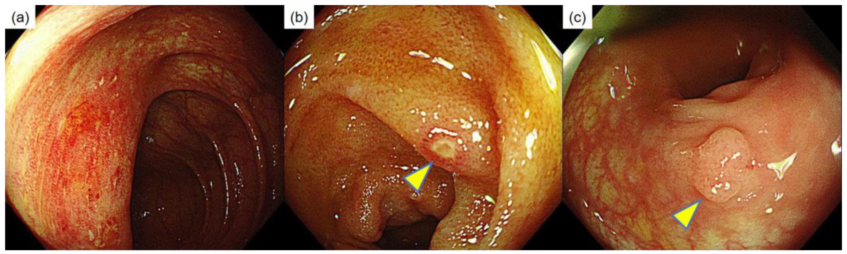
Figure 2.
Histopathological findings of the colorectal mucosa in the background biopsy. (a) Histological appearance of resolving stage UC. The biopsy showed crypt distortion and reduced crypt density; however, basal plasmacytosis and diffuse inflammatory cell infiltration in the stroma were not identified (bar = 100 μm). (b) An increase in eosinophils was observed in the stroma (bar = 20 μm). (c) Some crypts showed cryptitis (bar = 20 μm). (d) Histopathological findings of the SuSA of the rectum (bar = 100 μm). (e) Serration is confined to the upper one-third to one-half of the glands, whereas the deep crypt bases lack it (bar = 50 μm). (f) The proliferative zone is located in the basal to middle portions of the crypts, and the overall glandular architecture resembles that of a low-grade tubular adenoma (bar = 50 μm).
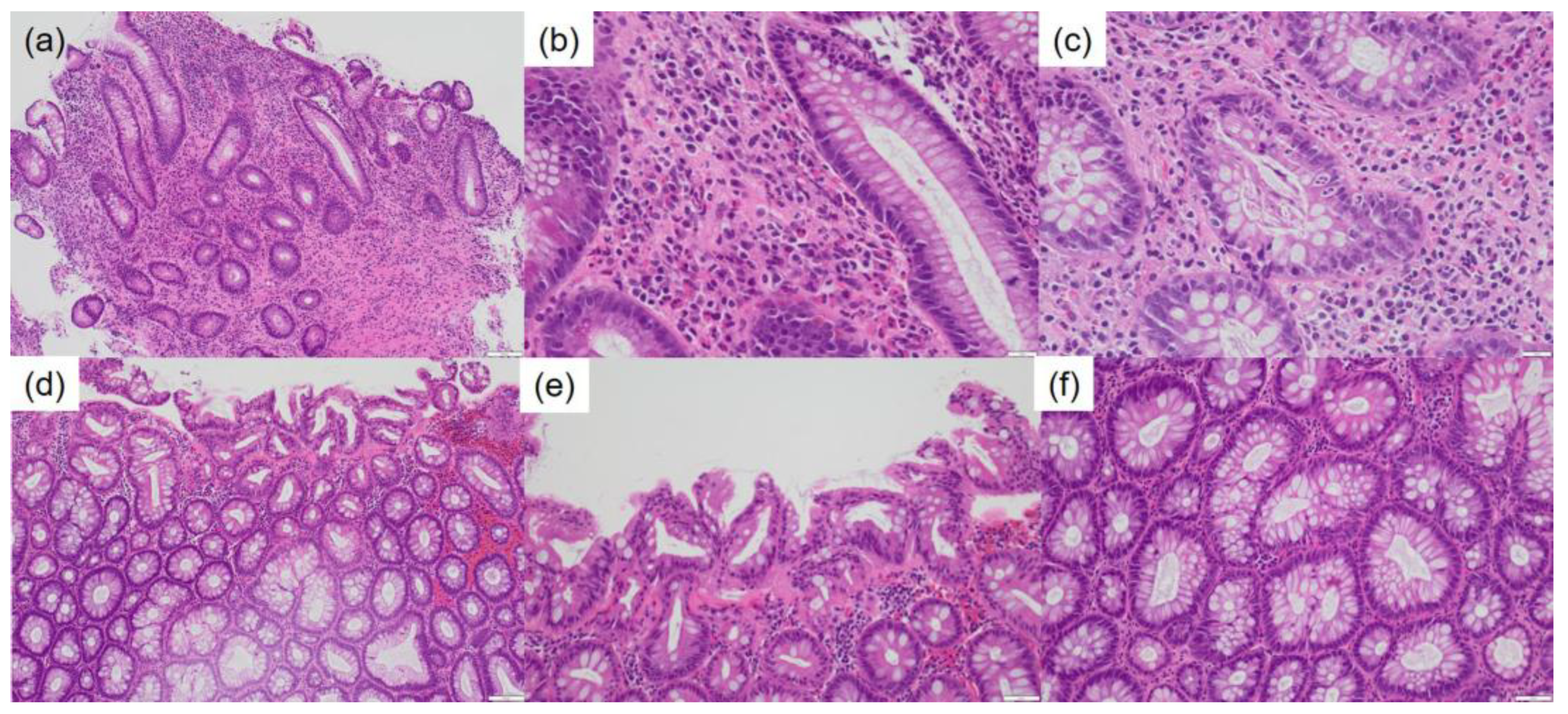
Figure 3.
Molecular findings. PTPRK::RSPO3 formed fusion genes at two junctions: between exon 1 of PTPRK and exon 2 of RSPO3, and between exon 1 of PTPRK and exon 3 of RSPO3.
Figure 3.
Molecular findings. PTPRK::RSPO3 formed fusion genes at two junctions: between exon 1 of PTPRK and exon 2 of RSPO3, and between exon 1 of PTPRK and exon 3 of RSPO3.
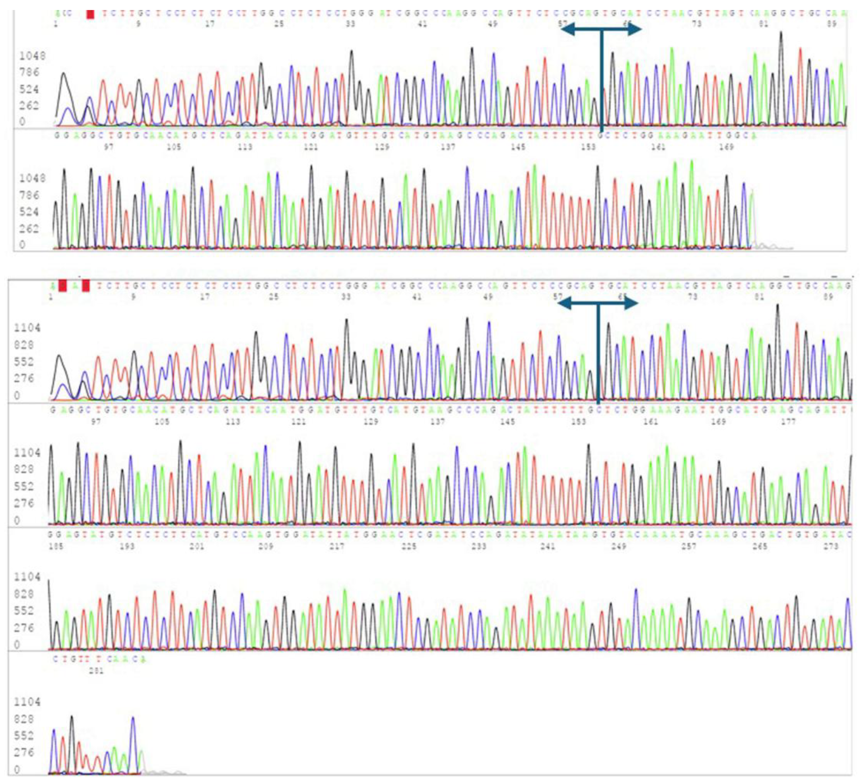

Table 1.
Clinicopathological and molecular features of superficially serrated adenomas.
| Author | Year | Age/sex | Location | Size | Morphology | Molecular findings | Follow up | ||
| (mm) | * | ** | *** | ||||||
| Hashimoto [1] | 2018 | 62/F | Rectum | 5 | N/A | RSPO3 fusion | KRAS mut | Negative | N/A |
| 68/F | Rectum | 15 | N/A | RSPO3 fusion | KRAS mut | Negative | N/A | ||
| 77/F | Sigmoid | 20 | N/A | RSPO3 overexpression | KRAS mut | Negative | N/A | ||
| 63/F | Rectum | 10 | N/A | RSPO3 overexpression | KRAS mut | Negative | N/A | ||
| 72/F | Sigmoid | 5 | N/A | RSPO3 fusion | KRAS mut | Negative | N/A | ||
| 62/M | Sigmoid | 18 | N/A | RSPO2 overexpression | KRAS mut | Negative | N/A | ||
| 66/M | Rectum | 5 | N/A | RSPO3 fusion | BRAF mut | Negative | N/A | ||
| 78/F | Transverse | 3 | N/A | RSPO3 fusion | KRAS mut | Negative | N/A | ||
| 79/M | Sigmoid | 4 | N/A | RSPO3 overexpression | KRAS mut | Negative | N/A | ||
| 69/M | Rectum | 9 | N/A | APC mut | KRAS mut | GNAS mut | N/A | ||
| 62/F | Ascending | 2 | N/A | RSPO3 overexpression | KRAS mut | Negative | N/A | ||
| 71/M | Sigmoid | 5 | N/A | RSPO3 fusion | KRAS mut | Negative | N/A | ||
| 65/F | Rectum | 9 | N/A | RSPO3 fusion | KRAS mut | Negative | N/A | ||
| 87/M | Rectum | 5 | N/A | RSPO3 fusion | KRAS mut | Negative | N/A | ||
| 83/M | Sigmoid | 2 | N/A | RSPO3 fusion | KRAS mut | Negative | N/A | ||
| 76/M | Rectum | 5 | N/A | RSPO3 overexpression | KRAS mut | Negative | N/A | ||
| 71/F | Rectum | 4 | N/A | RSPO3 fusion | KRAS mut | Negative | N/A | ||
| 64/F | Rectum | 4 | N/A | RSPO2 overexpression | KRAS mut | Negative | N/A | ||
| 72/M | Rectum | 3 | N/A | RSPO3 fusion | KRAS mut | Negative | N/A | ||
| 70/F | Sigmoid | 3 | N/A | APC mut | KRAS mut | Negative | N/A | ||
| Mizuguchi [10] | 2022 | 62/F | Rectum | 7 | Slightly elevated | RSPO3 fusion | KRAS mut | Negative | N/A |
| 68/F | Rectum | 15 | Slightly elevated | RSPO3 fusion | KRAS mut | Negative | N/A | ||
| 77/F | Sigmoid | 20 | Slightly elevated | RSPO3 overexpression | KRAS mut | Negative | N/A | ||
| 63/F | Rectum | 10 | Slightly elevated | RSPO3 overexpression | KRAS mut | Negative | N/A | ||
| 72/F | Sigmoid | 6 | Slightly elevated | RSPO3 fusion | KRAS mut | Negative | N/A | ||
| 62/M | Sigmoid | 20 | Slightly elevated | RSPO2 overexpression | KRAS mut | Negative | N/A | ||
| 66/M | Rectum | 5 | Slightly elevated | RSPO3 fusion | BRAF mut | Negative | N/A | ||
| 78/F | Transverse | 6 | Slightly elevated | RSPO3 fusion | KRAS mut | Negative | N/A | ||
| 79/M | Sigmoid | 4 | Slightly elevated | RSPO3 overexpression | KRAS mut | Negative | N/A | ||
| 69/M | Rectum | 9 | Slightly elevated | APC mut | KRAS mut | GNAS mut | N/A | ||
| 62/F | Ascending | 3 | Slightly elevated | RSPO3 overexpression | KRAS mut | Negative | N/A | ||
| 65/F | Rectum | 9 | Sessile | RSPO3 fusion | KRAS mut | Negative | N/A | ||
| 87/M | Rectum | 8 | Sessile | RSPO3 fusion | KRAS mut | Negative | N/A | ||
| 76/M | Rectum | 6 | Sessile | RSPO3 overexpression | KRAS mut | Negative | N/A | ||
| 71/F | Rectum | 5 | Sessile | RSPO3 fusion | KRAS mut | Negative | N/A | ||
| 64/F | Rectum | 6 | Slightly elevated | RSPO2 overexpression | KRAS mut | Negative | N/A | ||
| 72/M | Rectum | 3 | Slightly elevated | RSPO3 fusion | KRAS mut | Negative | N/A | ||
| 70/F | Sigmoid | 5 | Sessile | APC mut | KRAS mut | Negative | N/A | ||
| 71/F | Rectum | 9 | Slightly elevated | RSPO3 fusion | KRAS mut | Negative | N/A | ||
| 69/F | Descending | 10 | Slightly elevated | RSPO3 fusion | KRAS mut | Negative | N/A | ||
| 54/M | Rectum | 4 | Sessile | RSPO3 fusion | KRAS mut | Negative | N/A | ||
| 74/M | Sigmoid | 12 | Slightly elevated | RSPO3 fusion | KRAS mut | Negative | N/A | ||
| 71/M | Sigmoid | 18 | Sessile | RSPO3 fusion | KRAS mut | Negative | N/A | ||
| 80/F | Rectum | 5 | Sessile | RSPO2 overexpression | KRAS mut | Negative | N/A | ||
| 74/F | Sigmoid | 5 | Sessile | RSPO3 fusion | KRAS mut | Negative | N/A | ||
| Chen [12] | 2024 | 68/F | Sigmoid | 10 | Sessile | N/A | N/A | N/A | N/A |
| Gao [6] | 2025 | 38/M | Rectum | 21 | Slightly elevated | N/A | N/A | N/A | 11 months No recurrence |
| Present case | 2026 | 57/F | Rectum | 7 | Sessile | RSPO3 fusion | KRAS mut | Negative | 28 months No recurrence |
*WNT pathway gene alterations; **KRAS/BRAF mutations; ***GNAS mutations; F: female; M: male; N/A: not applicable; mut: mutation.
Disclaimer/Publisher’s Note: The statements, opinions and data contained in all publications are solely those of the individual author(s) and contributor(s) and not of MDPI and/or the editor(s). MDPI and/or the editor(s) disclaim responsibility for any injury to people or property resulting from any ideas, methods, instructions or products referred to in the content. |
© 2026 by the authors. Licensee MDPI, Basel, Switzerland. This article is an open access article distributed under the terms and conditions of the Creative Commons Attribution (CC BY) license.
Copyright: This open access article is published under a Creative Commons CC BY 4.0 license, which permit the free download, distribution, and reuse, provided that the author and preprint are cited in any reuse.