

Submitted:
14 August 2026
Posted:
18 August 2026
You are already at the latest version
Abstract
Chronic kidney disease (CKD) now affects roughly 850 million people worldwide, and both its prevalence and its death toll are expected to keep climbing over the next few decades. We understand CKD’s underlying biology far better than we did a generation ago, yet the tools used to catch it early haven’t kept pace, and the drugs available to slow its progression remain only partly effective. Extracellular vesicles (EVs)- the nanoscale, membrane-bound particles that nearly every cell type sheds into its surroundings have stepped into this gap as candidates for both diagnosis and treatment. Pairing EV biology with artificial intelligence has given rise to what we call “Artificial Intelligence–Virtual Extracellular Vesicles” (AIVEVs): a computational approach that uses deep learning and multi-omics integration to design, predict, and fine-tune EV-based interventions for kidney disease. This review walks through where AIVEV technology stands today, covering AI-driven biomarker discovery in urinary and circulating EVs, computationally guided engineering of kidney-targeted therapeutic EVs, machine learning strategies for classifying EV subtypes, generative approaches to virtual EV design, physiologically based pharmacokinetic (PBPK) modeling for renal drug delivery, and the regulatory groundwork still needed before any of this reaches the clinic. As the global CKD burden grows and AI methods mature in parallel, AIVEVs look increasingly like a genuine shift in how nephrologists might one day diagnose, stage, and treat kidney disease.
Keywords:
artificial intelligence
; extracellular vesicles
; chronic kidney disease
; biomarkers
; drug delivery
; machine learning
; precision medicine
; exosomes
; microvesicles
Introduction
Chronic kidney disease used to sit fairly low on the list of global health priorities. That has changed. Data from the Global Burden of Disease Study 2023 put CKD’s reach at nearly 800 million adults worldwide, with an age-standardized prevalence of about 14% among people 20 and older [1].
By 2023, CKD had become the ninth leading cause of death globally, and current projections suggest it could climb to third place in Western Europe by 2050 if nothing changes course [2]. The burden isn’t spread evenly, North Africa and the Middle East report the highest rates (18.0%), followed by South Asia (15.8%) and sub-Saharan Africa (15.6%), a pattern that reflects a tangle of genetic, environmental, and socioeconomic factors rather than any single cause [3].
The gaps in how we currently manage CKD are well known by now. Serum creatinine–based estimated glomerular filtration rate (eGFR) and the urine albumin-to-creatinine ratio (UACR) remain the workhorses of clinical practice, but both only flag kidney damage after a meaningful chunk of function has already been lost. What nephrology actually needs are biomarkers that can catch tubular injury early, forecast how a given patient’s disease will progress, and steer treatment accordingly. Just as pressing is the need for therapies that target the processes driving decline , fibrosis, chronic inflammation, endothelial dysfunction ,without the collateral damage that comes with conventional, systemically delivered drugs.
Extracellular vesicles have drawn a great deal of attention as a possible answer to both problems at once. These are heterogeneous nanoparticles, ranging anywhere from 30 nm to 5 μm, and they’re grouped by how they’re made: exosomes (30–150 nm, born from the endosomal pathway), microvesicles (100–1000 nm, budded directly off the plasma membrane), and apoptotic bodies (1–5 μm) [4]. Their cargo, proteins, lipids, metabolites, and nucleic acids, including miRNAs, mRNAs, and various non-coding RNAs ,carries a kind of molecular signature of whatever cell released them, healthy or not. In the kidney specifically, urinary EVs (uEVs) function almost like a liquid biopsy, picking up signals from the glomeruli, the tubules, and the interstitium alike [5].
Where things get interesting is at the intersection of EV biology and AI. Machine learning can now sort EV subpopulations out of messy imaging data, predict disease states from layered multi-omic EV profiles, and even design engineered EVs with a target therapeutic cargo already baked into the model [6]. Deep learning architectures ;convolutional neural networks (CNNs) for image work, transformer models for sequence data have turned in genuinely strong results on EV-related tasks [7]. And generative approaches, variational autoencoders (VAEs) and diffusion models among them, allow researchers to computationally design “virtual” EVs with predicted properties before anything touches a bench. We refer to this whole cluster of methods as Artificial Intelligence–Virtual Extracellular Vesicles, or AIVEVs.
This review takes stock of where AIVEV technology stands as it applies to CKD. We look at recent progress in AI-enabled EV biomarker discovery, at computational approaches to engineering therapeutic EVs, at the role PBPK modeling plays in renal drug delivery, and at what still needs to happen regulatory and otherwise before AIVEV-based interventions can move from the lab into routine clinical use.
The Global CKD Burden and Unmet Needs
Understanding just how large the CKD epidemic has grown helps put the promise of AIVEV technology in context. The Global Burden of Disease Study 2021 counted 359 million prevalent cases worldwide, along with 11.13 million new diagnoses, 1.53 million deaths, and 44.45 million disability-adjusted life years (DALYs) attributable to the disease in that year alone [8]. Prevalence crept up by about 0.92% annually between 1990 and 2021, but mortality rose more than twice as fast, at 2.66% a year , a sign that even as supportive care has improved, CKD itself has grown deadlier over time [9].

Table 1.
Global Epidemiology of Chronic Kidney Disease (2021–2023).
| Parameter | Estimate | Source/Year |
| Global prevalence | ~850 million adults | GBD 2023 [1] |
| Age-standardized prevalence | 14% (adults ≥20 years) | GBD 2023 [1] |
| Annual deaths | 1.5 million (one every 20 seconds) | WHO/KDIGO 2025 [2] |
| Patients on kidney replacement therapy | 4.6 million | ERA Registry 2025 [2] |
| Projected prevalence rate (2032) | 8,774 per 100,000 | GBD projection [9] |
| Highest regional prevalence | North Africa & Middle East (18.0%) | GBD 2023 [3] |
The forces pushing CKD prevalence upward — an aging population, rising rates of obesity and diabetes, more widespread hypertension , hit hardest in low- and middle-income countries, precisely where access to kidney replacement therapy is thinnest. That mismatch is really the crux of the problem: the places that need early detection and disease-modifying treatment most are also the places least equipped to deliver either at scale [10]. (Figure 1)
On the treatment side, current CKD management still leans heavily on renin-angiotensin-aldosterone system inhibitors, SGLT2 inhibitors, and mineralocorticoid receptor antagonists. These drugs slow the disease down; none of them stop it. For patients who progress all the way to end-stage kidney disease, dialysis and transplantation carry enormous personal and financial costs. Developing therapies that can actually interrupt fibrotic signaling, encourage tubular regeneration, and restore some immune balance remains one of nephrology’s biggest open problems [11,12,13].
Extracellular Vesicles in CKD: Biology and Clinical Relevance
EV Biogenesis and Heterogeneity
EVs aren’t one thing ;they’re a mixed population with different origins, sizes, surface markers, and functions. Exosomes form inside the endosomal system, budding inward within multivesicular bodies (MVBs) before those bodies fuse with the plasma membrane and release their contents. Microvesicles take a more direct route, budding straight off the plasma membrane’s outer surface. Apoptotic bodies are the largest of the three, released as fragments during programmed cell death [14].
This diversity cuts both ways for CKD research. Different EV subpopulations trace back to different renal cell types ; podocytes, tubular epithelial cells, endothelial cells, interstitial fibroblasts ; so in principle each carries a molecular fingerprint specific to whatever compartment produced it. In practice, though, EV subtypes overlap physically with lipoproteins and protein aggregates enough to make clean isolation and characterization genuinely difficult [6].
EVs as Diagnostic Biomarkers in CKD
The case for EVs as diagnostic tools in CKD has been building for a while. Urinary EVs are especially appealing since they can be collected without any invasive procedure and are naturally enriched for kidney-derived vesicles. Some of the more established EV-associated biomarkers for CKD include:
Table 2.
EV-Derived Biomarkers for CKD Diagnosis and Prognosis.
| Biomarker | EV Source | Expression in CKD | Clinical Application | Key Reference |
| miR-21 | Urinary exosomes | Upregulated | Fibrosis severity, progression marker | [16] |
| miR-29c | Urinary exosomes | Downregulated | Tubulointerstitial fibrosis prediction | [17] |
| miR-200b | Urinary exosomes | Downregulated | Early and late CKD staging | [18] |
| KIM-1 | Urinary EVs | Upregulated | Tubular injury, AKI-to-CKD transition | [19] |
| NGAL | Urinary EVs | Upregulated | Acute kidney injury, CKD progression | [20] |
| CCL2 mRNA | Tubular epithelial cell EVs | Upregulated | Interstitial inflammation, proteinuria | [21] |
| EGF | Urinary EVs | Downregulated | GFR decline prediction | [22] |
| PON1 | Serum EVs | Downregulated | CKD progression, cardiovascular risk | [5] |
| circRNA | Urinary exosomes | Correlated with fibrosis degree | Renal fibrosis specificity | [23] |
| CKD273 peptide panel | Urinary EVs/peptides | Differential pattern | CKD progression prediction | [24] |
Part of what makes miRNAs such durable biomarkers is that the lipid bilayer surrounding an EV shields them from RNase degradation, so they hold up well in stored or transported samples. The proteins anchored to EV surfaces offer a similar advantage, giving affinity-based assays something stable to grab onto. Even so, getting these biomarkers into actual clinical use has been slow going, mostly because of pre-analytical variability, a lack of standardized isolation protocols, and thin validation across large, diverse patient cohorts [25].
EVs as Therapeutic Agents in CKD
Beyond diagnostics, EVs — particularly those derived from mesenchymal stem cells (MSCs) — have shown real promise as treatments in preclinical CKD models. MSC-derived EVs (MSC-EVs) appear to help injured kidneys through several overlapping mechanisms: dialing down inflammation and apoptosis, encouraging angiogenesis and cell proliferation, reducing oxidative stress, and shifting immune cell polarization toward a less destructive state [26].
Recent single-cell RNA sequencing work has started to clarify how this actually happens at the cellular level. In one diabetic kidney disease (DKD) model, MSC-derived small EVs were shown to slow renal fibrosis by delivering CK1δ/β-TRCP, which pushes YAP toward ubiquitination and degradation and, in doing so, interrupts the TGF-β1/Smad2/3/YAP signaling axis that drives fibrosis [27].
That same study also flagged fibrosis-associated macrophages as key players in DKD progression, noting that MSC-EV treatment helped restore a more normal distribution of renal cell populations.
This isn’t purely a preclinical story anymore, either. In one landmark trial, 20 patients with stage III or IV CKD received two doses of umbilical cord MSC-EVs (100 μg/kg). Over a year of follow-up, they showed improved GFR, lower serum creatinine and blood urea nitrogen, reduced albuminuria, and more favorable circulating cytokine profiles — with no significant adverse effects reported [26]. It’s a small study, but it’s proof that EV-based CKD therapy can work in people, not just in animal models.
Artificial Intelligence in EV Research: Foundational Concepts
AI has been brought to bear on EV research largely because the field has run into a handful of stubborn problems: EV populations are extraordinarily heterogeneous, the omics data describing them is high-dimensional, image-based characterization is technically demanding, and any useful model needs to generalize across widely varying experimental conditions [6].
Machine Learning for EV Classification and Biomarker Discovery
Combining several of these approaches tends to outperform any single method on its own. In hepatocellular carcinoma work, for instance, a DNA cascade reaction–triggered individual EV nanoencapsulation (DCR-IEVN) assay paired with linear discriminant analysis, SVM, and logistic regression reached 93.3% overall accuracy in separating cancer patients from cirrhotic and healthy controls, beating out conventional biomarkers in the process. In a similar vein, total internal reflection fluorescence (TIRF) imaging combined with deep learning hit 100% prediction accuracy for cancer classification based on single-vesicle miRNA profiling [28].
Table 3.
AI/ML Techniques Applied to EV Research in CKD.
| AI Model | Application Domain | Primary Use Case | Data Modality | Representative Performance |
| Convolutional Neural Networks (CNNs) | EV imaging | Automated detection, segmentation, phenotypic characterization | TEM, cryo-EM, fluorescence microscopy | >95% classification accuracy for cancer EV subtypes [28] |
| Random Forests (RF) | Multi-omics | Biomarker discovery, feature importance ranking | Proteomics, transcriptomics, lipidomics | AUROC 0.82–0.88 for CKD progression prediction [29] |
| XGBoost | Clinical + biomarker data | Risk stratification, progression modeling | Structured clinical data, EV biomarker panels | AUROC 0.84–0.87, externally validated [29] |
| Support Vector Machines (SVM) | Flow cytometry, proteomics | EV phenotype classification, biomarker detection | High-dimensional flow, peptide fingerprinting | >90% accuracy for tumor EV detection [6] |
| Variational Autoencoders (VAEs) | Generative modeling | Virtual EV design, latent feature learning | Multi-modal EV characterization | Latent representations for cargo prediction [30] |
| Graph Neural Networks (GNNs) | Molecular interaction | Receptor-ligand prediction, targeting optimization | Protein-protein interaction networks | Emerging applications in EV engineering [30] |
| Transformers | Genomics, proteomics | Sequence interpretation, contextual embedding | Long-read sequencing, proteogenomics | State-of-the-art for sequence-based EV cargo prediction [6] |
| U-Net | Imaging | Semantic segmentation of EVs in complex backgrounds | Cryo-EM, super-resolution microscopy | Precise boundary delineation for size/shape analysis [6] |
Deep Learning for Single-Vesicle Analysis
Technologies for analyzing individual vesicles ;nanoparticle tracking analysis (NTA), single-particle interferometric reflectance imaging (SP-IRIS), high-sensitivity flow cytometry — now generate data at a resolution that was simply unavailable a decade ago. Deep learning is well suited to this kind of data. Autoencoders and VAEs learn compressed latent representations that treat phenotype as a continuous landscape rather than forcing vesicles into rigid bins, which makes it possible to pick out rare EV subpopulations ,sometimes less than 0.1% of the total vesicle count , that nonetheless carry outsized biological significance [30].
Attention-based transformers and graph neural networks are starting to be used to learn the relationship between phenotype and function directly from integrated data, which means predicting what a novel EV subset does biologically without necessarily running a separate functional assay for it. That alone could meaningfully speed up how quickly new biomarker candidates get screened [30].
Table 4.
AI Applications in Therapeutic EV Engineering for CKD.
| Engineering Stage | AI Approach | Application | Expected Outcome |
| Target identification | scRNA-seq + pathway mining | Identify dysregulated pathways and receptors in CKD kidneys | Precision target selection for cargo and surface modification |
| Cargo selection | NLP on biomedical literature + GNNs | Predict optimal miRNA/protein cargo for pathway modulation | Enhanced therapeutic efficacy |
| Surface modification | Molecular docking + RL | Design targeting peptides (e.g., KIM-1, RGD) for renal accumulation | Increased kidney-to-liver ratio |
| Loading optimization | VAEs + experimental design | Predict optimal loading conditions (electroporation, sonication) | Higher cargo-to-EV ratio |
| Stability prediction | Physics-informed neural networks | Model EV membrane dynamics, protein corona formation | Extended circulation time |
| PK/PD modeling | PBPK + ML | Predict tissue distribution, clearance, dose-response | Optimized dosing regimens |
| Safety profiling | Toxicity prediction models | Assess immunogenicity, off-target effects | Reduced adverse events |
Table 5.
Key Challenges and AI-Driven Solutions in AIVEV Clinical Translation.
| Challenge | Traditional Limitation | AI-Driven Solution | Implementation Status |
| EV isolation standardization | High inter-laboratory variability | ML-based quality control and batch effect correction | Emerging (MISEV2023 guidelines) |
| EV population heterogeneity | Bulk analysis masks rare subpopulations | Deep learning subtype classification; single-vesicle analysis | Validated in cancer; CKD application ongoing |
| Limited clinical validation data | Small cohorts, single-center studies | Federated learning across institutions; synthetic data generation | Pilot programs initiated |
| Kidney-specific targeting | Low therapeutic index | GNN-based receptor mapping; computational peptide design | Preclinical validation |
| Manufacturing scalability | Batch-to-batch inconsistency | Process analytical technology (PAT) with real-time ML monitoring | Industry adoption beginning |
| Regulatory approval pathways | Unclear classification (biologic vs. drug vs. device) | Explainable AI for regulatory submission; digital evidence packages | Guideline development in progress |
| Cost-effectiveness | High production costs | AI-optimized bioprocess design; predictive maintenance | Economic modeling phase |
AIVEVs for CKD Diagnosis: From Biomarker Panels to Digital Pathology
Multi-Omics Integration and AI-Enhanced Biomarker Discovery
Combining multi-omics data with EV analysis is one of the more promising directions for CKD biomarker discovery. Proteomic profiling of EVs can surface proteins tied to inflammation, oxidative stress, extracellular matrix remodeling, and metabolic dysfunction ,all pathways that matter for how CKD progresses [31].
Transcriptomic data adds another layer, revealing EV-associated miRNAs and non-coding RNAs that regulate gene expression in whatever cell picks the EV up. Metabolomic and lipidomic profiling round out the picture further.
None of this is useful without a way to integrate it, which is where AI comes in. Multi-omics factor analysis (MOFA) and related methods can pull out latent variables that capture variation shared across data types, while deep learning architectures build hierarchical representations that move progressively from raw molecular features up to disease-relevant patterns. The DIABLO framework (Data Integration Analysis for Biomarker discovery using Latent variable approaches for Omics studies) is a good example of this kind of integrative approach, and it has helped identify multi-omic signatures with stronger predictive power than any single data type could provide on its own [30].
AI-Powered Risk Stratification and Progression Modeling
Several groups have already shown that AI models built on EV biomarkers can meaningfully stratify CKD risk. Bienaimé and colleagues, for example, applied LASSO logistic regression and random forest ensembles to five validated urinary biomarkers ; CCL2, EGF, KIM-1, NGAL, and TGF-α , alongside standard clinical variables, and reached an AUROC of 0.88 for predicting rapid CKD progression (defined as a ≥40% eGFR decline or progression to end-stage kidney disease) in their derivation cohort, with 0.83 holding up in external validation. The CKD273 urinary peptide classifier, built using capillary electrophoresis–mass spectrometry and tested across more than 1,200 samples, achieved AUROCs between 0.85 and 0.93 for progression prediction [32].
More recently, XGBoost models that combine EV-associated biomarkers (TNFR1, TNFR2, KIM-1) with electronic health record data have reached AUROCs of 0.87 in derivation and 0.85 in external validation for predicting progression to kidney failure in diabetic kidney disease. The Klinrisk model, which relies on routine clinical features fed into an XGBoost architecture, has posted AUROCs of 0.84–0.86 across two randomized controlled trial cohorts [32]. (Figure 2)
Digital Pathology and Computational Renal Histology
It’s worth mentioning AI-enhanced renal histopathology even though it isn’t strictly EV-based, because it complements EV diagnostics by supplying the spatial context that molecular data alone can’t. Convolutional neural networks trained on periodic acid–Schiff–stained whole-slide images have reached AUCs above 0.80 for grading diabetic nephropathy severity and for picking out specific lesions like glomerulosclerosis and Kimmelstiel-Wilson nodules [32]. Bringing histological AI together with EV biomarker profiles ,something that might fairly be called “multi-modal AIVEV diagnostics” , looks like one of the more promising paths toward a genuinely comprehensive picture of CKD.
AIVEVs for CKD Treatment: Computational Design of Therapeutic EVs
The Therapeutic EV Engineering Pipeline
Turning EVs into real therapeutics for CKD means solving several engineering problems at once: getting cargo loaded efficiently, making the EVs home in on the kidney specifically, keeping them in circulation long enough to work, and manufacturing all of it at scale. AI is being applied at nearly every stage of that pipeline [6]. (Figure 3)
Kidney-Targeted EV Surface Engineering
Getting a therapeutic dose into the kidney while keeping systemic exposure low is still one of the harder problems here. A few AI-guided targeting strategies have emerged as front-runners:
KIM-1 Targeting. Kidney injury molecule-1 (KIM-1/HAVCR1) is strongly upregulated on injured tubular epithelial cells, which makes it a natural target for CKD therapy. Red blood cell–derived EVs conjugated with KIM-1–targeting LTH peptides have shown enhanced accumulation at sites of renal tubular injury [4]. AI-driven peptide design can push binding affinity and specificity higher while keeping immunogenicity down.
RGD Peptide Engineering. The arginine-glycine-aspartic acid (RGD) motif binds integrins found on activated endothelial cells and fibroblasts in fibrotic kidneys. Supramolecular nanofibers carrying RGD peptides have improved MSC-EV delivery of let-7a-5p miRNA, reducing apoptosis and activating autophagy via CASP3 and RragD [14]. Generative AI models can design novel RGD variants with better pharmacokinetic properties than the naturally occurring peptide.
Neutrophil Membrane Camouflage. EVs engineered with neutrophil membranes borrow the natural homing instincts neutrophils have for inflamed tissue. These hybrid vesicles concentrate noticeably in damaged renal tissue and have improved outcomes in acute kidney injury models [14]. AI models trained on neutrophil surface proteomics can help fine-tune membrane composition for CKD-specific applications.
Glycoengineering. Glycoengineered stem cell–derived EVs represent a newer approach, using optimized carbohydrate-mediated recognition of tubular epithelial cells and macrophages to achieve targeted therapy for acute kidney injury [33]. Machine learning models that predict glycan-receptor interactions can guide these glycoengineering strategies rather than leaving them to trial and error.
Generative Models for Virtual EV Design
The idea of a “virtual” EV , a computational stand-in that can be designed, tested, and refined in silico before anything reaches a lab bench ,represents a real shift in how therapeutic development could work. VAEs and generative adversarial networks (GANs) can learn the latent distribution of EV properties from existing experimental data and then generate new configurations predicted to work better [6].
A generative AI platform built for EV design would likely draw on:
- Protein structure prediction (AlphaFold) for modeling cargo and surface proteins
- Molecular dynamics simulations to predict membrane behavior
- Reinforcement learning for balancing competing objectives ,targeting, loading, stability, at once
- Digital twin frameworks that simulate how an individual patient might respond to a given engineered EV therapy
Together, these tools could cut down substantially on the experimental legwork needed at each iteration, potentially shrinking development timelines for personalized formulations from around 18 months down to 6–8 [34].
Physiologically Based Pharmacokinetic Modeling for Renal EV Delivery
Physiologically based pharmacokinetic (PBPK) modeling gives researchers a mechanistic way to predict how EVs are absorbed, distributed, metabolized, and excreted ,which matters enormously when it comes to translating results from animal studies into safe human dosing [35].
PBPK Model Structure for EVs
A whole-body PBPK model for EVs typically includes compartments for the major organs , kidney, liver, spleen, lung, heart, brain ,connected by blood flow. Key parameters feeding into such a model include:
- EV size distribution and zeta potential, both of which affect opsonization and clearance
- Organ blood flow rates and vascular permeability
- Macrophage-mediated uptake kinetics, the dominant clearance mechanism
- Renal filtration threshold (roughly 6–8 nm; most EVs are larger than this, which limits how much glomerular filtration actually contributes to their clearance)
For EVs specifically engineered to target the kidney, the model also needs to account for:
- Peritubular capillary fenestration, which allows access to the interstitial space
- Tubular epithelial cell uptake through endocytosis
- Excretion of intraluminal urinary EVs
For early-stage development, a simplified three-compartment model , circulation, target tissue, non-target tissue , with first-order clearance and sigmoidal concentration-effect relationships is usually enough to capture the essential dynamics [35].
AI-Enhanced PBPK Parameterization
Machine learning helps address several persistent PBPK challenges specific to EVs:
- Parameter estimation: Bayesian neural networks can infer tissue partition coefficients even from sparse experimental data
- Allometric scaling: ML models improve how well findings translate across species by learning nonlinear scaling relationships that simple formulas miss
- Population variability: Gaussian process models capture how much EV pharmacokinetics vary between individual subjects
- Disease state effects: Deep learning can predict how CKD-altered physiology — reduced renal blood flow, uremic toxins interfering with membrane interactions — changes how EVs behave in the body
Clinical Translation: Challenges and Regulatory Considerations
The Translation Funnel
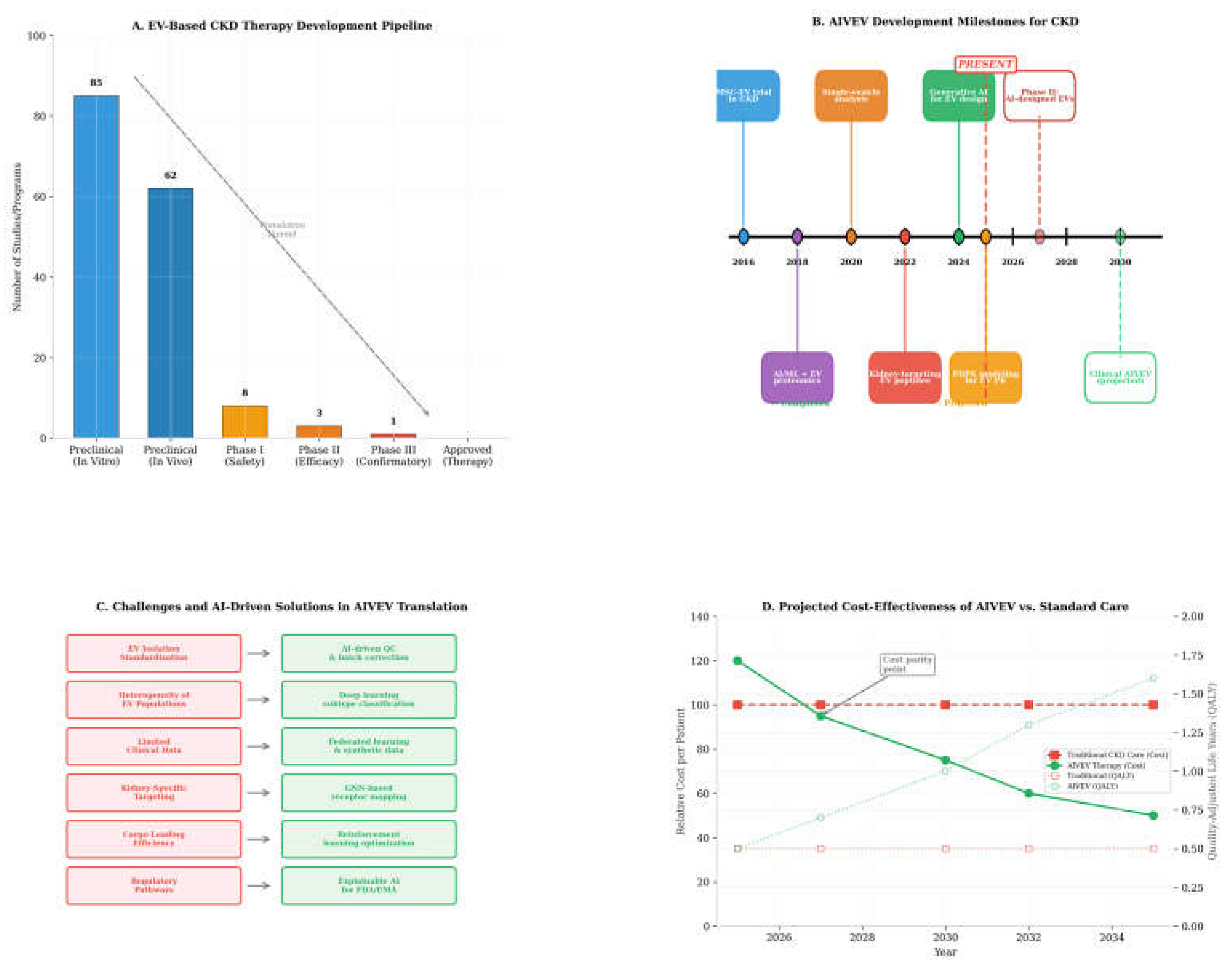
Even with encouraging preclinical results, moving EV-based therapies toward approved CKD treatments remains genuinely difficult. Of roughly 85 preclinical in vitro studies and 62 in vivo programs, only 8 have made it to Phase I trials, 3 to Phase II, and just 1 to Phase III — and no EV-based CKD therapy has been approved yet [36,37,38,39]. (Figure 4) That attrition rate reflects both the inherent complexity of biological therapeutics and the fact that EV manufacturing and its regulatory framework are both still relatively young.
Regulatory Pathways
The regulatory landscape for EV-based therapeutics is still taking shape. The International Society for Extracellular Vesicles (ISEV) has put out MISEV2023 guidelines calling for better data annotation, orthogonal quantification methods, and clearer validation metrics [6].
Where AI/ML components are involved, regulators are working out frameworks for software as a medical device (SaMD), with particular attention to:
- Algorithm transparency and explainability
- Whether training data actually represents the populations it will be used on, and how bias gets mitigated
- Protocols for continuous learning and model updating over time
- How AI and clinicians are meant to interact in a decision-support setting
Explainable AI (XAI) methods , SHAP (SHapley Additive exPlanations) values for feature importance, attention visualization for deep learning models — will likely be essential for earning both regulatory approval and clinical trust [6].
Future Directions and Concluding Perspectives
The AIVEV paradigm sits at the intersection of several trends that happen to be converging at once: deeper understanding of EV biology, a steady rise in available computational power, increasingly capable deep learning architectures, and a growing recognition that CKD deserves to be treated as a global health priority. A few developments seem likely to shape where the field goes over the next decade:
Digital Twins for Precision Nephrology. Bringing together a patient’s multi-omic data, imaging, and clinical history into a single computational model would allow AIVEV therapies to be tested virtually before ever being administered. Digital twin approaches like this are already being explored in oncology and cardiovascular medicine, and something similar could help identify the right EV formulation for a given CKD patient based on their molecular disease subtype, pharmacogenomic profile, and predicted response to treatment [6].
Federated Learning Networks. Because some CKD etiologies are relatively rare and training data needs to be diverse to be useful, federated learning — training AI models across multiple institutions without ever centralizing patient data , is likely to speed up both biomarker validation and therapeutic optimization while keeping patient privacy intact [41].
Real-World Evidence Integration. Feeding electronic health record data, wearable device outputs, and patient-reported outcomes into AIVEV models opens the door to dynamic treatment adjustment and better long-term outcome prediction. Convolutional autoencoder models applied to longitudinal EMR time-series data have already shown this is feasible, having been used to predict worsening diabetic kidney disease [29].
Synthetic Biology and Cell-Free Manufacturing. Progress in cell-free EV production, paired with AI-optimized bioprocess parameters, could eventually make therapeutic EV manufacturing scalable and genuinely affordable. Cutting production costs through AI-guided process optimization will matter most in resource-limited settings, which is precisely where the CKD burden is heaviest.
Taken together, AIVEVs represent a real attempt to bring artificial intelligence and nanoscale biological therapeutics together in a way that could actually change outcomes. For the hundreds of millions of people living with CKD worldwide, that convergence offers something concrete to hope for: earlier detection, sharper risk stratification, and treatment that finally does more than just slow the disease down. Getting there will take sustained investment in interdisciplinary research, real international cooperation on data sharing and standardization, and a regulatory approach that takes innovation seriously without losing sight of patient safety. It will take clinicians, scientists, data engineers, and patient advocates working together ,the whole kidney community ,to carry AIVEV technology from computational concept to something that actually reaches patients.
Author Contributions
K.D. (Kumar Digvijay): Conceptualization, methodology, investigation, writing, original draft, writing, review and editing, visualization, project administration;C.R. (Claudio Ronco): Conceptualization, supervision, writing,review and editing,H.B. (Henrik Birn): Writing,review and editing, supervision. All authors have read and agreed to the published version of the manuscript.
Funding
This research received no external funding.
Institutional Review Board Statement
Not applicable. This is a narrative review article based entirely on previously published data and does not involve new human subject research.
Informed Consent Statement
Not applicable.
Data Availability Statement
No new data were created or analyzed in this study. Data sharing is not applicable to this article.
Conflicts of Interest
The authors declare no conflicts of interest.
Abbreviations
AI — Artificial Intelligence
AIVEV(s) — Artificial Intelligence–Virtual Extracellular Vesicle(s)
AKI — Acute Kidney Injury
AUC — Area Under the Curve
AUROC — Area Under the Receiver Operating Characteristic curve
CCL2 — C-C motif Chemokine Ligand 2
circRNA — Circular RNA
CKD — Chronic Kidney Disease
CKD273 — A 273-peptide urinary classifier panel for CKD progression prediction
CNN(s) — Convolutional Neural Network(s)
cryo-EM — Cryogenic Electron Microscopy
DALYs — Disability-Adjusted Life Years
DCR-IEVN — DNA Cascade Reaction-triggered Individual EV Nanoencapsulation
DIABLO — Data Integration Analysis for Biomarker discovery using Latent variable approaches for Omics studies
DKD — Diabetic Kidney Disease
EGF — Epidermal Growth Factor
eGFR — estimated Glomerular Filtration Rate
EMR — Electronic Medical Record
ERA — European Renal Association
EV(s) — Extracellular Vesicle(s)
GBD — Global Burden of Disease (Study)
GFR — Glomerular Filtration Rate
GNN(s) — Graph Neural Network(s)
HAVCR1 — Hepatitis A Virus Cellular Receptor 1 (gene encoding KIM-1)
ISEV — International Society for Extracellular Vesicles
KDIGO — Kidney Disease: Improving Global Outcomes
KIM-1 — Kidney Injury Molecule-1
LASSO — Least Absolute Shrinkage and Selection Operator
LTH peptide — KIM-1-targeting peptide sequence used for EV surface conjugation
MISEV2023 — Minimal Information for Studies of Extracellular Vesicles (2023 guidelines)
ML — Machine Learning
MOFA — Multi-Omics Factor Analysis
mRNA — messenger RNA
MSC(s) — Mesenchymal Stem Cell(s)
MSC-EV(s) — Mesenchymal Stem Cell-derived Extracellular Vesicle(s)
MVBs — Multivesicular Bodies
NGAL — Neutrophil Gelatinase-Associated Lipocalin
NLP — Natural Language Processing
NTA — Nanoparticle Tracking Analysis
PAS — Periodic Acid–Schiff (stain)
PAT — Process Analytical Technology
PBPK — Physiologically Based Pharmacokinetic
PD — Pharmacodynamic
PK — Pharmacokinetic
PON1 — Paraoxonase 1
RF — Random Forest
RGD — Arginine-Glycine-Aspartic acid (peptide motif)
RL — Reinforcement Learning
RNase — Ribonuclease
SaMD — Software as a Medical Device
scRNA-seq — single-cell RNA sequencing
SGLT2 — Sodium-Glucose Cotransporter-2
SHAP — SHapley Additive exPlanations
SP-IRIS — Single-Particle Interferometric Reflectance Imaging Sensor
SVM — Support Vector Machine
TEM — Transmission Electron Microscopy
TGF-α — Transforming Growth Factor alpha
TGF-β1 — Transforming Growth Factor beta 1
TIRF — Total Internal Reflection Fluorescence
TNFR1 / TNFR2 — Tumor Necrosis Factor Receptor 1 / 2
UACR — Urine Albumin-to-Creatinine Ratio
uEVs — urinary Extracellular Vesicles
UMAP — Uniform Manifold Approximation and Projection
VAE(s) — Variational Autoencoder(s)
WHO — World Health Organization
XAI — Explainable Artificial Intelligence
XGBoost — eXtreme Gradient Boosting
YAP — Yes-Associated Protein
References
- GBD 2023 Chronic Kidney Disease Collaborators. Global, regional, and national burden of chronic kidney disease in adults, 1990-2023, and its attributable risk factors: A systematic analysis for the Global Burden of Disease Study 2023. Lancet 2025, 406, 2461–2482. [Google Scholar] [CrossRef] [PubMed]
- Ortiz, A.; Lees, J.S.; Torra, R.; Stel, V.S.; Kramer, A.; Mark, P.B. The updated global burden of chronic kidney disease: One death every 20 seconds. Nephrol. Dial. Transplant. 2026. [Google Scholar] [CrossRef] [PubMed]
- GBD 2023 Kidney Failure with Replacement Therapy Collaborators. Global, regional, and national prevalence of kidney failure with replacement therapy and associated aetiologies, 1990-2023: A systematic analysis for the Global Burden of Disease Study 2023. Lancet Glob. Health 2025, 13, e1378–e1395. [CrossRef] [PubMed]
- Zheng, Y.; Wang, H.; Li, X.; Xie, J.; Fan, J.; Ren, S. Extracellular vesicles in chronic kidney disease: Diagnostic and therapeutic roles. Front Pharmacol. 2024, 15, 1371874. [Google Scholar] [CrossRef] [PubMed]
- Garcia, N.A.; Gonzalez-King, H.; Mellergaard, M.; Nair, S.; Salomon, C.; Handberg, A. Comprehensive strategy for identifying extracellular vesicle surface proteins as biomarkers for chronic kidney disease. Front Physiol. 2024, 15, 1328362. [Google Scholar] [CrossRef] [PubMed]
- Tiwari, A.; Widodo Krisnawati, D.I.; Tzou, K.Y.; Kuo, T.R. Machine learning for extracellular vesicles enables diagnostic and therapeutic nanobiotechnology. J. Nanobiotechnology 2026, 24, 153. [Google Scholar] [CrossRef] [PubMed]
- Tran, H.L.; Zheng, W.; Issadore, D.A.; et al. Extracellular Vesicles for Clinical Diagnostics: From Bulk Measurements to Single-Vesicle Analysis. ACS Nano 2025, 19, 28021–28109. [Google Scholar] [CrossRef] [PubMed]
- Guo, J.; Liu, Z.; Wang, P.; et al. Global, regional, and national burden inequality of chronic kidney disease, 1990-2021: A systematic analysis for the global burden of disease study 2021. Front. Med. 2025, 11, 1501175. [Google Scholar] [CrossRef] [PubMed]
- Xie, K.; Cao, H.; Ling, S.; et al. Global, regional, and national burden of chronic kidney disease, 1990-2021: A systematic analysis for the global burden of disease study 2021. Front. Endocrinol. 2025, 16, 1526482. [Google Scholar] [CrossRef] [PubMed]
- Ameh, O.I.; Ekrikpo, U.E.; Kengne, A.P. Preventing CKD in Low- and Middle-Income Countries: A Call for Urgent Action. Kidney Int. Rep. 2019, 5, 255–262. [Google Scholar] [CrossRef] [PubMed]
- Muhammad Daniyal, S.; Ajaz, H.; Riaz, M.; et al. Combination therapy with mineralocorticoid receptor antagonists and SGLT2 inhibitors versus SGLT2 inhibitor monotherapy in chronic kidney disease: An updated meta-analysis of randomized controlled trials. BMC Nephrol. 2025, 27, 69. [Google Scholar] [CrossRef] [PubMed]
- Fountoulakis, N.; Miyamoto, Y.; Pavkov, M.E.; Karalliedde, J.; Maltese, G. Pathophysiology of vascular ageing and the effect of novel cardio-renal protective medications in preventing progression of chronic kidney disease in people living with diabetes. Diabet. Med. 2025, 42, e15464. [Google Scholar] [CrossRef] [PubMed]
- Zhao, Z.; Lin, Y.; Shen, P.; et al. Medical Cost and Healthcare Utilization in Adults with Chronic Kidney Disease and Type 2 Diabetes in a Chinese Regional Population. Adv. Ther. 2025, 42, 6118–6131. [Google Scholar] [CrossRef] [PubMed]
- Li, B.; Qi, C.; Zhang, Y.; et al. Frontier role of extracellular vesicles in kidney disease. J. Nanobiotechnology 2024, 22, 583. [Google Scholar] [CrossRef] [PubMed]
- Cao, H.; Li, Z.; Ye, J.; et al. Emerging roles of exosomes in the diagnosis and treatment of kidney diseases. Front. Pharmacol. 2025, 16, 1525314. [Google Scholar] [CrossRef] [PubMed]
- Glowacki, F.; Savary, G.; Gnemmi, V.; et al. Increased circulating miR-21 levels are associated with kidney fibrosis. PLoS ONE 2013, 8, e58014. [Google Scholar] [CrossRef] [PubMed]
- Lv, L.L.; Cao, Y.H.; Ni, H.F.; et al. MicroRNA-29c in urinary exosome/microvesicle as a biomarker of renal fibrosis. Am. J. Physiol. Ren. Physiol. 2013, 305, F1220–F1227. [Google Scholar] [CrossRef] [PubMed]
- Yu, Y.; Bai, F.; Qin, N.; et al. Non-Proximal Renal Tubule-Derived Urinary Exosomal miR-200b as a Biomarker of Renal Fibrosis. Nephron 2018, 139, 269–282. [Google Scholar] [CrossRef] [PubMed]
- Li, S.; Zhou, L.; Huang, Y.; Tang, S. Emerging Frontiers in acute kidney injury: The role of extracellular vesicles. Bioact. Mater. 2025, 48, 149–170. [Google Scholar] [CrossRef] [PubMed]
- Devarajan, P. Neutrophil gelatinase-associated lipocalin: A promising biomarker for human acute kidney injury. Biomark. Med. 2010, 4, 265–280. [Google Scholar] [CrossRef] [PubMed]
- Selvaskandan, H.; Shi, S.; Twaij, S.; Cheung, C.K.; Barratt, J. Monitoring Immune Responses in IgA Nephropathy: Biomarkers to Guide Management. Front Immunol. 2020, 11, 572754. [Google Scholar] [CrossRef] [PubMed]
- Rende, U.; Guller, A.; Goldys, E.M.; Pollock, C.; Saad, S. Diagnostic and prognostic biomarkers for tubulointerstitial fibrosis. J. Physiol. 2023, 601, 2801–2826. [Google Scholar] [CrossRef] [PubMed]
- Cao, Y.; Shi, Y.; Yang, Y.; et al. Urinary exosomes derived circRNAs as biomarkers for chronic renal fibrosis. Ann. Med. 2022, 54, 1966–1976. [Google Scholar] [CrossRef] [PubMed]
- Schanstra, J.P.; Zürbig, P.; Alkhalaf, A.; et al. Diagnosis and Prediction of CKD Progression by Assessment of Urinary Peptides. J. Am. Soc. Nephrol. 2015, 26, 1999–2010. [Google Scholar] [CrossRef] [PubMed]
- Sun, I.O.; Lerman, L.O. Urinary Extracellular Vesicles as Biomarkers of Kidney Disease: From Diagnostics to Therapeutics. Diagnostics 2020, 10, 311. [Google Scholar] [CrossRef] [PubMed]
- Ceccotti, E.; Quaglia, M.; Camussi, G.; Bruno, S. Mesenchymal stem cells derived extracellular vesicles for chronic kidney disease: Pleiotropic mechanisms of actions of a versatile therapy. Front Bioeng. Biotechnol. 2025, 13, 1612193. [Google Scholar] [CrossRef] [PubMed]
- Ji, C.; Zhang, J.; Shi, H.; et al. Single-cell RNA transcriptomic reveal the mechanism of MSC derived small extracellular vesicles against DKD fibrosis. J. Nanobiotechnology 2024, 22, 339. [Google Scholar] [CrossRef] [PubMed]
- Tran, H.L.; Zheng, W.; Issadore, D.A.; et al. Extracellular Vesicles for Clinical Diagnostics: From Bulk Measurements to Single-Vesicle Analysis. ACS Nano 2025, 19, 28021–28109. [Google Scholar] [CrossRef] [PubMed]
- Abbasi, T.; Pinky, L. Personalized Prediction in Nephrology: A Comprehensive Review of Artificial Intelligence Models Using Biomarker Data. BioMedInformatics 2025, 5, 67. [Google Scholar] [CrossRef]
- Ardic, N.; Dinc, R. Artificial Intelligence-Enabled Bioengineering of Extracellular Vesicle Platforms in Cardiovascular Medicine. Bioengineering 2026, 13, 573. [Google Scholar] [CrossRef] [PubMed]
- Li, Y.; Taherkhani, S.; Saidov, K.; Matniyozova, M. Integrative multi-omics profiling for early diagnosis, stratification and personalized management of chronic kidney disease: A new paradigm. Clin. Exp. Med. 2025, 26, 94. [Google Scholar] [CrossRef] [PubMed]
- Abbasi, T.; Pinky, L. Personalized Prediction in Nephrology: A Comprehensive Review of Artificial Intelligence Models Using Biomarker Data. BioMedInformatics 2025, 5, 67. [Google Scholar] [CrossRef]
- Kim, S.H.; Kim, C.H.; Lee, C.H.; et al. Glycoengineered stem cell-derived extracellular vesicles for targeted therapy of acute kidney injury. Biomaterials 2025, 318, 123165. [Google Scholar] [CrossRef] [PubMed]
- Cluitmans, M.J.M.; Plank, G.; Heijman, J. Digital twins for cardiac electrophysiology: State of the art and future challenges. Herzschrittmacherther Elektrophysiol. 2024, 35, 118–123. [Google Scholar] [CrossRef] [PubMed]
- Kumar, P.; Mehta, D.; Bissler, J.J. Physiologically Based Pharmacokinetic Modeling of Extracellular Vesicles. Biology 2023, 12, 1178. [Google Scholar] [CrossRef] [PubMed]
- Aghajani Nargesi, A.; Lerman, L.O.; Eirin, A. Mesenchymal stem cell-derived extracellular vesicles for kidney repair: Current status and looming challenges. Stem Cell Res. Ther. 2017, 8, 273. [Google Scholar] [CrossRef] [PubMed]
- Delrue, C.; De Bruyne, S.; Speeckaert, R.; Speeckaert, M.M. Urinary Extracellular Vesicles in Chronic Kidney Disease: From Bench to Bedside? Diagnostics 2023, 13, 443. [Google Scholar] [CrossRef] [PubMed]
- Erdbrügger, U.; Hoorn, E.J.; Le, T.H.; Blijdorp, C.J.; Burger, D. Extracellular Vesicles in Kidney Diseases: Moving Forward. Kidney360 2023, 4, 245–257. [Google Scholar] [CrossRef] [PubMed]
- Centre Hospitalier Universitaire, Amiens. Extracellular Vesicles as Biomarkers for Chronic Renal Failure (VE-IRC). ClinicalTrials.gov identifier: NCT04700631. Updated September 19, 2025. Available online: https://clinicaltrials.gov/study/NCT04700631 (accessed on 15 July 2026).
- García-Barberán, V.; Gómez Del Pulgar, M.E.; Guamán, H.M.; Benito-Martin, A. The times they are AI-changing: AI-powered advances in the application of extracellular vesicles to liquid biopsy in breast cancer. Extracell. Vesicles Circ. Nucl. Acids 2025, 6, 128–140. [Google Scholar] [CrossRef] [PubMed]
Figure 1.
The Artificial Intelligence–Virtual Extracellular Vesicle (AIVEV) Paradigm for CKD. (A) Global CKD prevalence by region (2023 data from GBD Study). (B) Projected global CKD mortality trend (1990–2032). (C) Distribution of CKD stages globally. (D) Conceptual framework showing patient data input, AI processing engine with specific methodologies, AIVEV output generation, and clinical application domains with continuous learning feedback.
Figure 1.
The Artificial Intelligence–Virtual Extracellular Vesicle (AIVEV) Paradigm for CKD. (A) Global CKD prevalence by region (2023 data from GBD Study). (B) Projected global CKD mortality trend (1990–2032). (C) Distribution of CKD stages globally. (D) Conceptual framework showing patient data input, AI processing engine with specific methodologies, AIVEV output generation, and clinical application domains with continuous learning feedback.
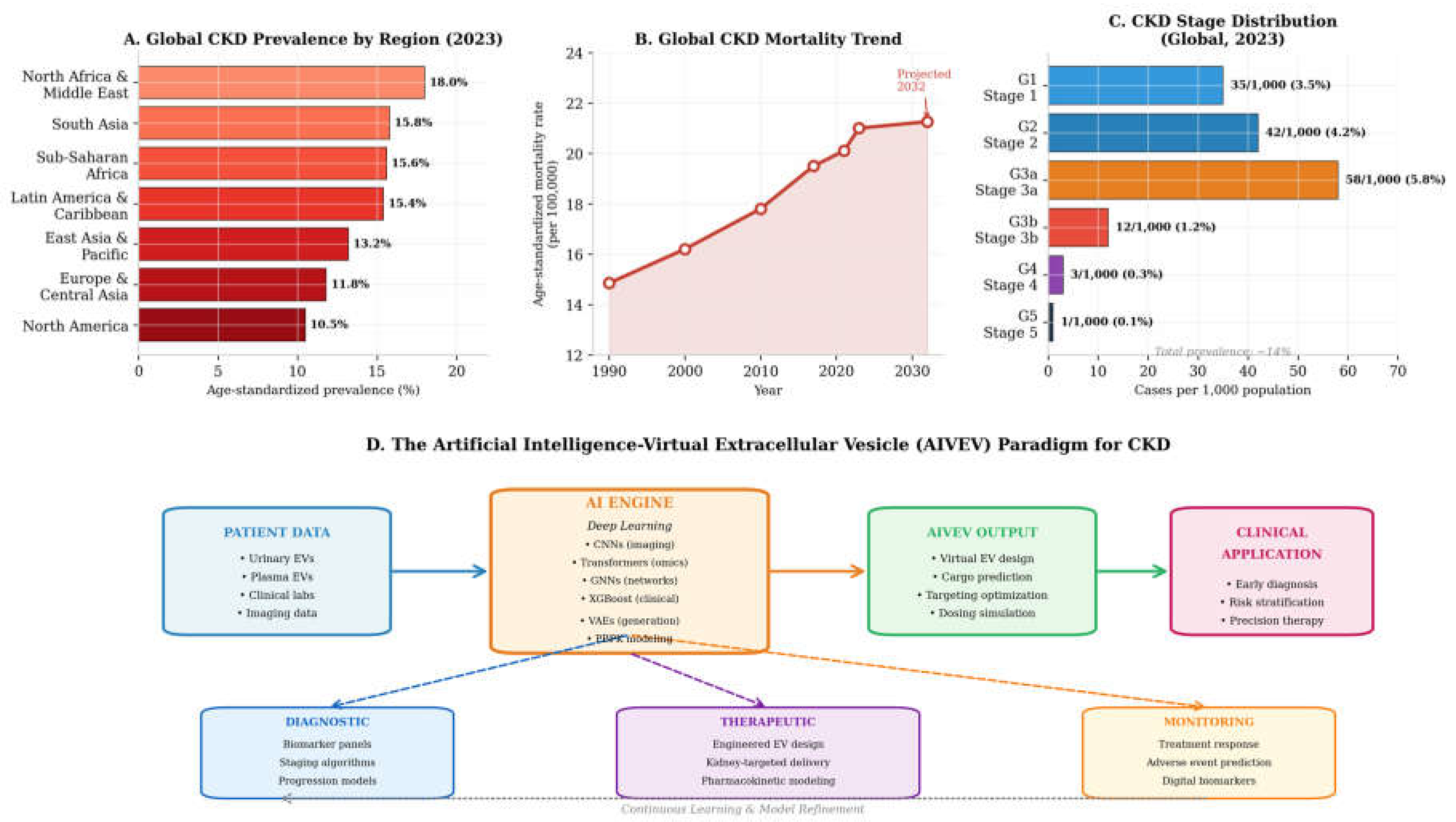
Figure 2.
AI-Enabled EV Biomarker Discovery for CKD. (A) Comparative performance of AI models for CKD EV-based diagnosis. (B) AI-ranked EV biomarkers by SHAP feature importance values. (C) UMAP visualization of urinary EV subpopulations across CKD stages. (D) AI-predicted CKD progression trajectories with confidence intervals.
Figure 2.
AI-Enabled EV Biomarker Discovery for CKD. (A) Comparative performance of AI models for CKD EV-based diagnosis. (B) AI-ranked EV biomarkers by SHAP feature importance values. (C) UMAP visualization of urinary EV subpopulations across CKD stages. (D) AI-predicted CKD progression trajectories with confidence intervals.
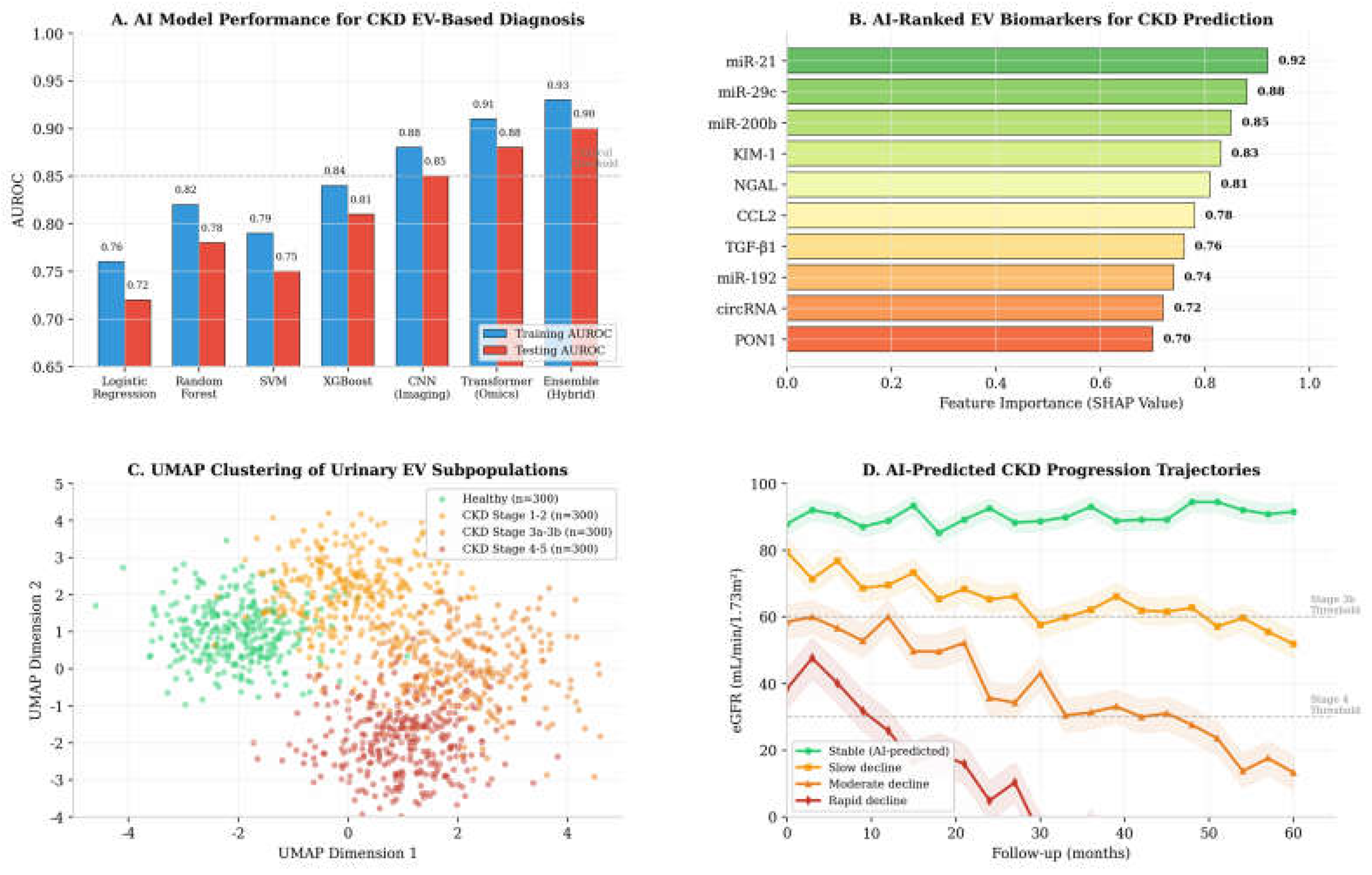
Figure 3.
AIVEV Therapeutic Design and PBPK Modeling. (A) Five-step AI-guided engineered EV design workflow with key computational tools. (B) PBPK simulation of EV tissue distribution following intravenous administration. (C) Comparative renal targeting efficiency of different EV surface modifications.
Figure 3.
AIVEV Therapeutic Design and PBPK Modeling. (A) Five-step AI-guided engineered EV design workflow with key computational tools. (B) PBPK simulation of EV tissue distribution following intravenous administration. (C) Comparative renal targeting efficiency of different EV surface modifications.
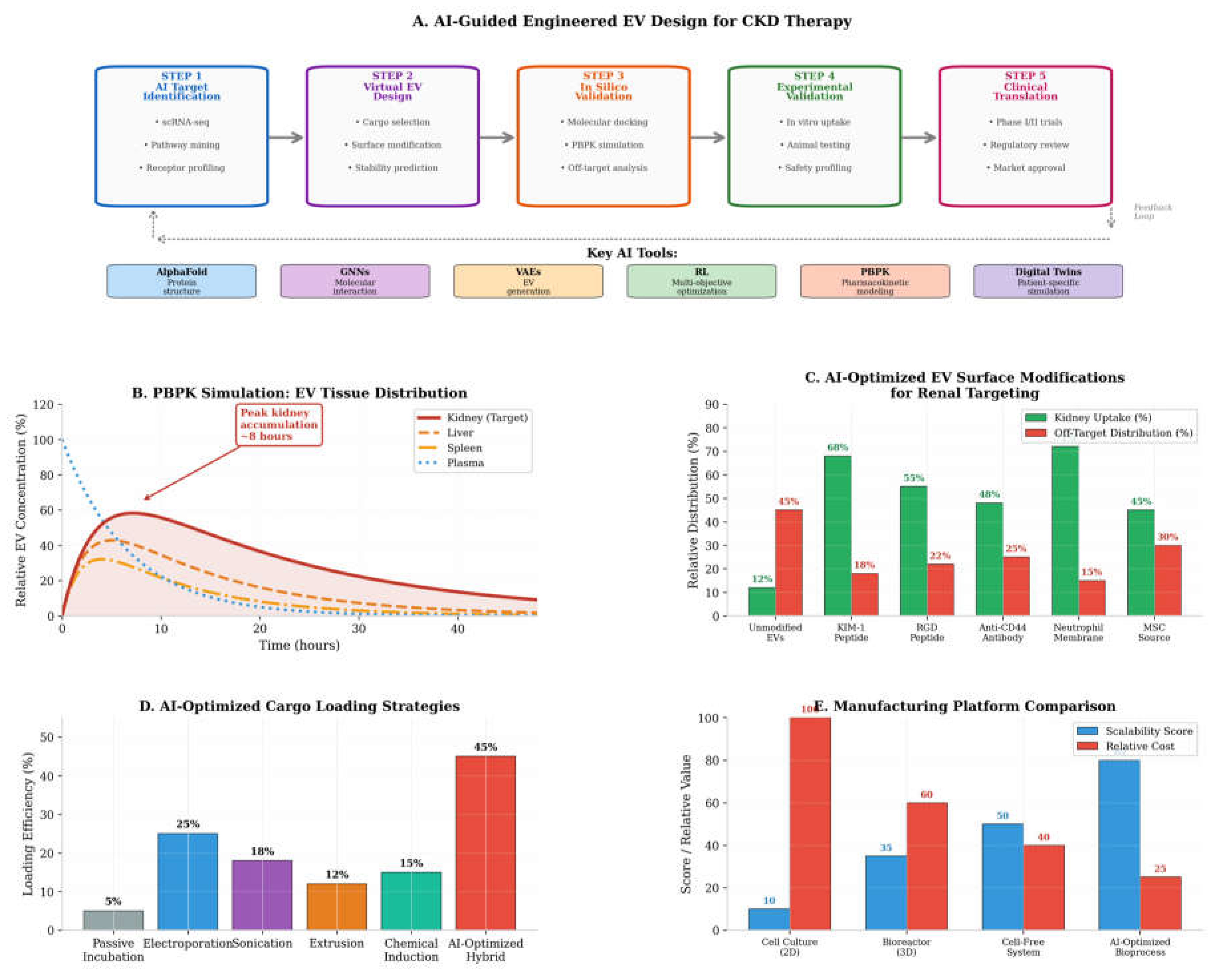
Figure 4.
Clinical Translation and Future Directions. (A) EV-based CKD therapy development pipeline showing translation funnel. (B) Timeline of AIVEV development milestones (2016–2030, projected). (C) Challenges and AI-driven solutions in AIVEV translation. (D) Projected cost-effectiveness of AIVEV versus standard CKD care.
Figure 4.
Clinical Translation and Future Directions. (A) EV-based CKD therapy development pipeline showing translation funnel. (B) Timeline of AIVEV development milestones (2016–2030, projected). (C) Challenges and AI-driven solutions in AIVEV translation. (D) Projected cost-effectiveness of AIVEV versus standard CKD care.
Disclaimer/Publisher’s Note: The statements, opinions and data contained in all publications are solely those of the individual author(s) and contributor(s) and not of MDPI and/or the editor(s). MDPI and/or the editor(s) disclaim responsibility for any injury to people or property resulting from any ideas, methods, instructions or products referred to in the content. |
© 2026 by the authors. Licensee MDPI, Basel, Switzerland. This article is an open access article distributed under the terms and conditions of the Creative Commons Attribution (CC BY) license (http://creativecommons.org/licenses/by/4.0/).
Copyright: This open access article is published under a Creative Commons CC BY 4.0 license, which permit the free download, distribution, and reuse, provided that the author and preprint are cited in any reuse.