

Submitted:
16 August 2026
Posted:
17 August 2026
You are already at the latest version
Abstract
Pathogenic variants in ADPRS, encoding ADP-ribosylhydrolase 3 (ARH3), cause stress-induced childhood-onset neurodegeneration with variable ataxia and seizures (CONDSIAS; OMIM: 618170), an ultra-rare autosomal recessive disorder with fewer than 61 reported cases worldwide. Here we report a previously undiagnosed 36-year-old who we analysed using trio exome sequencing analysis. We identified a homozygous pathogenic ADPRS variant (NM_017825.3:c.1004T>G; p.Val335Gly), bringing to an end to a 33-year diagnostic odyssey. The patient presented with neurological features consistent with CONDIAS including childhood-onset gait instability, distal sensory-motor axonopathy, and later-emerging cerebellar ataxia with consistently normal brain MRI. However, he also showed with severe primary hypogonadism, including markedly reduced testosterone, elevated gonadotropins, microorchidism, and secondary osteopenia. Although neurological features are well-characterised in CONDSIAS, endocrine involvement has not been emphasized as part of the clinical phenotype. We conducted a literature review of CONDSIAS and report an update on the mutation and clinical spectrum. Our review highlights that endocrine involvement can be part of the phenotype in CONDSIAS . The patient described here has the most severe gonadal phenotype reported to date. Treatment of the patient with testosterone replacement precipitated neurological deterioration on two separate occasions, suggesting that sex hormone supplementation should be carefully considered for ARH3-deficient individuals.
Keywords:
CONDSIAS
; ADPRS
; childhood-onset neurodegeneration
; hypogonadism
; trio exome sequencing
Introduction
Stress-induced childhood-onset neurodegeneration with variable ataxia and seizures (CONDSIAS; OMIM: 618170) is an ultra-rare autosomal recessive disorder caused by biallelic loss-of-function variants in ADPRS (also known as ADPRHL2), located at 1p34.3, which encodes ADP-ribosylhydrolase 3 (ARH3). First described in 2018 [1,2] approximately 60 cases have been reported globally to date. The disorder typically presents in the first decade of life following a period of normal development, and symptoms are frequently triggered or worsened by physiological stressors such as fever, infection, or physical trauma [3,4].
ARH3 is an eraser enzyme that removes ADP-ribose modifications from protein residues and DNA, thereby regulating the cellular ADP-ribosylation cycle, a post-translational signalling pathway critical to DNA repair, apoptosis, transcription, and telomere maintenance [5,6]. When ARH3 function is lost, poly(ADP-ribose) (PAR) accumulates pathologically on histones, triggering parthanatos, a form of PAR-driven programmed cell death that preferentially affects neurons and other non-dividing cells [7].
Core clinical features of CONDSIAS include developmental delay or regression in childhood, cerebellar ataxia, stress-sensitive seizures, axonal peripheral neuropathy, and cognitive decline, though the presentation is highly variable across individuals [1,2,3,8,9,10]. The defining clinical hallmark is episodic neurological deterioration, often precipitated by illness or physical stress, with periods of partial recovery between episodes. Endocrine, musculoskeletal, and autonomic involvement are being increasingly recognised as additional components of the phenotype.
Here we report a 36-year-old man who remained molecularly undiagnosed for more than three decades, despite undergoing extensive clinical and genetic investigation. Using trio exome sequencing analysis we identified a homozygous pathogenic ADPRS variant (p.Val335Gly), providing a molecular diagnosis of CONDSIAS, ending his diagnostic odyssey. While the patient showed core neurological features of CONDSIAS, he also presented with severe primary hypogonadism, a feature not previously described in CONDSIAS. We describe the clinical features of this patient which are novel to CONDSIAS, review the mutational and phenotypic spectra across all published cases of ADPRS-related CONDSIAS, and discuss the diagnostic and clinical implications.
Materials and Methods
Ethics and Patient Consent
The study was approved by the Human Research Ethics Committees of the Clinical Centre Split, Croatia (Klasa: 500-03/23-01/200; Ur. broj: 2181-147-01-06/LJ.Z.-23-02) and Adelaide University (HREC protocol 000000329987). The study was conducted in accordance with the Declaration of Helsinki. Written informed consent for genetic analysis, data publication, and clinical material disclosure was provided by the patient.
Clinical Investigation
A 36-year-old Caucasian male born to non-consanguineous European parents underwent comprehensive neurological, electrophysiological, neuroimaging, and endocrine assessment across multiple time points. Serial neurological examinations at ages 22, 27, and 32 included evaluations of motor function (proximal and distal strength), cranial and peripheral nerve status, deep tendon reflexes, sensory testing, gait assessment, and cerebellar signs. Nerve conduction studies (NCS) and electromyography (EMG) were used to characterise the neuropathy pattern. Brain and spine MRI, including fluid-attenuated inversion recovery (FLAIR) sequences, and CT imaging were performed at multiple time points. Endocrine investigations comprised serial measurements of serum testosterone, follicle-stimulating hormone (FSH), and luteinizing hormone (LH) at ages 22 and 34; testicular ultrasound at age 21; testicular MRI at age 34; and dual-energy X-ray absorptiometry (DEXA) at age 34. A complete clinical timeline and detailed examination findings are provided in Supplementary Appendix S1–S3.
Genetic Investigation
Karyotyping
Conventional G-banded karyotype analysis was performed on short-term peripheral blood mononuclear cell cultures following standard phytohemagglutinin stimulation and colchicine treatment.
Trio Exome Sequencing
Genomic DNA was extracted from peripheral blood of the patient and both parents. Libraries were prepared using SureSelect Human All Exon V7 (Agilent, Santa Clara, CA) and sequenced on the Illumina NovaSeq platform. Reads were aligned to GRCh38 using novoAlign v4.0 [11] and variant calling used the GATK HaplotypeCaller v4 [12]. Variants were annotated with SnpEff/SnpSift v5.0e [13] against dbNSFP v4 [14]. Variant prioritisation used Slivar v0.2.7 [15] across multiple inheritance models. Variant filtering additionally searched for and excluded pathogenic or likely pathogenic variants in established hypogonadism and disorders of sex development (DSD) genes as a cause, including NR5A1, KAL1, FGFR1, PROKR2, SOX9, DMRT1, SRY, AR, CYP11A1, HSD17B3, SRD5A2, AMH, AMHR2, LHCGR, FSHR, and STAR. A candidate homozygous ADPRS variant was confirmed by bidirectional direct Sanger sequencing of proband and parental genomic DNA. Variant classification followed ACMG/AMP guidelines.
Literature Review
A review of published ADPRS-related disease was performed. PubMed, Google Scholar, and the Google search engine were searched using the terms “CONDSIAS,” “ADPRS,” “ADPRHL2,” “ARH3,” and “childhood-onset neurodegeneration with variable ataxia and seizures.” No language or date restrictions were applied. Reference lists of retrieved articles were screened for additional cases. Genotype, phenotype, age of onset, endocrine findings, and outcome data were extracted and tabulated for all identified cases.
Results
Clinical Findings
Early motor development of the patient was normal until 3.5 years of age. The patient developed progressive gait instability, distal muscle weakness, and a characteristic head deviation during walking, with recurrent falls that resulted in fractures on two occasions. Right-hand weakness and impaired fine motor function became apparent by age 7, and urinary continence was not achieved until age 12. Despite this burden of disability, the patient completed a university degree. Formal cognitive assessment at age 32 yielded a borderline-range IQ of 79.
Serial neurological examinations revealed a progressive deteriorating course of the disease (Table S2). By age 27, the patient had developed global areflexia, with worsening distal limb weakness, predominantly distal muscle atrophy, and hypotonia. Examination at age 32 revealed ataxic gait, a positive Romberg sign, and strabismus, while proximal muscle strength remained relatively preserved. At each assessment, electromyography analysis revealed chronic axonal sensory-motor neuropathy, characterised by spontaneous denervation activity and polyphasic, high-amplitude motor unit potentials, with preserved or near-normal nerve conduction velocities, indicating a primary axonopathy rather than a demyelinating process. Serial brain and spine MRI and CT, including FLAIR sequences, were consistently unremarkable. The evolving clinical picture led to successive differential diagnoses: distal spinal muscular atrophy (SMA) (age 22), SMA type 3/Kugelberg–Welander syndrome (age 27), and hereditary motor neuropathy or motor neuron disease (age 32).
Severe primary hypogonadism in the patient was first identified at age 11 during investigation for gynecomastia. Biochemical confirmation at age 22 (Table S3) revealed profoundly low serum testosterone 0.55 nmol/L (0.16 ng/mL; reference range 8.64–29.0 nmol/L [2.8–8.0 ng/mL]), markedly elevated FSH 87.5 mIU/mL (reference 0.7–11.1 mIU/mL), and elevated LH 18.4 mIU/mL (reference 0.8–7.6 mIU/mL). At age 34, similar abnormalities persisted: testosterone 0.65 nmol/L (0.19 ng/mL; reference 8.64–29.0 nmol/L), FSH 90.5 mIU/mL (reference 1.5–12.4 mIU/mL), and LH 11.46 mIU/mL (reference 1.7–8.6 mIU/mL). Testicular imaging confirmed microorchidism on ultrasound at age 21 (right: 10.3 × 5.7 mm; left: 13.0 × 6.6 mm), and MRI at age 34 showed complete absence of right testicular parenchyma and a hypoplastic left testis. DEXA scanning at age 34 demonstrated low bone mineral density consistent with osteopenia at multiple sites (L1–L4 T-score -2.4 SD; femoral neck T-score -2.4 SD), with L4 Z-score -2.8 SD indicating bone density substantially below expected for age. By WHO criteria, T-scores >-2.5 SD define osteopenia rather than osteoporosis. Testosterone replacement therapy was attempted at ages 11 and 36; on both occasions, it produced severe acne and a clear worsening of neurological function, prompting discontinuation.
Genetic Findings
The patient was suspected to have spinal muscular dystrophy, however, SMN1 deletion testing at age 25 was negative. Clinical exome sequencing of a 4,800-gene panel at age 29 identified heterozygous variants in TK2 (paternal) and HSPB1 (maternal), with neither genetic finding being consistent with the patient’s phenotype. Karyotype analysis was normal (46,XY), excluding Klinefelter syndrome and other sex chromosome aneuploidies as causes of primary hypogonadism.
Using trio exome sequencing analysis, we identified a homozygous variant in ADPRS exon 6: GRCh38 Chr1:g.36093298T>G; NM_017825.3:c.1004T>G (p.Val335Gly). Both parents were shown to be heterozygous carriers (Figure 1). This variant is classified as pathogenic (ClinVar: VCV000599343; OMIM: 610624.0007; dbSNP: rs201735454) and has been identified in other individuals with CONDSIAS, across different genetic ancestries [1,16,17,18]. Direct Sanger sequencing confirmed the genotype in all three family members.
Discussion
Inheritance and the ADPRS Variant Landscape
CONDSIAS is an autosomal recessive disorder due to variants in ADPRS [1,2,4,8]. Consistent with this, both parents of our patient were asymptomatic carriers of the pathogenic variant and the patient was homozygous. To date, 26 different pathogenic ADPRS variants have been identified in 60 individuals with CONDSIAS, inclusive of the current case (Figure 2; Table 1). Variant classes include missense, nonsense, frameshift, splice-site, and in-frame deletion/insertion changes. Mapping of patient variants onto the ARH3 protein structure reveals clustering within α-helices of quasi-domains A and C, which are involved in Mg2+-dependent catalysis, as well as domains B and D, which contribute to structural integrity and protein folding [6].
Missense variants in ADPRS have been found in 31 of the 60 patients with CONDSIAS reported to date. The most common variant reported so far is p.Val335Gly (NM_017825.3:c.1004T>G), identified in the patient reported here and 11 other previously reported patients. The pathogenic variant has been shown to be associated with childhood-to-adult onset of the disorder and in patients with diverse ancestries. The variant Val335Gly has been associated with ataxia, seizures, and progressive axonal neuropathy as core features [1,16,17]. An important outlier is p.Ala280Thr, associated with adult-onset PAMP syndrome (episodic psychosis, ataxia, motor neuropathy, and pyramidal signs), demonstrating that ADPRS variants show variable expressivity [19]. Nonsense variants, found in 14 patients, produce truncated, non-functional proteins and are typically associated with earlier age of onset and a more rapidly progressive disease course [1,2,3,20]. Frameshift variants (9 patients) also abolish ARH3 activity and have been linked to deep white-matter abnormalities, cerebellar atrophy, and direct biochemical evidence of PAR accumulation in patient cells [8]. Compound heterozygous variants have been reported in 3 patients whose phenotypes extend beyond the core neurological syndrome to include autonomic dysfunction and endocrine disturbance [4,21,22].
Genotype-phenotype correlations for ADPRS variants remain preliminary given the rarity of CONDSIAS, but a pattern may be emerging where loss-of-function variants (nonsense and frameshift variants) are associated with earlier-onset, more severe, and more rapidly progressive disease. Missense or splice-site variants are more often associated with slower disease progression, selective organ involvement, or adult-onset presentations [8,9,22], potentially reflecting residual activity [23]. As genome sequencing becomes more widely applied in clinical practice, detection of structural variants and deep intronic changes in ADPRS are predicted to expand the known mutational landscape.
The Multisystem Clinical Spectrum of CONDSIAS
CONDSIAS displays a variable clinical picture, reflecting the widespread consequences of ARH3 dysfunction across the nervous system and multiple organ systems. Onset is typically in early childhood following a period of normal development, and symptoms are often precipitated or worsened by physiological stressors such as infection, trauma, or fever [3,4]. Core neurological features include developmental delay or regression (affecting motor and language milestones) [2,8], cerebellar ataxia with dysmetria and wide-based gait [4], stress-sensitive seizures that may be poorly controlled with standard antiepileptic therapy [22], distal axonal peripheral neuropathy with weakness and atrophy [17] and cognitive decline ranging from static intellectual disability to progressive deterioration [9].
The neurological course of the patient reported here exemplifies several hallmarks of CONDSIAS. Childhood-onset gait instability and distal sensory-motor axonopathy preceded cerebellar signs by approximately 25 years, a sequence that is well recognised in the disorder, but that in isolation closely mimics hereditary motor neuropathy or spinal muscular atrophy [1]. Crucially, two features distinguished this case from the differential diagnoses: the later-emerging cerebellar involvement, and the episodic exacerbation of motor function triggered by testosterone replacement, which mirrors the physiological stress-induced neurological deterioration documented in other descriptions of patients with CONDSIAS [2,3,4].
While static intellectual disability is reported in approximately 65% of published CONDSIAS cases, our patient’s cognitive function (IQ of 79), though below average, has remained stable enough to permit completion of a university degree, suggesting a milder neurocognitive spectrum than is typical for complete loss-of-function alleles.
A distinctive and previously unreported clinical feature of the patient presented here is severe primary hypogonadism. This led us to review any previous reports of hypogonadism in patients with CONDSIAS. Prior reports of endocrine involvement in CONDSIAS are limited: one male patient carried mild hypogonadism with elevated FSH [2], a 27-year-old female with the p.Val98Trpfs*23 variant experienced premature menopause [1] and isolated cases have shown reduced TSH levels [22] or hypothyroidism, with one also developing diabetes insipidus [9]. To our knowledge, our patient has the most severe primary hypogonadism described in a patient with CONDSIAS, as assessed by testosterone levels, gonadotropin elevation, testicular size, and consequent osteopenia (T-score -2.4 SD; low bone mineral density secondary to androgen deficiency) (Table 2). The mechanism underlying primary gonadal failure in CONDSIAS is not established. ARH3 is expressed in testicular tissue, and ADP-ribosylation is implicated in gonadal steroidogenesis and germ cell maintenance; it is plausible that ARH3 dysfunction disrupts these processes, though direct evidence is lacking. Systematic endocrine evaluation has not been performed in most published patients with CONDSIAS, suggesting that gonadal and metabolic involvement may be underreported.
Before attributing the severe primary hypogonadism in this patient to ARH3 dysfunction, well-established alternative causes were considered and excluded. Klinefelter syndrome (47,XXY), the most common genetic cause of primary hypogonadism in males, was excluded by genetic testing, with the patients having a normal 46,XY karyotype. There was no history of gonadotoxic chemotherapy, radiotherapy, or orchidectomy. Congenital cryptorchidism was not documented in clinical records. The 47,XYY syndrome and other sex chromosome aneuploidies were also excluded by the normal karyotype result. Trio exome sequencing analysis also excluded pathogenic or likely pathogenic variants in established hypogonadism and genes associated with differences in sex development, including NR5A1, KAL1, FGFR1, PROKR2, SOX9, DMRT1, SRY, AR, CYP11A1, HSD17B3, SRD5A2, AMH, AMHR2, LHCGR, FSHR, and STAR. Taken together, the early age of onset (gynecomastia at age 11), the progressive severity of gonadal failure, and the absence of any alternative genetic or environmental aetiology support attribution of the severe hypogonadism to ARH3 dysfunction.
Given the patients’s very low tesoterone levels, treatment with testosterone replacement was trialled twice, when the patient was 11 and 36 years old. Neurological deterioration was seen to follow both administrations (Table S1 and Note). The reasons for the adverse effects are not known, but exogenous testosterone is itself a physiological stressor, capable of inducing anabolic and metabolic surges [24]. In an ARH3-deficient nervous system, such demands may trigger the same cascade of PAR accumulation and neuronal injury as infections or physical trauma [5,7]. If hormone replacement is considered for treatment of patients with CONDSIAS in the future, conservative dosing, gradual titration, and close neurological monitoring may be advisable.
Differential Diagnosis and the Challenge of Clinical Recognition
The broad phenotypic overlap between CONDSIAS and other inherited neurological conditions creates substantial diagnostic challenges (Table 3). Our patient’s clinical course prompted successive consideration of SMA type 3, hereditary motor neuropathy, Kennedy disease, Charcot-Marie-Tooth disease, and mitochondrial cytopathy, none of which were confirmed by genetic testing. The resulting 33-year gap between symptom onset and the molecular diagnosis achieved in this study illustrates the diagnostic vulnerability inherent to clinically heterogeneous, recently characterised genetic disorders that lack a distinctive biochemical or imaging biomarker.
A consistently normal brain MRI across more than three decades of disease progression is a notable negative finding that distinguishes CONDSIAS from progressive hereditary ataxias, leukodystrophies, and mitochondrial cytopathies. These conditions usually show cerebellar or white-matter abnormalities early in the disease course. CONDSIAS should be considered in the differential diagnosis of a patient presenting with childhood-onset neurological decline who shows normal or near-normal neuroimaging,
Several clinical features should also prompt clinicians to consider CONDSIAS in the differential diagnosis: childhood onset of symptoms following a period of normal development; episodic or stress-triggered neurological deterioration with partial inter-episode recovery; chronic axonal peripheral neuropathy with later emergence of cerebellar features; multisystem involvement including endocrine or autonomic dysfunction; and normal or near-normal neuroimaging, particularly in earlier disease stages.
Diagnostic Value of Trio Exome Sequencing
In this patient, the analysis of targeted spinal muscular atrophy testing and a 4,800-gene clinical exome panel failed to provide a molecular diagnosis. Trio exome sequencing resolved the diagnostic odyssey by identifying the homozygous ADPRS variant in an unbiased, exome-wide analysis. Parental trio exome sequencing data enabled rapid segregation confirmation and definitive phasing of the homozygous variant call. The diagnostic yield of trio exome sequencing in childhood-onset neurological disorders consistently exceeds that of targeted gene panels [25].
Beyond establishing the diagnosis, the molecular result had immediate practical consequences for the patient and his family. It enabled informed genetic counselling to be offered to the family, regarding a 25% recurrence risk to siblings and allowed the clinical trajectory to be anticipated based on the ADPRS p.Val335Gly literature.
Conclusions
CONDSIAS is a rare autosomal recessive neurodegenerative disorder caused by ADPRS variants, which impair ARH3 function and disrupt cellular ADP-ribosylation homeostasis. The clinical phenotype includes childhood onset ataxia, stress sensitive seizures, axonal neuropathy, and developmental delay or regression. Our patient showed severe primary hypogonadism with microorchidism and secondary osteopenia. Other common causes of primary hypogonadism (Klinefelter syndrome, cryptorchidism, anorchia, gonadotoxic exposure) were excluded, suggesting that hypogonadism may be part of the CONDSIAS phenotypic spectrum. However, confirmation will require identification of similar gonadal findings in additional cases.
Several clinical observations may be useful. In patients with childhood onset neurological decline that is episodic, stress triggered, or multisystem, and particularly when neuroimaging is normal, CONDSIAS can be considered in the differential diagnosis. Endocrine assessment, including measurement of gonadotropins and testosterone, may be informative in confirmed or suspected ADPRS related disease. Testosterone replacement should be approached with caution, as it was associated with neurological worsening in our patient. When targeted genetic testing is unrevealing, trio exome sequencing can help establish a molecular diagnosis, which facilitates genetic counselling and clinical management.
Further prospective studies and case registries are needed to define the full phenotypic spectrum of CONDSIAS, determine the prevalence of endocrine involvement, and clarify genotype-phenotype correlations.
Supplementary Materials
The following supporting information can be downloaded at the website of this paper posted on Preprints.org.
Author Contributions
A.S.: investigation, bioinformatic analysis, formal analysis, writing: original draft. T.A.: clinical investigation, data curation. Z.S.: variant validation. R.H.: variant validation. G.N.: clinical investigation. Z.G.: clinical investigation. M.G.R.: genetic investigation, methodology, writing: review and editing. L.M.D.: conceptualisation, genetic investigation, supervision, writing: review and editing. All authors reviewed and approved the final manuscript.
Funding
This study was supported by National and Health Medical Research Council of Australia (Senior Research Fellowship: 1104718; Project Grant: 1125523 to L.M.D.) and institutional funding from the University of South Australia (now Adelaide University). A.S. was supported by an IRTS fee-waiver scholarship from the University of South Australia (now Adelaide University) and is an employee of Novocraft Technologies.
Ethics Approval
The study was conducted in accordance with the Declaration of Helsinki and approved by the Human Research Ethics Committees of the Clinical Centre Split, Croatia (Klasa: 500-03/23-01/200, Ur. broj: 2181-147-01-06/LJ.Z.-23-02), and Adelaide University (HREC protocol 000000329987).
Consent to Participate
Written informed consent was obtained from the patient.
Consent to Publish
The patient (or parent/guardian, where applicable) provided written consent for publication of clinical, genetic, and imaging findings; consent-to-disclose documentation is available on request.
Data Availability
Original contributions are included in this article. Additional data are available from the corresponding author on reasonable request.
Competing Interests
A.S. is a PhD student at the University of South Australia (now Adelaide University) and an employee of Novocraft Technologies, the maker and distributor of novoAlign software referenced in this study. This presents no perceived or actual conflict of interest but is disclosed for transparency. The remaining authors report no disclosures relevant to the manuscript.
References
- Danhauser, K.; Alhaddad, B.; Makowski, C.; Piekutowska-Abramczuk, D.; Syrbe, S.; Gomez-Ospina, N.; et al. Bi-allelic ADPRHL2 Mutations Cause Neurodegeneration with Developmental Delay, Ataxia, and Axonal Neuropathy. Am. J. Hum. Genet. 2018, 103(5), 817–25. [Google Scholar] [CrossRef]
- Ghosh, S.G.; Becker, K.; Huang, H.; Dixon-Salazar, T.; Chai, G.; Salpietro, V.; et al. Biallelic Mutations in ADPRHL2, Encoding ADP-Ribosylhydrolase 3, Lead to a Degenerative Pediatric Stress-Induced Epileptic Ataxia Syndrome. Am. J. Hum. Genet. 2018, 103(3), 431–9. [Google Scholar] [CrossRef] [PubMed]
- Bajaj, S.; Shah, P.; Shah, A.; Setty, P.N.; Seenappa, V.; Hingwala, D. An Indian Child with CONDSIAS Due to a Novel Variant in ADPRHL2 Gene. Ann. Indian Acad. Neurol. 2022, 25(6), 1190–2. [Google Scholar] [CrossRef] [PubMed]
- Lindskov, F.O.; Karlsson, W.K.; Skovbolling, S.L.; Nielsen, E.N.; Duno, M.; Stokholm, J.; et al. Expanding the Spectrum of Stress-Induced Childhood-Onset Neurodegeneration with Variable Ataxia and Seizures (CONDSIAS). Cerebellum 2024, 23(2), 861–71. [Google Scholar] [CrossRef] [PubMed]
- Mueller-Dieckmann, C.; Kernstock, S.; Lisurek, M.; von Kries, J.P.; Haag, F.; Weiss, M.S.; et al. The structure of human ADP-ribosylhydrolase 3 (ARH3) provides insights into the reversibility of protein ADP-ribosylation. Proc. Natl. Acad. Sci. U S A 2006, 103(41), 15026–31. [Google Scholar] [CrossRef] [PubMed]
- Suskiewicz, M.J.; Prokhorova, E.; Rack, J.G.M.; Ahel, I. ADP-ribosylation from molecular mechanisms to therapeutic implications. Cell 2023, 186(21), 4475–95. [Google Scholar] [CrossRef] [PubMed]
- McGurk, L.; Rifai, O.M.; Bonini, N.M. Poly(ADP-Ribosylation) in Age-Related Neurological Disease. Trends Genet. 2019, 35(8), 601–13. [Google Scholar] [CrossRef] [PubMed]
- Aryan, H.; Razmara, E.; Farhud, D.; Zarif-Yeganeh, M.; Zokaei, S.; Hassani, S.A.; et al. Novel imaging and clinical phenotypes of CONDSIAS disorder caused by a homozygous frameshift variant of ADPRHL2: a case report. BMC Neurol. 2020, 20(1), 291. [Google Scholar] [CrossRef] [PubMed]
- Oz Yildiz, S.; Yalnizoglu, D.; Simsek Kiper, P.O.; Gocmen, R.; Sogukpinar, M.; Utine, G.E.; et al. Delineation of ADPRHL2 Variants: Report of Two New Patients with Review of the Literature. Neuropediatrics 2024, 55(3), 156–65. [Google Scholar] [CrossRef] [PubMed]
- Yan, S.; Ren, J.; Su, H.; Ma, J.; He, W.; Cai, X.; et al. Novel heterozygous variant of ADPRHL2 causes pathogenic variation in CONDSIAS. Heliyon 2024, 10(14), e32945. [Google Scholar] [CrossRef] [PubMed]
- Hercus, C. novoAlign v4. 4 ed2022. [PubMed]
- Van der Auwera, G.A.; Carneiro, M.O.; Hartl, C.; Poplin, R.; Del Angel, G.; Levy-Moonshine, A.; et al. From FastQ data to high confidence variant calls: the Genome Analysis Toolkit best practices pipeline. Curr. Protoc. Bioinform. 2013, 43(1110), 11 0 1–0 33. [Google Scholar] [CrossRef] [PubMed]
- Cingolani, P.; Platts, A.; Wang le, L.; Coon, M.; Nguyen, T.; Wang, L.; et al. A program for annotating and predicting the effects of single nucleotide polymorphisms, SnpEff: SNPs in the genome of Drosophila melanogaster strain w1118; iso-2; iso-3. Fly 2012, 6(2), 80–92. [Google Scholar] [CrossRef] [PubMed]
- Liu, X.; Li, C.; Mou, C.; Dong, Y.; Tu, Y. dbNSFP v4: a comprehensive database of transcript-specific functional predictions and annotations for human nonsynonymous and splice-site SNVs. Genome Med. 2020, 12(1), 103. [Google Scholar] [CrossRef] [PubMed]
- Pedersen, B.S.; Brown, J.M.; Dashnow, H.; Wallace, A.D.; Velinder, M.; Tristani-Firouzi, M.; et al. Effective variant filtering and expected candidate variant yield in studies of rare human disease. npj Genom. Med. 2021, 6(1), 60. [Google Scholar] [CrossRef] [PubMed]
- Mahungu, A.C.; Steyn, E.; Floudiotis, N.; Wilson, L.A.; Vandrovcova, J.; Reilly, M.M.; et al. The mutational profile in a South African cohort with inherited neuropathies and spastic paraplegia. Front Neurol. 2023, 14, 1239725. [Google Scholar] [CrossRef] [PubMed]
- Beijer, D.; Agnew, T.; Rack, J.G.M.; Prokhorova, E.; Deconinck, T.; Ceulemans, B.; et al. Biallelic ADPRHL2 mutations in complex neuropathy affect ADP ribosylation and DNA damage response. Life Sci. Alliance 2021, 4(11). [Google Scholar] [CrossRef] [PubMed]
- Tompa, M.; Tolvaj, B.; Vadvari, A.; Feher, N.; Kiss, Z.; Kalman, B. Clinical, Genetic, and Pathological Studies in Two Brothers with Stress-Induced Childhood-Onset Neurodegeneration with Variable Ataxia and Seizures: A Case Report. Case Rep. Neurol. 2024, 16(1), 188–95. [Google Scholar] [CrossRef]
- Durmus, H.; Mertoglu, E.; Sticht, H.; Ceylaner, S.; Kulaksizoglu, I.B.; Hashemolhosseini, S.; et al. Episodic psychosis, ataxia, motor neuropathy with pyramidal signs (PAMP syndrome) caused by a novel mutation in ADPRHL2 (AHR3). Neurol. Sci. 2021, 42(9), 3871–8. [Google Scholar] [CrossRef] [PubMed]
- Modage, A.; Gaonkar, V.; Pereira, M.I. Recurrent Paroxysmal Torticollis as a Presenting Feature of Stress-Induced Childhood-Onset Neurodegeneration with Variable Ataxia and Seizures (CONDSIAS). Indian Pediatr. 2025, 62(6), 468–9. [Google Scholar] [CrossRef] [PubMed]
- Lu, A.; Dong, C.; Chen, B.; Xie, L.; Hu, H. Case Report: Stress-Induced Childhood-Onset Neurodegeneration With Ataxia-Seizures Syndrome Caused by a Novel Compound Heterozygous Mutation in ADPRHL2. Front Neurol. 2022, 13, 807291. [Google Scholar] [CrossRef] [PubMed]
- Ma, J.; Qian, Q.; Yan, S.; Dou, H.; Li, C.; Sun, D. Child-Onset Cerebellar Ataxia Caused by Two Compound Heterozygous Variants in ADPRS Gene: A Case Report. Front Genet. 2021, 12, 788702. [Google Scholar] [CrossRef] [PubMed]
- Bannister, M.; Bray, S.; Aggarwal, A.; Billington, C., Jr.; Nguyen, H.D. An ADPRS variant disrupts ARH3 stability and subcellular localization in children with neurodegeneration and respiratory failure. HGG Adv. 2025, 6(1), 100386. [Google Scholar] [CrossRef] [PubMed]
- Bhasin, S.; Wang, C.; Chandra, M.S.; Gagliano-Juca, T.; Jasuja, R. Mechanisms of Testosterone’s Anabolic Effects on Muscle and Function: Controversies and New Insights. Endocr. Rev. 2026, 47(2), 280–300. [Google Scholar] [CrossRef] [PubMed]
- Chang, Y.M.; Huang, Y.T.; Lai, P.C. Genetic testing for diagnosing neurodevelopmental disorders and epilepsy: a systematic review and meta-analysis. Syst. Rev. 2025, 14(1), 155. [Google Scholar] [CrossRef] [PubMed]
- Mishra, B.; Fatima, S.; Agarwal, A.; Radhakrishnan, D.M.; Garg, A.; Srivastava, A.K. Dystonia and Myelopathy in a Case of Stress-Induced Childhood-Onset Neurodegeneration with Ataxia and Seizures (CONDSIAS). Mov. Disord. Clin. Pract. 2021, 8(1), 156–8. [Google Scholar] [CrossRef] [PubMed]
- Nandana, J.; Fasaludeen, A.; Kumar, A.A.; Vijayaraghavan, A.; Nair, S.S.; Cherian, A.; et al. Phenotypic Diversity in Stress-induced Childhood-Onset Neurodegeneration with Variable Ataxia and Seizures: Novel Associations with Parkinsonism, Icthyosis and Cataract. Cerebellum 2025, 24(4), 112. [Google Scholar] [CrossRef] [PubMed]
- Paketci, C.; Karakaya, M.; Edem, P.; Bayram, E.; Keller, N.; Daimaguler, H.S.; et al. Clinical, electrophysiological and genetic characteristics of childhood hereditary polyneuropathies. Rev. Neurol. 2020, 176(10), 846–55. [Google Scholar] [CrossRef] [PubMed]
- Ozturk, G.; Ayaz, A.; Topcu, Y.; Akyuz, G.; Unver, O.; Akbeyaz, I.H.; et al. Stress-induced Childhood Onset Neurodegeneration with Ataxia and Seizures (CONDSIAS) Presenting with Torticollis Attacks: Phenotypic Variability of the Same Mutation in Two Turkish Patients. Ann. Indian Acad. Neurol. 2022, 25(2), 292–4. [Google Scholar] [CrossRef] [PubMed]
- Mashimo, M.; Bu, X.; Aoyama, K.; Kato, J.; Ishiwata-Endo, H.; Stevens, L.A.; et al. PARP1 inhibition alleviates injury in ARH3-deficient mice and human cells. JCI Insight 2019, 4(4). [Google Scholar] [CrossRef]
- Eslamiyeh, H.; Vahidi Mehrjardi, M.Y.; Poursalehi, N.; Dehghan Tezerjani, M. Repurposing doxycycline for a case of CONDSIAS Syndrome with a novel ADPRHL2 missense mutation. Acta Neurol. Belg. 2025, 125(1), 241–2. [Google Scholar] [CrossRef] [PubMed]
- de Mattos, P.D.; de Moura, R.D.; Bertolino, M.F.; Valente, P.F.; Matos, I.A.; Caldas, V.M.; et al. Novel ADPRS Missense Variant (p.Leu162Pro) Causes Stress-Induced Childhood-Onset Neurodegeneration With Ataxia and Seizures. Neurol. Genet. 2026, 12(3), e200375. [Google Scholar] [CrossRef] [PubMed]
Figure 1.
Pedigree and Sanger sequencing confirmation of the ADPRS variant in the proband and parents. The patient (II-1) is homozygous for the ADPRS variant NM_017825.3:c.1004T>G (p.Val335Gly) [GRCh38 Chr1:g.36093298T>G]; the father (I-1) and mother (I-2) are each heterozygous carriers. Sanger chromatogram traces for each individual are shown, with the sequence variant position indicated by an arrow.
Figure 1.
Pedigree and Sanger sequencing confirmation of the ADPRS variant in the proband and parents. The patient (II-1) is homozygous for the ADPRS variant NM_017825.3:c.1004T>G (p.Val335Gly) [GRCh38 Chr1:g.36093298T>G]; the father (I-1) and mother (I-2) are each heterozygous carriers. Sanger chromatogram traces for each individual are shown, with the sequence variant position indicated by an arrow.
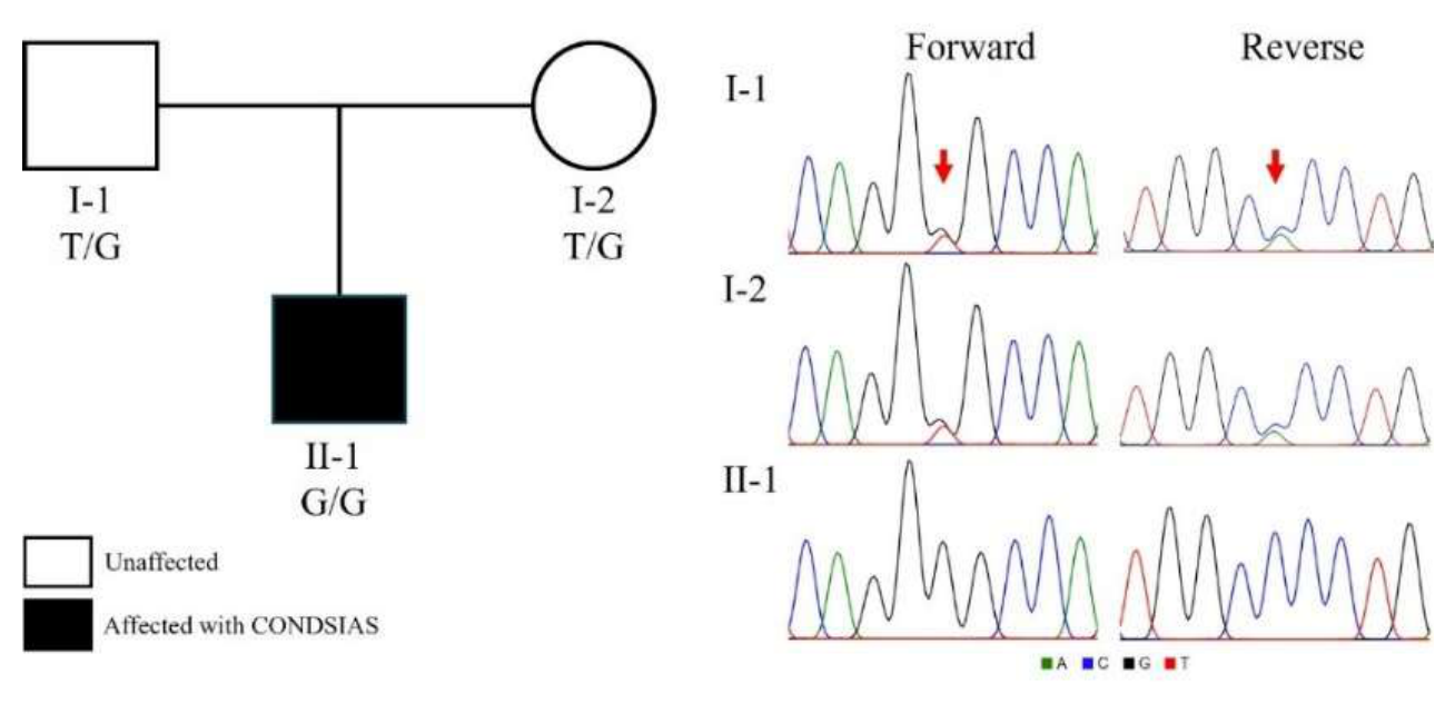
Figure 2.
Distribution and structural context of ADPRS variants reported in CONDSIAS. (a) Schematic representation of ADPRS exons illustrating the position and class of all reported pathogenic variants. Missense variants are shown in blue; nonsense in red; frameshift in purple; splice-site in black; in-frame deletion/insertion in orange. The variant associated with the highest number of reported cases (p.Val335Gly, observed in this study) is highlighted with a yellow background. Orange dashed lines indicate variants observed in compound-heterozygous genotypes. (b) Topology map of the ARH3 protein showing α-helix arrangement and quasi-domain assignments (Domain A in red; B in pink; C in orange; D in green). The two catalytic Mg²⁺ ions (MgI and MgII) are indicated. (c) Three-dimensional structure of ARH3 with missense variants mapped and colour-coded by quasi-domain. (d) Linear amino acid sequence of ARH3 with secondary-structure elements and quasi-domain boundaries. Reported pathogenic variants are highlighted and colour-coded by variant class as in panel A. Figure B & D were adapted from Mueller-Dieckmann et al. (2006) [5].
Figure 2.
Distribution and structural context of ADPRS variants reported in CONDSIAS. (a) Schematic representation of ADPRS exons illustrating the position and class of all reported pathogenic variants. Missense variants are shown in blue; nonsense in red; frameshift in purple; splice-site in black; in-frame deletion/insertion in orange. The variant associated with the highest number of reported cases (p.Val335Gly, observed in this study) is highlighted with a yellow background. Orange dashed lines indicate variants observed in compound-heterozygous genotypes. (b) Topology map of the ARH3 protein showing α-helix arrangement and quasi-domain assignments (Domain A in red; B in pink; C in orange; D in green). The two catalytic Mg²⁺ ions (MgI and MgII) are indicated. (c) Three-dimensional structure of ARH3 with missense variants mapped and colour-coded by quasi-domain. (d) Linear amino acid sequence of ARH3 with secondary-structure elements and quasi-domain boundaries. Reported pathogenic variants are highlighted and colour-coded by variant class as in panel A. Figure B & D were adapted from Mueller-Dieckmann et al. (2006) [5].
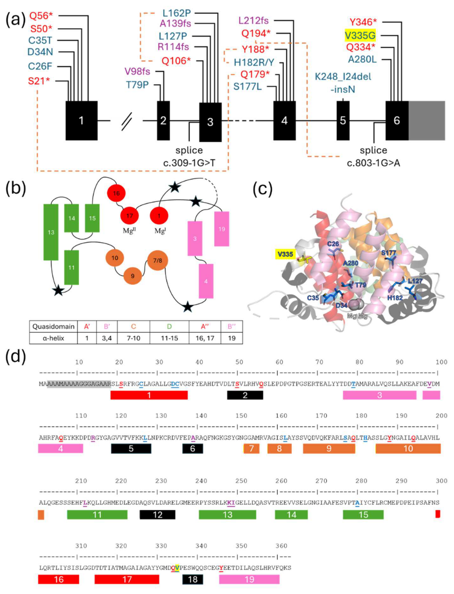

Table 1.
Summary of all reported pathogenic ADPRS variants and associated primary phenotypes. Variant numbering follows NM_017825.3. Phenotype columns reflect counts per variant class: A = Ataxia; S = Seizures; GA = Gait Abnormalities; CD = Cognitive Decline/Intellectual Disability; DD = Developmental Delay; CA = Cerebellar Atrophy; ED = Endocrine Disorder. ★ = variant observed in this case report.
Table 1.
Summary of all reported pathogenic ADPRS variants and associated primary phenotypes. Variant numbering follows NM_017825.3. Phenotype columns reflect counts per variant class: A = Ataxia; S = Seizures; GA = Gait Abnormalities; CD = Cognitive Decline/Intellectual Disability; DD = Developmental Delay; CA = Cerebellar Atrophy; ED = Endocrine Disorder. ★ = variant observed in this case report.
| Exon | Variant (Protein / cDNA) | Type | n | Onset | Deaths | A | S | GA | CD | DD | CA | ED | (n)Ref. |
| 1 | p.Cys26Phe (c.77G>T) | Missense | 1 | 15 m | 1 (16 y) | 0 | 1 | 0 | 1 | 1 | 0 | 0 | (1) [17] |
| — | p.Asp34Asn (c.100G>A) | Missense | 2 | 3 y | 0 | 2 | 0 | 2 | 2 | 1 | 2 | 0 | (1) [2]; (1) [26] |
| — | p.Cys35Tyr (c.104G>A) | Missense | 1 | 4 y | 0 | 1 | 1 | 1 | 0 | 0 | 1 | 0 | (1) [27] |
| — | p.Ser50* (c.149C>G) | Nonsense | 1 | 6 y | 0 | 1 | 0 | 1 | 0 | 0 | 0 | 0 | (1) [27] |
| — | p.Gln56* (c.166C>T) | Nonsense | 2 | 1.5–2 y | 2 (3–12 y) | 2 | 1 | 2 | 1 | 2 | 1 | 0 | (1) [3]; (1) [20] |
| 2 | p.Thr79Pro (c.235A>C) | Missense | 6 | <1–4 y | 1 (3 y) | 4 | 3 | 6 | 4 | 3 | 3 | 1 | (1) [2]; (1) [28]; (2) [29]; (2) [9] |
| — | p.Val98Trpfs*23 (c.292delG) | Frameshift | 2 | 1–2 y | 2 (5–13 y) | 2 | 2 | 2 | 2 | 2 | 2 | 0 | (2) [1] |
| 3 | c.309−1G>T | Splice-site | 2 | 14–15 m | 1 (4 y) | 2 | 2 | 2 | 2 | 2 | 0 | 0 | (2) [1] |
| — | p.Gln106* (c.316C>T) | Nonsense | 1 | 2 y | 0 | 0 | 0 | 1 | 1 | 1 | 1 | 1 | (1) [2] |
| — | p.Arg114fs* (c.340_341del) | Frameshift | 3 | 18 m | 1 (4 y) | 0 | 0 | 1 | 3 | 2 | 0 | 1 | (3) [30] |
| — | p.Leu127Pro (c.380T>C) | Missense | 1 | 2 y 8m | 0 | 1 | 1 | 1 | 1 | 1 | 0 | 0 | (1) [31] |
| — | p.Ala139Glyfs*4 (c.414_418del) | Frameshift | 3 | 9 m–15 y | 0 | 1 | 3 | 2 | 2 | 1 | 2 | 0 | (2) [2]; (1) [27] |
| 4 | p.Ser177Leu (c.530C>T) | Missense | 2 | 1–1.5 y | 1 (6 y) | 0 | 2 | 2 | 2 | 2 | 0 | 0 | (2) [2] |
| — | p.His182Arg (c.545A>G) | Missense | 2 | 24–28 m | 1 (5 y 2m) | 0 | 2 | 2 | 2 | 1 | 0 | 0 | (2) [23] |
| — | p.Leu212fs (c.636_639del) | Frameshift | 1 | 4 y | 1 (4 y) | 1 | 1 | 1 | 0 | 1 | 1 | 0 | (1) [8] |
| 5 | p.Lys248_Ile249delinsAsn (c.744_746del) | In-frame del/ins | 1 | 4 y | 1 (17 y) | 1 | 1 | 1 | 1 | 1 | 1 | 0 | (1) [1] |
| 6 | p.Ala280Thr (c.838G>A) | Missense | 4 | 20–32 y | 0 | 4 | 0 | 4 | 0 | 0 | 1 | 0 | (4) [19] |
| — | p.Gln334* (c.1000C>T) | Nonsense | 9 | 15–24 m | 9 (2–15 y) | 1 | 9 | 5 | 6 | 7 | 2 | 0 | (9) [2] |
| — | p.Val335Gly (c.1004T>G) ★ | Missense | 12 | 1–29 y | 6 (11–50 y) | 9 | 7 | 11 | 6 | 6 | 5 | 1 | (6) [1]; (2) [17]; (1) [16]; (2) [18]; (1) This study |
| — | p.Tyr346* (c.1038C>G) | Nonsense | 1 | 2 y | 1 (12 y) | 1 | 0 | 1 | 1 | 1 | 1 | 0 | (1) [1] |
| Compound Heterozygous | |||||||||||||
| 1,4 | p.Ser21*; p.Gln179* (c.62C>A; c.535C>T) | Nonsense/ Nonsense | 1 | 3 y | 0 | 1 | 1 | 1 | 1 | 1 | 1 | 0 | (1) [21] |
| 3 | p.Gln106*; p.Leu162Pro (c.316C>T; c.485T>C) | Nonsense/ Missense | 1 | 1 y | 0 | 1 | 1 | 1 | 1 | 1 | 1 | 0 | (1) [32] |
| 4 | p.His182Tyr; p.Tyr188* (c.544C>T; c.564C>A) | Missense/ Nonsense | 1 | 5 y | 0 | 1 | 0 | 1 | 0 | 0 | 1 | 0 | (1) [4] |
| 4,6 | p.Gln194Ter; Intron5 splice (c.580C>T; c.803-1G>A) | Missense/ Splice | 1 | 30 m | 0 | 0 | 1 | 1 | 1 | 0 | 1 | 1 | (1) [22] |
| TOTAL | 61 |
28 (45.9%) |
36 (59.0%) |
39 (63.9%) |
52 (85.2%) |
40 (65.6%) |
37 (60.6%) |
27 (44.2%) | 5 (8.2%) | — | |||
Table 2.
Reported endocrine and gonadal findings associated with specific ADPRS variants in CONDSIAS. ★ = variant observed in this case report.
Table 2.
Reported endocrine and gonadal findings associated with specific ADPRS variants in CONDSIAS. ★ = variant observed in this case report.
| Variant (Protein/cDNA) | Functional Consequence | Endocrine Findings | Gonadal Findings | Case Reference |
| p.Thr79Pro (c.235A>C) | Reduces thermal stability; severely unstable protein with markedly reduced cellular levels | Central diabetes insipidus, adrenal insufficiency, hypothyroidism | Not reported | Case 2 [9] |
| p.Val98Trpfs*23 (c.292delG) | Early truncation (136 aa); complete loss of enzymatic activity | Hypothyroidism (sister) | Premature menopause (27-year-old sister) | Sister [1] |
| p.Gln106* (c.316C>T) | Complete absence of ARH3 protein | Elevated FSH; gynecomastia onset age 10 | Mild hypogonadism | F2-II-2 [2] |
| p.Gln194Ter + c.803-1G>A (compound het.) | Nonsense-mediated mRNA decay; intron 5 retention; reduced ARH3 expression | Reduced TSH levels | Not reported | Proband [22] |
| p.Val335Gly (c.1004T>G)★ | Reduced steady-state protein levels; altered nuclear localisation; catalytic activity retained in vitro | Very low testosterone; markedly elevated LH and FSH; osteoporosis secondary to hypogonadism | Severe primary hypogonadism; microorchidism; absence of right testicular parenchyma | This study |
Table 3.
Disorders with phenotypic overlap with CONDSIAS and key shared clinical features. SCAR26 = spinocerebellar ataxia, autosomal recessive, 26; AOA4 = ataxia with oculomotor apraxia type 4.
Table 3.
Disorders with phenotypic overlap with CONDSIAS and key shared clinical features. SCAR26 = spinocerebellar ataxia, autosomal recessive, 26; AOA4 = ataxia with oculomotor apraxia type 4.
| Disorder | Overlapping Features with CONDSIAS | Reference |
| Friedreich Ataxia | Progressive ataxia, kyphoscoliosis, dorsal column degeneration, axonal neuropathy | [4] |
| Mitochondrial Disorders | Episodic worsening with physiological stress, multisystemic involvement, developmental delay | [2,3,26] |
| Charcot-Marie-Tooth Disease | Axonal motor/sensory polyneuropathy, distal weakness, pes cavus | [3,17] |
| Episodic Ataxia | Stress-induced episodes of truncal ataxia, vertigo, incoordination | [4,9,19] |
| GLUT1 Deficiency Syndrome | Paroxysmal exertion-induced dystonia, early-onset seizures, movement disorder | [4,9] |
| Guillain-Barré Syndrome | Acute motor-predominant peripheral polyneuropathy, respiratory failure | [23] |
| SCAR26 / AOA4 | Progressive ataxia, oculomotor abnormalities, peripheral sensorimotor neuropathy | [1,2] |
| Juvenile ALS | Upper and lower motor neuron signs, progressive weakness | [3] |
| Ataxia Telangiectasia | Early childhood-onset ataxic neuropathy without telangiectasia | [9] |
| Autoimmune Encephalitis | Acute encephalopathy, loss of consciousness, respiratory failure | [9] |
| Wilson Disease | Progressive neurological decline, speech delay | [1,2] |
| Leigh Syndrome | Severe paediatric neurodegeneration, respiratory failure, developmental delay | [1] |
| Niemann-Pick Type C | Cerebellar eye movement disorder, intention tremor, ataxia | [2] |
| Spinal Muscular Atrophy Type 3 | Proximal/distal motor weakness, chronic neurogenic EMG pattern | This study |
Disclaimer/Publisher’s Note: The statements, opinions and data contained in all publications are solely those of the individual author(s) and contributor(s) and not of MDPI and/or the editor(s). MDPI and/or the editor(s) disclaim responsibility for any injury to people or property resulting from any ideas, methods, instructions or products referred to in the content. |
© 2026 by the authors. Licensee MDPI, Basel, Switzerland. This article is an open access article distributed under the terms and conditions of the Creative Commons Attribution (CC BY) license (http://creativecommons.org/licenses/by/4.0/).
Copyright: This open access article is published under a Creative Commons CC BY 4.0 license, which permit the free download, distribution, and reuse, provided that the author and preprint are cited in any reuse.