

Submitted:
05 August 2026
Posted:
14 August 2026
You are already at the latest version
Abstract
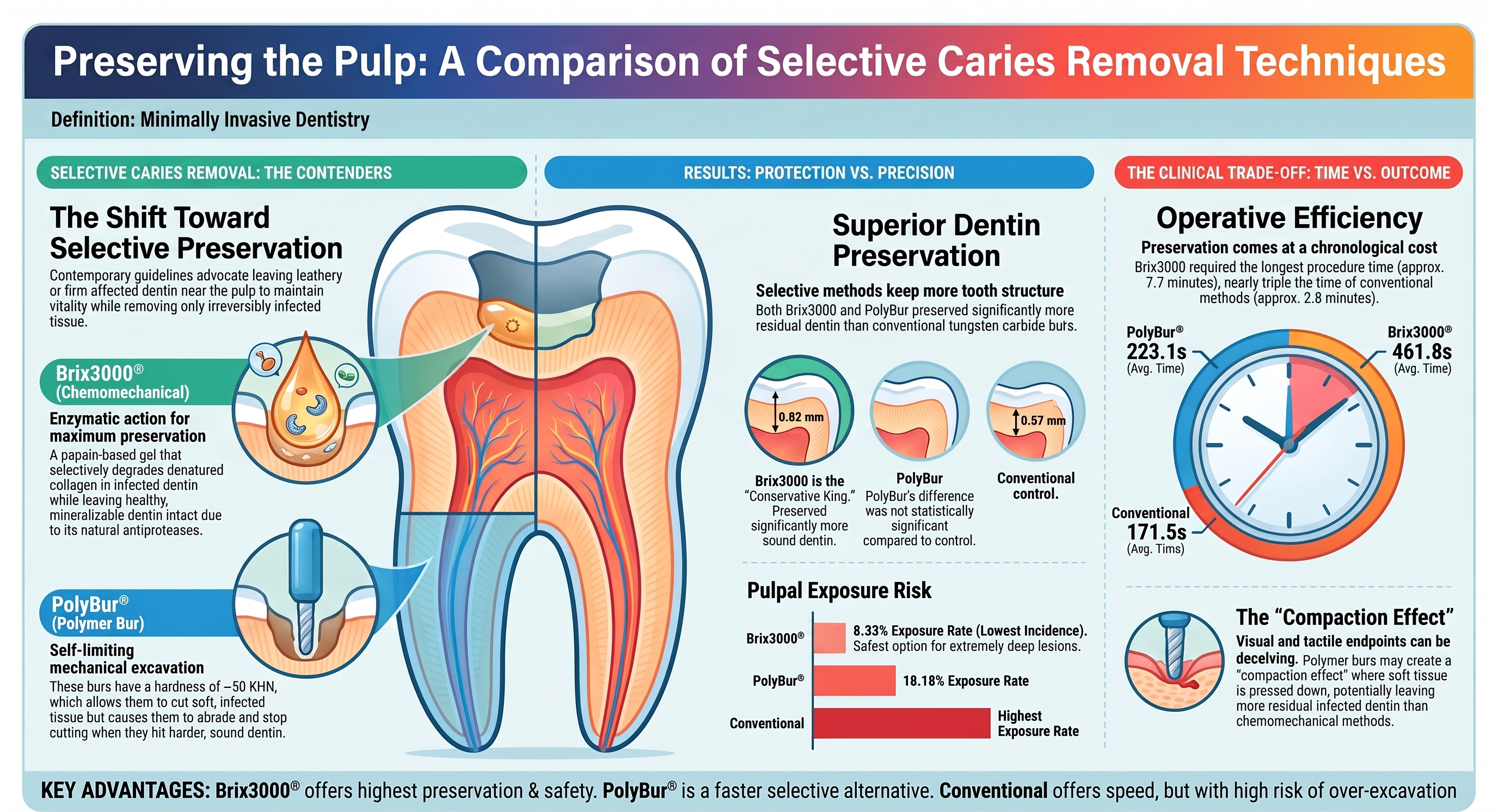
Background/objective: Minimally invasive dentistry advocates selective caries removal to preserve affected dentin, protect pulpal vitality, and maintain tooth structure. To compare selective caries removal using Brix3000® and polymer burs (PolyBur®) with conventional caries removal in deep dentinal lesions. Methods: Thirty extracted human molars with deep carious lesions were sectioned using a split-tooth design (n = 15). One half underwent selective caries removal with either Brix3000® or PolyBur®, while the corresponding half received conventional caries removal. Caries removal was completed according to predefined visual and tactile endpoints. Residual dentin thickness was assessed radiographically and stereomicroscopically. Operative time, residual infected dentin, and pulpal exposure were also evaluated. Statistical significance was set at p < 0.05. Results: Pulpal exposure occurred in one Brix3000® specimen, two PolyBur® specimens, and four corresponding controls, resulting in final sample sizes of 12 and 11 specimens, respectively. Both selective techniques preserved significantly more dentin than conventional caries removal. Radiographically, residual dentin thickness was greater with Brix3000® than with its corresponding control (p = 0.02), whereas the difference between PolyBur® and its control was not significant (0p = 0.06). No significant differences were found in residual infected dentin between Brix3000® and PolyBur®. Brix3000® preserved significantly more sound dentin but required the longest operative time (p < 0.001). Conclusions: Both selective caries removal techniques preserved more dentin than conventional caries removal. Brix3000® was the most conservative technique, preserving significantly more sound dentin than PolyBur®, although at the expense of longer operative times. These findings support selective caries removal as a minimally invasive approach for managing deep dentinal lesions.
Keywords:
dental caries
; selective caries removal
; chemomechanical caries removal
; polymer bur
1. Introduction
Dental caries remains the most prevalent non-communicable disease worldwide and continues to represent a major public health burden [1]. In 2016, the International Caries Consensus Collaboration (ICCC) defined deep caries as a condition that extends radiographically beyond the inner third or quarter of the dentin [2].
Deep and extremely deep carious lesions constitute a major clinical challenge because extensive tissue removal may compromise pulpal vitality, whereas insufficient caries removal may leave residual infected dentin beneath the restoration. Radiographically defined deep lesions are generally associated with bacterial invasion confined to the dentin, whereas extremely deep lesions are more frequently associated with severe pulpal inflammation and bacterial contamination. Therefore, the radiographic distinction between deep and extremely deep carious lesions may serve as an indicator of both the extent of bacterial penetration and the severity of the pulpal response prior to intervention [3].
Contemporary minimally invasive dentistry advocates selective caries removal strategies that preserve remineralizable dentin while minimizing the risk of pulpal exposure [2]. Current evidence recommends leaving leathery or firm carious dentin adjacent to the pulp in deep carious lesions. Specifically, when lesions radiographically extend into the inner third or quarter of the dentin, selective removal should be limited to soft dentin to reduce the likelihood of pulpal exposure while maintaining pulpal vitality [4].
Different selective caries removal strategies have been proposed, including chemomechanical systems based on papain-derived enzymatic activity and self-limiting polymer burs designed to selectively remove softened dentin according to substrate hardness [5]. Although both techniques aim to preserve dentin structure, their biological behavior regarding residual infected dentin and dentin preservation remains insufficiently understood.
Brix3000® contains 30,000 U/mg of papain, a proteolytic enzyme that selectively degrades partially denatured type I collagen fibers within carious dentin. The absence of the antiprotease α1-antitrypsin in infected tissues allows papain to break down collagen molecules that have been partially degraded by the carious process, facilitating the removal of the fibrinous layer formed during lesion progression. In contrast, healthy dentin contains antiproteases that inhibit papain activity, preventing proteolytic degradation of sound tissue and thereby conferring selectivity to the removal process. In addition, Brix3000® incorporates Encapsulating Buffer Emulsion (EBE) technology, which maintains an optimal pH for enzyme stabilization and promotes the controlled release of papain during its proteolytic action on collagen [6,7].
Studies comparing the biological effects of Brix3000® with chemomechanical caries removal agents containing chloramines have demonstrated lower cytotoxicity and reduced induction of apoptosis in pulp cells [8]. These findings suggest that papain-based formulations may be more biocompatible and less detrimental to the dentin–pulp complex than chloramine-based alternatives. Such characteristics support the clinical use of Brix3000® for selective caries removal in deep dentinal lesions, where maintaining pulpal vitality is of paramount importance.
Dentin hardness has been proposed as a reliable criterion for distinguishing remineralizable from non-remineralizable carious dentin [9]. Sound dentin exhibits a Knoop hardness ranging from approximately 51 to 65 KHN, depending on its depth and anatomical location, whereas active carious dentin is considerably softer, with values between 4.4 and 11.2 KHN. Arrested carious dentin shows greater variability, with reported hardness values ranging from 16.0 to 61.0 KHN [10].
Based on these differences, polymer burs were developed with a hardness of approximately 50 KHN, intermediate between sound and infected dentin [11]. This characteristic enables selective caries removal by preferentially cutting soft, infected tissue while becoming progressively abraded upon contact with harder, potentially remineralizable dentin. As a result, the excavation process is self-limiting and ceases when the bur becomes visibly worn and no longer effectively removes dentin [12]. Consequently, polymer burs do not necessarily eliminate all infected dentin, a feature that is consistent with the principles of minimally invasive caries management.
PolyBur® is specifically indicated for the management of deep carious lesions approaching the pulp, with the aim of preserving sound dentin and reducing the risk of pulpal exposure[12]. The instrument is typically operated at low rotational speeds, ranging from 500 to 800 rpm [13].
Previous studies have mainly focused on excavation efficacy or operative time [14,15]. However, limited evidence directly compares chemomechanical and polymer bur selective excavation techniques using paired split-tooth experimental models combined with radiographic and stereomicroscopic assessment of residual dentin [16].
Therefore, the aim of the present in vitro study was to compare selective chemomechanical excavation using Brix3000® and polymer bur excavation with conventional non-selective caries removal in deep dentinal lesions. The evaluated outcomes included operative time, residual dentin thickness, presence of residual infected dentin, and the incidence of pulpal exposure.
The null hypothesis was that no significant differences would exist among the evaluated excavation techniques regarding dentin preservation and pulpal exposure.
2. Materials and Methods
Study Design and Ethical Approval
An in vitro split-tooth experimental study was conducted to compare selective and conventional caries excavation techniques in deep dentinal lesions. The study protocol was approved by the Human Research Ethics Committee of the Universitat de València (Reference number: 2025-ODON-3865315).
Sample Size Calculation and Sample Selection
The sample size was calculated assuming a statistical power of 80%, a significance level of 5% (α = 0.05), and an expected effect size (Cohen’s d) ≥ 0.7, resulting in a minimum sample of 30 teeth.
Extracted permanent maxillary and mandibular molars presenting deep carious lesions were collected and stored in saline solution at 4 °C until completion of sample collection no longer than 2 weeks after extraction. Subsequently, the specimens were disinfected in 0.1% thymol solution for 24 hours and cleaned using curettes and a prophylaxis brush.
The inclusion criteria were: teeth presenting coronal carious lesions clinically classified as International Caries Detection and Assessment System (ICDAS) scores 4 to 6; radiographic extension reaching the middle or inner third of dentin (RB4–RC5 classification) [10]. Teeth presenting enamel structural defects, previous endodontic treatment, loss of more than half of the coronal structure, or lesions not radiographically extending to the middle or inner third of dentin were excluded.
Radiographic assessment was performed using a CCD digital sensor (3 × 4 cm). The X-ray tube was positioned perpendicular to the tooth and sensor at a 90° angle and a 20 cm focal distance using the following parameters: 60 kV, 7 mA, and 0.03 s exposure time. A buccolingual projection was obtained for all specimens.
Sample selection was independently performed by two calibrated examiners, obtaining a Cohen’s kappa coefficient of 0.89. The maximum interval between extraction and experimental use was 3 months.
Split-Tooth Preparation and Radiographic Measurements
Each tooth was sectioned mesiodistally through the center of the carious lesion using a diamond disc (DTX932D.HP.220, Jota AG, Rüthi, Switzerland) mounted on a handpiece under continuous water cooling (KaVo, Biberach, Germany). During sectioning, teeth were stabilized using a precision parallel vice (Proxxon 28602, JB Tools, Barcelona, Spain), obtaining two halves with similar lesion extension.
A new radiograph was obtained for each sectioned specimen. Images were exported to Rhinoceros 8 software (Robert McNeel & Associates, Seattle, USA) to calculate percentage of lesion penetration and residual dentin thickness before excavation.
Lesion penetration was calculated as the ratio between the distance from the amelodentinal junction to the pulpal limit of radiolucency and the total dentin thickness. Following caries excavation, residual dentin thickness was measured again radiographically. For image calibration, the actual tooth dimensions were measured using a millimetric ruler and transferred to the corresponding radiographic image to establish a real-scale reference. Figure 1
Randomization and Experimental Groups
A split-tooth paired design was used, in which one half of each tooth was randomly assigned to the experimental excavation technique and the other half to the conventional control group. The specimens were distributed into two experimental groups, Brix3000® followed by hand excavation versus conventional excavation and PolyBur® versus conventional excavation. To identify the control specimens, a small notch was created on the root surface using a round diamond bur mounted on a contra-angle handpiece. Each tooth pair was stored in an individual container throughout the experimental procedures.
Selective Caries Removal Procedures
Initial cavity access and excavation of the lateral cavity walls were performed using a pear-shaped diamond bur (822-01, Ø 010, Intensive SA, Grancia, Switzerland) until hard dentin was achieved. Conventional caries removal of the pulpal floor was performed using a tungsten carbide bur (H1SEM.204, Komet, Lemgo, Germany) mounted on a low-speed handpiece (KaVo, Biberach, Germany) using light circular movements until hard dentin was clinically detected.
Chemomechanical caries removal was performed using Brix3000® gel (Brix Medical Science, Santa Fe, Argentina), a papain-based agent that selectively degrades denatured collagen in infected dentin while preserving affected dentin with remineralization potential. After the peripheral cavity walls had been cleaned to hard dentin, approximately 2 mm of gel was applied to the pulpal floor for 2 minutes. The softened dentin was removed using a blunt double-ended excavator (1.4 mm diameter) with pendulum movements. The procedure was repeated until no soft, easily detachable dentin remained on the pulpal floor, the remaining dentin exhibited a firm consistency on gentle probing, and the gel no longer became turbid after application.
Selective excavation with polymer burs was performed using PolyBur® P1204014 burs (Komet, Lemgo, Germany) mounted on a low-speed handpiece. After excavation of the cavity walls, infected dentin was removed from the center of the lesion outward without applying pressure. Excavation was considered complete when the bur no longer removed dentin substrate. The bur was replaced after every three specimens.
Removal Endpoint Criteria
The removal endpoint was determined according to visual and tactile criteria consistent with minimally invasive dentistry recommendations [2]. Visually, the endpoint corresponded to the presence of yellowish or light brown dentin. Tactilely, the residual dentin surface was considered acceptable when the excavator passed smoothly without sticking or producing a tug-back sensation, while maintaining a leathery or firm consistency [17].
Following excavation, the cavity was cleaned using cotton pellets soaked in 0.12% chlorhexidine solution.
Operative time was recorded in seconds using a digital chronometer, including washing, drying, and cavity reassessment procedures.
Stereomicroscopic Analysis
After caries removal, all specimens were examined using a stereomicroscope (Leica M165 C, Leica Microsystems, Wetzlar, Germany) equipped with a digital camera (Leica DMC 2900). Specimens were positioned according to the mesiodistal sectioning plane and examined at ×16 magnification, including the pulpal floor and pulp chamber. Digital images were exported to ImageJ software (National Institutes of Health, Bethesda, MD, USA) for morphometric analysis.
Morphologically distinct dentin layers were identified according to differences in colour, translucency, and anatomical location. Following previously described stereomicroscopic criteria, darker brown opaque areas were classified as residual infected dentin, paler brown translucent areas as affected dentin, and yellowish-white areas as sound dentin [18]. Reparative dentin was identified by its characteristic morphology and its anatomical location adjacent to the pulp chamber. The interfaces between adjacent dentin layers were manually delineated, and the thickness of each layer was measured by drawing a line perpendicular to the interface between consecutive layers. Residual dentin thickness was measured as the distance from the deepest point of the cavity floor to the roof of the pulp chamber and corresponded to the sum of the residual infected, affected, sound, and reparative dentin layers.
Image calibration was performed using the scale integrated into the stereomicroscope images, where a predefined distance corresponded to 0.2 mm. Figure 2 illustrates the stereomicroscopic appearance of the different dentin layers and the measurement protocol used.
Dental sections were examined under a stereomicroscope at ×16 magnification, including the pulpal floor of the cavity and the pulp chamber. In each image, a straight line was drawn from the roof of the pulp chamber to the deepest point of the cavity floor, corresponding to the area of minimum remaining dentin thickness. The thickness of each morphologically distinct dentin layer was measured along this line. The different dentin layers are indicated as follows: red, infected dentin; blue, affected dentin; green, sound dentin; and black, reparative dentin.
All caries removal procedures were performed by a single calibrated operator, thereby eliminating inter-operator variability. Stereomicroscopic and radiographic measurements were independently performed by two calibrated investigators blinded to the caries removal technique. In cases of disagreement regarding layer identification or measurements, the images were re-evaluated until consensus was reached.
The primary outcome variables were: Residual dentin thickness, presence of residual infected dentin and pulpal exposure occurrence. The secondary outcomes included operative time, stereomicroscopic characterization of residual dentin.
Statistical Analysis
Statistical analysis was performed using SPSS version 29.0 (IBM Corp., Armonk, NY, USA).Data normality was assessed using the Shapiro–Wilk test. Paired comparisons between experimental and control halves were performed using paired statistical tests. Comparisons between the Brix3000® and PolyBur® experimental groups were performed using independent statistical tests. A significance level of p < 0.05 was adopted for all analyses.
3. Results
Sample Characteristics
A total of 30 molars presenting deep dentinal carious lesions were included in the study. Of these, 56.5% corresponded to maxillary molars and 43.5% to mandibular molars. Regarding lesion location, 47.8% were occlusoproximal lesions, 26.1% were exclusively occlusal lesions, and the remaining lesions were proximal.
According to the ICDAS classification, 6.7% of the teeth were classified as ICDAS 4, 80.0% as ICDAS 5, and 13.3% as ICDAS 6. Radiographically, two lesions clinically classified as ICDAS 4 and seven lesions classified as ICDAS 5 corresponded to category RB4. The remaining sixteen ICDAS 5 lesions and all five ICDAS 6 lesions were categorized as RC5. The distribution of the RC5 lesions was comparable between the experimental groups, 49.8% were allocated to the Brix3000® group and 50.2% to the PolyBur® group.
Pulpal exposure occurred in one specimen treated with Brix3000® and in two corresponding control specimens. In the PolyBur® group, pulpal exposure was observed in two specimens and in two corresponding controls. All specimens with mechanical pulpal exposure were excluded from subsequent analyses because disruption of the pulp chamber prevented measurement of an intact residual dentin barrier. Consequently, residual dentin thickness could not be reliably quantified, and inclusion of these specimens would have introduced substantial bias into the quantitative and stereomicroscopic analyses.
The final sample size for the analyses was 12 specimens in the Brix3000® group and 11 specimens in the PolyBur® group, post-hoc sensitivity analysis demonstrated that the statistical power was adequately maintained. For the primary outcome of postoperative sound dentin thickness, the achieved large effect sizes (Cohen’s d > 0.8) compensated for the sample reduction, ensuring that the paired and independent comparisons remained statistically robust (p < 0.05) and capable of identifying true significant differences without introducing statistical artifacts.
Comparison Between Each Experimental Group and Their Control
For the Brix3000® group, no significant differences were observed between the experimental and control halves regarding radiographic lesion depth (70.57 ± 6.79% vs. 66.92 ± 9.30%; p = 0.16) or preoperative sound dentin thickness (1.07 ± 0.27 mm vs. 1.19 ± 0.49 mm; p = 0.28). Following caries removal, residual sound dentin thickness measured radiographically was significantly greater in the Brix3000® group than in the corresponding control group (0.82 ± 0.39 mm vs. 0.57 ± 0.52 mm; p = 0.02).
Stereomicroscopic analysis revealed significantly greater affected dentin thickness in specimens treated with Brix3000® compared with their paired controls (1.13 ± 0.50 mm vs. 0.72 ± 0.30mm; p = 0.04). Similarly, total residual dentin thickness was significantly greater in the experimental group (1.46 ± 0.63 mm vs. 1.11 ± 0.58 mm; p = 0.01). No significant differences were observed in reparative dentin thickness (0.41 ± 0.30 mm vs. 0.61 ± 0.34 mm; p = 0.20). Residual infected dentin was identified in five specimens treated with Brix3000®, with a mean thickness of 0.72 ± 0.30 mm, whereas no residual infected dentin was detected in the corresponding control specimens. (Table 1)
No significant differences were observed between the experimental and control halves regarding radiographic lesion depth in the PolyBur® group (78.06 ± 10.52% vs. 76.65 ± 6.77%; p = 0.63) or preoperative sound dentin thickness (0.79 ± 0.32 mm vs. 0.81 ± 0.205 mm; p = 0.75). Following excavation, residual sound dentin thickness was greater in the PolyBur® group than in the corresponding control group (0.35± 0.15 mm vs. 0.26 ± 0.32 mm); however, the difference did not reach statistical significance (p = 0.06).
Stereomicroscopic evaluation demonstrated significantly greater affected dentin thickness in specimens treated with PolyBur® than in their paired controls (0.99 ± 0.32 mm vs. 0.74 ± 0.41 mm; p = 0.04). No significant differences were found in reparative dentin thickness (0.40 ± 0.07 mm vs. 0.38 ± 0.11 mm; p = 0.66) or total residual dentin thickness (1.29 ± 0.46 mm vs. 1.03 ± 0.74 mm; p = 0.43).
Residual infected dentin was detected in three PolyBur®-treated specimens, with a mean thickness of 0.23 ± 0.14 mm, whereas no residual infected dentin was observed in the corresponding control specimens. Sound dentin was not identified in any specimen treated with PolyBur®, whereas two control specimens exhibited a mean sound dentin thickness of 0.52 ± 0.13 mm. Statistical comparisons were not performed for variables with insufficient observations in one of the groups. (Table 2)
Comparison Between Selective Excavation Techniques
Direct comparison between the two selective excavation techniques showed significantly greater preoperative and postoperative sound dentin thickness in the Brix3000® group than in the PolyBur® group (p = 0.03 and p =0.019, respectively). No significant differences were observed between the two techniques regarding residual infected dentin, affected dentin, reparative dentin, or total residual dentin thickness (all p > 0.05). (Table 3)
Sound dentin was identified in only two specimens from the Brix3000® group, whereas no sound dentin was observed in the PolyBur® group. Owing to the limited number of observations, no statistical comparison was performed.
Operative Time
Operative time differed significantly among the evaluated caries removal techniques (Table 4). The longest time was recorded for the chemomechanical technique using Brix3000® (461.81 ± 115.31 s), followed by PolyBur® (223.06 ± 64.95 s). Both selective techniques required significantly longer operative times than their corresponding conventional controls (p < 0.05).
Direct comparison between the two selective techniques showed that Brix3000® required significantly more time than PolyBur® (p<0.001). No significant differences were observed between the two conventional control groups.
4. Discussion
The present study compared two selective caries removal techniques, a chemomechanical method based on papain gel (Brix3000®) and a self-limiting polymer bur system (PolyBur®), with conventional non-selective excavation in deep dentinal lesions. The null hypothesis was rejected. Significant differences were observed among the evaluated caries removal techniques regarding dentin preservation and pulpal exposure. Both selective approaches demonstrated a more conservative behavior than conventional excavation preserving greater amounts of residual dentin.
To the authors’ knowledge, few studies have directly compared a papain-based chemomechanical caries removal system and polymer burs using a split-tooth design while simultaneously evaluating residual dentin thickness, residual infected dentin, operative time, and pulpal exposure. Therefore, the present study provides a comprehensive assessment of the biological and operative consequences of two contemporary selective caries removal strategies.
Furthermore, a methodological aspect that warrants discussion is the reduction in the final sample size due to accidental pulpal exposures during caries removal. Although the initial sample size calculation determined a minimum requirement of 30 teeth, the strict exclusion of specimens that suffered pulp chamber breach resulted in final group sizes of 12 specimens for Brix3000®, and 11 specimens for PolyBur®. This exclusion was methodologically mandatory, an anatomical exposure renders the residual dentin thickness physically non-existent at that specific site, and including a value of zero would have severely skewed the data variance and mispresented the structural measurements.
The primary outcomes were residual dentin thickness, residual infected dentin thickness, and pulpal exposure, whereas operative time and stereomicroscopic characterization of the residual dentin were considered secondary outcomes. Overall, both selective caries removal techniques preserved significantly more dentin than conventional caries removal. Among the selective approaches, Brix3000® proved to be more conservative than PolyBur®, preserving greater amounts of residual dentin and showing a lower incidence of pulpal exposure (8.33% vs. 18.18%), although this was achieved at the expense of a significantly longer operative time. Direct comparison of these findings with previous studies is limited because the incidence of pulpal exposure has rarely been reported as an outcome when evaluating chemo-mechanical agents or polymer burs. Most investigations have focused on caries removal efficacy, operative time, microbiological outcomes, or patient-related variables, while specimens with pulpal exposure were either excluded from the analysis or the frequency of this event was not reported. Nevertheless, the lower exposure rate observed with Brix3000® is consistent with the tissue-preserving mechanism of papain-based chemo-mechanical caries removal and with the principles of selective caries removal, which aim to minimize unnecessary dentin removal and reduce the risk of pulpal exposure in deep lesions.
Current concepts in cariology emphasize selective caries removal as a biologically based approach aimed at preserving pulp vitality and maximizing tooth structure preservation. Rather than removing all softened dentin, contemporary management strategies advocate the selective elimination of irreversibly infected dentin while retaining affected dentin with remineralization potential [19].
To minimize variability between specimens, a split-tooth design was adopted, in which each tooth served as its own control. This methodology has been widely used in studies evaluating caries excavation techniques because it substantially reduces inter-specimen variability related to lesion morphology, dentin structure, and caries progression [16].
In the present study, the endpoint of excavation was determined using visual and tactile criteria, reflecting routine clinical practice. Although these criteria are inherently subjective, they remain the most widely used method for selective caries removal. Previous studies have demonstrated that dentin hardness and moisture content correlate more closely with bacterial invasion than dentin color, which may persist even in arrested lesions [11,20]. Consequently, tactile assessment remains a clinically relevant indicator for identifying the transition between infected and affected dentin.
One of the main findings of the present study was that both selective caries removal techniques preserved significantly greater amounts of residual dentin than conventional caries removal. This finding agrees with previous studies showing that chemomechanical and other minimally invasive caries removal techniques are more conservative than rotary instrumentation, reducing unnecessary removal of sound dentin. Celiberti et al. demonstrated that chemomechanical caries removal preserved larger amounts of dentin and resulted in less over-preparation than conventional burs [21].
Beyond the overall preservation of dentin, both Brix3000® and PolyBur® retained significantly greater amounts of affected dentin than their respective controls. Preservation of affected dentin is a key objective of selective caries removal because this tissue remains structurally intact, contains potentially remineralizable collagen, and can contribute to maintaining the dentinal barrier while reducing the risk of pulpal exposure [22].
Radiographic analysis demonstrated significantly greater postoperative sound dentin thickness following Brix3000® caries removal, whereas PolyBur® showed a similar trend without reaching statistical significance. Likewise, stereomicroscopic evaluation revealed a significantly greater amount of residual dentin in the Brix3000® group than in its corresponding control group. Nevertheless, these findings should be interpreted in light of the baseline differences observed between the experimental groups, as lesions allocated to the PolyBur® group tended to be deeper and exhibited lower preoperative sound dentin thickness. Therefore, part of the observed differences may reflect initial lesion characteristics rather than the excavation technique alone.
The conservative behavior observed with Brix3000® may be explained by the proteolytic activity of papain, which selectively degrades partially denatured collagen while preserving intact collagen fibrils [23]. Since the organic matrix plays a fundamental role in dentin remineralization and apatite crystal maturation, preservation of structurally intact collagen may contribute to maintaining the regenerative potential of affected dentin [24].
A clinically relevant outcome was pulpal exposure. Selective caries removal with Brix3000® resulted in fewer pulpal exposures than conventional excavation, supporting previous evidence indicating that minimally invasive approaches reduce the risk of accidental pulp exposure in deep lesions. These findings are in agreement with the recent meta-analysis which reported significantly lower pulpal exposure rates for selective caries removal techniques compared with conventional excavation [25]
A key concern regarding selective caries removal is the potential persistence of residual infected dentin. In the present study, residual infected dentin was identified in specimens treated with both selective techniques, although the frequency and thickness were greater in the Brix3000® group. This finding agrees with previous reports indicating that highly conservative excavation methods frequently leave residual infected dentin within the cavity floor [21]. The presence of residual infected dentin after caries removal with polymer burs may be partially explained by the so-called compaction effect described by Banerjee et al., whereby softened carious tissue becomes compressed against the cavity floor, creating an apparently firm surface despite the persistence of infected dentin underneath [26].
Nevertheless, the clinical significance of residual infected dentin should be interpreted cautiously. Systematic reviews have consistently demonstrated that complete removal of infected dentin is not required for successful management of deep carious lesions provided that an adequate coronal seal is achieved [27]. Arrest of bacterial activity following restoration placement appears to be more important than complete elimination of microorganisms.
From a clinical perspective, a major advantage of the Brix 3000 chemomechanical caries removal gel is the preservation of well-preserved, uncollapsed collagen fibers. This outcome, attributable to the selective proteolytic action of the encapsulated papain which degrades only the denatured collagen of infected dentine, offers a significant ultrastructural benefit. By avoiding the mechanical compaction and thermal trauma typical of rotary instruments, it yields a biologically receptive substrate[28]. Consequently, this suggests that self-etch adhesive systems can achieve a deeper, more homogeneous monomer infiltration into the intertubular spaces, facilitating the formation of a highly stable, predictable, and durable hybrid layer over time[29].
Based on the results of the present study, the use of Brix3000® for selective caries removal could serve as a viable alternative in treating deep caries lesions. Additional research should be conducted to determine the most appropriate adhesive techniques for restoration.
With respect to treatment time, both selective caries removal techniques required significantly longer procedures than conventional caries removal, with Brix3000® showing the greatest increase in duration. These findings are consistent with the systematic review by Hamama et al., which concluded that chemomechanical caries removal is an effective minimally invasive approach but generally requires more time than rotary instrumentation [30]. Similar results have been reported in recent in vitro studies comparing Brix3000®, ceramic burs, and diamond burs, in which chemomechanical caries removal consistently showed longer treatment times [31].
The extended treatment time associated with Brix3000® is most likely attributable to its mechanism of action. As a papain-based agent, it requires successive application cycles to selectively degrade denatured collagen before the softened dentin can be removed. In contrast, although PolyBur® also required significantly more time than conventional caries removal, its self-limiting cutting mechanism allows continuous removal of softened dentin without repeated application steps, which may explain its shorter treatment time compared with Brix3000®.
However, from a clinical decision-making perspective, this additional investment of operative time (approximately 7.7 minutes for Brix3000® versus 2.8 minutes for conventional removal) is fully justified by the biologically oriented benefits achieved. The substantial reduction in accidental pulpal exposure rates observed in the selective groups directly minimizes the subsequent need for complex, costly, and invasive endodontic procedures or unpredictable vital pulp therapies. Therefore, the trade-off between clinical time and tissue preservation favors the implementation of these minimally invasive strategies when managing deep dentinal lesions.
Limitations and Future Directions
Several limitations should be considered when interpreting the findings of the present study. First, different calibration procedures were required for radiographic and stereomicroscopic measurements, which may have introduced minor differences in absolute dimensional values. Therefore, the results should be interpreted primarily as relative comparisons between groups rather than as absolute measurements.
Although determination of the caries removal endpoint and stereomicroscopic classification of residual dentin involved an inherent subjective component, several measures were implemented to improve methodological consistency. All caries removal procedures were performed by a single calibrated operator, thereby eliminating inter-operator variability [20]. In addition, radiographic and stereomicroscopic measurements were independently assessed by two blinded investigators, with disagreements resolved by consensus. This approach is widely accepted to reduce observer bias and improve reproducibility in morphometric studies [32]. Nevertheless, some degree of observer-dependent variability cannot be completely excluded. Furthermore, the identification of the different dentin layers was based exclusively on stereomicroscopic morphological characteristics and was not validated by histological, microbiological, or microhardness analyses. Consequently, some degree of misclassification between adjacent dentin layers cannot be ruled out.
Another limitation is the absence of microbiological assessment. Therefore, residual infected dentin was identified according to stereomicroscopic characteristics rather than direct evaluation of bacterial viability. Likewise, although the use of natural carious lesions enhances the clinical relevance of the present findings, it precludes complete standardization of lesion characteristics, including lesion activity, tissue composition, and lesion depth. This limitation was reflected in the baseline differences between the experimental groups, as lesions allocated to the PolyBur® group tended to be radiographically deeper (78.1% vs. 70.6% of total dentin thickness; p = 0.05), whereas specimens assigned to the Brix3000® group presented significantly greater preoperative sound dentin thickness. These differences may have influenced the postoperative measurements and should therefore be considered when interpreting comparisons between the two selective caries removal techniques.
Finally, a small reduction in sample size occurred during specimen processing. Although this may have reduced statistical power and increased the risk of Type II error, the split-tooth paired design used in the present study maximized statistical efficiency by minimizing inter-specimen biological variability [33,34]. Consequently, the reduced sample size should be considered when interpreting non-significant findings. Future studies should include larger, fully completed cohorts with better matching of lesion characteristics and should combine stereomicroscopic evaluation with histological, microbiological, and microhardness analyses to further validate the morphologic classification of residual dentin and provide a more comprehensive characterization of the tissue remaining after selective caries removal.
5. Conclusions
Within the limitations of this in vitro study, selective caries removal techniques preserved significantly more residual dentin than conventional caries removal. Brix3000® was the most conservative technique, preserving the greatest amount of dentin, although at the expense of longer operative times and a greater amount of residual infected dentin. A lower frequency of pulpal exposure was observed only with Brix3000®. Overall, these findings support selective caries removal as a biologically based and minimally invasive approach for the management of deep dentinal lesions.
Author Contributions
“Conceptualization, C.LL. and M.M.; methodology, M.P and J.G.; software, S.F..; validation, C.L., M.M..; formal analysis, S.F.; investigation, M.P.; resources, J.G.; and M.P. data curation, C.L.; writing—original draft preparation, M.P.; writing—review and editing, S.F.; supervision, C.L..
Funding
This research received no external funding.
Institutional Review Board Statement
“The study was conducted in accordance with the Declaration of Helsinki, and approved by the Ethics Committee of University of Valencia (protocol code 2025-ODON-3865315 and 08/04/2025).
Informed Consent Statement
Informed consent was obtained from all subjects involved in the study.
Conflicts of Interest
Declare conflicts of interest or state.
References
- Balaji, S.M.; Seeberger, G.K.; Hennedige, O. Burden of oral diseases and noncommunicable diseases: An Asia-Pacific perspective. Indian J. Dent. Res. 2018, 29, 820–829. [Google Scholar] [CrossRef] [PubMed]
- Innes, N.P.T.; Frencken, J.E.; Bjørndal, L.; Maltz, M.; Manton, D.J.; Ricketts, D.; Van Landuyt, K.; Banerjee, A.; Campus, G.; Doméjean, S.; Fontana, M.; Leal, S.; Lo, E.; Machiulskiene, V.; Schulte, A.; Splieth, C.; Zandona, A.N.; Schwendicke, F. Managing carious lesions: Consensus recommendations on terminology. Adv. Dent. Res. 2016, 28, 49–57. [Google Scholar] [CrossRef] [PubMed]
- Demant, S.; Dabelsteen, S.; Bjørndal, L. A macroscopic and histological analysis of radiographically well-defined deep and extremely deep carious lesions: Carious lesion characteristics as indicators of the level of bacterial penetration and pulp response. Int. Endod. J. 2021, 54, 319–330. [Google Scholar] [CrossRef] [PubMed]
- Schwendicke, F.; Kosan, E.; Banerjee, A.; Baysan, A.; Bjørndal, L.; Ceballos, L.; Duncan, H.F.; Herbst, S.; Neuhaus, K.W.; O Apos Connell, A.C.; Paris, S.; Dujic, H. Deep caries management: EFCD-ESE-ORCA S3-level clinical practice guideline. Caries Res. 2026, 22, 1–19. [Google Scholar] [CrossRef] [PubMed]
- Inamdar, M.S.; Chole, D.G.; Bakle, S.S.; Gandhi, N.P.; Hatte, N.R.; Rao, M.P. Comparative evaluation of BRIX3000, CARIE CARE, and SMART BURS in caries excavation: An in vivo study. J. Conserv. Dent. 2020, 23, 163–168. [Google Scholar] [CrossRef] [PubMed]
- AlHumaid, J. Efficacy and efficiency of Papacarie versus conventional method in caries removal in primary teeth: An SEM study. Saudi J. Med. Med. Sci. 2020, 8, 41–45. [Google Scholar] [CrossRef] [PubMed]
- Abinaya, R.; Nagar, P.; Urs, P.; Janani, J.; Smitha, S. Comparing the efficacy of three minimally invasive techniques on demineralized dentin in primary teeth and evaluating its residual dentin and microhardness levels: An in vitro study. Int. J. Clin. Pediatr. Dent. 2020, 13, 585–589. [Google Scholar] [CrossRef] [PubMed]
- López-García, S.; Pérez-Guzmán, N.; Rodríguez-Lozano, F.J.; Pecci-Lloret, M.P.; García-Bernal, D.; Murcia, L.; et al. Biological effects of new chemical-mechanical caries removal products on human dental pulp stem cells. Caries Res. 2024, 58, 612–624. [Google Scholar] [CrossRef] [PubMed]
- Widbiller, M.; Weiler, R.; Knüttel, H.; Galler, K.M.; Buchalla, W.; Scholz, K.J. Biology of selective caries removal: A systematic scoping review protocol. BMJ Open 2022, 12, e061119. [Google Scholar] [CrossRef] [PubMed]
- Martignon, S.; Pitts, N.B.; Goffin, G.; Mazevet, M.; Douglas, G.V.A.; Newton, J.T.; Twetman, S.; Deery, C.; Doméjean, S.; Jablonski-Momeni, A.; et al. CariesCare practice guide: Consensus on evidence into practice. Br. Dent. J. 2019, 227, 353–362. [Google Scholar] [CrossRef] [PubMed]
- Prabhakar, A.; Kiran, N.K. Clinical evaluation of polyamide polymer burs for selective dentin removal. J. Contemp. Dent. Pract. 2009, 10, 26–34. [Google Scholar] [CrossRef]
- Dammaschke, T.; Rodenberg, T.N.; Schäfer, E.; Ott, K.H.R. Efficiency of the polymer bur SmartPrep compared with conventional tungsten carbide bur in dentin caries excavation. Oper. Dent. 2006, 31, 256–260. [Google Scholar] [CrossRef] [PubMed]
- Silva, N.R.F.A.; Carvalho, R.M.; Pegoraro, L.F.; Tay, F.R.; Thompson, V.P. Evaluation of a self-limiting concept in dentinal caries removal. J. Dent. Res. 2006, 85, 282–286. [Google Scholar] [CrossRef] [PubMed]
- Al-Badri, H.; Al-Taee, L.A.; Banerjee, A.; Al-Shammaree, S.A. An in vitro evaluation of residual dentin retained after using novel enzymatic-based chemomechanical caries removal agents. Sci. Rep. 2024, 14, 19223. [Google Scholar] [CrossRef] [PubMed]
- Al-Sagheer, R.M.; Addie, A.J.; Al-Taee, L.A. An in vitro assessment of the residual dentin after using three minimally invasive caries removal techniques. Sci. Rep. 2024, 14, 1–12. [Google Scholar] [CrossRef] [PubMed]
- Banerjee, A.; Kidd, E.A.M.; Watson, T.F. In vitro evaluation of five alternative methods of carious dentine excavation. Caries Res. 2000, 34, 144–150. [Google Scholar] [CrossRef] [PubMed]
- Gupta, N.; Chowdhary, N.; Reddy, V.R.; NK, K.; Peddi, R.; Kumar, R. Evaluation of caries removal efficacy using BRIX3000 and atraumatic restorative treatment in primary molars: A clinical comparative study. J. Contemp. Dent. Pract. 2022, 23, 419–424. [Google Scholar] [CrossRef]
- Katirci, G.; Ermis, R.B. Microindentation hardness and calcium/phosphorus ratio of dentin following excavation of dental caries lesions with different techniques. SpringerPlus 2016, 5, 1641. [Google Scholar] [CrossRef] [PubMed]
- Schwendicke, F.; Göstemeyer, G. Understanding dentists’ management of deep carious lesions in permanent teeth: A systematic review and meta-analysis. Implement. Sci. 2016, 11, 142. [Google Scholar] [CrossRef] [PubMed]
- Schwendicke, F.; Frencken, J.; Innes, N. Clinical recommendations on carious tissue removal in cavitated lesions. Monogr. Oral Sci. 2018, 27, 162–166. [Google Scholar] [CrossRef] [PubMed]
- Celiberti, P.; Francescut, P.; Lussi, A. Performance of four dentine excavation methods in deciduous teeth. Caries Res. 2006, 40, 117–123. [Google Scholar] [CrossRef] [PubMed]
- Elmarakby, A.M.; Habib, S.F.K.; Alkhaldy, I.M.; AlJasser, R.M.; Elsebaey, L.M.L. Comparative study for assessment of two different minimally invasive caries removal techniques. BMC Oral Health 2025, 25, 196. [Google Scholar] [CrossRef] [PubMed]
- Maashi, M.S.; Elkhodary, H.M.; Alamoudi, N.M.; Bamashmous, N.O. Chemomechanical caries removal methods: A literature review. Saudi Dent. J. 2023, 35, 233–243. [Google Scholar] [CrossRef] [PubMed]
- Pires, M.L.B.A.; Mendes Soares, I.P.; Fernandes, L.O.; Anselmi, C.; Gibin, M.S.; Baesso, M.L.; et al. Mineral-to-matrix ratio of caries-affected dentin treated with biomimetic analogs using dentin fluid and a remineralizing solution as ion donors. J. Dent. 2025, 161, 105993. [Google Scholar] [CrossRef] [PubMed]
- González-Gil, D.; Flores-Fraile, J.; Vera-Rodríguez, V.; Martín-Vacas, A.; López-Marcos, J. Comparative meta-analysis of minimally invasive and conventional approaches for caries removal in permanent dentition. Medicina 2024, 60, 402. [Google Scholar] [CrossRef] [PubMed]
- Banerjee, A.; Watson, T.F.; Kidd, E.A.M. Dentine caries: Take it or leave it? Dent. Update 2000, 27, 272–276. [Google Scholar] [CrossRef] [PubMed]
- Ricketts, D.; Lamont, T.; Innes, N.P.; Kidd, E.; Clarkson, J.E. Operative caries management in adults and children. Cochrane Database Syst. Rev. 2013, 3, CD003808. [Google Scholar] [CrossRef] [PubMed]
- Ghilotti, J.; Manzano, P.; Melo, M.; Lozano, A.; Llena, C. Dentine surface morphology according to caries removal method and subsequent acid etching. Eur. J. Paediatr. Dent. 2024, 25, 208–213. [Google Scholar] [CrossRef] [PubMed]
- Sakoolnamarka, R.; Burrow, M.F.; Tyas, M.J. Morphological study of demineralized dentine after caries removal using two different methods. Aust. Dent. J. 2002, 47, 116–122. [Google Scholar] [CrossRef] [PubMed]
- Hamama, H.H.H.; Yiu, C.K.Y.; Burrow, M.F.; King, N.M. Systematic review and meta-analysis of randomized clinical trials on chemomechanical caries removal. Oper. Dent. 2015, 40, 167–178. [Google Scholar] [CrossRef] [PubMed]
- Elfeel, N.M.; Aboelmaaty, M.M.; Mostafa, M.H.; El-Sayed, S.R. Evaluation of the efficacy of minimally invasive methods versus conventional methods for caries removal in primary molars. Cureus 2023, 15, e50803. [Google Scholar] [CrossRef] [PubMed]
- Koo, T.K.; Li, M.Y. A guideline of selecting and reporting intraclass correlation coefficients for reliability research. J. Chiropr. Med. 2016, 15, 155–163. [Google Scholar] [CrossRef] [PubMed]
- Hujoel, P.; DeRouen, T.A. Validity issues in split-mouth trials. J. Clin. Periodontol. 1992, 19, 625–627. [Google Scholar] [CrossRef] [PubMed]
- Zhu, H.; Zhang, S.; Ahn, C. Sample size considerations for split-mouth design. Stat. Methods Med. Res. 2015, 26, 2543–2551. [Google Scholar] [CrossRef] [PubMed]
- Lakens, D. Sample size justification. Collabra Psychol. 2022, 8, 33267. [Google Scholar] [CrossRef]
Figure 1.
Radiographic measurement of caries lesion depth and remaining dentin thickness. (a) Determination of lesion penetration and remaining dentin thickness. (b) Image calibration for linear measurements. A, amelodentinal junction (ADJ); B, pulpal limit of the radiolucent lesion; C, roof of the pulp chamber. AB/AC, lesion penetration ratio; BC, remaining dentin thickness (RDT).
Figure 1.
Radiographic measurement of caries lesion depth and remaining dentin thickness. (a) Determination of lesion penetration and remaining dentin thickness. (b) Image calibration for linear measurements. A, amelodentinal junction (ADJ); B, pulpal limit of the radiolucent lesion; C, roof of the pulp chamber. AB/AC, lesion penetration ratio; BC, remaining dentin thickness (RDT).
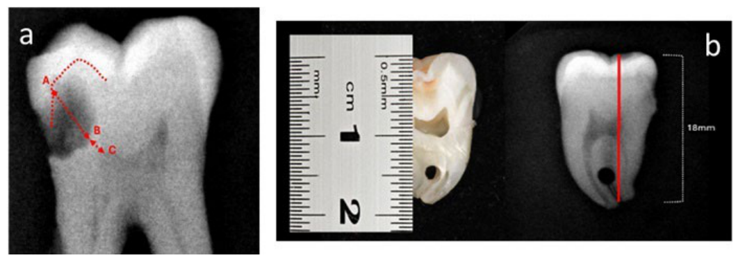
Figure 2.
Representative image illustrating the measurement of the different dentin types identified after caries excavation.
Figure 2.
Representative image illustrating the measurement of the different dentin types identified after caries excavation.
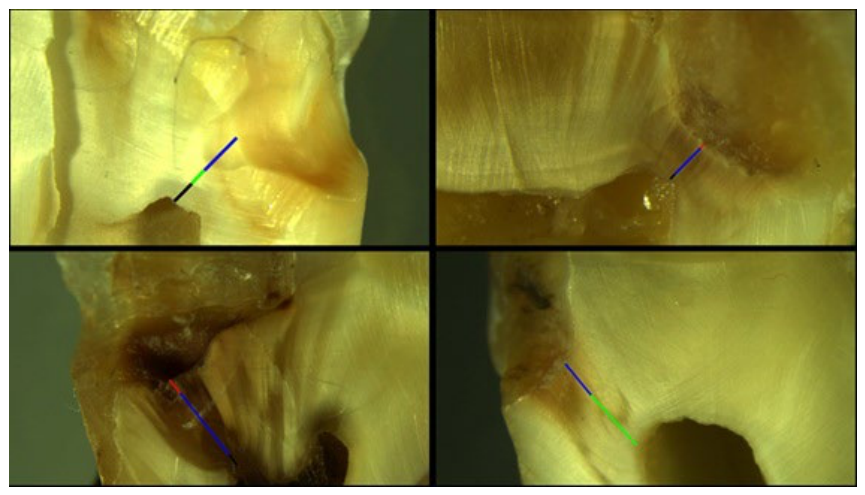

Table 1.
Brix3000 versus conventional excavation.
| Variable | Brix3000 mean ± SD | Control mean ± SD | Test | p |
|---|---|---|---|---|
| Percentage thickness lesion Rx | 70.57 ± 6.79 | 66.92 ± 9.30 | Paires t-Test | 0.16 |
| SD pre-removal X-ray (mm) | 1.079 ± 0.27 | 1.190 ± 0.49 | Paires t-Test | 0.28 |
| SD post-removal X-ray (mm) | 0.823 ± 0.39 | 0.573 ± 0.52 | Paires t-Test | 0.02 |
| AD stereomicroscope (mm) | 1.138 ± 0.50 | 0.720 ± 0.30 | Paires t-Test | 0.04 |
| SD stereomicroscope (mm) | 0.920 ± 0.46 | 0.875 ± 0.55 | Wilcoxon test | 1.00 |
| RD stereomicroscope (mm) | 0.412 ± 0.30 | 0.617 ± 0.34 | Paires t-Test | 0.20 |
| RMD stereomicroscope (mm) | 1.462 ± 0.63 | 1.110 ± 0.58 | Paires t-Test | 0.01 |
mm= millimeters, SD: sound dentin, ID: infected dentin, AD affected dentin, RD: reparative dentin, RMD: remaining dentin.
Table 2.
PolyBur® versus conventional excavation.
| Variable | PolyBur mean ± SD | Control mean ± SD | Test | p |
|---|---|---|---|---|
| Percentage thickness lesion Rx | 78.06 ± 10.52 | 76.65 ± 6.77 | Paires t-Test | 0.63 |
| SD pre-removal X-ray (mm) | 0.79 ± 0.32 | 0.81 ± 0.20 | Paires t-Test | 0.75 |
| SD post-removal X-ray (mm) | 0.35 ± 0.15 | 0.26 ± 0.32 | Wilcoxon test | 0.06 |
| AD stereomicroscope (mm) | 0.99 ± 0.329 | 0.74 ± 0.41 | Paires t-Test | 0.04 |
| RD stereomicroscope (mm) | 0.40 ± 0.070 | 0.38 ± 0.11 | Paires t-Test | 0.66 |
| RMD stereomicroscope (mm) | 1.29 ± 0.466 | 1.03 ± 0.74 | Wilcoxon test | 0.43 |
mm= millimeters, SD: sound dentin, ID: infected dentin, AD affected dentin, RD: reparative dentin, RMD: remaining dentin.
Table 3.
Comparison between the two selective excavation technique.
| Variable | Brix3000 mean ± SD | Polybur mean± SD | Test | p |
|---|---|---|---|---|
| % lesion thickness on X-ray | 70.5 ± 6.79 | 78.06 ± 10.52 | t-test | 0.05 |
| SD pre-removal X-ray (mm) | 1.07 ± 0.27 | 0.79 ± 0.32 | t-test | 0.03 |
| SD post-removal X-ray (mm) | 0.82 ± 0.39 | 0.33 ± 0.35 | Mann-Whitney | 0.01 |
| ID stereomicroscope (mm) | 0.27 ± 0.15 | 0.22 ± 0.14 | t-test | 0.66 |
| AD stereomicroscope (mm) | 0.83 ± 0.40 | 0.87 ± 0.31 | Mann-Whitney | 0.73 |
| RD stereomicroscope (mm) | 0.43 ± 0.28 | 0.40 ± 0.07 | t-test | 0.86 |
| RMD stereomicroscope (mm) | 1.39 ± 0.64 | 1.18 ± 0.49 | t-test | 0.50 |
mm= millimeters, SD: sound dentin, ID: infected dentin, AD affected dentin, RD: reparative dentin, RMD: remaining dentin.
Table 4.
Working times in seconds.
| mean | SD | p | |
|---|---|---|---|
| Removal time Brix3000® | 461.81a | 115.31 | <0.001 |
| Removal time control | 171.54 | 65.31 | |
| Removal time control Polybur® | 223.06a | 64.95 | 0.003 |
| Removal time control | 181.81 | 46.11 |
The same letter in the superscript indicates significant differences between the two experimental groups.
Disclaimer/Publisher’s Note: The statements, opinions and data contained in all publications are solely those of the individual author(s) and contributor(s) and not of MDPI and/or the editor(s). MDPI and/or the editor(s) disclaim responsibility for any injury to people or property resulting from any ideas, methods, instructions or products referred to in the content. |
© 2026 by the authors. Licensee MDPI, Basel, Switzerland. This article is an open access article distributed under the terms and conditions of the Creative Commons Attribution (CC BY) license (http://creativecommons.org/licenses/by/4.0/).
Copyright: This open access article is published under a Creative Commons CC BY 4.0 license, which permit the free download, distribution, and reuse, provided that the author and preprint are cited in any reuse.