

Submitted:
06 August 2026
Posted:
14 August 2026
You are already at the latest version
Abstract
Background: Computer-aided diagnosis (CAD) algorithms may be useful for the detection of pulmonary nodules, some of them early-stage primary lung cancers (LCs). Method: A CAD algorithm for the identification of pulmonary nodules in chest x-rays was used in a prospective cohort of patients without respiratory symptoms who underwent an x-ray. Chest x-rays were sorted according to the probability of the presence of a pulmonary nodule, and images placed in the highest percentiles by the algorithm were read by a radiologist, checked for previous diagnoses, and assessed for new primary LCs. Results: The radiological study confirmed the presence of a round opacities in 88 out of the 1842 chest x-rays placed in the highest percentiles (4.8%), 44 of which were found to be malignant (50%), and 7 of them undiagnosed primary LCs (7.9%). The CAD algorithm prompted a new diagnosis of primary LC in 0.4% of the sorted chest x-rays. Conclusion: A CAD algorithm prospectively identified chest x-rays with a high probability of pulmonary nodules among patients without respiratory symptoms. Radiologist-confirmed nodules were found in 4.8% of high-probability images, and previously undiagnosed primary lung cancers accounted for near 8% of confirmed round opacities and 1% of high-probability chest x-rays.
Keywords:
chest x-ray
; computed-assisted diagnosis
; lung cancer
; early diagnosis
Introduction
Lung cancer (LC) is a frequent medical condition and has a low survival, mainly attributable to the late diagnosis of the disease, given that more than three quarters of cases already present regional progression or metastasis when identified [1,2,3,4,5,6]. Early detection improves life expectancy [7], but diagnosis during the initial period of the disease is difficult due to the absence or non-specificity of symptoms [8]. When a chest radiograph is performed in patients without respiratory symptoms for unrelated reasons and a LC is seen, the disease is often in a preclinical phase (9] and when in early stage a LC may be surgically treatable and potentially curable without additional therapies [10]. However, when the diagnosis is delayed and is made when symptoms appear the disease is often advanced and presents lower survival rates [11,12,13,14].
Chest x-ray is the most frequent radiologic exam in thoracic diseases. However, up to three quarters of these tests are performed for non-respiratory diseases in the absence of respiratory symptoms, and often are only examined by non-radiologists. In this situation a pulmonary nodule that may be an early-stage LC can be missed [10,11,12,13,15,16,17], causing a clinically significant delay in the diagnosis [8], that will have a negative impact on survival [11,12,13,15,16,17,18]. Nowadays it is possible to integrate computer-aided diagnosis (CAD) algorithms in centralized digital medical imaging repositories, where they may be able to identify pulmonary nodules [19,20,21,22], some of which would be early-stage LCs [23].
A CAD algorithm based on convolutional neural networks [24,25] recently demonstrated its usefulness for the identification of round opacities in a retrospective study of chest x-rays performed for non-respiratory diseases and examined only by physicians untrained in lung imaging [26]. The algorithm sorted the baseline chest x-rays according to the probability of the presence of a pulmonary nodule, and images considered in the higher range were subsequently read by a radiologist. One tenth of the chest x-rays pointed by the algorithm with a round opacity subsequently confirmed by the radiologist had a LC diagnosed months after the performance of the baseline x-ray. The current investigation comprised a prospective internal validation of the CAD algorithm used in that retrospective study. The frequency of round opacities in chest x-rays considered in the higher probability range by the algorithm was determined, together with the proportion of LCs in this group, so as to validate the previous results of that retrospective analysis in real-time clinical practice.
Methods
Rationale
A CAD algorithm for the identification of pulmonary nodules was created from an anonymized chest x-ray repository available in Hospital Universitari Parc Taulí, and was used for the first time in a clinical retrospective study of chest x-rays performed in 2008 for non-respiratory reasons, which had been examined only by clinical physicians. In that study a radiologist confirmed a round opacity in one quarter of the images placed in the highest probability percentile by the algorithm, and three per cent of these images corresponded to a LC undiagnosed at baseline and only correctly labelled with a delay of one year [26]. The present manuscript reports the results of a prospective observational internal validation study following the same approach.
Prospective Validation Study
To assess the performance of a CAD algorithm for the identification of early-stage LC in real-time clinical practice, posteroanterior chest x-rays performed in the emergency rooms of the Hospital Universitari Parc Taulí during one semester in patients without respiratory symptoms were prospectively sorted by the algorithm according to the probability of the presence of a pulmonary nodule. The studied chest x-rays were all baseline images obtained in the emergency rooms for any clinically significant non-respiratory disease that required a diagnosis, which were read by the clinical physician attending the patient. An operational approach was followed every working day, with the selection of the 20 chest x-rays with the highest probability of a pulmonary nodule, according the CAD algorithm, that were subsequently examined by one chest radiologist for confirmation [27]. This approach was followed over the course of 99 working days. Duplicates and x-rays performed in patients who died in the following month and before diagnosis were not considered for further study. Known cancers were identified from clinical records. Patients with a chest x-ray considered by the algorithm to present a pulmonary nodule who had a round opacity confirmed by a radiologist that had not been previously studied were referred to a fast-track LC clinic for diagnosis. New cases were confirmed by biopsy and/or a subsequent increase in size of the image in accordance with the natural history of LC. The final diagnoses of the round opacities confirmed by the radiologist were subsequently recorded, to determine the frequency of primary and secondary LCs among them, either previously diagnosed or undiagnosed. The data obtained were anonymized prior to analysis.
The present study was reviewed and approved by the Comitè de Ètica de la Investigación con Medicamentos Parc Taulí de Sabadell. The guidelines followed in this study complied with the applicable Spanish regulations (Biomedical Research Law 14/2007).
The research performed involved human subjects, and the investigations were carried out following the rules of the Declaration of Helsinki of 1975, revised in 2013, and after the approval from the Institutional Ethics Committee (Project Identification code 2018619 / date of approval October 22th 2018), that confirmed that the study meets national and international guidelines. The research performed did not include procedures outside the common practice, and all obtained data had been anonymized before its inclusion on the database of the project. Accordingly, all subsequent analyses had not included any personal information of the studied patients. The Institutional Ethics Committee accepted the anonimizing procedure followed and, considering that the study did not require additional procedures ahead of the usual practice, did not require an informed consent signed the participants, in accordance with the local legislation (BPC (CPMP/ICH/135/95).
Statistical Analysis
Results for categorical variables are expressed as absolute and relative frequencies and results for continuous variables as medians and ranges when required. In accordance with the operational approach followed in the study, on each working day the chest x-rays performed in patients attending the hospital for non-respiratory illnesses were identified, and the 20 chest x-rays considered by the CAD algorithm at the highest probability for a pulmonary nodule after sorting were selected, expressing their placement in the probability scale as percentile. The percentile that included more than 90% of the images in the operational database was identified and used as a cut-off, and the subgroup of chest x-rays above this percentile was selected as the sample to be studied from the original data set. Clinical records from patients with radiologist-confirmed round opacities were examined for previous LC or other primary cancers. For undiagnosed images, a new primary LC diagnosis was accepted when a confirmative biopsy was obtained and/or the subsequent growth of the round opacity was compatible with the natural history of LC, in the absence of an active cancer elsewhere. The prevalences of confirmed round opacities in the high probability chest x-rays and that of LC diagnoses, both previously known and new, were determined. The size of the primary LCs diagnosed after the CAD algorithm identification was measured in order to establish the proportion of LCs with diameters characteristic of early-stage LC, according the 9th edition of the TNM classification, which would be surgically treatable in the absence of a regional or distant dissemination (stages I or IIa / <5 cm diameter). All analyses were performed using SPSS statistical software package version 28.0.1.0 (SPSS, IBM, Armonk, New York, USA).
Results
During the study period, the CAD algorithm prospectively selected the 20 chest x-rays with the highest estimated probability of a pulmonary nodule on each working day. This operational approach was maintained for 99 working days, resulting in 1980 selected chest x-rays. Five images were excluded from subsequent analysis because they were duplicates (n=2) or corresponded to patients who died shortly after the x-ray and before a diagnostic assessment could be completed (n=3). Therefore, 1975 chest x-rays were evaluable (Figure 1). The evaluable chest x-rays had been placed between percentiles 99 and 34 by the CAD algorithm after daily sorting according to the estimated probability of a pulmonary nodule (median 88, IQR 81–93). Most selected images were included when a percentile >70 was used as the cut-off for the main analysis and conform the analysed sample (1842/1975, 93.3%). On 70 of the 99 working days (70.7%), all selected chest x-rays were placed above this percentile.
A round opacity was confirmed by the radiologist in 88 cases among the 1842 analysed high-probability chest x-rays, and correspond to 4.8% of the high-probability images. Of the 88 radiologist-confirmed round opacities, 44 were malignant, corresponding to 50.0% of confirmed images and 2.4% of the high-probability chest x-rays (Figure 1). Fourteen of the 88 confirmed round opacities corresponded to previously undiagnosed cancers, representing 15.9% of round images and 0.8% of the high-probability chest x-rays. These cancers included seven new metastases in the lung and seven undiagnosed primary lung cancers, which corresponded to 7.9% of the confirmed round opacities and 0.4% of the high-probability chest x-rays (Figure 1).
The clinical and radiological characteristics of the 14 previously undiagnosed cancers are shown in Table 1. Among the seven newly identified primary lung cancers, three had histological confirmation, including two small cell lung cancers and one non-small cell lung cancer, whereas four were diagnosed clinically based on progressive enlargement compatible with the natural history of lung cancer. Five of the seven newly identified primary lung cancers measured ≤5 cm, a size compatible with an earlier-stage presentation, in the absence of nodal or distant spread. Overall, the CAD-based strategy identified previously undiagnosed primary lung cancer in 0.4% of high-probability chest x-rays performed in patients without respiratory symptoms.
Discussion
In this prospective study we validated the use of a CAD algorithm created for the identification of pulmonary nodules in chest x-rays, which has previously shown its usefulness on a retrospective set of thoracic images [26]. The CAD algorithm was used to prospectively sort chest x-rays of patients without respiratory symptoms who attended the emergency rooms for unrelated diseases, and identified images with the highest probability of a pulmonary nodule. The radiologist confirmed the presence of a round opacity in five per cent of chest x-rays pointed by the CAD algorithm, and near eight percent of these images were undiagnosed primary LCs. Overall, the use of the CAD algorithm in clinical practice was able to identify new pulmonary malignancies in up to one per cent of chest x-rays considered by the algorithm at high probability for a pulmonary nodule.
This prospective study confirmed in real clinical practice that the use of a CAD algorithm designed to detect pulmonary nodules in chest x-rays is able to identify high-probability images in patients attending the emergency rooms for non-respiratory diseases. A half of the images pointed by the CAD algorithm with a round opacity subsequently confirmed by the radiologist were malignant, and up to eight percent of these images were primary LC, a result that confirms a previous retrospective analysis with the same CAD algorithm [26]. Missing pulmonary nodules in the chest x-ray is a major cause of delay in the diagnosis of primary LC, and has demonstrated a negative impact on the prognosis of the disease [11,12,13]. Examining 587 patients with LC, Singh and cols found missed pulmonary nodules in previously available chest x-rays in one third of patients in whom a LC was identified half a year after the first abnormal x-ray [8]; and similar results were obtained in a retrospective assessment of the CAD algorithm used in the present study [26]. Nam and cols. have also reported that a CAD algorithm was able to improve nodule identification in chest x-rays [28], doubling the detection rate of pulmonary nodules, part of them primary LCs, when compared with usual care, and similar results have been reported by other authors [29,30]. Our results confirmed that the use of a CAD algorithm designed to detect pulmonary nodules facilitates the recognition of round opacities in patients attending the emergency rooms for non-respiratory diseases, some of them undiagnosed primary LCs.
This study has several limitations. First, it was a single-center internal validation study performed in one hospital, and external validation in other institutions, imaging systems, and clinical workflows would be required before the results can be generalized. Second, the study followed an operational approach in which only the 20 chest x-rays with the highest estimated probability of a pulmonary nodule were reviewed each working day. Therefore, the present design allows assessment of the clinical yield of the CAD-based strategy, but does not allow estimation of sensitivity, specificity, negative predictive value, or the number of pulmonary nodules and lung cancers that may have been present among lower-ranked images. Third, the main analysis focused on chest x-rays placed above percentile 70 by the CAD algorithm. This cut-off was selected pragmatically because it included most images in the operational database, but it was not designed to optimize diagnostic accuracy. Fourth, the study included posteroanterior chest x-rays performed in patients without respiratory symptoms attending the emergency room for non-respiratory diseases. Therefore, the results may not apply to symptomatic patients. Finally, the study was designed to identify previously undiagnosed malignancies among CAD-selected high-probability images, but it did not assess downstream outcomes such as treatment changes, surgical resection rates, stage migration, survival, cost-effectiveness, or the potential burden of false-positive findings.
Conclusion
A CAD algorithm focused on the detection of pulmonary nodules identified a subgroup of chest x-rays from patients without respiratory symptoms in whom round opacities were subsequently confirmed by a radiologist in 4.8% of cases. Half of these radiologist-confirmed nodules were malignant, and previously undiagnosed primary lung cancers accounted for near 8.0% of confirmed nodules and 0.4% of high-probability chest x-rays. These findings support the potential role of CAD algorithms as complementary safety tools for identifying clinically relevant unsuspected pulmonary nodules in routine chest x-rays performed for non-respiratory reasons.
Funding
The computer-aided diagnosis algorithm creation, clinical research and manuscript preparation have been partly funded by Instituto de Salud Carlos III DTS21/00135, Asociación Española Contra el Cáncer, Ciber de Enfermedades Respiratorias - Ciberes and Agència de Gestió d'Ajuts Universitaris i de Recerca – AGAUR and CERCA Programme - Generalitat de Catalunya.
Acknowledgments
The authors thank Michael Maudsley for providing an outline for this manuscript.
Data Availability Statement
The datasets generated during and/or analysed during the current study are available from the corresponding author on reasonable request.
Code Availability
The custom computer code / algorithms used in this study are not publicly available. Notwithstanding, the corresponding author may make them available on reasonable request.
References
- Global Cancer Observatory: Cancer Today. Lyon, France: International Agency for Research on Cancer. In https://gco.iarc.fr/today/home. Accessed in September 2025.
- Ferlay J, Colombet M, Soerjomataram I, Parkin DM, Piñeros M, Znaor A, et al. Cancer statistics for the year 2020: An overview. Int J Cancer 2021; 149: 778-789. [CrossRef]
- Filho AM, Laversanne M, Ferlay J, Colombet M, Piñeros M, Znaor A, et al.The GLOBOCAN 2022 cancer estimates: Data sources, methods, and a snapshot of the cancer burden worldwide. Int J Cancer 2025; 156:1336-1346. [CrossRef]
- Detterbeck FC, Woodard GA, Bader AS et al. The Proposed Ninth Edition TNM Classification of Lung Cancer. Chest 2024; 166:882–895. [CrossRef]
- Huang J, Deng Y, Tin MS, Lok V, Ngai CH, Zhang L, et al. Distribution, Risk Factors, and Temporal Trends for Lung Cancer Incidence and Mortality. A Global Analysis Chest 2022; 161:1101-1111. [CrossRef]
- Rami-Porta R, Nishimura KK, Giroux DJ, Detterbeck F, Cardillo G, Edwards JG, et al. The International Association for the Study of Lung Cancer Lung Cancer Staging Project: Proposals for Revision of the TNM Stage Groups in the Forthcoming (Ninth) Edition of the TNM Classification for Lung Cancer. J Thorac Oncol 2024; 19:1007–1027. [CrossRef]
- Olsson JK, Schultz EM, Gould MK. Timeliness of care in patients with lung cancer: a systematic review. Thorax 2009; 64:749-56. [CrossRef]
- Singh H, Hirani K, Kadiyala H, et al. Characteristics and predictors of missed opportunities in lung cancer diagnosis: an electronic health record-based study. J Clin Oncol 2010; 28:3307-15. [CrossRef]
- Ades A, Biswas M, Welton NJ and Hamilton W. Symptom lead time distribution in lung cancer: natural history and prospects for early diagnosis. Int J Epidemiol 2014; 43:1865-73. [CrossRef]
- Sakai M, Kato A, Kobayashi N, Nakamura R, Okawa S and Sato Y. Improved lung cancer detection in cardiovascular outpatients by the pulmonologist-based interpretation of chest radiographs. Intern Med 2015; 54:2991-7. [CrossRef]
- Turkington PM, Kennan N, Greenstone MA. Misinterpretation of the chest x ray as a factor in the delayed diagnosis of lung cancer. Postgrad Med J 2002; 78:158-60. [CrossRef]
- Quekel LGBA, Kessels AGH, Goei R and Van Engelshoven JMA. Miss rate of lung cancer on the chest radiograph in clinical practice. Chest 1999; 115:720-4.
- Kashiwabara K, Koshi S, Itonaga K, Nakahara O, Tanaka M, Toyonaga M. Outcome in patients with lung cancer found on lung cancer mass screening roentgenograms, but who did not subsequently consult a doctor. Lung Cancer 2003; 40:67-72. [CrossRef]
- Kanashiki M, Satoh H, Ishikawa H, Yamashita YT, Ohtsuka M, Sekizawa K. Time from finding abnormality on mass-screening to final diagnosis of lung cancer. Oncol Rep 2003; 10:649-52.
- Homayounieh F, Digumarthy S, Ebrahimian S Rueckel J, Hoppe BJ, Sabel BO, et al. An Artificial Intelligence–Based Chest X-ray Model on Human Nodule Detection Accuracy From a Multicenter Study JAMA Netw Open 2021; 4:e2141096.
- Graber ML, Winters BD, Matin R, Cholankeril RT, Murphy DR, Singh H, Bradford A. Interventions to improve timely cancer diagnosis: an integrative review. Diagnosis (Berl) 2024; 12:153-162. [CrossRef]
- Gatt ME, Spectre G, Paltiel O, Hiller N, Stalnikowicz R. Chest radiographs in the emergency department: is the radiologist really necessary? Postgrad Med J 2003; 79:214-7. [CrossRef]
- Borg M, Kristensen K, Alstrup G, Mamaeva T, Arshad A, Laursen CB, et al. Consequences of Losing Incidental Pulmonary Nodules to Follow-Up: Unmonitored Nodules Progressing to Stage IV Lung Cancer. Respiration 2024; 103:53-59. [CrossRef]
- Ueda D, Yamamoto A, Shimazaki A, Walston SL, Matsumoto T, Izumi N, et al. Artificial intelligence-supported lung cancer detection by multi-institutional readers with multi-vendor chest radiographs: a retrospective clinical validation study. BMC Cancer 2021; 21:1120. [CrossRef]
- Robert D, Sathyamurthy S, Singh AK, Matta SA, Tadepalli M, Tanamala S, et al. Effect of Artificial Intelligence as a Second Reader on the Lung Nodule Detection and Localization Accuracy of Radiologists and Non-radiology Physicians in Chest Radiographs: A Multicenter Reader Study. Acad Radiol 2025; 32:1706–1717. [CrossRef]
- Topff L, Steltenpool S, Ranschaert ER, Ramanauskas N, Menezes R, Visser JJ, et al. Artificial intelligence-assisted double reading of chest radiographs to detect clinically relevant missed findings: a two-centre evaluation. Eur Radiol 2024; 34:5876-5885. [CrossRef]
- Wang C, Elazab A, Wu J, Hu Q. Lung nodule classification using deep feature fusion in chest radiography. Comput Med Imaging Graph 2017; 57:10-18. [CrossRef]
- Horry M, Chakraborty S, Pradhan B, Paul M, Gomes D, Ul-Haq A, et al. Deep Mining Generation of Lung Cancer Malignancy Models from Chest X-ray Images. Sensors (Basel) 2021; 21:6655. [CrossRef]
- Chen S, Han Y, Lin J, Zhao X, Kong P. Pulmonary nodule detection on chest radiographs using balanced convolutional neural network and classic candidate detection. Artif Intell Med 2020; 107:101881. [CrossRef]
- Lo SB, Lou SA, Lin JS, Freedman MT, Chien MV, Mun SK. Artificial convolution neural network techniques and applications for lung nodule detection. IEEE Trans Med Imaging 1995; 14:711-8. [CrossRef]
- Juan J, Monso E, Lozano C, Cufí M, Subías-Beltrán P, Ruiz-Dern L, et al. Computer-assisted diagnosis for an early identification of lung cancer in chest X rays. Sci Rep 2023; 13:7720. [CrossRef]
- Kirshenboim Z, Gilat EK, Carl L, Bekker E, Tau N, Klug M, et al. Retrospectively assessing evaluation and management of artificial-intelligence detected nodules on uninterpreted chest radiographs in the era of radiologists shortage. Eur J Radiol 2024;170:111241. [CrossRef]
- Nam JG, Hwang EJ, Kim J, Park N, Lee EH, Kim HJ. AI Improves Nodule Detection on Chest Radiographs in a Health Screening Population: A Randomized Controlled Trial. Radiology 2023; 307:e221894.
- Essa MEA. Diagnostic accuracy of AI in chest radiography for pneumonia and lung cancer: A meta-analysis. Eur J Radiol Open 2025; 15:100701. [CrossRef]
- Robert D, Sathyamurthy S, Singh AK, Matta SA, Tadepalli M, et al. Detection and Localization Accuracy of Radiologists and Non-radiology Physicians in Chest Radiographs: A Multicenter Reader Study. Acad Radiol 2025; 32:1706–17. [CrossRef]
Figure 1.
Flowchart of computer-assisted diagnosis selected chest x-rays and diagnostic yield.
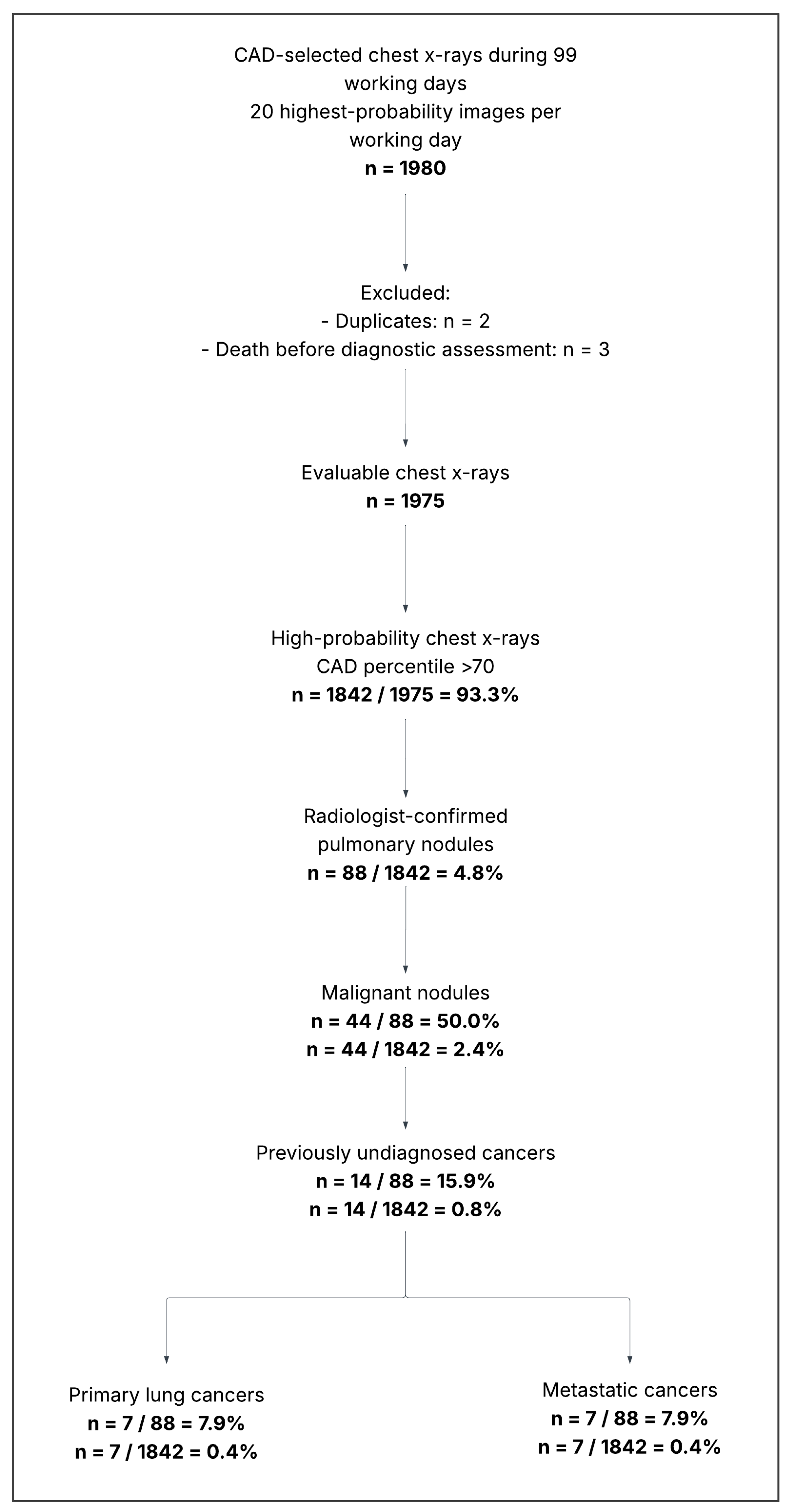

Table 1.
- New Metastasic And Primary Lung Cancers, Identified Through Computed-Assisted Diagnosis. *Clinical diagnosis based on progressive enlargement compatible with the natural history of lung cancer (see text for details).
Table 1.
- New Metastasic And Primary Lung Cancers, Identified Through Computed-Assisted Diagnosis. *Clinical diagnosis based on progressive enlargement compatible with the natural history of lung cancer (see text for details).
| Case | Chest x-ray finding | Size (cm) | Diagnosis |
|---|---|---|---|
| New metastatic cancers | |||
| 1 | Left lung nodule | 2 | Colorectal cancer |
| 2 | Bilateral nodules | Up to 3 | Colorectal cancer |
| 3 | Right lung nodule | 3 | Breast cancer |
| 4 | Bilateral round lung opacities | Up to 6 | Laryngeal cancer |
| 5 | Right lung nodule | 3 | Colorectal cancer |
| 6 | Left lung nodule | 2 | Colorectal cancer |
| 7 | Right lung nodule | 2 | Colorectal cancer |
| New primary lung cancers | |||
| 8 | Upper left lobe round opacity | 4 | Clinical diagnosis |
| 9 | Upper right lobe mass | 7 | Clinical diagnosis |
| 10 | Lower left lobe round opacity | 5 | Clinical diagnosis |
| 11 | Round mediastinal enlargement | 3 | Small cell lung cancer |
| 12 | Right pulmonary round opacity | 6 | Clinical diagnosis |
| 13 | Upper left lobe round opacity | 4 | Non-small cell lung cancer |
| 14 | Round right hilar image | 5 | Small cell lung cancer |
Disclaimer/Publisher’s Note: The statements, opinions and data contained in all publications are solely those of the individual author(s) and contributor(s) and not of MDPI and/or the editor(s). MDPI and/or the editor(s) disclaim responsibility for any injury to people or property resulting from any ideas, methods, instructions or products referred to in the content. |
© 2026 by the authors. Licensee MDPI, Basel, Switzerland. This article is an open access article distributed under the terms and conditions of the Creative Commons Attribution (CC BY) license (http://creativecommons.org/licenses/by/4.0/).
Copyright: This open access article is published under a Creative Commons CC BY 4.0 license, which permit the free download, distribution, and reuse, provided that the author and preprint are cited in any reuse.