

Submitted:
10 August 2026
Posted:
12 August 2026
You are already at the latest version
Abstract
Background/Objectives: Ultrasound-guided percutaneous lavage (UGPL) is an established minimally invasive treatment for symptomatic calcific tendinitis of the shoulder after failure of conservative management. However, treatment outcomes are variable, and the prognostic significance of symptom duration and radiographic characteristics of calcific deposits remains uncertain. This study evaluated the clinical and radiographic outcomes of UGPL and identified predictors of treatment failure. Methods: A retrospective analysis of prospectively collected data was performed on 65 patients who underwent UGPL for rotator cuff calcific tendinitis between 2018 and 2022. Patients were stratified into an acute group (symptom duration < 7 days, severe pain, Gärtner type III deposits) and a chronic group (symptom duration >15 days, moderate pain, Gärtner type I–II deposits). Clinical outcomes included pain assessed with the visual analog scale (VAS), shoulder range of motion, and Rotator Cuff Quality of Life (RC-QOL) scores. Radiographic and ultrasound examinations evaluated calcification morphology, size, and resorption before treatment and at 3 months. Treatment failure was defined as persistent pain (VAS >5 at 6 months) and/or the need for arthroscopic surgery. Results: Patients in the acute group demonstrated significantly greater improvements in pain, shoulder mobility, and RC-QOL scores at all follow-up evaluations compared with the chronic group (all p < 0.001). Complete radiographic resorption of calcifications occurred in 77% of acute cases, whereas 68% of chronic cases showed no radiographic change. Calcification size decreased significantly more in the acute group (p < 0.001). Arthroscopic surgery was required in 15% of acute patients compared with 41% of chronic patients (p = 0.017). Chronic symptom duration, dense Gärtner type I–II calcifications, and smaller baseline deposits were associated with an increased risk of treatment failure. Conclusions: UGPL provides excellent clinical and radiographic outcomes in patients with acute, resorptive-phase calcific tendinitis. Chronic symptoms and dense calcific deposits are associated with reduced treatment success and higher rates of surgical intervention. Combining symptom duration with radiographic morphology may improve patient selection and optimize outcomes following UGPL.
Keywords:
calcific tendinitis
; rotator cuff
; ultrasound-guided percutaneous lavage
; radiographic resorption
; prognostic factors
1. Introduction
Calcific tendinitis of the shoulder is a common cause of pain and disability, characterized by the deposition of calcium hydroxyapatite crystals within the rotator cuff (RC) tendons, most frequently the supraspinatus tendon [1,2]. The condition typically affects adults between the fourth and sixth decades of life and is more prevalent in women [1,3].
Although its etiology remains incompletely understood, calcific tendinitis is currently regarded as a cell-mediated process rather than a purely degenerative disorder. The pathogenetic model proposed by Uhthoff and Loehr describes a sequence of precalcific, calcific, and post-calcific stages, with the calcific phase including formative, resting, and resorptive phases[4]. It is in the reabsorptive phase the patients are mostly symptomatic. The postcalcific phase is the healing phase in which there is reabsorption of the deposit. Rui et al. postulated that incorrect differentiation of stem cells, Tendon-Derived Stem Cells, into osteoblasts or chondrocytes, could be the basis of calcification[5].
Clinical manifestations vary according to disease stage, ranging from mild chronic symptoms to severe acute pain associated with spontaneous calcium resorption.
Conservative treatment, including nonsteroidal anti-inflammatory drugs (NSADs), physiotherapy, and corticosteroid injections, represents the first-line approach for most patients. Although spontaneous resolution may occur, a substantial proportion of patients experience persistent pain and functional limitation despite prolonged nonoperative management[6]. In these cases, surgical removal of calcific deposits has traditionally been considered the definitive treatment, providing reliable pain relief and functional improvement [1,7]. However, surgery is associated with higher costs, operative morbidity, and longer recovery periods.
Ultrasound-guided percutaneous needling and lavage (UGPL), also known as barbotage, has emerged as a minimally invasive alternative for symptomatic calcific tendinitis. By allowing real-time visualization of the deposit, ultrasound guidance facilitates fragmentation and aspiration of the calcium material while minimizing soft-tissue damage. Several studies have demonstrated significant improvements in pain, shoulder function, and patient satisfaction following UGPL [8,9,10]. Furthermore, randomized controlled trials have shown that UGPL provides superior outcomes compared with isolated corticosteroid injections and may achieve results comparable to surgical treatment in selected patients[6,11]. Consequently, UGPL is currently considered one of the preferred interventional treatments for patients who fail conservative therapy.
Despite its overall effectiveness, treatment outcomes after UGPL are not uniform[12,13,14]. A proportion of patients continue to experience persistent symptoms or require additional procedures, including repeat lavage or arthroscopic removal of the deposit [13]. Therefore, identifying predictors of treatment success and failure has become increasingly important.
Among the factors potentially influencing outcomes, the radiographic characteristics of calcium deposits have received considerable attention. The classification proposed by Gärtner and Heyer categorizes calcifications according to their radiographic appearance and density[15]. Dense, homogeneous, and sharply demarcated deposits (Type I) are believed to represent the formative phase of calcification, whereas less dense and heterogeneous deposits (Types II and III) are more frequently associated with the resorptive phase. Previous studies have suggested that dense deposits may be more difficult to fragment and aspirate, resulting in less favorable clinical and radiographic outcomes[12,16]. However, evidence regarding the prognostic value of deposit morphology remains inconsistent.
Symptom duration may also influence treatment effectiveness. Acute presentations are commonly associated with active calcium resorption and a softer deposit consistency, potentially facilitating aspiration during UGPL[17]. Conversely, chronic symptoms may reflect more mature and organized calcifications that are less responsive to percutaneous treatment. Although chronicity has been proposed as a negative prognostic factor, available evidence remains limited and conflicting[18,19].
Given the growing use of UGPL and the lack of consensus regarding predictors of outcome, further investigation is warranted. The purpose of this prospective comparative study was to evaluate the clinical and radiographic outcomes of UGPL in patients with calcific tendinitis of the shoulder and to identify predictors of treatment failure. Particular attention was directed toward the radiological status of calcium deposits and symptom duration. We hypothesized that dense calcific deposits and chronic symptom duration are associated with a higher likelihood of treatment failure following UGPL.
2. Materials and Methods
This retrospective analysis was based on prospectively collected data from patients who underwent UGPL for calcific tendinitis of the shoulder at our Shoulder and Elbow Unit between January 2018 and December 2022. Given the retrospective nature of the analysis, Institutional Review Board approval was not required.
Patients were eligible if they were aged ≥18 years, had a clinical diagnosis of RC calcific tendinitis confirmed by radiography and ultrasound (US), had not received subacromial or local corticosteroid injections within the preceding 4 months, and were available for follow-up throughout the study period.
Patients were excluded if they had a history of shoulder trauma, previous arthroscopic or open shoulder surgery, shoulder infection or neoplasm, rheumatoid arthritis or other autoimmune diseases, severe systemic medical conditions, or pregnancy. Additional exclusion criteria included the need for concomitant surgical procedures, cognitive impairment that could compromise the ability to provide informed consent or complete the clinical and patient-reported assessments, and a history of allergic or adverse reactions to NSADs or hyaluronic acid injections.
An a priori power analysis was performed assuming a 4-point difference in the visual analog scale (VAS) pain score between groups, a standard deviation of 3.5 points, and a minimal clinically important difference (MCID) of 1.8 points. With a statistical power of 90%, the required sample size was calculated to be at least 17 patients per group.
The diagnosis of calcific tendinitis was established based on the presence of shoulder pain, tenderness, and restricted range of motion, and was confirmed by radiographic and ultrasound examinations.
2.1. Imaging Evaluation
2.1.1. Standard Radiographs
Standard shoulder radiographs in antero-posterior Grashey, outlet and axillary views were used to allow localization and assessing the texture and morphology of the deposit according to Gartner criteria as follow[20,21]: type I (calcific deposit clearly circumscribed with a dense appearance); type II (hybrid type, clearly circumscribed and translucent, cloudy and dense); type III (cloudy and translucent appearance without clear circumscription). Types I and III correspond to the phormative and resorptive phase, respectively; type II is an hybrid texture.
All patients in the study population underwent radiographic evaluation before and 90 days (89 ± 2 days) after the procedure of UGPN to assess the evolution of calcium deposit (resorption – complete or partial – or unchanged).
2.1.2. US Assessment
US examinations were performed by two orthopedic surgeons with 10 years of experience in shoulder ultrasonography, using a GE Logiq 7 system equipped with a high-frequency 7.5–14 MHz linear transducer. We examined the affected shoulder according to guidelines issued by the European Society of Musculoskeletal Radiology [22]. US confirmed the presence of calcific deposits identified on radiographs and was used to determine their location within the RC tendons, as well as their size (mm) and echotexture. Deposit morphology and stage were further characterized based on posterior acoustic shadowing patterns, as previously described[1,23,24]. In the resting (formative) phase, deposits appear hyperechoic with a continuous arc-shaped shadow, whereas in the resorptive phase they appear non-arc-shaped, including fragmented, punctate, cystic, or nodular patterns. These sonographic appearances have been correlated with symptomatic and asymptomatic stages of calcific tendinopathy.
The subscapularis tendon was assessed with the arm in external rotation and the elbow resting on the iliac crest. It was evaluated in both longitudinal (transverse probe orientation) and short-axis (sagittal probe orientation) planes during passive internal and external rotation with the arm in a neutral hanging position.
The supraspinatus tendon was evaluated with the arm positioned posteriorly, which brings the tendon into a more anterior position and allows optimal probe alignment. The transducer was oriented approximately vertically and moved superiorly and posteriorly over the tendon while maintaining its orientation to obtain long-axis views. In this view, the subacromial–subdeltoid bursa appears as a thin hypoechoic layer between the supraspinatus tendon and the deltoid muscle. For short-axis evaluation, the tendon was scanned from the biceps tendon landmark posteriorly for approximately 2 cm, beyond which the infraspinatus tendon is visualized.
For longitudinal assessment of the infraspinatus and teres minor tendons, the patient’s arm was placed across the chest in slight flexion and internal rotation, allowing visualization from a posterior-superior approach.
As with radiographic assessment, US was performed before and 90 days (89 ± 2 days) after UGPN to evaluate changes in the size and echotexture of any residual calcific deposits.
2.1.3. UGPN Procedure
The procedure is typically performed with the patient in the beach-chair position under sterile conditions. After aseptic preparation of the skin and ultrasound transducer, the calcific deposit is identified along its longitudinal axis. Local anesthesia is then administered by infiltrating 8–10 mL of lidocaine into the subacromial bursa and surrounding peri-tendinous soft tissues, including the area adjacent to the calcific deposit.
Two 20-gauge needles are used. The first needle is advanced into the inferior portion of the calcific deposit, while the second is inserted parallel and slightly more superficial, with the bevel oriented in the opposite direction to establish a lavage circuit. Saline solution is intermittently injected into the deposit to increase intralesional pressure, facilitating fragmentation and dissolution of the calcific material. The resulting chalk-like suspension is aspirated through the second needle, and lavage is continued until ultrasound demonstrates adequate decompression of the deposit.
At the end of the procedure, a subacromial injection of 4 mg betamethasone is administered. The entire procedure typically requires approximately 15 minutes.
Patients are advised to rest the treated shoulder for the first 48 hours following the procedure, after which a supervised rehabilitation program with gentle active-assisted range-of-motion exercises is initiated.
2.1.4. Treatment Groups
We initially recruited an intention-to-treat population of 68 patients to be allocated to two treatment groups. Of these, 2 declined participation due to logistical issues and 3 were excluded because of incomplete clinical and radiographic data. The final study population comprised 65 patients with complete datasets, who were assigned to groups (34 in the acute group and 24 in the chronic group) according to symptom duration, pain intensity, and radiographic appearance of the calcific deposit.
Patients reporting severe pain (VAS >7), acute symptom onset (<7 days), and Gärtner Type III radiographic morphology were classified as the acute group. Patients with moderate pain (VAS score ≤ 7), chronic symptom duration (>15 days), and Gärtner Type I–II radiographic appearance were assigned to the chronic group.
A subgroup of patients who declined the proposed treatment and opted for periodic follow-up at our outpatient clinic served as the control group. These patients were not available to undergo additional radiographic examinations after the initial assessment.
2.1.5. Outcome Measures
The primary outcome measure was pain intensity, assessed using the VAS, a patient-reported scale ranging from 0 (no pain) to 10 (worst imaginable pain).
Secondary outcome measures included active shoulder range of motion and the Italian version of the Rotator Cuff Quality of Life (RC-QL) questionnaire [25,26]. Active shoulder mobility was assessed using a goniometer and included active anterior elevation (AAE), active lateral elevation (ALE), internal rotation (IR), and external rotation (ER) with the arm at the side. Internal rotation was additionally scored on a 0–10 scale based on the ability to reach the T7 vertebral level using the Apley scratch test.
The RC-QL is a disease-specific instrument designed to evaluate patients’ perception of the impact of rotator cuff disorders on quality of life. It consists of 34 items divided into five domains: symptoms and physical complaints (16 items), work-related concerns (4 items), sports and recreation (4 items), lifestyle issues (5 items), and social and emotional aspects (5 items). Each item is scored on a 100-point scale, yielding a total score ranging from 0 (worst) to 3400 (best), which is subsequently converted into a percentage score.
2.1.6. Follow-Up
Patients in the acute and chronic groups were evaluated for pain, shoulder mobility, and RC-QL score. Assessments were performed at baseline and at 1 month (30 ± 1 days), 3 months (90 ± 2 days), 6 months (180 ± 10 days), and 12 months (350 ± 14 days) after UGPN.
In the control group, pain scores were recorded at baseline, 6 months (178 ± 8 days), and 12 months (348 ± 12 days) after the index evaluation.
2.1.7. Statistical Analysis
Descriptive statistics (absolute and percentage frequency, mean, median with interquartle range [IQR], standard deviation [SD], and range) for each group were calculated for all variables.
For all outcome measures, delta score variables were calculated as the difference between the baseline score and the corresponding follow-up scores at 1, 3, 6, and 12 months.
All variables were summarized separately for each study group. Associations between individual variables and the two groups were assessed using the chi-square test for categorical variables, Student's t-test for normally distributed continuous variables, and non-parametric tests (Mann–Whitney U test) for score and delta score variables.
The relationship between all delta score variables and the clinical and demographic characteristics was evaluated using non-parametric correlation analysis (Spearman's rank correlation coefficient) and non-parametric rank-based tests (Mann–Whitney U test or Kruskal–Wallis test), according to the type of variable.
Treatment failure was defined as the need for arthroscopic surgery and/or a pain score greater than 5 points at the 6-month follow-up.
Prognostic factors associated with treatment failure were analyzed in relation to all clinical and demographic variables as well as baseline scores using the chi-square test, Student's t-test, and the Mann–Whitney U test (multivariate analysis).
3. Results
The acute and chronic groups were comparable in terms of sample size, age, sex distribution, body mass index (BMI), and dominant side. No significant differences were observed between either treatment group and the control group (Table 1).
The mean symptom duration in the affected shoulder was 4.7 days in the acute group and 34 days in the chronic group (p < 0.001).
3.1. Clinical Outcomes
Baseline pain scores were higher in the acute group (p=0.041).
In the acute group, VAS pain scores were significantly lower than baseline at 1, 3, 6, and 12 months (all p < 0.001). In contrast, pain scores in the chronic group remained higher throughout follow-up compared with baseline and were significantly higher than those of the acute group at all follow-up time points (all p < 0.001). Pain scores in the acute group were also significantly lower than those in the control group at 6 and 12 months (both p < 0.001), whereas no significant differences were observed between the chronic and control groups (Table 2).
At baseline, AAE, ALE, ER, and IR were significantly lower in the acute group compared with the chronic group (all p < 0.001). The acute group showed significant improvement in all planes of motion at 1 month (p < 0.001), with sustained improvements at 3, 6, and 12 months compared with baseline (all p < 0.001).
In the chronic group, shoulder mobility did not significantly change at any follow-up time point compared with baseline and remained significantly lower than in the acute group throughout follow-up (all p < 0.001) (Table 3)
Similarly, the RC-QL score improved significantly in the acute group at 1, 3, 6, and 12 months compared with baseline (all p < 0.001) and was significantly higher than in the chronic group at all follow-up assessments (all p < 0.001) (Table 4)
3.2. Radiographic Outcomes
The distribution of radiographic calcification type and location prior to the index procedure across the three groups is reported in Table 5.
Post-UGPN radiographs demonstrated complete resorption of calcifications in 26 shoulders (77%) and partial resorption in 8 (23%) in the acute group (Figure 1 a-b). In the chronic group, calcific deposits remained unchanged in 20 cases (68%) and showed partial resorption in 9 cases (32%) (Figure 2 a-b).
On ultrasound evaluation, Gärtner Type I–II calcific deposits were characterized by a dense, arc-shaped calcific plaque with complete acoustic shadowing, whereas Gärtner Type III calcific deposits appeared as softer, irregular deposits with partial or absent acoustic shadowing.
Calcification size in the acute group decreased from a median of 13.1 mm (IQR 12–14 mm) pre-UGPN to 0 mm (IQR 0–0.4 mm) post-procedure. In the chronic group, size decreased from 10 mm (IQR 8.2–11.3 mm) to 7 mm (IQR 0.5–10 mm) (p<0.001).
Pre-treatment prognostic factors associated with failure following UGPN for calcific tendinitis of the shoulder, as identified in multivariate analysis, are reported in Table 6.
Due to persistent pain and limited shoulder mobility, arthroscopic removal of the calcific deposit and rotator cuff repair was required in 5 patients in the acute group (15%) and in 12 patients in the chronic group (41%) (p = 0.017).
Figure 1.
(a) Anteroposterior (A–P) radiograph of the right shoulder demonstrating a Gärtner type III calcific deposit before UGPN. The patient presented with severe pain (VAS score, 10) and shoulder pseudoparalysis of 48-hour duration. (b) AP radiograph obtained 89 days after UGPN demonstrating almost complete resorption and fragmentation of the calcific deposit. At follow-up, the patient was pain-free and had regained full shoulder range of motion.
Figure 1.
(a) Anteroposterior (A–P) radiograph of the right shoulder demonstrating a Gärtner type III calcific deposit before UGPN. The patient presented with severe pain (VAS score, 10) and shoulder pseudoparalysis of 48-hour duration. (b) AP radiograph obtained 89 days after UGPN demonstrating almost complete resorption and fragmentation of the calcific deposit. At follow-up, the patient was pain-free and had regained full shoulder range of motion.
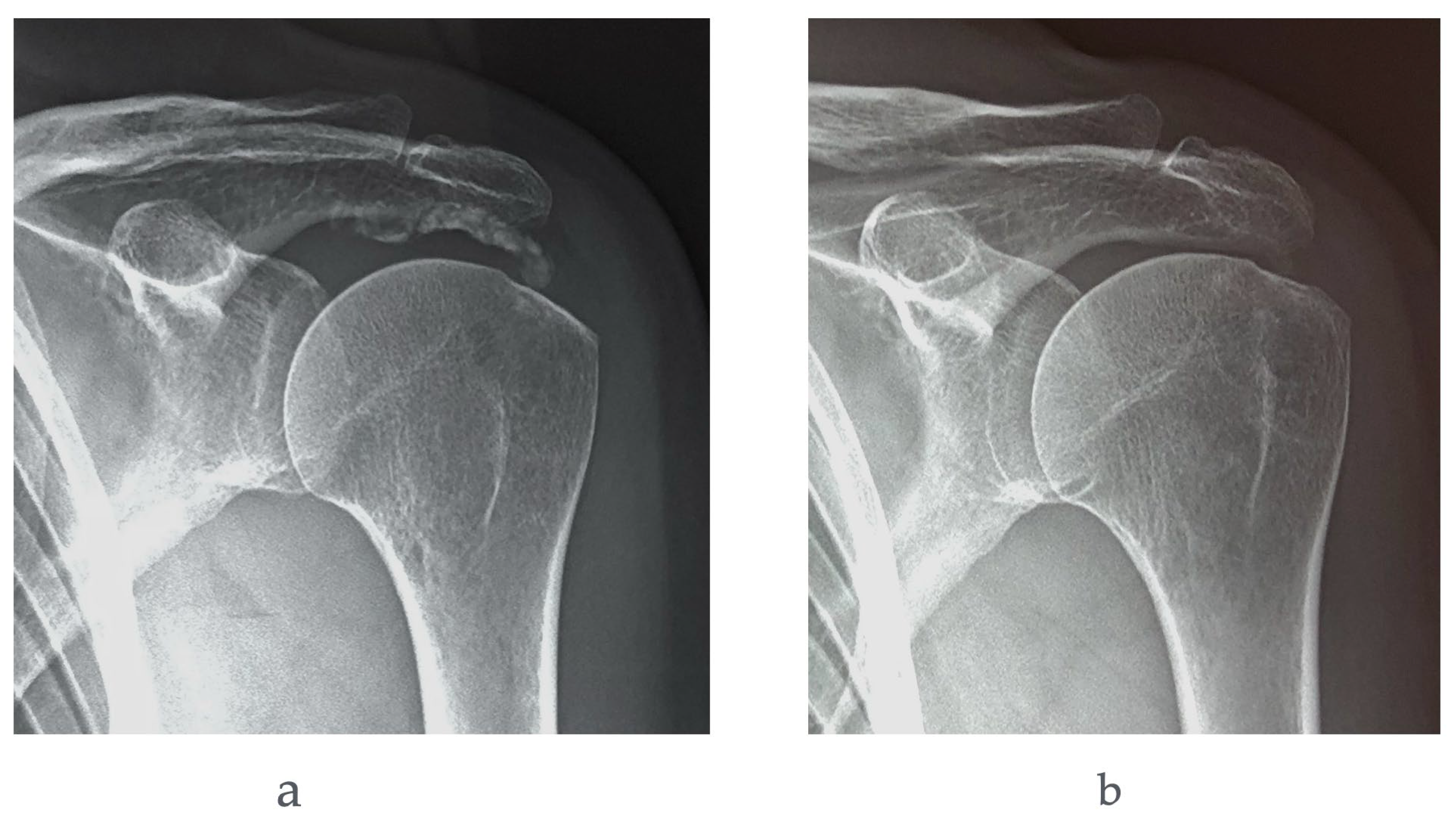
Abbreviations: A–P, anteroposterior; UGPN, ultrasound-guided percutaneous needling; VAS, visual analog scale.
Figure 2.
(a) Anteroposterior (A–P) radiograph of the left shoulder demonstrating a Gärtner type I calcific deposit before UGPN. The patient reported moderate pain (VAS score, 5) and had a full range of shoulder motion. (b) Anteroposterior radiograph obtained 91 days after UGPN showing persistence of the calcific deposit with minimal radiographic changes. The patient continued to experience persistent shoulder pain.
Figure 2.
(a) Anteroposterior (A–P) radiograph of the left shoulder demonstrating a Gärtner type I calcific deposit before UGPN. The patient reported moderate pain (VAS score, 5) and had a full range of shoulder motion. (b) Anteroposterior radiograph obtained 91 days after UGPN showing persistence of the calcific deposit with minimal radiographic changes. The patient continued to experience persistent shoulder pain.
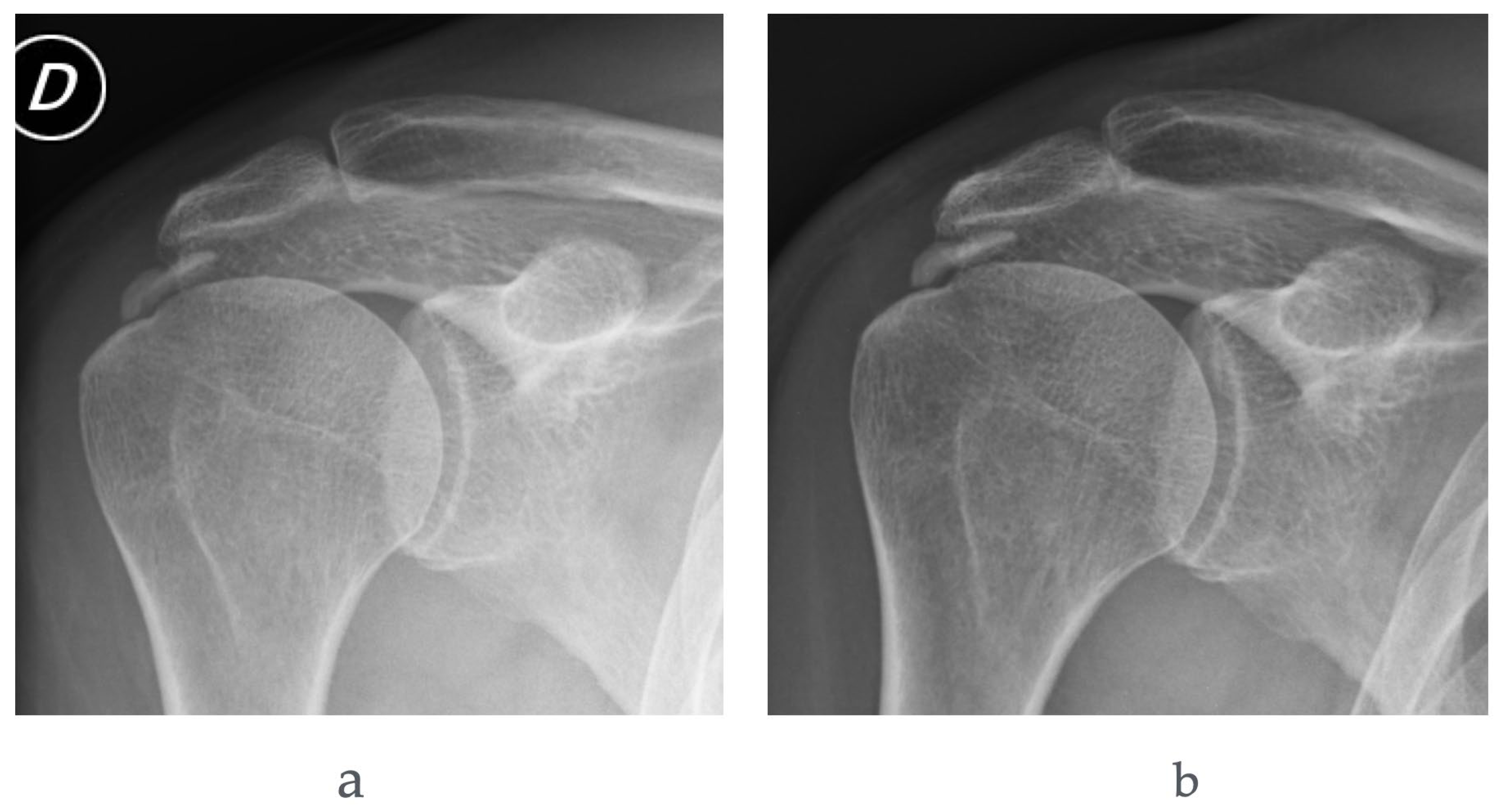
Abbreviations: A–P, anteroposterior; UGPN, ultrasound-guided percutaneous needling; VAS, visual analog scale.
4. Discussion
UGPL has been identified as an effective non-surgical interventional technique for reducing pain and improving function in patients with RC calcific tendinopathy[10,14,27,28,29]. However, controversies remain regarding the low quality of the available evidence, as well as the need to optimize patient selection and further refine the treatment protocol[30,31].
This prospective comparative study evaluated the clinical and radiographic outcomes of UGPL in patients with calcific tendinitis of the shoulder, with particular emphasis on the influence of symptom duration and radiographic characteristics of calcific deposits. The main findings of the present study are that (i) patients with acute symptom onset and radiographic features consistent with resorptive-phase calcifications achieved significantly better clinical and radiographic outcomes compared with those with chronic symptoms; (ii) the acute group demonstrated greater improvements in pain, shoulder mobility, and quality of life at all follow-up time points; and (iii) chronic symptom duration and dense calcific morphology were associated with a higher risk of treatment failure.
Overall, our findings confirm and extend previous evidence supporting the effectiveness of UGPL in calcific tendinitis. Several early studies demonstrated that ultrasound-guided barbotage provides substantial pain relief and functional improvement through mechanical disruption and aspiration of calcium deposits[8,32]. Similarly, del Cura et al. reported significant clinical improvement in patients treated with percutaneous lavage, with favorable medium-term outcomes[33]. More recent randomized controlled trials have further established UGPL as an effective minimally invasive treatment option, often superior to corticosteroid injection alone and comparable in selected cases to surgical management[6,11]. Our results are consistent with these findings, demonstrating significant and sustained improvements in pain and function in the majority of patients undergoing UGPL.
A key contribution of the present study is the stratification of outcomes according to clinical and radiographic disease status. Patients in the acute group—characterized by short symptom duration, higher pain intensity, and Gärtner Type III morphology—experienced rapid and sustained improvement following UGPL. This finding is in line with the pathophysiological model of Uhthoff and Loehr, who described the resorptive phase of calcific tendinitis as the most symptomatic stage, characterized by increased intratendinous pressure and inflammatory activity[4]. During this phase, calcific deposits are often softer and partially fragmented, which may facilitate mechanical disruption and aspiration during lavage. Our ultrasound findings support this hypothesis, as Type III deposits demonstrated more irregular and less dense echotexture, consistent with a more resorptive stage.
Conversely, patients in the chronic group showed limited improvement in both clinical and radiographic outcomes. Pain scores, range of motion, and RC-QL scores remained significantly worse than those of the acute group throughout follow-up, and radiographic resolution was less frequent. These findings suggest that chronic calcific tendinitis may represent a more stable and organized form of hydroxyapatite deposition, less responsive to percutaneous intervention. Similar observations have been reported by Ogon et al., who found that denser and well-defined calcifications were associated with poorer response to non-surgical treatments[16]. Louwerens et al. also highlighted that radiographic morphology may predict treatment response, with more homogeneous deposits showing lower rates of complete resorption after intervention[34]. Our findings reinforce these observations and suggest that radiographic classification may be a clinically useful tool for pre-treatment stratification. Only a minority of authors reported that type I calcifications were the most frequently aspirated, and the reason for this discrepancy remains unclear[35].
In addition to radiographic morphology, symptom duration emerged as a relevant prognostic factor. Patients with acute onset (<7 days) demonstrated significantly better outcomes than those with chronic symptoms (>15 days), supporting the hypothesis that earlier-stage disease is more amenable to percutaneous treatment. This is consistent with earlier histopathological and clinical observations suggesting that acute episodes correspond to active calcium resorption and increased vascularity, which may facilitate fragmentation and clearance of calcific material[17]. In contrast, chronic tendinopathy may reflect a more fibrotic and organized environment, reducing the effectiveness of lavage procedures. Although some previous studies have reported conflicting results regarding the influence of symptom duration[36], our data support its role as a meaningful predictor of outcome when considered alongside radiographic characteristics.
Radiographic outcomes further corroborated the clinical findings. The acute group exhibited a high rate of complete or near-complete resorption of calcifications (77%), whereas the chronic group showed predominantly partial or absent resorption. These results are comparable to those reported in previous observational studies, where complete resorption rates after UGPL ranged between 60% and 80%, depending on deposit characteristics and technique[9,32]. The limited resorption observed in chronic cases suggests that mechanical lavage alone may be insufficient in more organized calcifications, potentially requiring adjunctive or alternative treatments such as repeated lavage or arthroscopic removal or association with high energy extracorporeal shock wave therapy[37].
Importantly, our study also identified pre-treatment prognostic factors associated with failure after UGPL. Although multivariate analysis is reported separately, the overall pattern suggests that dense radiographic morphology (Gärtner Type I–II), chronic symptom duration, and small baseline calcification size are associated with poorer outcomes. These findings align with previous reports indicating that Type I deposits are more resistant to percutaneous fragmentation due to their high density and lower internal fluid content[16,38]. A recent study further confirmed the efficacy of UGPN for the treatment of calcific deposits larger than 10 mm[39]. Other authors have suggested that the less favorable outcomes reported after UGPN in patients with smaller calcific deposits may be confounded by the fact that these patients tend to be less symptomatic at baseline and, consequently, are less likely to demonstrate clinically meaningful improvement[40]. This hypothesis is consistent with our findings.
Therefore, patient selection appears to play a critical role in optimizing outcomes following UGPL. The reduced efficacy of UGPL in the chronic group was further supported by the higher rate of patients requiring an arthroscopic approach compared with the acute group (41% vs 15%, respectively).
The present study has several clinical implications. First, it supports the use of UGPL as an effective first-line interventional treatment for calcific tendinitis, particularly in patients with acute or resorptive-phase disease. Second, it highlights the importance of integrating clinical presentation and imaging findings to guide treatment decisions. Third, it suggests that patients with chronic symptoms and dense calcifications may require alternative or more aggressive treatment strategies.
However, some limitations should be acknowledged. The study design included a control group composed of patients who declined intervention, which may introduce selection bias. In addition, although follow-up extended to 12 months, longer-term outcomes remain unknown. Finally, while radiographic and ultrasound classifications were used to stratify patients, histological confirmation of calcification phase was not available.
5. Conclusions
This study confirms the hypothesis that dense calcific deposits and longer symptom duration are associated with a higher likelihood of treatment failure following UGPN. Conversely, patients with resorptive-phase calcifications achieved the greatest clinical and radiographic improvement, supporting the role of calcification morphology as a key determinant of treatment response. Together with symptom duration, radiographic characteristics should therefore be considered when selecting candidates for UGPN. Future studies should aim to refine predictive models and validate patient selection criteria to further optimize outcomes with this minimally invasive treatment.
Author Contributions
Conceptualization, G.M.; Formal analysis, G.M. and F.P.; Resources, G.M. and P.P.; Data curation, G.M. and F.P..; Writing—original draft, G.M.; Writing—review & editing, G.M. and F.P. All authors have read and agreed to the published version of the manuscript.
Funding
This research received no external funding.
Informed Consent Statement
Informed consent was obtained from all subjects involved in the study.
Data Availability Statement
The original contributions presented in this study are included in the article. Further inquiries can be directed to the corresponding author.
Acknowledgments
The authors gratefully acknowledge Dr. Elisabetta Fabbri for her valuable assistance with the statistical analysis.
Conflicts of Interest
The authors declare no conflicts of interest.
References
- Merolla, G.; Singh, S.; Paladini, P.; Porcellini, G. Calcific Tendinitis of the Rotator Cuff: State of the Art in Diagnosis and Treatment. J. Orthop. Traumatol. 2016, 17, 7–14. [Google Scholar] [CrossRef]
- Merolla, G.; Bhat, M.G.; Paladini, P.; Porcellini, G. Complications of Calcific Tendinitis of the Shoulder: A Concise Review. J. Orthop. Traumatol. 2015, 16, 175–183. [Google Scholar] [CrossRef]
- Barile, A.; La Marra, A.; Arrigoni, F.; Mariani, S.; Zugaro, L.; Splendiani, A.; Di Cesare, E.; Reginelli, A.; Zappia, M.; Brunese, L.; et al. Anaesthetics, Steroids and Platelet-Rich Plasma (PRP) in Ultrasound-Guided Musculoskeletal Procedures. Br. J. Radiol. 2016, 89, 20150355. [Google Scholar] [CrossRef] [PubMed]
- Uhthoff, H.K.; Loehr, J.W. Calcific Tendinopathy of the Rotator Cuff: Pathogenesis, Diagnosis, and Management. J. Am. Acad. Orthop. Surg. 1997, 5, 183–191. [Google Scholar] [CrossRef] [PubMed]
- RUI Yun-feng Does Erroneous Differentiation of Tendon-Derived Stem Cells Contribute to the Pathogenesis of Calcifying Tendinopathy? 中华医学杂志(英文版) 2011, 124, 606. [CrossRef]
- De Witte, P.B.; Selten, J.W.; Navas, A.; Nagels, J.; Visser, C.P.J.; Nelissen, R.G.H.H.; Reijnierse, M. Calcific Tendinitis of the Rotator Cuff: A Randomized Controlled Trial of Ultrasound-Guided Needling and Lavage Versus Subacromial Corticosteroids. Am. J. Sports Med. 2013, 41, 1665–1673. [Google Scholar] [CrossRef] [PubMed]
- Lorbach, O.; Haupert, A.; Berger, C.; Brockmeyer, M. Clinical and Structural Results of Rotator Cuff Repair Compared With Rotator Cuff Debridement in Arthroscopic Treatment of Calcifying Tendinitis of the Shoulder. Am. J. Sports Med. 2021, 49, 3196–3201. [Google Scholar] [CrossRef] [PubMed]
- Farin, P.U.; Räsänen, H.; Jaroma, H.; Harju, A. Rotator Cuff Calcifications: Treatment with Ultrasound-Guided Percutaneous Needle Aspiration and Lavage. Skelet. Radiol. 1996, 25, 551–554. [Google Scholar] [CrossRef] [PubMed]
- Del Cura, J.L.; Torre, I.; Zabala, R.; Legórburu, A. Sonographically Guided Percutaneous Needle Lavage in Calcific Tendinitis of the Shoulder: Short- and Long-Term Results. Am. J. Roentgenol. 2007, 189, W128–W134. [Google Scholar] [CrossRef] [PubMed]
- Moggio, L.; Marotta, N.; De Sire, A.; Benedetto, G.L.; Gasparini, G.; Ammendolia, A.; Parrotta, E.I.; Mercurio, M. Efficacy of Conservative Approaches on Pain Relief and Function in Patients With Rotator Cuff Calcific Tendinopathy: Which Is the Best Option? A Systematic Review and Network Meta-Analysis. Orthop. Surg. 2025, 17, 3048–3066. [Google Scholar] [CrossRef] [PubMed]
- De Witte, P.B.; Kolk, A.; Overes, F.; Nelissen, R.G.H.H.; Reijnierse, M. Rotator Cuff Calcific Tendinitis: Ultrasound-Guided Needling and Lavage Versus Subacromial Corticosteroids: Five-Year Outcomes of a Randomized Controlled Trial. Am. J. Sports Med. 2017, 45, 3305–3314. [Google Scholar] [CrossRef] [PubMed]
- Louwerens, J.K.G.; Sierevelt, I.N.; Van Noort, A.; Van Den Bekerom, M.P.J. Evidence for Minimally Invasive Therapies in the Management of Chronic Calcific Tendinopathy of the Rotator Cuff: A Systematic Review and Meta-Analysis. J. Shoulder Elb. Surg. 2014, 23, 1240–1249. [Google Scholar] [CrossRef] [PubMed]
- Song, H.S. The Efficacy of Repeated Needling for Calcific Tendinitis of the Rotator Cuff. Clin. Shoulder Elb. 2021, 24, 53–54. [Google Scholar] [CrossRef] [PubMed]
- Yoo, J.C.; Koh, K.H.; Park, W.H.; Park, J.C.; Kim, S.M.; Yoon, Y.C. The Outcome of Ultrasound-Guided Needle Decompression and Steroid Injection in Calcific Tendinitis. J. Shoulder Elb. Surg. 2010, 19, 596–600. [Google Scholar] [CrossRef] [PubMed]
- Gärtner, J.; Heyer, A. Tendinosis calcarea der Schulter. Orthopäde 1995, 24, 284–302. [Google Scholar] [PubMed]
- Ogon, P.; Suedkamp, N.P.; Jaeger, M.; Izadpanah, K.; Koestler, W.; Maier, D. Prognostic Factors in Nonoperative Therapy for Chronic Symptomatic Calcific Tendinitis of the Shoulder. [CrossRef] [PubMed]
- Uhthoff, H.K.; Sarkar, K.; Maynard, J.A. Calcifying Tendinitis: A New Concept of Its Pathogenesis. Clin. Orthop. 1976, 118, 164–168. [Google Scholar] [CrossRef]
- Cho, N.S.; Lee, B.G.; Rhee, Y.G. Radiologic Course of the Calcific Deposits in Calcific Tendinitis of the Shoulder: Does the Initial Radiologic Aspect Affect the Final Results? J. Shoulder Elb. Surg. 2010, 19, 267–272. [Google Scholar] [CrossRef] [PubMed]
- Cacchio, A.; Rompe, J.D. US-Guided Percutaneous Treatment of Shoulder Calcific Tendonitis: Some Clarifications Are Needed. Radiology 2010, 254, 990–990. [Google Scholar] [CrossRef] [PubMed]
- Louwerens_Radiographic Assessment of Calcifying Tendinitis of the Rotator Cuff - an Inter- and Intraobserver Study
- Gärtner, J.; Heyer, A. Tendinosis calcarea der Schulter. [CrossRef]
- Beggs, I.; Bianchi, S.; Bueno, A.; Cohen, M.; Court-Payen, M.; Grainger, A.; Kainberger, F.; Klauser, A.; Martinoli, C.; McNally, E.; et al. Musculoskeletal Ultrasound Technical Guidelines II; Elbow.
- Lin, C.-H.; Chao, H.-L.; Chiou, H.-J. Calcified Plaque Resorptive Status as Determined by High-Resolution Ultrasound Is Predictive of Successful Conservative Management of Calcific Tendinosis. Eur. J. Radiol. 2012, 81, 1776–1781. [Google Scholar] [CrossRef] [PubMed]
- Chiou, H.-J.; Chou, Y.-H.; Wu, J.-J.; Hsu, C.-C.; Huang, D.-Y.; Chang, C.-Y. Evaluation of Calcific Tendonitis of the Rotator Cuff: Role of Color Doppler Ultrasonography. J. Ultrasound Med. 2002, 21, 289–295. [Google Scholar] [CrossRef] [PubMed]
- Hollinshead, R.M.; Mohtadi, N.G.H.; Vande Guchte, R.A.; Wadey, V.M.R. Two 6-Year Follow-up Studies of Large and Massive Rotator Cuff Tears: Comparison of Outcome Measures. J. Shoulder Elb. Surg. 2000, 9, 373–379. [Google Scholar] [CrossRef] [PubMed]
- Papalia, R.; Osti, L.; Leonardi, F.; Denaro, V.; Maffulli, N. RC-QOL Score for Rotator Cuff Pathology: Adaptation to Italian. Knee Surg. Sports Traumatol. Arthrosc. 2010, 18, 1417–1424. [Google Scholar] [CrossRef] [PubMed]
- Moggio, L.; Mercurio, M.; Marotta, N.; Longo, U.G.; Gasparini, G.; Ammendolia, A.; De Sire, A. Effectiveness of Ultrasound-Guided Lavage for Rotator Cuff Calcific Tendinopathy: A Case Series Study from a Clinical and Radiological Perspective. J. Clin. Med. 2025, 14, 5376. [Google Scholar] [CrossRef] [PubMed]
- Lee, J.P.; Kim, D.S.; Han, Jin Young; Baik, S.H.; Kwak, J.W.; Kim, S.H. Clinical and Radiological Outcomes of Ultrasound-Guided Barbotage Using a Spinal Needle and Subacromial Steroid Injection for Calcific Tendinitis of the Shoulder. Clin. Shoulder Elb. 2022, 25, 140–144. [Google Scholar] [CrossRef] [PubMed]
- Sconfienza, L.M.; Bandirali, M.; Serafini, G.; Lacelli, F.; Aliprandi, A.; Di Leo, G.; Sardanelli, F. Rotator Cuff Calcific Tendinitis: Does Warm Saline Solution Improve the Short-Term Outcome of Double-Needle US-Guided Treatment? Radiology 2012, 262, 560–566. [Google Scholar] [CrossRef] [PubMed]
- Carvalho, C.P.; Xavier, M.G.; Pereira, A.R.; Silva, S.G.; Cruz, A.O. Ultrasound-Guided Percutaneous Lavage for the Treatment of Calcific Tendinopathy of the Supraspinatus: A Comprehensive Approach. Cureus 2025. [Google Scholar] [CrossRef] [PubMed]
- Vignesh, K.N.; McDowall, A.; Simunovic, N.; Bhandari, M.; Choudur, H.N. Efficacy of Ultrasound-Guided Percutaneous Needle Treatment of Calcific Tendinitis. Am. J. Roentgenol. 2015, 204, 148–152. [Google Scholar] [CrossRef] [PubMed]
- Aina, R.; Cardinal, E.; Bureau, N.J.; Aubin, B.; Brassard, P. Calcific Shoulder Tendinitis: Treatment with Modified US-Guided Fine-Needle Technique. Radiology 2001, 221, 455–461. [Google Scholar] [CrossRef] [PubMed]
- Del Cura, J.L.; Torre, I.; Zabala, R.; Legórburu, A. Sonographically Guided Percutaneous Needle Lavage in Calcific Tendinitis of the Shoulder: Short- and Long-Term Results. Am. J. Roentgenol. 2007, 189, W128–W134. [Google Scholar] [CrossRef] [PubMed]
- Louwerens, J.K.G.; Sierevelt, I.N.; Van Noort, A.; Van Den Bekerom, M.P.J. Evidence for Minimally Invasive Therapies in the Management of Chronic Calcific Tendinopathy of the Rotator Cuff: A Systematic Review and Meta-Analysis. J. Shoulder Elb. Surg. 2014, 23, 1240–1249. [Google Scholar] [CrossRef] [PubMed]
- Chen, H.; Cheng, J.; Tsai, J.; Rothman, R.D.; Bonanno, J.; Mintz, D.N.; Kirschner, J.S.; Wyss, J.F. Correlation between Aspiration of Calcific Rotator Cuff Tendinopathy and Morphology on Imaging. PM&R 2025, 17, 782–792. [Google Scholar] [CrossRef] [PubMed]
- Cho, N.S.; Lee, B.G.; Rhee, Y.G. Radiologic Course of the Calcific Deposits in Calcific Tendinitis of the Shoulder: Does the Initial Radiologic Aspect Affect the Final Results? J. Shoulder Elb. Surg. 2010, 19, 267–272. [Google Scholar] [CrossRef] [PubMed]
- Zhang, H.; Wang, Y.; Yang, J.; Zhao, Z.; Zhang, W.; Liu, X. Ultrasound-Guided Needling Combined With High- or Low-Energy Extracorporeal Shock Wave Therapy Versus Needling Alone for Calcified Rotator Cuff: A Retrospective Study. Orthop. J. Sports Med. 2024, 12, 23259671241275078. [Google Scholar] [CrossRef] [PubMed]
- Gartner, J.; Simons, B. Analysis of Calcific Deposits in Calcifying Tendinitis. Orthop. 1990, 254, 111–120. [Google Scholar] [CrossRef]
- Ten Hove, F.L.; Bas De Witte, P.; Reijnierse, M.; Navas, A. Needling and Lavage in Rotator Cuff Calcific Tendinitis: Ultrasound-Guided Technique. JBJS Essent. Surg. Tech. 2024, 14. [Google Scholar] [CrossRef] [PubMed]
- De Witte, P.B.; Van Adrichem, R.A.; Selten, J.W.; Nagels, J.; Reijnierse, M.; Nelissen, R.G.H.H. Radiological and Clinical Predictors of Long-Term Outcome in Rotator Cuff Calcific Tendinitis. Eur. Radiol. 2016, 26, 3401–3411. [Google Scholar] [CrossRef] [PubMed]

Table 1.
Demographic data of the study population.
| Data | ||||
| Variable | Acute group | Chronic group | Control Group | P value |
| Patients (%) | 34 (39.1) | 29 (33.3) | 24 (27.6) | 0.42 |
| Mean age (years + SD) | 45.1 +6.9 | 45.2 +6.4 | 44.6 +6.3 | 0.93 |
| Gender (Males) (%) | 12 (35) | 10 (34.48) | 7 (29.17) | 0.89 |
| BMI | 22.9 + 2.9 | 23.3 + 2.6 | 23.1 + 2.5 | 0.91 |
| Dominant side (no.) (%) | 22 (64.7) | 16 (55.2) | 14 (58.3) | 0.74 |
| Symptom onset (days) (mean, range)* | 4.7 (1-15) | 34 (20-90) | 33 (26-92) | <0.001 |
| Follow-up months (days/weeks)** | 1 (28-32 days) 3 (12-15) 6 (25-27) 12 (51-53) |
1 (28-32 days) 3 (12-15) 6 (25-27) 12 (52-54) |
6 months (25-27) 12 months (52-54) |
|
SD: standard deviation. BMI: body mass index. * Symptoms included pain and limited mobility in the affected shoulder.** Clinical outcome measures included pain score, shoulder range of motion, and the RC-QOL index in the acute and chronic groups. Patients in the control group were assessed for pain score only.
Table 2.
Comparison of overall pain scores in acute, chronic and control groups.
| Acute group | Chronic group | p-values | |||||||
| Variable | Median (IQR) | Mean (SD) | Delta Score | Median (IQR) | Mean (SD) | Delta Score | Acute vs Chronic |
Acute vs Control |
Chronic vs Control |
| Baseline | 8 (8-9) | 8.4 (0.7) | 6 (6-7) | 6.4 (0.7) | 0.041 | 0.06 | 0.27 | ||
| 1 month | 4 (2-4) | 3.7 (2.2) | -3.29 p<0.001* |
7 (6-8) | 6.6 (1.5) | -0.43 p=0.84 |
<0.001 | ||
| 3 months | 3 (2-4) | 3.6 (1.9) | -3.85 p<0.001* | 7 (6-8) | 6.6 (1.6) | -0.45 p=0.11* |
<0.001 | ||
| 6 months | 3 (3-4) | 3.7 (1.8) |
-3.71 p<0.001* |
7(7-7) | 7.2 (0.8) |
+0.01 p=0.92* |
<0.001 | <0.001 | 0.047 |
| 12 months | 3 (3-4) | 3.7 (1.9) |
-3.68 p<0.001* |
8(7-8) | 7.7 (0.7) |
+0.05 p=0.94* |
<0.001 | <0.001 | 0.041 |
IQR: interquartile range (25th-75th percentile). SD: standard deviation. *Comparison between baseline and follow-up (Mann-Whitney test).
Table 3.
Comparison of active shoulder mobility across acute and chronic group at each follow-up time point.
Table 3.
Comparison of active shoulder mobility across acute and chronic group at each follow-up time point.
| Acute group | Chronic group | P value | |||
| Variable | Median (IQR) | Mean (SD) | Median (IQR) | Mean (SD) | |
| AAE | |||||
| Baseline | 75 (60-90) | 71.8 (23.9) | 130 (130-135) | 131.7 (4.9) | <0.001 |
| 1 month | 160 (158-162) | 155 (16.2) | 140 (130-160) | 143.8 (17.9) | <0.001 |
| 3 months | 170 (160-172) | 143.8 (14.9) | 140 (130-150) | 164.1 (14.6) | <0.001 |
| 6 months | 170 (160-172) | 164.4 (14) | 140 (130-150) | 144.8 (15.3) | <0.001 |
| 12 months | 170 (160-170) | 165 (13.4) | 140 (130-150) | 143.8 (15.2) | <0.001 |
| ALE | |||||
| Baseline | 75 (50-84) | 64.1 (27.6) | 130 (120-130) | 127.9 (6.6) | <0.001 |
| 1 month | 160 (158-160) | 155.8 (13.2) | 140 (130-160) | 143.4 (17.6) | <0.001 |
| 3 months | 160 (158-160) | 156.8 (13.1) | 140 (130-150) | 143.1 (15.1) | <0.001 |
| 6 months | 160 (158-160) | 157.1 (12.6) | 140 (130-150) | 144.1 (15.5) | <0.001 |
| 12 months | 160 (158-160) | 157.4 (10.2) | 140 (130-150) | 143.4 (15.1) | <0.001 |
| ER | |||||
| Baseline | 20 (10-20) | 18.4 (12.2) | 30 (30-35) | 31.4 (5.3) | |
| 1 month | 60 (60-70) | 61.4 (14.8) | 60 (60-70) | 62.7 (11.9) | <0.001 |
| 3 months | 70 (60-79) | 66.1 (16.1) | 60 (60-70) | 64.8 (11.5) | <0.001 |
| 6 months | 70 (60-80) | 67.3 (13.7) | 70 (60-70) | 65.8 (10.8) | <0.001 |
| 12 months | 70 (60-79) | 67.4 (13.7) | 70 (60-80) | 68.3 (11.7) | 0.002 |
| IR | |||||
| Baseline | 2 (2-2) | 1.8 (1.1) | 4 (4-4) | 4.5 (0.9) | <0.001 |
| 1 month | 8 (8-8) | 7.64 (1) | 6 (4-6) | 5.4 (0.9) | <0.001 |
| 3 months | 8 (8-8) | 7.8 (1) | 6 (6-6) | 6.1 (0.5) | <0.001 |
| 6 months | 8 (8-8) | 7.9 (1) | 6 (6-6) | 6.1 (0.5) | <0.001 |
| 12 months | 8 (8-8) | 7.8 (1.2) | 6 (6-6) | 6.2 (0.4) | <0.001 |
IQR: interquartile range. SD: standard deviation. AAE: Active Anterior Elevation. ALE: Active Lateral Elevation. ER: External Rotation. IR: Internal Rotation. AAE, ALE and ER are reported in degrees. IR is reported in points: 0 = dorsum of hand to lateral thigh to 10 = dorsum of hand to interscapular region.
Table 4.
Comparison of Rotator Cuff Quality of Life Index across acute and chronic group.
| Acute group | Chronic group | P value | |||||
| Variable | Median (IQR) | Mean (SD) | Delta Score | Median (IQR) | Mean (SD) | Delta Score | |
| Baseline | 28 (14-28) | 23.8 (8.6) | 34 (32-36) | 33.8 (3.8) | p=0.006 | ||
| 1 month | 70 (70-71) | 66.3 (15.1) | +42 (30-46) | 34 (28-34) | 34.5 (3.6) | 4 (2-4) | P<0.001 |
| 3 months | 80 (66-80) | 71.3 (6.9) | +52 (40-58) | 34 (32-38) | 34.5 (3.6) | 0 (0-2) | p<0.001 |
| 6 months | 80 (66-80) | 71.1 (16.7) |
+50 (40-56) | 34 (32-38) | 34.4 (3.6) | 0 (0-2) | p< 0.001 |
| 12 months | 80 (66-80) | 71.3 (16) | +52 (44-55) | 34 (32-38) | 34.5 (3.6) | 0 (0-2) | p<0.001 |
IQR: interquartile range. SD: standard deviation.
Table 5.
Radiographic morphology and anatomical location of calcific deposits within the rotator cuff tendons in the acute, chronic, and control groups.
Table 5.
Radiographic morphology and anatomical location of calcific deposits within the rotator cuff tendons in the acute, chronic, and control groups.
| X-ray morphology* | Location in the RC tendons** | |||||
| Type I | Type II | Type III | Supraspinatus | Infraspinatus | Subscapularis | |
| Acute group (n°) (%) | 0 (0) | 7 (20) | 27 (80) | 28 (82) | 6 (18) | 0 (0) |
| Chronic group (n°) (%) | 24 (84) | 5 (16) | 0 (0) | 22 (76) | 6 (21) | 1 (3) |
| Control group (n°) (%) | 17 (71) | 4 (17) | 3 (12) | 19 (79) | 4 (17) | 1 (4) |
RC: rotator cuff. *Morphology of calcium deposit according to Gartner criteria. **Location of calcium deposit was assessed by ultrasound.
Table 6.
Multivariate analysis of pre-treatment prognostic factors for failure after ultrasound-guided percutaneous needling in calcific tendinitis of the shoulder.
Table 6.
Multivariate analysis of pre-treatment prognostic factors for failure after ultrasound-guided percutaneous needling in calcific tendinitis of the shoulder.
| Variable | P value |
| Symptom duration > 15 days | <0.001 |
| AAE > 90° | 0.035 |
| ALE > 84° | 0.024 |
| IR > 2 points | 0.013 |
| Gärtner type I calcific deposit* | <0.001 |
| Calcification size <10 mm | 0.002 |
| Solid texture of calcific deposit* | <0.001 |
AAE: Active Anterior Elevation. ALE: Active Lateral Elevation. ER: External Rotation. IR: Internal Rotation. *Radiographic classification. **Ultrasound assessment.
Disclaimer/Publisher’s Note: The statements, opinions and data contained in all publications are solely those of the individual author(s) and contributor(s) and not of MDPI and/or the editor(s). MDPI and/or the editor(s) disclaim responsibility for any injury to people or property resulting from any ideas, methods, instructions or products referred to in the content. |
© 2026 by the authors. Licensee MDPI, Basel, Switzerland. This article is an open access article distributed under the terms and conditions of the Creative Commons Attribution (CC BY) license (http://creativecommons.org/licenses/by/4.0/).
Copyright: This open access article is published under a Creative Commons CC BY 4.0 license, which permit the free download, distribution, and reuse, provided that the author and preprint are cited in any reuse.