

Submitted:
10 August 2026
Posted:
11 August 2026
You are already at the latest version
Abstract
The G protein-coupled seven-transmembrane receptor, CXC chemokine receptor 5 (CXCR5), is predominantly expressed on B cells located in the secondary lymphoid tissues, follicular helper T cells, and lymphoma cells. Binding to its ligand, CXCL13, mediates cell migration and regulates lymphocyte trafficking. Aberrant CXCL13/CXCR5 expression and signaling have been implicated in tumor progression, autoimmune diseases, and chronic inflammatory disorders. Therefore, specific mAbs against CXCR5 are expected to be useful for diagnosis and therapeutic applications. In this study, novel anti-human CXCR5 mAbs (Cx5Mabs) were developed through flow cytometry-based high-throughput screening. One clone, Cx5Mab-6 (IgG2b, κ), recognized CXCR5-overexpressed Chinese hamster ovary (CHO)-K1 cells but did not react with the other 5 CXCR receptors-overexpressed CHO-K1 cells in flow cytometry. Additionally, Cx5Mab-6 recognized endogenous CXCR5 in the human Burkitt lymphoma Raji cell line. The dissociation constant (KD) values of Cx5Mab-6 for CHO/CXCR5 and Raji were 3.4 × 10⁻⁹ M and 1.2 × 10⁻10 M, respectively. Furthermore, Cx5Mab-6 is useful for Western blotting and can detect CXCR5 in human lymphoma tissue by immunohistochemistry. These findings suggest that Cx5Mab-6 is versatile for basic research and has potential applications in clinical diagnosis.
Keywords:
monoclonal antibody
; CXCR5
; cell-based immunization and screening
; flow cytometry
; Western blotting
; immunohistochemistry
1. Introduction
Chemokines are classified into four subfamilies (CC, CXC, CX3C, and XC) based on the number and arrangement of conserved N-terminal cysteine residues [1,2]. Chemokine receptors are seven-transmembrane G protein-coupled receptors that initiate intracellular signaling upon ligand binding [1,3,4]. The chemokine-receptor system plays essential roles in lymphocyte proliferation, differentiation, organization, and immune responses [5,6].
CXCR5, formerly known as Burkitt lymphoma receptor 1, is predominantly expressed on normal B cells in secondary lymphoid tissues, including the spleen, lymph nodes, tonsil, and Peyer’s patches [7]. CXCR5 is also expressed on follicular helper T (Tfh) cells and lymphoma cells [8]. Upon binding its ligand, CXCL13, CXCR5 mediates cell migration and plays a critical role in lymphocyte trafficking [9,10]. CXCL13 is produced by multiple cell types, including follicular dendritic cells, Tfh cells, macrophages, and epithelial cells [11]. In secondary lymphoid tissues, CXCL13 orchestrates lymphoid architecture by recruiting B cells into follicles. During immune responses, CXCL13 guides Tfh and B cells to the light zone of germinal centers (GCs), where Tfh cells help B cells undergo affinity maturation and differentiate into long-lived plasma cells or memory B cells [11].
The CXCL13-CXCR5 axis has been implicated in the pathogenesis of several autoimmune diseases, including rheumatoid arthritis, systemic lupus erythematosus (SLE), multiple sclerosis, Sjögren’s disease (SjD), and inflammatory bowel disease (IBD) [12,13]. In autoimmune diseases, aberrant CXCL13 expression induces ectopic lymphoid proliferation and promotes the production of pathogenic autoantibodies [14]. In rheumatoid arthritis, CXCL13 enhances endothelial progenitor cell recruitment and vascular endothelial growth factor expression, thereby promoting synovial angiogenesis [15]. In lupus nephritis, CXCL13 stimulates mesangial cell proliferation and transforming growth factor-β1 production, while inducing podocytes to secrete pro-inflammatory mediators including CXCL1, CXCL12, macrophage migration inhibitory factor, leukemia inhibitory factor, and soluble intercellular adhesion molecule-1 [11]. In patients with IBD, serum CXCL13 levels are significantly elevated compared with those in healthy controls [16]. In a dextran sodium sulfate-induced mouse model of colitis, CXCL13 expression was markedly increased in the colon. Genetic deficiency of CXCL13 suppressed colitis development by reducing the migration of CD4+CXCR5+ T cells to the mesenteric lymph nodes and promoting the accumulation of regulatory B cells in the colon [16]. These results support the CXCL13-CXCR5 axis as a potential therapeutic target for IBD.
Aberrant CXCL13/CXCR5 expression and signaling have been implicated in tumor progression [17]. CXCR5 is expressed in several B-cell lymphomas and in a subset of T-cell lymphomas, generally reflecting the tumor’s normal cellular origin [18]. CXCR5 is also expressed in solid tumors, including lung cancer [19], breast cancer [20], colorectal cancer [21], and prostate cancer [22]. The CXCL13-CXCR5 axis promotes tumor proliferation and invasion by activating the phosphatidylinositol 3-kinase, Raf/MEK/ERK, focal adhesion kinase/paxillin, and DOCK2/Rac/JNK pathways [23]. These findings suggest that CXCR5 is a promising therapeutic target, and that an antibody capable of selectively eliminating CXCR5-positive malignant tumors may be an effective treatment strategy [17]. Anti-CXCR5 chimeric antigen receptor T therapies targeting B- and T-cell lymphomas have been developed in preclinical studies [24,25].
The Cell-Based Immunization and Screening (CBIS) method is a high-throughput flow cytometry–based approach for generating monoclonal antibodies (mAbs). Using the CBIS method, we generated several anti-chemokine receptor mAbs suitable for flow cytometry (https://www.med-tohoku-antibody.com/topics/antibody_bank.htm, accessed on 13 August 2026). Most mAbs that recognize conformational epitopes are not suitable for Western blotting and immunohistochemistry (IHC). A few mAbs, such as the anti-mouse CCR1 mAb (C1Mab-6), can be used in Western blotting [26]. In this study, we employed the CBIS method to develop highly versatile anti-CXCR5 mAbs for flow cytometry, Western blotting, and IHC.
2. Materials and Methods
2.1. Cell Lines
Chinese hamster ovary (CHO)-K1, human glioblastoma (GBM) LN229, and mouse myeloma P3X63Ag8U.1 (P3U1) cell lines were obtained from the American Type Culture Collection (ATCC, Manassas, VA, USA). Human Burkitt lymphoma Raji cell line was obtained from RIKEN BioResource Research Center (Ibaraki, Japan). The Cell lines were maintained as described previously. P3U1, Raji, and CHO-K1 were cultured in Roswell Park Memorial Institute-1640 medium (Nacalai Tesque, Inc., Kyoto, Japan), supplied with 100 U/mL penicillin, 100 μg/mL streptomycin, 0.25 μg/mL amphotericin B (Nacalai Tesque, Inc., Kyoto, Japan), and 10% heat-inactivated fetal bovine serum (FBS; Thermo Fisher Scientific, Inc., Waltham, MA, USA). LN229 were cultured in Dulbecco’s Modified Eagle Medium (Nacalai Tesque, Inc.) supplied with above supplements. All the cells were cultured in a humidified incubator at 37 °C with 5% CO2.
2.2. Plasmid Construction and Establishment of Stable Transfectants
The cDNAs of human CXCR1 (NM_000634.3), CXCR2 (NM_001557.4), CXCR5 (NM_ 001716.5) were synthesized by Eurofins Genomics KK (Tokyo, Japan). Genes encoding human CXCR3 (NM_001504.2), CXCR4 (NM_003467.3), and CXCR6 (NM_006564.2) were obtained from RIKEN BioResource Research Center. The cDNAs were subcloned into the pCAG-Ble vector. These plasmids were transfected into CHO-K1 or LN229 cells, and stable transfectants were sorted using anti-CXCR1 mAb (clone 8F1/CXCR1, BioLegend, San Diego, CA, USA), anti-CXCR2 mAb (clone 5E8/CXCR2, BioLegend), anti-CXCR3 mAb (clone G025H7, BioLegend), anti-CXCR4 mAb (clone 2B11/CXCR4, BD Biosciences, Franklin Lakes, NJ, USA), anti-CXCR5 mAb (J252D4, BioLegend), and anti-CXCR6 mAb (clone K041E5, BioLegend) using the Neon transfection system (Thermo Fisher Scientific, Inc.). Finally, CXCRs overexpressed CHO-K1 (e.g., CHO/CXCR5) and CXCR5-overexpressed LN229 (LN229/CXCR5) were established. The stable transfectants were maintained as described previously [27].
2.3. Production of Hybridomas
Female BALB/cAJcl mice (CLEA Japan, Tokyo, Japan) were intraperitoneally immunized with LN229/CXCR5 (1 × 10⁸ cells/injection) mixed with 2% Alhydrogel adjuvant (InvivoGen, San Diego, CA, USA). Following three additional weekly immunizations (1.0 × 10⁸ cells/injection), a booster dose (1 × 10⁸ cells/injection) was administered two days before spleen excision. Hybridomas were produced as previously described [28]. A mAb, Cx5Mab-6, was purified from hybridoma culture supernatant maintained in serum-free Hybridoma-SFM medium (Thermo Fisher Scientific, Inc.) using Ab-Catcher Extra affinity resin (ProteNova, Kagawa, Japan).
2.4. Flow Cytometric Analysis
Cells were harvested with 1 mM EDTA and washed with blocking buffer [0.1% bovine serum albumin in phosphate-buffered saline (PBS)]. The cells were incubated with primary mAbs for 30 min at 4 °C. The cells were then stained with Alexa Fluor 488-conjugated anti-mouse IgG (1:2000; Cell Signaling Technology, Inc., Danvers, MA, USA). Isotype control mouse IgG1 and IgG2b mAbs (CvMab-62 and RdMab-20, respectively) were described previously [29,30]. Flow cytometric data were acquired on an SA3800 Cell Analyzer (Sony Corp., Tokyo, Japan). Cells were gated on forward scatter (FSC) and side scatter (SSC), and fluorescence intensity was analyzed using FlowJo software (BD Biosciences).
2.5. Determination of Dissociation Constant Values Using Flow Cytometry
To evaluate the binding affinity of Cx5Mab-6 and J252D4, cells were incubated with serially diluted mAbs, followed by staining with Alexa Fluor 488-conjugated anti-mouse IgG (1:200 dilution). Fluorescence signals were acquired by flow cytometry, and the geometric mean fluorescence intensity (GeoMean) was analyzed using FlowJo software. The dissociation constant (KD) was estimated by fitting the binding curves (antibody concentration versus GeoMean) to a one-site binding model implemented in GraphPad Prism 6 (GraphPad Software, Inc., La Jolla, CA, USA).
2.6. Western Blotting
Western blotting was performed as described previously [29]. Cx5Mab-6 (1 μg/mL), J252D4 (1 μg/mL), and an anti-isocitrate dehydrogenase 1 (IDH1) mAb (clone RcMab-1-mG1, 1 μg/mL) were used as primary mAbs. Horseradish peroxidase-conjugated anti-mouse IgG (1:1000; Agilent Technologies Inc., Santa Clara, CA, USA) was used as the secondary mAb. Chemiluminescence signals were developed using Pierce™ ECL Plus (Thermo Fisher Scientific, Inc.) or ImmunoStar LD (FUJIFILM Wako Pure Chemical Corporation, Osaka, Japan). The signals were imaged using ChemiDoc Touch MP (Bio-Rad Laboratories, Inc., Berkeley, CA, USA).
2.7. Immunohistochemistry Using Cell Blocks and Tissue Arrays
All IHC procedures were performed on the VENTANA BenchMark ULTRA PLUS (Roche Diagnostics, Indianapolis, IN, USA). Formalin-fixed, paraffin-embedded (FFPE) cell sections were prepared as described previously [27]. A human lymphoma tissue microarray (LM241b) was purchased from US Biomax Inc. (Rockville, MD, USA). Sections were stained with 0.5 μg/mL of Cx5Mab-6, RdMab-20, or J252D4 using the BenchMark ULTRA PLUS with the ultraView Universal DAB Detection Kit (Roche Diagnostics, Indianapolis).
3. Results
3.1. Development of Anti-CXCR5 mAb by the CBIS Method
As described in section 2.2, CHO/CXCR5 was prepared as an immunogen. CHO/CXCR5 (1 × 10⁸ cells/mouse) was immunized five times into two BALB/cAJcl mice (Figure 1A). Subsequently, hybridomas were generated by fusing the splenocytes with myeloma P3U1 (Figure 1B). The supernatants of hybridoma were screened to identify those positive for LN229/CXCR5 and negative for parental LN229 (Figure 1C). As a result, 13 positive wells out of 958 (1.4%) were identified. Limiting dilution was performed to clone hybridomas producing anti-CXCR5 mAb (Figure 1D). After validation of specificity and other applications, such as Western blotting and immunohistochemistry, a clone Cx5Mab-6 (IgG2b, κ) was finally selected and purified as described in section 2.3.
3.2. Flow Cytometry Analysis of Cx5Mab-6 and J252D4 Against CXCR5-Overexpressed Cells
We next performed flow cytometry using Cx5Mab-6 against CHO/CXCR5, CHO-K1, LN229/CXCR5, and LN229. Cx5Mab-6 reacted with CHO/CXCR5 in a dose-dependent manner from 10 to 0.01 μg/mL (Figure 2A). In contrast, Cx5Mab-6 did not recognize CHO-K1 even at 10 μg/mL (Figure 2B). A commercially available anti-CXCR5 mAb (clone J252D4) also recognized CHO/CXCR5 but not CHO-K1 (Figure 2A and B). At the lower concentrations (0.1 and 0.01 μg/mL), the reactivity of Cx5Mab-6 was superior to that of J252D4. Furthermore, Cx5Mab-6 and J252D4 reacted with LN229/CXCR5 in a dose-dependent manner (Supplementary Figure S1A). Cx5Mab-6 also showed superior reactivity to that of J252D4. In contrast, Cx5Mab-6 and J252D4 did not recognize LN229 (Supplementary Figure S1B).
3.3. The Specificity of Cx5Mab-6 Using CXCRs-Overexpressed CHO-K1
To investigate the specificity of Cx5Mab-6 among CXCR members, CHO-K1 cell lines which overexpressed other CXCRs, including CXCR1, CXCR2, CXCR3, CXCR4, or CXCR6, were additionally established. As shown in Figure 3, Cx5Mab-6 reacted with CHO/CXCR5 but did not react with other CXCRs-overexpressed in CHO-K1. The cell surface expression of CXCRs was confirmed by each mAbs. These results indicate that Cx5Mab-6 is a specific mAb to CXCR5 among those CXCRs.
3.4. Flow Cytometry Analysis of Cx5Mab-6 and J252D4 Against an Endogenous CXCR5-Expressing Cell Line
We next investigated the endogenous CXCR5 in a human Burkitt lymphoma cell line, Raji. Cx5Mab-6 reacted with Raji in a dose-dependent manner from 1 to 0.01 μg/mL. In contrast, an IgG2b isotype control mAb (RdMab-20) did not recognize Raji even at 1 μg/mL (Figure 4A). Clone J252D4 also recognized Raji at 1 μg/mL but an IgG1 isotype control mAb (CvMab-62) did not (Figure 4B). These results indicate that Cx5Mab-6 also showed superior reactivity to Raji compared with J252D4.
3.5. Determination of Binding Affinity of Cx5Mab-6 to CXCR5-Positive Cells
The binding affinity of Cx5Mab-6 was measured with CHO/CXCR5 and Raji using flow cytometry. The KD values for Cx5Mab-6 with CHO/CXCR5 and Raji were 3.4 (± 0.8) × 10−9 M and 1.2 (± 0.3) × 10−10 M, respectively (Figure 5). The KD values of CHO/CXCR5 and Raji in J252D4 showed lower binding affinity compared to Cx5Mab-6 (1.6 × 10⁻⁸ M for CHO/CXCR5 and 1.2 × 10⁻⁸ M for Raji, Supplementary Figure S2). These results indicated that Cx5Mab-6 has high binding affinity for exogenous and endogenous CXCR5.
3.6. Western Blotting Using Cx5Mab-6 and J252D4
Western blotting was performed using Cx5Mab-6 and J252D4. Whole-cell lysates from CHO-K1 and CHO/CXCR5 were analyzed. Cx5Mab-6 mainly detected 60 and 120 kDa bands in CHO/CXCR5, but not in CHO-K1 (Figure 6A). Although J252D4 showed similar band pattern, the intensity was weak compared to that of Cx5Mab-6 (Figure 6B). Figure 6C shows an internal control, isocitrate dehydrogenase 1 (IDH1), detected by RcMab-1-mG1. These results demonstrate that Cx5Mab-6 can detect CXCR5 with high-sensitivity in Western blotting.
3.7. Immunohistochemistry Using Cx5Mab-6 and J252D4 in Formalin-Fixed Paraffin-Embedded Cell Blocks
We assessed whether Cx5Mab-6 and J252D4 is suitable for IHC of FFPE sections from CHO-K1 and CHO/CXCR5. Cx5Mab-6 and J252D4 showed strong membranous and cytoplasmic staining in CHO/CXCR5 but not in CHO-K1 (Figure 7A and B). These results suggest that Cx5Mab-6 can detect CXCR5 in IHC of FFPE sections of cultured cells.
3.8. Immunohistochemistry Using Cx5Mab-6 in Formalin-Fixed Paraffin-Embedded Tumor Tissue
CXCR5 is predominantly expressed on tonsillar B cells and lymphoma cells. Using a lymphoma tissue array, IHC was next performed. Cx5Mab-6 showed positive staining in lymphoid follicle of tonsil (Figure 8A) and a case of plasmacytic lymphoma (Figure 8B), but the isotype control mAb did not. The high-magnification images inserted at the top right corners indicated the presence of CXCR5-positive and negative cells. Similar staining was observed in diffuse large-B cell lymphoma and diffuse T cell lymphoma (Supplementary Figure S3). Table 1 summarized the result. This result indicates that Cx5Mab-6 is suitable to detect endogenous CXCR5 in FFPE lymphoma tissues by IHC.
4. Discussion
This study demonstrated a novel anti-CXCR5 mAb, Cx5Mab-6, established by the CBIS method (Figure 1). Cx5Mab-6 recognized both exogenous and endogenous CXCR5 in flow cytometry (Figure 2 and Figure 4) without cross-reactivity to other CXCRs (Figure 3), indicating that Cx5Mab-6 is useful for the specific detection and isolation of the CXCR5-positive cells by fluorescence-activated cell sorting. CXCR5 typically positions CD4+ Tfh cells within the B cell zone to aid in antibody production [31]. Secondary lymphoid organs also harbor a functionally distinct subset of memory CD8+ T cells defined by the expression of CXCR5 [32]. A landmark study identified CXCR5+ CD8+ T cells in human tonsils and showed that they promoted IgG production by B cells in vitro, indicating a direct B-cell helper function [33]. Furthermore, the CXCR5+ CD8+ T cells are emerging as important mediators of antiviral immunity during chronic infection [34]. CXCR5+ CD8+ T cells enriched in human tonsils exhibited tissue-resident characteristics and express Granzyme K [35]. CXCR5 expression is especially enriched in tonsillar CD8+ T cells specific for latent Epstein–Barr virus (EBV) antigens and is associated with a programmed death 1 (PD-1) + stem-like tissue-resident phenotype, suggesting that this subset contributes to local immune surveillance against EBV [35]. Cx5Mab-6 is suitable for IHC in cell blocks (Figure 7) and human tonsil (Figure 8) using an automated slide-staining system, VENTANA BenchMark ULTRA PLUS. Through multiple staining technique, Cx5Mab-6 would contribute to the identification of CXCR5+ CD4+ T cells or CXCR5+ CD8+ T cells in IHC.
The tumor immune contexture reflects the density, composition, functional status, and spatial organization of tumor-infiltrating leukocytes and can serve as a source of information for assessing prognosis and predicting therapeutic response to immunotherapy [36]. Immunohistochemical studies have demonstrated the favorable clinical implications of the presence of CD8+ T cells in many solid tumors [36]. The tumor infiltrating CD8+ T cells are largely dysfunctional [37], and further single cell sequencing of the whole transcriptome analysis identified CXCR5+ CD8+ T cells, which possess gene networks responsible for stem-like plasticity and cytotoxicity with a partial exhausted phenotype [38]. The presence of CXCR5+ CD8+ T cells is associated with better overall survival in patients with lung cancer [38], pancreatic cancer [39], gastric cancer [40], hepatocellular carcinoma [41], and colorectal cancer [42]. Furthermore, the presence of CXCR5+ CD8+ T cells indicates high responsiveness to immune checkpoint blockade therapy in some tumors [43,44,45]. Therefore, Cx5Mab-6 is thought to be useful for diagnosis of CXCR5+ CD8+ T cells in solid tumors and would contribute to isolate CXCR5-positive cells from tumors.
SjD and SLE exhibit several common serological characteristics indicative of systemic autoimmune disease, including dysregulated B-cell responses, the generation of autoantibodies such as antinuclear antibodies and anti-Ro/SSA antibodies, a pronounced interferon-related gene signature, and persistent production of proinflammatory cytokines in circulation [46,47,48]. Beyond these systemic abnormalities, SjD is characterized by tissue- and organ-specific infiltration of lymphocytes, local production of proinflammatory cytokines, and impairment of glandular function [49,50]. SjD and SLE have been implicated to share genetic risk at 11q23.3 (DDX6-CXCR5 locus) [51]. The functional single nucleotide polymorphisms (SNPs) analyses revealed that the SNPs showed cell type-specific and allele-specific effects on nuclear protein binding and enhancer/promoter activity in immune and salivary gland/kidney epithelial cells. These results suggest that the SNPs at the DDX6-CXCR5 locus influences the common mechanisms of autoimmunity including interferon signaling (DDX6) and lymphocytic infiltration of disease-target tissues (CXCR5) in SjD and SLE [52]. Cx5Mab-6 would be useful for diagnosis to detect the aberrant B-cell activation and ectopic lymphoid organization in SjD and SLE.
As shown in the result of Western blotting, Cx5Mab-6 mainly detected two major bands (60 and 120 kDa) in CHO/CXCR5 lysate (Figure 6A). As shown in a previous study, in vitro translated unmodified CXCR5 in rabbit reticulocytes revealed 38 kDa protein, and approximately 60 kDa CXCR5 band was detected in CXCR5-overexpressed 293 cell lysate [53]. Growing evidence has demonstrated that GPCRs are able to work as dimers or oligomers in addition to the monomer and revealed the physiological and pathological role in receptors oligomerization [54,55]. These results suggest that monomeric (60 kDa) and dimeric (120 kDa) forms were present in CHO/CXCR5. Additionally, more than 120 kDa band was mainly detected by Cx5Mab-6 in Raji cell lysate. Further generation of CXCR5-knockout Raji is essential to clarify the endogenous CXCR5 expression and the status of CXCR5 oligomerization in Raji cells.
Identification of mAb epitope is essential to understand the property of mAb. We previously reported the flow cytometry-based epitope mapping using extracellular domain-substituted mutants and point mutants. Using the strategy, we identified an epitope in the extracellular loop 2 in an anti-mouse CCR1 mAb (clone S15040E) [56] and a epitope in the N-terminal region in another anti-mouse CCR1 mAb (clone C1Mab-6) [57]. The strategy would contribute to the identification of Cx5Mab-6 epitope, which may help the understanding of structure or biological property of Cx5Mab-6 in the future.
Supplementary Materials
The following supporting information can be downloaded at: https://www.mdpi.com/article/doi/s1, Figure S1: Flow cytometric analysis using Cx5Mab-6 and J252D4 in LN229/CXCR5 and LN229., Figure S2: Determination of the binding affinity of J252D4 by flow cytometry., Figure S3: Immunohistochemistry using Cx5Mab-6 in a lymphoma tissue array.
Author Contributions
Conceptualization, M.K.K. and Y.K.; investigation, A.N., H.Y., R.I., Y.O., K.S., S.O., T.N., M.Y., S.H., and H.S.; writing—original draft preparation, H.S.; writing—review and editing, Y.K.; project administration, Y.K.; funding acquisition, H.S. and Y.K. All authors have read and agreed to the manuscript.
Funding
This research was supported in part by the Japan Agency for Medical Research and Development (AMED) under Grant Numbers JP26am0521010 (to Y.K.), JP26ama121008 (to Y.K.), JP25ama221153 (to Y.K.), and JP25ama221339 (to Y.K.), and by the Japan Society for the Promotion of Science (JSPS) Grants-in-Aid for Scientific Research (KAKENHI) grant no. 25K10553 (to Y.K.) and 26K02289 (to H.S.).
Institutional Review Board Statement
The animal study protocol was approved by the Animal Care and Use Committee of Tohoku University (Permit number: 2022MdA-001; Approval Date: 1 April 2022) for studies involving animals.
Informed Consent Statement
Not applicable.
Data Availability Statement
The data presented in this study are available in the article.
Conflicts of Interest
The authors declare no conflicts of interest.
References
- Hughes, C.E.; Nibbs, R.J.B. A guide to chemokines and their receptors. FEBS J. 2018, 285, 2944–2971. [Google Scholar] [CrossRef] [PubMed]
- Zlotnik, A.; Yoshie, O. The chemokine superfamily revisited. Immunity 2012, 36, 705–716. [Google Scholar] [CrossRef] [PubMed]
- Gustavsson, M. New insights into the structure and function of chemokine receptor:chemokine complexes from an experimental perspective. J. Leukoc. Biol. 2020, 107, 1115–1122. [Google Scholar] [CrossRef] [PubMed]
- Kufareva, I.; Salanga, C.L.; Handel, T.M. Chemokine and chemokine receptor structure and interactions: implications for therapeutic strategies. Immunol. Cell Biol. 2015, 93, 372–383. [Google Scholar] [CrossRef] [PubMed]
- Sokol, C.L.; Luster, A.D. The chemokine system in innate immunity. Cold Spring Harb. Perspect. Biol. 2015, 7. [Google Scholar] [CrossRef] [PubMed]
- Luther, S.A.; Cyster, J.G. Chemokines as regulators of T cell differentiation. Nat. Immunol. 2001, 2, 102–107. [Google Scholar] [CrossRef] [PubMed]
- Schulz, O.; Hammerschmidt, S.I.; Moschovakis, G.L.; Förster, R. Chemokines and Chemokine Receptors in Lymphoid Tissue Dynamics. Annu Rev. Immunol. 2016, 34, 203–242. [Google Scholar] [CrossRef] [PubMed]
- Hsieh, C.H.; Jian, C.Z.; Lin, L.I.; Low, G.S.; Ou, P.Y.; Hsu, C.; Ou, D.L. Potential Role of CXCL13/CXCR5 Signaling in Immune Checkpoint Inhibitor Treatment in Cancer. Cancers 2022, 14. [Google Scholar] [CrossRef] [PubMed]
- Legler, D.F.; Loetscher, M.; Roos, R.S.; Clark-Lewis, I.; Baggiolini, M.; Moser, B. B cell-attracting chemokine 1, a human CXC chemokine expressed in lymphoid tissues, selectively attracts B lymphocytes via BLR1/CXCR5. J. Exp. Med. 1998, 187, 655–660. [Google Scholar] [CrossRef] [PubMed]
- Gunn, M.D.; Ngo, V.N.; Ansel, K.M.; Ekland, E.H.; Cyster, J.G.; Williams, L.T. A B-cell-homing chemokine made in lymphoid follicles activates Burkitt’s lymphoma receptor-1. Nature 1998, 391, 799–803. [Google Scholar] [CrossRef] [PubMed]
- Pan, Z.; Zhu, T.; Liu, Y.; Zhang, N. Role of the CXCL13/CXCR5 Axis in Autoimmune Diseases. Front Immunol. 2022, 13, 850998. [Google Scholar] [CrossRef] [PubMed]
- Yao, S.Y.; Peng, Y.; Zhang, X.; Yang, H.; Kazuo, S.; Liu, J.; Du, M.Q.; Lin, L.X.; Chen, Q.; Jin, H. The CXCL13-CXCR5 axis and follicular cytotoxic T cells: Linking T-B crosstalk in autoimmune diseases. BioMed Pharmacother. 2025, 193, 118784. [Google Scholar] [CrossRef] [PubMed]
- Wang, B.; Wang, M.; Ao, D.; Wei, X. CXCL13-CXCR5 axis: Regulation in inflammatory diseases and cancer. Biochim Biophys. Acta Rev. Cancer 2022, 1877, 188799. [Google Scholar] [CrossRef] [PubMed]
- Nerviani, A.; Pitzalis, C. Role of chemokines in ectopic lymphoid structures formation in autoimmunity and cancer. J. Leukoc. Biol. 2018, 104, 333–341. [Google Scholar] [CrossRef] [PubMed]
- Tsai, C.H.; Chen, C.J.; Gong, C.L.; Liu, S.C.; Chen, P.C.; Huang, C.C.; Hu, S.L.; Wang, S.W.; Tang, C.H. CXCL13/CXCR5 axis facilitates endothelial progenitor cell homing and angiogenesis during rheumatoid arthritis progression. Cell Death Dis. 2021, 12, 846. [Google Scholar] [CrossRef] [PubMed]
- Liu, T.; Liu, Y.; Liu, C.X.; Jiang, Y.M. CXCL13 is elevated in inflammatory bowel disease in mice and humans and is implicated in disease pathogenesis. Front Immunol. 2022, 13, 997862. [Google Scholar] [CrossRef] [PubMed]
- Argyris, D.G.; Johnson, L.; Hägglöf, T.; Filippou, P.S.; Karagiannis, G.S. Emerging involvement of CXCL13 in cancer development and progression. Cytokine Growth Factor Rev. 2026, 87, 73–88. [Google Scholar] [CrossRef] [PubMed]
- Kazanietz, M.G.; Durando, M.; Cooke, M. CXCL13 and Its Receptor CXCR5 in Cancer: Inflammation, Immune Response, and Beyond. Front Endocrinol. 2019, 10, 471. [Google Scholar] [CrossRef] [PubMed]
- Singh, R.; Gupta, P.; Kloecker, G.H.; Singh, S.; Lillard, J.W., Jr. Expression and clinical significance of CXCR5/CXCL13 in human non-small cell lung carcinoma. Int. J. Oncol. 2014, 45, 2232–2240. [Google Scholar] [CrossRef] [PubMed]
- Biswas, S.; Sengupta, S.; Roy Chowdhury, S.; Jana, S.; Mandal, G.; Mandal, P.K.; Saha, N.; Malhotra, V.; Gupta, A.; Kuprash, D.V.; et al. CXCL13-CXCR5 co-expression regulates epithelial to mesenchymal transition of breast cancer cells during lymph node metastasis. Breast Cancer Res. Treat. 2014, 143, 265–276. [Google Scholar] [CrossRef] [PubMed]
- Qi, X.W.; Xia, S.H.; Yin, Y.; Jin, L.F.; Pu, Y.; Hua, D.; Wu, H.R. Expression features of CXCR5 and its ligand, CXCL13 associated with poor prognosis of advanced colorectal cancer. Eur. Rev. Med. Pharmacol. Sci. 2014, 18, 1916–1924. [Google Scholar] [PubMed]
- Garg, R.; Blando, J.M.; Perez, C.J.; Abba, M.C.; Benavides, F.; Kazanietz, M.G. Protein Kinase C Epsilon Cooperates with PTEN Loss for Prostate Tumorigenesis through the CXCL13-CXCR5 Pathway. Cell Rep. 2017, 19, 375–388. [Google Scholar] [CrossRef] [PubMed]
- Itatani, Y.; Kawada, K.; Inamoto, S.; Yamamoto, T.; Ogawa, R.; Taketo, M.M.; Sakai, Y. The Role of Chemokines in Promoting Colorectal Cancer Invasion/Metastasis. Int. J. Mol. Sci. 2016, 17. [Google Scholar] [CrossRef] [PubMed]
- Zschummel, M.; Bunse, M.; Spierling, A.L.; Li, A.; Joedicke, J.J.; Margineanu, A.; Blachut, S.; Lindberg, E.L.; Ruiz-Orera, J.; Hübner, N.; et al. Engineered CCR7 Overexpression Enhances Nodal CAR T-cell Homing and Cytotoxicity toward B-cell Lymphoma. Cancer Immunol. Res. 2026, 14, 827–844. [Google Scholar] [CrossRef] [PubMed]
- Bunse, M.; Pfeilschifter, J.; Bluhm, J.; Zschummel, M.; Joedicke, J.J.; Wirges, A.; Stark, H.; Kretschmer, V.; Chmielewski, M.; Uckert, W.; et al. CXCR5 CAR-T cells simultaneously target B cell non-Hodgkin’s lymphoma and tumor-supportive follicular T helper cells. Nat. Commun. 2021, 12, 240. [Google Scholar] [CrossRef] [PubMed]
- Ouchida, T.; Isoda, Y.; Nakamura, T.; Yanaka, M.; Tanaka, T.; Handa, S.; Kaneko, M.K.; Suzuki, H.; Kato, Y. Establishment of a Novel Anti-Mouse CCR1 Monoclonal Antibody C(1)Mab-6. Monoclon. Antib. Immunodiagn. Immunother. 2024, 43, 67–74. [Google Scholar] [CrossRef] [PubMed]
- Ubukata, R.; Suzuki, H.; Tanaka, T.; Kaneko, M.K.; Kato, Y. Development of an anti-CDH15/M-cadherin monoclonal antibody Ca(15)Mab-1 for flow cytometry, immunoblotting, and immunohistochemistry. Biochem Biophys. Rep. 2025, 43, 102138. [Google Scholar] [CrossRef] [PubMed]
- Ubukata, R.; Suzuki, H.; Kaneko, M.K.; Kato, Y. Development of novel anti-CDH1/E-cadherin monoclonal antibodies for versatile applications. Biochem. Biophys. Rep. 2026, 45, 102401. [Google Scholar] [CrossRef] [PubMed]
- Shimizu, K.; Suzuki, H.; Kaneko, M.K.; Kato, Y. Ca13Mab-17, a Novel Anti-Cadherin-13 Monoclonal Antibody for Versatile Applications. Antibodies 2026, 15, 39. [Google Scholar] [CrossRef] [PubMed]
- Li, G.; Suzuki, H.; Kaneko, M.K.; Kato, Y. A Novel Anti-Cadherin-19 Monoclonal Antibody (Ca(19)Mab-8) for Flow Cytometry, Western Blotting, and Immunohistochemistry. Curr. Issues Mol. Biol. 2026, 48. [Google Scholar] [CrossRef] [PubMed]
- Morita, R.; Schmitt, N.; Bentebibel, S.E.; Ranganathan, R.; Bourdery, L.; Zurawski, G.; Foucat, E.; Dullaers, M.; Oh, S.; Sabzghabaei, N.; et al. Human blood CXCR5(+)CD4(+) T cells are counterparts of T follicular cells and contain specific subsets that differentially support antibody secretion. Immunity 2011, 34, 108–121. [Google Scholar] [CrossRef] [PubMed]
- Chu, F.; Li, H.S.; Liu, X.; Cao, J.; Ma, W.; Ma, Y.; Weng, J.; Zhu, Z.; Cheng, X.; Wang, Z.; et al. CXCR5(+)CD8(+) T cells are a distinct functional subset with an antitumor activity. Leukemia 2019, 33, 2640–2653. [Google Scholar] [CrossRef] [PubMed]
- Quigley, M.F.; Gonzalez, V.D.; Granath, A.; Andersson, J.; Sandberg, J.K. CXCR5+ CCR7- CD8 T cells are early effector memory cells that infiltrate tonsil B cell follicles. Eur. J. Immunol. 2007, 37, 3352–3362. [Google Scholar] [CrossRef] [PubMed]
- Nguyen, S.; Deleage, C.; Darko, S.; Ransier, A.; Truong, D.P.; Agarwal, D.; Japp, A.S.; Wu, V.H.; Kuri-Cervantes, L.; Abdel-Mohsen, M.; et al. Elite control of HIV is associated with distinct functional and transcriptional signatures in lymphoid tissue CD8(+) T cells. Sci. Transl. Med. 2019, 11. [Google Scholar] [CrossRef] [PubMed]
- Rivera Ballesteros, O.; Rieble, L.; Cai, C.; Sekine, T.; Nilsén, V.; Adamo, S.; Müller, T.R.; Constantz, C.; Niessl, J.; White, E.; et al. CXCR5 identifies stem-like resident memory CD8+ T cells enriched for latent EBV specificity in tonsils. Sci. Adv. 2026, 12, eady8316. [Google Scholar] [CrossRef] [PubMed]
- Fridman, W.H.; Zitvogel, L.; Sautès-Fridman, C.; Kroemer, G. The immune contexture in cancer prognosis and treatment. Nat. Rev. Clin. Oncol. 2017, 14, 717–734. [Google Scholar] [CrossRef] [PubMed]
- Philip, M.; Schietinger, A. CD8(+) T cell differentiation and dysfunction in cancer. Nat. Rev. Immunol. 2022, 22, 209–223. [Google Scholar] [CrossRef] [PubMed]
- Brummelman, J.; Mazza, E.M.C.; Alvisi, G.; Colombo, F.S.; Grilli, A.; Mikulak, J.; Mavilio, D.; Alloisio, M.; Ferrari, F.; Lopci, E.; et al. High-dimensional single cell analysis identifies stem-like cytotoxic CD8(+) T cells infiltrating human tumors. J. Exp. Med. 2018, 215, 2520–2535. [Google Scholar] [CrossRef] [PubMed]
- Bai, M.; Zheng, Y.; Liu, H.; Su, B.; Zhan, Y.; He, H. CXCR5(+) CD8(+) T cells potently infiltrate pancreatic tumors and present high functionality. Exp. Cell Res. 2017, 361, 39–45. [Google Scholar] [CrossRef] [PubMed]
- Wang, J.; Li, R.; Cao, Y.; Gu, Y.; Fang, H.; Fei, Y.; Lv, K.; He, X.; Lin, C.; Liu, H.; et al. Intratumoral CXCR5(+)CD8(+)T associates with favorable clinical outcomes and immunogenic contexture in gastric cancer. Nat. Commun. 2021, 12, 3080. [Google Scholar] [CrossRef] [PubMed]
- Jin, Y.; Lang, C.; Tang, J.; Geng, J.; Song, H.K.; Sun, Z.; Wang, J. CXCR5(+)CD8(+) T cells could induce the death of tumor cells in HBV-related hepatocellular carcinoma. Int. Immunopharmacol. 2017, 53, 42–48. [Google Scholar] [CrossRef] [PubMed]
- E, J.; Yan, F.; Kang, Z.; Zhu, L.; Xing, J.; Yu, E. CD8(+)CXCR5(+) T cells in tumor-draining lymph nodes are highly activated and predict better prognosis in colorectal cancer. Hum. Immunol. 2018, 79, 446–452. [Google Scholar] [CrossRef] [PubMed]
- Zhong, S.M.; Guo, S.Y.; Yang, Y.J.; Chen, C.T.; Zhang, D.D.; Wu, Y.L.; Li, Y.Q.; Jin, Z.Y.; Liu, S.M. Co-expression pattern of CD8, CXCR5 and CXCL13 in tumor microenvironment predicts favorable efficacy of immunotherapy in lung adenocarcinoma. Eur. J. Pharmacol. 2026, 1019, 178691. [Google Scholar] [CrossRef] [PubMed]
- Xu, S.; Li, D.; Ning, T.; Lu, Y.; Sun, Y.; Bai, H.; Qiao, L.; Deng, T.; Liu, Y. High expression of CXCL13 predicts a favorable response to immunotherapy by upregulating CXCR5+CD8+ T-cell infiltration in gastric cancer. Front Immunol. 2025, 16, 1551259. [Google Scholar] [CrossRef] [PubMed]
- Ding, L.; Sun, L.; Bu, M.T.; Zhang, Y.; Scott, L.N.; Prins, R.M.; Su, M.A.; Lechner, M.G.; Hugo, W. Antigen presentation by clonally diverse CXCR5+ B cells to CD4 and CD8 T cells is associated with durable response to immune checkpoint inhibitors. Front Immunol. 2023, 14, 1176994. [Google Scholar] [CrossRef] [PubMed]
- Thorlacius, G.E.; Björk, A.; Wahren-Herlenius, M. Genetics and epigenetics of primary Sjögren syndrome: implications for future therapies. Nat. Rev. Rheumatol. 2023, 19, 288–306. [Google Scholar] [CrossRef] [PubMed]
- Ortíz-Fernández, L.; Martín, J.; Alarcón-Riquelme, M.E. A Summary on the Genetics of Systemic Lupus Erythematosus, Rheumatoid Arthritis, Systemic Sclerosis, and Sjögren’s Syndrome. Clin. Rev. Allergy Immunol. 2023, 64, 392–411. [Google Scholar] [CrossRef] [PubMed]
- Guga, S.; Wang, Y.; Graham, D.C.; Vyse, T.J. A review of genetic risk in systemic lupus erythematosus. Expert Rev. Clin. Immunol. 2023, 19, 1247–1258. [Google Scholar] [CrossRef] [PubMed]
- Paredes, J.L.; Fernandez-Ruiz, R.; Niewold, T.B. T Cells in Systemic Lupus Erythematosus. Rheum. Dis. Clin. North Am. 2021, 47, 379–393. [Google Scholar] [CrossRef] [PubMed]
- Ríos-Ríos, W.J.; Sosa-Luis, S.A.; Torres-Aguilar, H. T Cells Subsets in the Immunopathology and Treatment of Sjogren’s Syndrome. Biomolecules 2020, 10. [Google Scholar] [CrossRef] [PubMed]
- Lessard, C.J.; Li, H.; Adrianto, I.; Ice, J.A.; Rasmussen, A.; Grundahl, K.M.; Kelly, J.A.; Dozmorov, M.G.; Miceli-Richard, C.; Bowman, S.; et al. Variants at multiple loci implicated in both innate and adaptive immune responses are associated with Sjögren’s syndrome. Nat. Genet 2013, 45, 1284–1292. [Google Scholar] [CrossRef] [PubMed]
- Wiley, M.M.; Radziszewski, M.; Khatri, B.; Joachims, M.L.; Tessneer, K.L.; Stolarczyk, A.M.; Yao, S.; Li, J.; Pritchett-Frazee, C.; Johnston, A.A.; et al. Variants in the DDX6-CXCR5 autoimmune disease risk locus influence the regulatory network in immune cells and salivary gland. Ann. Rheum. Dis. 2025, 84, 1512–1527. [Google Scholar] [CrossRef] [PubMed]
- Emrich, T.; Förster, R.; Lipp, M. Topological characterization of the lymphoid-specific seven transmembrane receptor BLR1 by epitope-tagging and high level expression. Biochem Biophys. Res. Commun. 1993, 197, 214–220. [Google Scholar] [CrossRef] [PubMed]
- Di Marino, D.; Conflitti, P.; Motta, S.; Limongelli, V. Structural basis of dimerization of chemokine receptors CCR5 and CXCR4. Nat. Commun. 2023, 14, 6439. [Google Scholar] [CrossRef] [PubMed]
- Milligan, G.; Ward, R.J.; Marsango, S. GPCR homo-oligomerization. Curr. Opin. Cell Biol. 2019, 57, 40–47. [Google Scholar] [CrossRef] [PubMed]
- Okada, A.; Suzuki, H.; Arimori, T.; Tanaka, T.; Kaneko, M.K.; Kato, Y. Epitope analysis of an anti-mouse CCR1 monoclonal antibody S15040E using flow cytometry. Biochem Biophys. Rep. 2025, 44, 102265. [Google Scholar] [CrossRef] [PubMed]
- Okada, A.; Suzuki, H.; Kaneko, M.K.; Kato, Y. Epitope Mapping of Anti-Mouse C-C Motif Chemokine Receptor 1 Monoclonal Antibodies Developed by the Cell-Based Immunization and Screening Method. Monoclon. Antib. Immunodiagn. Immunother. 2026, 45, 58–68. [Google Scholar] [CrossRef] [PubMed]
Figure 1.
Schematic representation of anti-CXCR5 mAb production. A) CHO/CXCR5 was injected into BALB/cAJcl mice intraperitoneally. (B) After five immunizations, spleen cells were fused with P3U1. (C) The supernatants from hybridomas were screened by flow cytometry using LN229/CXCR5 and LN229 cells. (D) An anti-CXCR5 mAb-producing hybridoma clone (Cx5Mab-6) was established through limiting dilution.
Figure 1.
Schematic representation of anti-CXCR5 mAb production. A) CHO/CXCR5 was injected into BALB/cAJcl mice intraperitoneally. (B) After five immunizations, spleen cells were fused with P3U1. (C) The supernatants from hybridomas were screened by flow cytometry using LN229/CXCR5 and LN229 cells. (D) An anti-CXCR5 mAb-producing hybridoma clone (Cx5Mab-6) was established through limiting dilution.
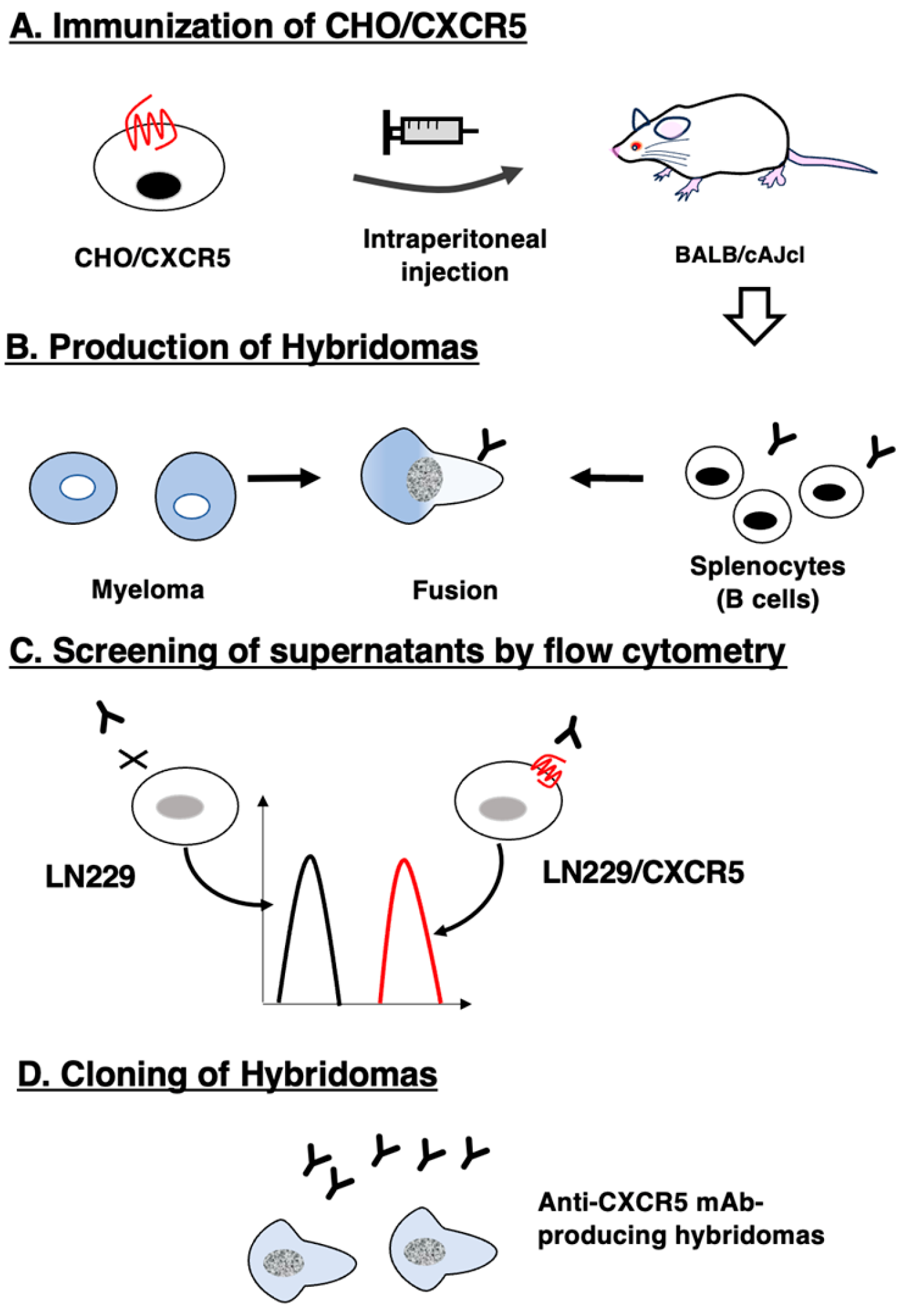
Figure 2.
Flow cytometric analysis using Cx5Mab-6 and J252D4. CHO/CXCR5 (A) and CHO-K1 (B) were treated with Cx5Mab-6 and J252D4 at the indicated concentrations (red) or with blocking buffer (black, negative control). The mAbs-treated cells were incubated with Alexa Fluor 488-conjugated anti-mouse IgG. Fluorescence data were collected using the SA3800 Cell Analyzer.
Figure 2.
Flow cytometric analysis using Cx5Mab-6 and J252D4. CHO/CXCR5 (A) and CHO-K1 (B) were treated with Cx5Mab-6 and J252D4 at the indicated concentrations (red) or with blocking buffer (black, negative control). The mAbs-treated cells were incubated with Alexa Fluor 488-conjugated anti-mouse IgG. Fluorescence data were collected using the SA3800 Cell Analyzer.
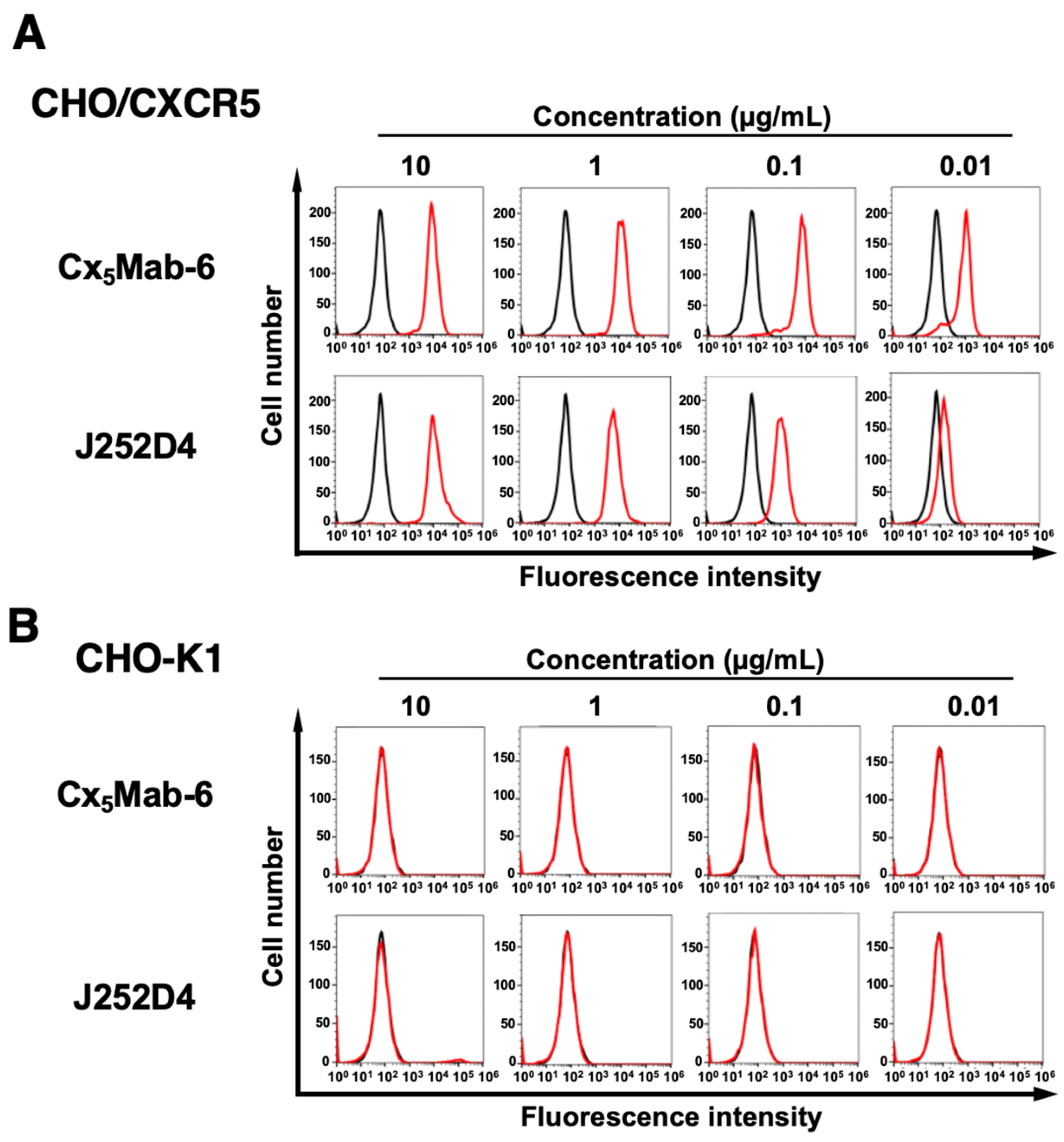
Figure 3.
Flow cytometry analysis of Cx5Mab-6 in CXCRs-overexpressed CHO-K1. (Upper panels) The CXCR1, CXCR2, CXCR3, CXCR4, CXCR5, and CXCR6-overexpressed CHO-K1 were treated with 1 µg/mL of Cx5Mab-6 (red) or with control blocking buffer (black, negative control), followed by treatment with anti-mouse IgG conjugated with Alexa Fluor 488. (Lower panels) Each CXCR expression was confirmed by 1 µg/mL of an anti-CXCR1 mAb (clone 8F1/CXCR1), an anti-CXCR2 mAb (clone 5E8/CXCR2), an anti-CXCR3 mAb (clone G025H7), an anti-CXCR4 mAb (clone 2B11/CXCR4), an anti-CXCR5 mAb (clone J252D4), and an anti-CXCR6 mAb (clone K041E5), followed by the treatment with Alexa Fluor 488-conjugated secondary mAbs. The fluorescence data were collected using the SA3800 Cell Analyzer.
Figure 3.
Flow cytometry analysis of Cx5Mab-6 in CXCRs-overexpressed CHO-K1. (Upper panels) The CXCR1, CXCR2, CXCR3, CXCR4, CXCR5, and CXCR6-overexpressed CHO-K1 were treated with 1 µg/mL of Cx5Mab-6 (red) or with control blocking buffer (black, negative control), followed by treatment with anti-mouse IgG conjugated with Alexa Fluor 488. (Lower panels) Each CXCR expression was confirmed by 1 µg/mL of an anti-CXCR1 mAb (clone 8F1/CXCR1), an anti-CXCR2 mAb (clone 5E8/CXCR2), an anti-CXCR3 mAb (clone G025H7), an anti-CXCR4 mAb (clone 2B11/CXCR4), an anti-CXCR5 mAb (clone J252D4), and an anti-CXCR6 mAb (clone K041E5), followed by the treatment with Alexa Fluor 488-conjugated secondary mAbs. The fluorescence data were collected using the SA3800 Cell Analyzer.
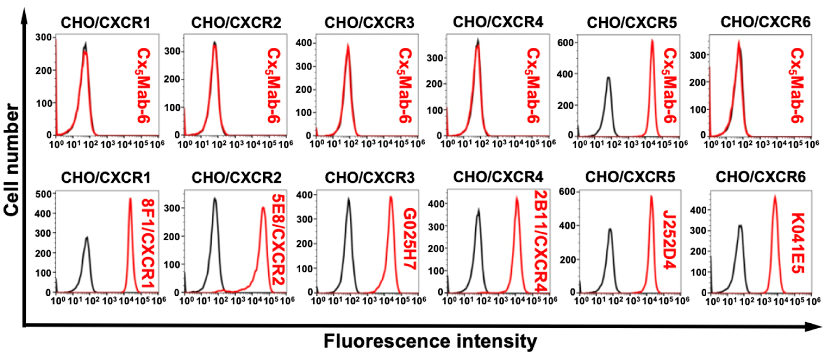
Figure 4.
Flow cytometric analysis using Cx5Mab-6 and J252D4 against an endogenous CXCR5-positive cell line. (A) Raji was treated with Cx5Mab-6, IgG2b isotype control (RdMab-20) at the indicated concentrations (red), or blocking buffer (black, negative control). (B) Raji was treated with J252D4, IgG1 isotype control (CvMab-62) at the indicated concentrations (red), or blocking buffer (black, negative control). The mAbs-treated cells were incubated with Alexa Fluor 488-conjugated anti-mouse IgG. Fluorescence data were collected using the SA3800 Cell Analyzer.
Figure 4.
Flow cytometric analysis using Cx5Mab-6 and J252D4 against an endogenous CXCR5-positive cell line. (A) Raji was treated with Cx5Mab-6, IgG2b isotype control (RdMab-20) at the indicated concentrations (red), or blocking buffer (black, negative control). (B) Raji was treated with J252D4, IgG1 isotype control (CvMab-62) at the indicated concentrations (red), or blocking buffer (black, negative control). The mAbs-treated cells were incubated with Alexa Fluor 488-conjugated anti-mouse IgG. Fluorescence data were collected using the SA3800 Cell Analyzer.
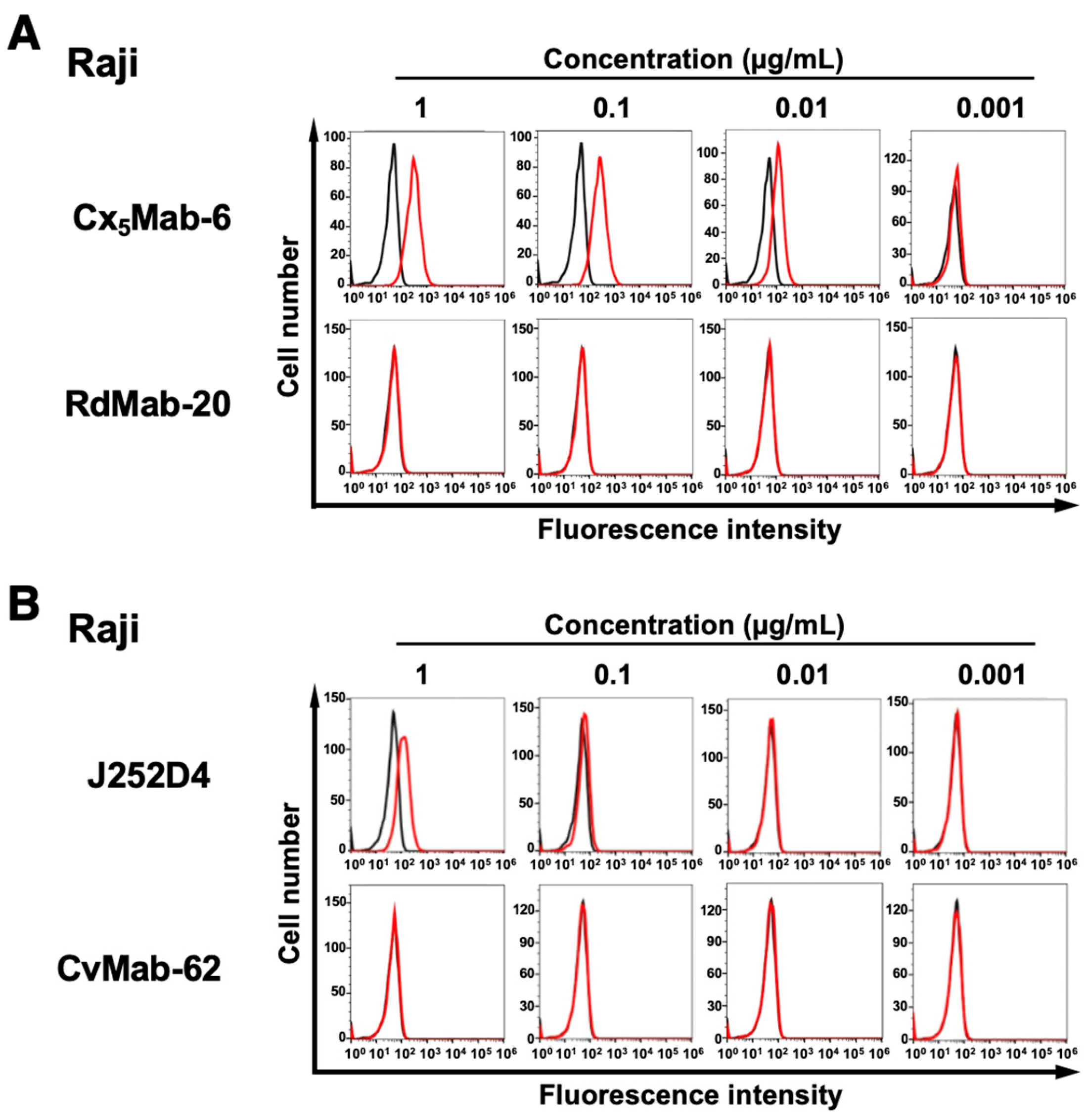
Figure 5.
Determination of the binding affinity of Cx5Mab-6 by flow cytometry. CHO/CXCR5 and Raji cells were incubated with serially diluted Cx5Mab-6, then reacted with Alexa Fluor 488-conjugated anti-mouse IgG. Geometric mean fluorescence values were measured using the SA3800 Cell Analyzer and FlowJo software. Average KD values (± standard deviation) from three independent measurements were calculated using GraphPad PRISM 6. Representative graphs are shown.
Figure 5.
Determination of the binding affinity of Cx5Mab-6 by flow cytometry. CHO/CXCR5 and Raji cells were incubated with serially diluted Cx5Mab-6, then reacted with Alexa Fluor 488-conjugated anti-mouse IgG. Geometric mean fluorescence values were measured using the SA3800 Cell Analyzer and FlowJo software. Average KD values (± standard deviation) from three independent measurements were calculated using GraphPad PRISM 6. Representative graphs are shown.
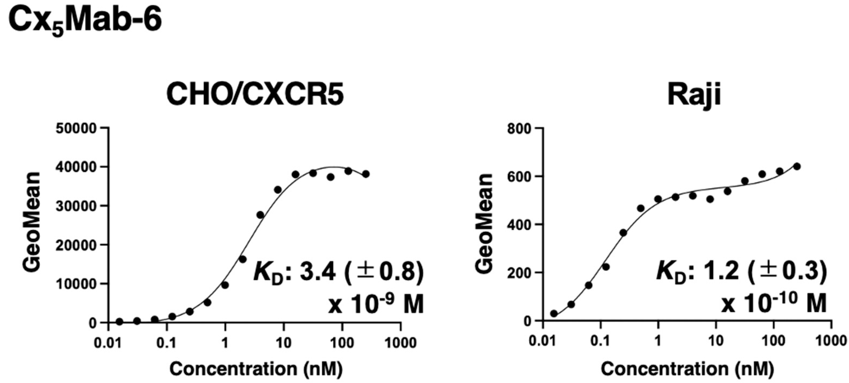
Figure 6.
Western blotting using Cx5Mab-6 and J252D4. Cell lysates (10 μg/lane) from CHO-K1 and CHO/CXCR5 were electrophoresed and transferred to polyvinylidene difluoride membranes. The membranes were incubated with 1 μg/mL of Cx5Mab-6 (A), 1 μg/mL of J252D4 (B), or 2 μg/mL of RcMab-1-mG1 (an anti-IDH1 mAb) (C), followed by the treatment with anti-mouse IgG-conjugated with horseradish peroxidase. The exposure condition in (A) and (B) was same.
Figure 6.
Western blotting using Cx5Mab-6 and J252D4. Cell lysates (10 μg/lane) from CHO-K1 and CHO/CXCR5 were electrophoresed and transferred to polyvinylidene difluoride membranes. The membranes were incubated with 1 μg/mL of Cx5Mab-6 (A), 1 μg/mL of J252D4 (B), or 2 μg/mL of RcMab-1-mG1 (an anti-IDH1 mAb) (C), followed by the treatment with anti-mouse IgG-conjugated with horseradish peroxidase. The exposure condition in (A) and (B) was same.
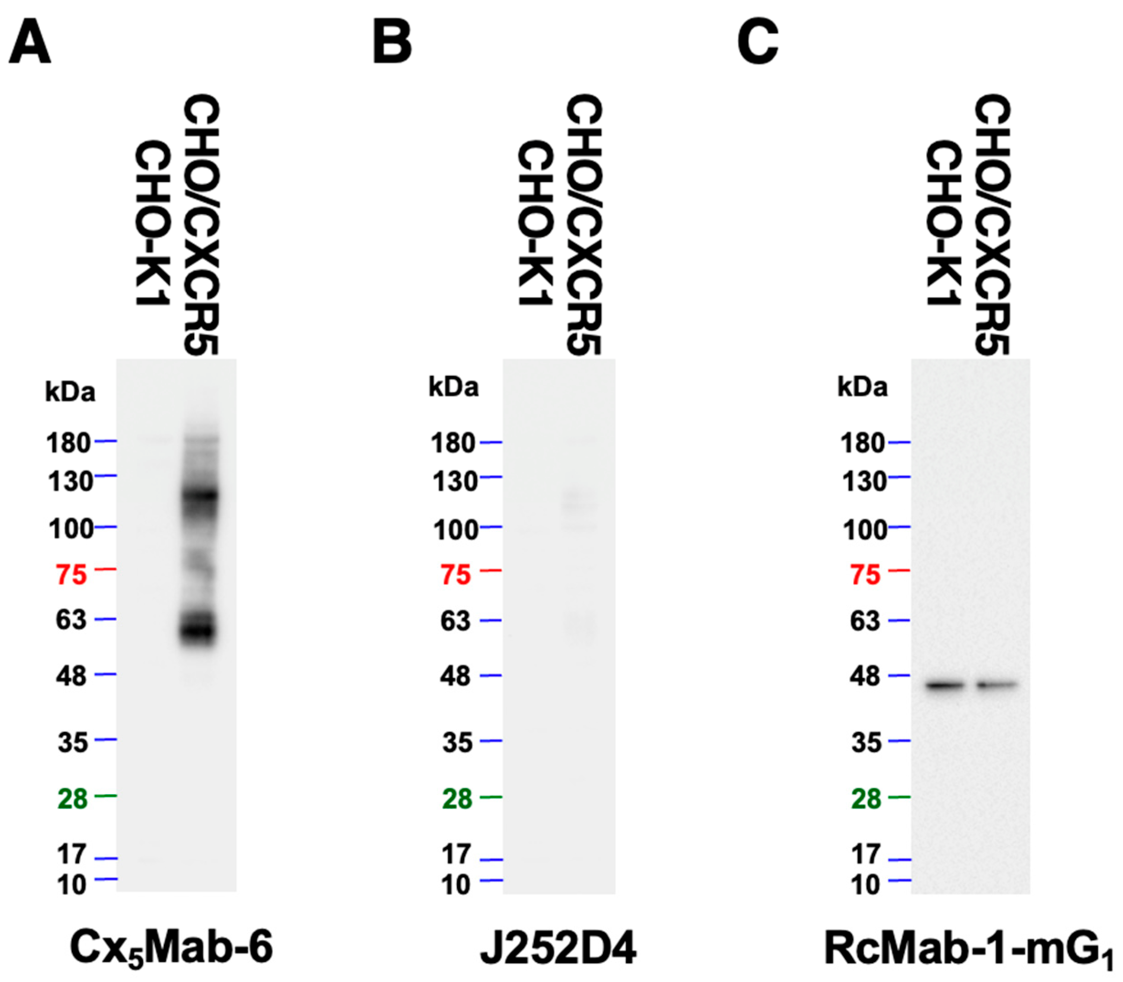
Figure 7.
Immunohistochemistry using Cx5Mab-6 and J252D4 in formalin-fixed paraffin-embedded cell blocks. CHO/CXCR5 and CHO-K1 sections were treated with 0.5 μg/mL of Cx5Mab-6 (A) or J252D4 (B). The staining was performed using BenchMark ULTRA PLUS with the ultraView Universal DAB Detection Kit, Scale bar = 100 μm.
Figure 7.
Immunohistochemistry using Cx5Mab-6 and J252D4 in formalin-fixed paraffin-embedded cell blocks. CHO/CXCR5 and CHO-K1 sections were treated with 0.5 μg/mL of Cx5Mab-6 (A) or J252D4 (B). The staining was performed using BenchMark ULTRA PLUS with the ultraView Universal DAB Detection Kit, Scale bar = 100 μm.
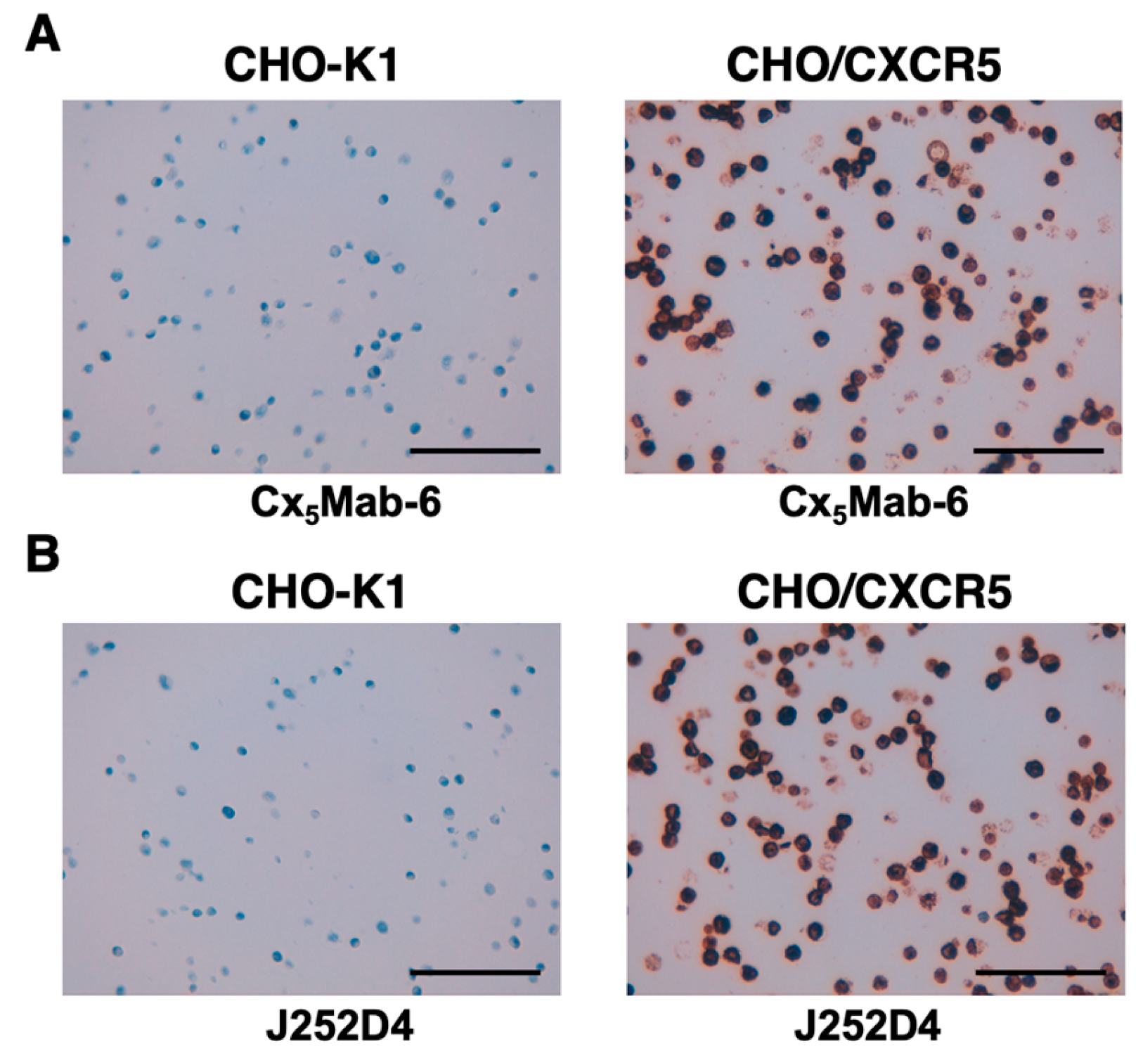
Figure 8.
Immunohistochemistry using Cx5Mab-6 in a lymphoma tissue array. Sequential sections of human lymphoma tissue array (LM241b) were treated with 0.5 μg/mL of Cx5Mab-6 or isotype control IgG2b (RdMab-20). lymphoid follicle of normal tonsil (A) and plasmacytic lymphoma (B) were shown. The high-magnification images inserted at the top right corners of each image. The staining was performed using BenchMark ULTRA PLUS with the ultraView Universal DAB Detection Kit, Scale bar = 100 μm.
Figure 8.
Immunohistochemistry using Cx5Mab-6 in a lymphoma tissue array. Sequential sections of human lymphoma tissue array (LM241b) were treated with 0.5 μg/mL of Cx5Mab-6 or isotype control IgG2b (RdMab-20). lymphoid follicle of normal tonsil (A) and plasmacytic lymphoma (B) were shown. The high-magnification images inserted at the top right corners of each image. The staining was performed using BenchMark ULTRA PLUS with the ultraView Universal DAB Detection Kit, Scale bar = 100 μm.
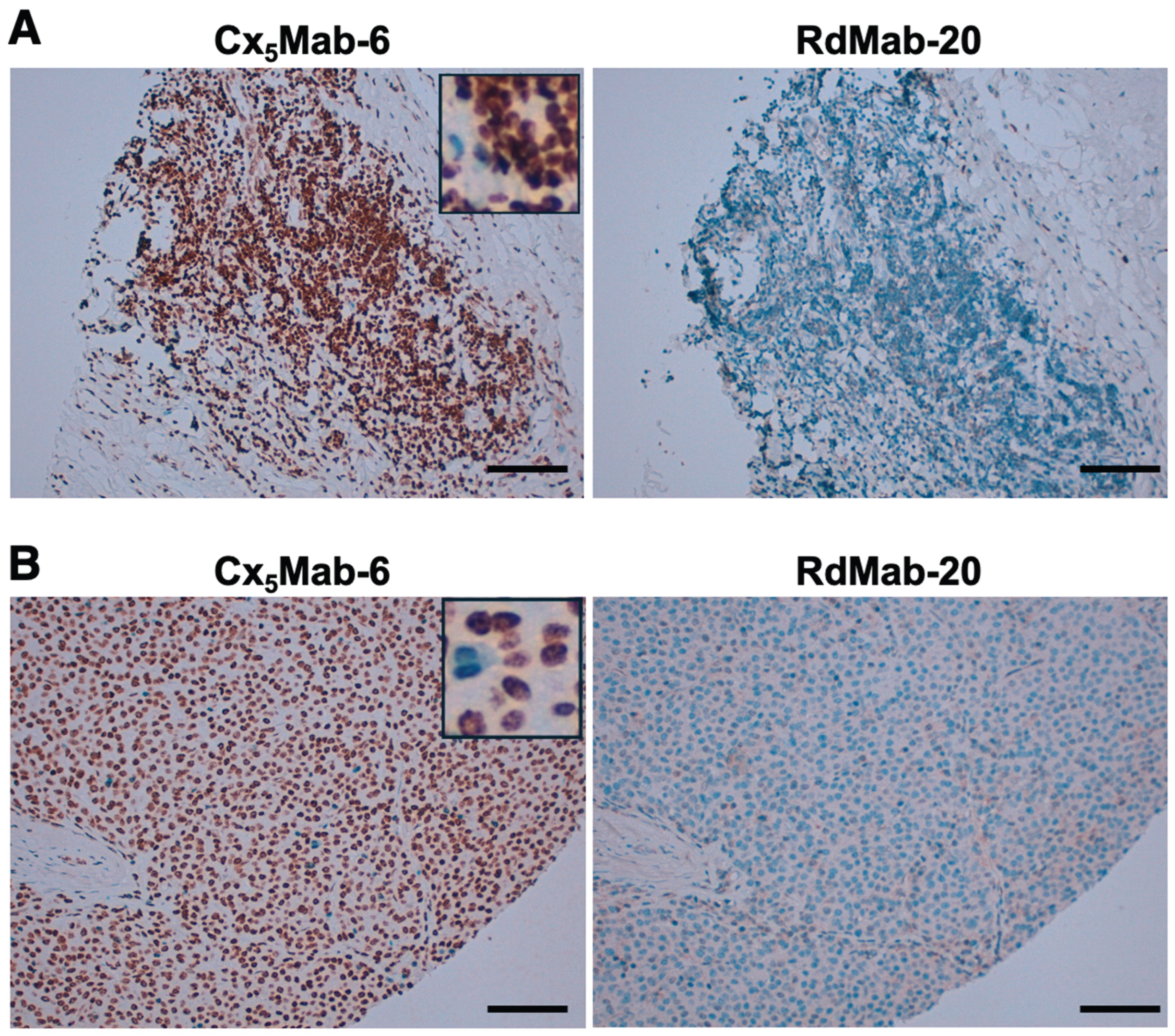

Table 1.
Immunohistochemistry of a lymphoma tissue array (LM241b) by Cx5Mab-6.
| No. | Age | Sex | Pathology diagnosis | Organ | Cx5Mab-6 |
| 1 | 71 | M | Diffuse B cell lymphoma of neck | LN | 1+ |
| 2 | 63 | M | Plasmacytic lymphoma of left armpit | LN | 3+ |
| 3 | 50 | M | Diffuse B cell lymphoma | Colon | 3+ |
| 4 | 64 | F | Diffuse large-B cell lymphoma of left groin | LN | 3+ |
| 5 | 43 | F | Diffuse large-B cell lymphoma of left groin | LN | 3+ |
| 6 | 16 | M | Mucosa-associated B-cell lymphoma | Colon | 1+ |
| 7 | 58 | M | Mantle cell lymphoma of neck | LN | 1+ |
| 8 | 64 | M | Lymphoid plasma cell lymphoma of neck | LN | 2+ |
| 9 | 51 | M | Diffuse large-B cell lymphoma of right armpit | LN | 2+ |
| 10 | 28 | M | Diffuse T cell lymphoma of left neck | LN | 3+ |
| 11 | 45 | M | Normal tonsil tissue | Tonsil | 3+ |
−, No stain; 1+, Weak intensity; 2+, Moderate intensity; 3+, Strong intensity. LN, Lymph node.
Disclaimer/Publisher’s Note: The statements, opinions and data contained in all publications are solely those of the individual author(s) and contributor(s) and not of MDPI and/or the editor(s). MDPI and/or the editor(s) disclaim responsibility for any injury to people or property resulting from any ideas, methods, instructions or products referred to in the content. |
© 2026 by the authors. Licensee MDPI, Basel, Switzerland. This article is an open access article distributed under the terms and conditions of the Creative Commons Attribution (CC BY) license (http://creativecommons.org/licenses/by/4.0/).
Copyright: This open access article is published under a Creative Commons CC BY 4.0 license, which permit the free download, distribution, and reuse, provided that the author and preprint are cited in any reuse.