

Submitted:
10 August 2026
Posted:
11 August 2026
You are already at the latest version
Preprints on COVID-19 and SARS-CoV-2
Abstract
The aim of our study is to retrospectively examine the frequency of rotavirus and adenovirus in acute gastroenteritis cases, their distribution according to age, season, and gender, and to investigate the epidemiological impact of COVID-19. We conducted a single-center, retrospective, observational cohort study over a four-year period. A total of 46.459 stool samples with a preliminary diagnosis of acute gastroenteritis from our hospital's wards and outpatient clinics were included in the study. Rotavirus and adenovirus antigens in stool samples were investigated using qualitative immunochromatographic diagnostic tests, and patient records were retrospectively evaluated. The distribution of antigen positivity according to seasons, patient ages, and genders was statistically evaluated using Pearson chi-square analysis. Rotavirus antigen was found in 10.7% of patients with gastroenteritis. 76.8% of rotavirus positive samples were taken from children aged 0-5 years, with the highest antigen positivity rate of 14.5% observed in children aged 13-24 months. Adenovirus antigen was detected in 3.2% of patients. Adenovirus positivity was highest in children aged 2-4 years (4.1%). There was no statistically significant difference between genders in terms of rotavirus and adenovirus prevalence. In our region, rotavirus and enteric adenoviruses, especially among pediatric gastroenteritis cases, maintain their importance as a significant public health problem. It has been observed that rotavirus gastroenteritis increases in winter and spring, while adenovirus gastroenteritis increases in spring and summer. In 2020, positivity rates were found to be very low, which has been interpreted as a result of the peak period of the COVID-19 pandemic.
Keywords:
adenovirus
; rotavirus
; gastroenteritis
; COVID-19
1. Introduction
Gastroenteritis may be associated with infectious and noninfectious causes; the causative agents of infectious gastroenteritis are microorganisms, including bacteria, viruses, fungi, or parasites. Viruses causing acute viral gastroenteritis belong to the families Reoviridae (rotavirus), Caliciviridae (norovirus and sapovirus), Astroviridae (astrovirus), and Adenoviridae (adenovirus types 40 and 41) [1,2].
Recently, it's been reported that viruses are the cause of 30 to 70% of infectious gastroenteritis cases in our country. Viruses such as rotavirus, enteric adenovirus, norovirus, astrovirus, and sapovirus are identified as the causes of viral gastroenteritis, especially in children [3,4,5]. Acute gastroenteritis is a significant health issue characterized by high morbidity and mortality rates in young children. Acute gastroenteritis in childhood is a major cause of hospital admissions. Adenoviruses and rotaviruses are causative agents of acute gastroenteritis in children under five in both developing and developed countries, leading to epidemics and deaths each year [6].
In acute gastroenteritis, a prevalent cause of morbidity and mortality worldwide, rapid and accurate identification of the causative agent will enable the selection of rapid and appropriate treatment options while preventing unnecessary antibiotic use. Immunochromatographic methods are widely used in many laboratories for the diagnosis of viral gastroenteritis due to their ease of use and interpretation [7].
The factors causing gastroenteritis vary depending on the patient's age, season, and geographical region. Being aware of the regional causes of acute gastroenteritis is crucial for taking infection control measures, obtaining epidemiological data on acute gastroenteritis infections caused by these factors, and initiating appropriate treatment. Our study investigates the frequency of rotavirus and adenovirus antigens, which are viral infection agents, in stool samples from patients referred to our laboratory with a preliminary diagnosis of acute gastroenteritis, using an immunochromatographic method.
2. Materials and Methods
2.1. Ethical Committee Approval
This study was conducted in accordance with the Declaration of Helsinki on Ethical Principles and was approved by the Ethics Committee of Ethics Committee of Sakarya University Faculty of Medicine, Health Sciences Scientific Research Ethics Committee, Sakarya, Turkey (approval number: 050.04-399710-68, dated: 19.09.2024).
2.2. Study Population
We conducted a single-center, retrospective, observational cohort study. A total of 46.459 stool samples with a preliminary diagnosis of acute gastroenteritis from various wards and outpatient clinics of Sakarya University Hospital's Medical Microbiology Laboratory were included in the study between January 1, 2020, and January 1, 2024.
2.3. Clinical Characteristics and Laboratory Methods
A total of 23.760 samples were tested for rotavirus, and 22.699 samples were tested for adenovirus. Patient records were reviewed for age, gender, time of hospital admission, and frequency of adenovirus and rotavirus positivity. Patients were divided into four groups according to age: 0 to 12 months, 13 to 24 months, 2 to 4 years, 5 to 16 years, and over 16 years, and their records were retrospectively analyzed.
Patients whose stool samples were sent to our laboratory with a preliminary diagnosis of acute gastroenteritis and who underwent rapid immunochromatographic testing for rotavirus and enteric adenovirus antigens were retrospectively evaluated. A rapid diagnostic test (Microcult Rota-Adenovirus Kombo Cassette Test, Tıpkimsan, Turkey) developed based on the principle of a qualitative immunochromatographic method capable of detecting both virus antigens in the same sample was used to detect the presence of rotavirus and adenovirus antigens in fresh stool samples. The sensitivity of the test for Rotavirus was 97.3, and specificity was 97.1, while for Adenovirus, sensitivity was 95.2 and specificity was 97.7. In accordance with the manufacturer's recommendations, quality control studies were conducted separately for each diagnostic kit using positive control antigens.
2.4. Statistical Analysis
The data collected in the study were analyzed using the IBM SPSS Statistics 26 (IBM SPSS, Chicago, IL, USA) program. Descriptive statistics for categorical and continuous variables were presented as numbers and percentages. The distribution of antigen positivity depending on seasons, patient ages, and genders was statistically evaluated using Pearson's chi-square analysis. A p-value < 0.05 was considered statistically significant for all tests.
3. Results
Four-year results indicate that rotavirus antigen positivity was detected in 2.539 (10.7%) of 23.760 stool samples tested for rotavirus as a cause of viral gastroenteritis. Rotavirus antigen was detected in 10.5% of male patients and 10.9% of female patients, and no statistically significant difference was found between the incidence of rotavirus and gender (p=0.306, p>0.05). Of the samples that tested positive for rotavirus antigen, 1.951 (76.8%) were from patients aged 0-5 years, and antigen positivity was most commonly detected in children aged 13-24 months (14.5%). Rotavirus antigen positivity in the group aged 13-24 months was found to be statistically significantly higher than in other groups (p <0.05).
The frequency of rotavirus-positive cases was highest in spring (18.3%) and winter (9.8%), and this result was statistically significant (p<0.05). An increase in acute gastroenteritis cases caused by rotavirus was observed in winter and spring. Over the years, rotavirus positivity was detected at 15.4% in 2022, peaking between February and May of the same year (ranging from 22.2% to 28%). Likewise, in 2021 and 2023, the highest positivity rates were detected between February and May, and in July and August 2023, antigen positivity rates of 16.6% and 11.8% were found, respectively, above the annual incidence rate. When examining the 2020 data, the annual positivity rate was 5.3%, with antigen positivity rates of 11.4% and 14.7% in January and March, respectively. The data for this year was interpreted as a result of the most intense period of the COVID-19 pandemic. Table 1 shows the distribution of rotavirus antigen positivity rates by gender, age group, and season, while Figure 1 provides detailed data on monthly rotavirus positivity rates by year.
Based on four-year results, adenovirus antigen positivity was detected in 734 (3.2%) of 22.699 stool samples tested for adenovirus as a cause of viral gastroenteritis. Adenovirus antigen positivity was detected in 3.2% of male patients and 3.3% of female patients, and no significant association was found between adenovirus antigen positivity and gender (p=0.892, p>0.05). Considering age distribution, the group with the highest rate of adenovirus antigen positivity was the group aged 2-4 years, with a rate of 4.1% (Table 2). Compared to other groups, this rate was found to be significantly higher (p<0.05).
The frequency of adenovirus-positive cases was highest in spring (3.4%) and summer (3.8%), and this trend was found to be statistically significant (p<0.05). It was observed that cases of acute gastroenteritis caused by adenoviruses increased in spring and summer. Over the years, adenovirus positivity was highest in 2022 at 6.7%, peaking in July-August (17.4% and 10.8%, respectively) of the same year. Throughout 2022, the highest rates of adenovirus positivity in the four-year period were detected from January to November. When examining the 2020 data, the annual positivity rate was 1.2%, with monthly incidence rates ranging from 0.0% to 2.2%. When examining the 2020 data, the annual positivity rate is 1.2%, and the monthly incidence rates vary between 0.0% and 2.2%. The highest rate of 2.2% was detected in February, and the data for this year has been interpreted as a result of the most intense period of the COVID-19 pandemic. According to 2021 data, the annual positivity rate is 1.7%, with monthly incidence rates ranging from 0.5% to 3.4%, with the highest rate of 3.4% observed in November. In 2023, the highest positivity rate was found in November at 4.3%. Table 2 shows the distribution of adenovirus antigen positivity rates by gender, age groups, and seasons, while Figure 2 provides detailed data on monthly adenovirus positivity rates by year.
4. Discussion
Rotavirus and enteric adenovirus are common causes of viral gastroenteritis. Transmission occurs via the fecal-oral route, particularly through shared items [8]. Studies in our country have indicated that the most common cause of acute gastroenteritis is viral, and among these, rotavirus and enteric adenovirus serotypes 40-41 are the most common [9,10,11,12]. Knowing the epidemiological characteristics of viral gastroenteritis causes is important because they can lead to high mortality and morbidity and unnecessary antibiotic use. Rapid detection of the viral agent in viral gastroenteritis is crucial in preventing unnecessary antibiotic use and hospital admissions [9].
The positivity rates for viral gastroenteritis agents are known to vary between different countries and regions within the same country. The main reason for the variation in the frequency of rotavirus and adenovirus in different regions and countries is the difference in research groups and methods. Variations in the prevalence of viral gastroenteritis may also be observed depending on socio-economic differences and the importance attached to hygiene conditions [11].
The incidence of rotavirus infections, which are the most common and severe cause of acute gastroenteritis in childhood, varies seasonally around the world. In temperate climate regions, rotavirus cases are generally more prevalent in spring and winter, while in hot and tropical regions, they are reported to be widespread throughout the year [12,13,14]. In a systematic review including 98 studies, Tapısız et al. [15] noted that rotavirus infection in children in Turkey is generally observed in winter. Vural et al. [16] reported an 8.9% positivity rate for rotavirus in acute gastroenteritis cases, with a positivity rate of 11.4% in spring and 12.0% in winter. Çaycı et al. [17] found an 11.7% positivity rate for rotavirus in their study of acute gastroenteritis cases, reporting that rotavirus was the most common infectious agent in spring (18.6%) and winter (17%). Genc et al. [18] detected rotavirus positivity in 13.6% of gastroenteritis cases in their study conducted in the Siirt region, and they reported that the highest positivity rate was 19.6% in children aged 13-24 months. Şenol et al. [19] reported a 9.6% rate of rotavirus positivity in their study conducted in the Elazığ region, with a particularly high rate of 13.8% among children aged 2-4 years. In our study, the rate of rotavirus positivity was 10.7% over a 4-year period, consistent with the literature. Rotavirus positivity was found to be highest seasonally in spring (18.3%) and winter (9.8%), and this trend was statistically significant (p<0.05). During the four-year observation period, the highest rotavirus positivity rates in our region were detected between February and May in 2021-2022 and 2023, and these high positivity rates were interpreted as indicating a rotavirus outbreak in our region during these months. Unlike what was expected, in 2023, antigen positivity rates were found to be 16.6% and 11.8% above the annual incidence rate in July and August, respectively, in line with the literature, and the high incidence during the summer months was interpreted as an epidemic situation (Figure 1).
Globally, the rate of rotavirus infection in children hospitalized for diarrhea is approximately 30 to 50%, and more than 90% of children with severe rotavirus infections live in developing countries [20,21]. Tapısız et al. [15] reported a rotavirus detection rate of 31.8% in children under 5 years old in Turkey in their systematic review. Barutçu et al. [21] detected rotavirus positivity in 40% of children with acute gastroenteritis with an average age of under 2 years old and reported that cases were frequently seen during the winter months. Vural et al. [16] reported that rotavirus positivity in acute gastroenteritis cases aged 0-5 years was 15.9%. In the same study, considering the age distribution of patients, rotavirus positivity was reported to be 10.5% in the group aged 0-12 months, 16.8% in the group aged 13-24 months, and 14.9% in the group aged 2-5 years. Çaycı et al. [17] reported in their study of acute gastroenteritis cases that rotavirus positivity was observed in 24.5% of children aged 13-24 months and 11.7% of children aged 3-5 years. Taşkın et al. [11] found a rotavirus positivity rate of 14.7% in their studies, with the highest positivity rates reported in children aged 0-2 years at 57.9%. Aytaç et al. [22] found rotavirus positivity in 9.5% of patients with a preliminary diagnosis of acute gastroenteritis in their study conducted in the Elazığ region, with the highest rates observed in spring and winter months. The highest incidence rates of rotavirus positivity were reported in children aged 0-12 months (12.9%) and 13-24 months (11.1%). The results of our study reveal that rotavirus antigen positivity was detected in 10.7% of cases. Of the samples that tested positive for rotavirus antigen, 1,951 (76.8%) were from patients aged 0-5 years, with rotavirus antigen positivity most commonly detected in children aged 13-24 months at 14.5%. Upon analyzing our study data by year, the highest antigen positivity was detected in the group aged 13-24 months in the three years except 2022, while in 2022, the highest positivity rate was found in the group aged 2-4 years at 21.5% (Table 1).
Enteric adenovirus serotypes 40 and 41 are serotypes associated with viral gastroenteritis, especially in young children [23]. Enteric adenoviruses are frequently observed among the causes of acute gastroenteritis in children under the age of four. Children aged 6 months to 2 years who attend daycare centers and nurseries are at high risk [12,24]. Vural et al. [16] reported adenovirus positivity in 4.4% of acute gastroenteritis cases, with adenovirus positivity occurring at a rate of 4.6% in summer and 5.1% in winter. Çaycı et al. [17] found adenovirus positivity in 3.3% of acute gastroenteritis cases in their study, reporting that adenoviruses were the most common cause of infection in winter (4.6%) and spring (3.4%). Tokak et al. [12] reported adenovirus positivity at a rate of 3.4% in children with acute gastroenteritis, noting that adenovirus was more frequently associated with infection in the fall months of October and November. Taşkın et al. [11] detected adenovirus positivity at a rate of 4.4% and reported that enteric adenovirus antigen positivity was highest in summer and autumn months. Vural et al. [16] noted that adenovirus positivity was 3.8% in acute gastroenteritis cases among children aged 0-5 years. In the same study, considering the age distribution of patients, 2.8% were aged 0-12 months, 3.2% were aged 13-24 months, and 4.9% were aged 2-5 years. Çaycı et al. [17] reported in their study of acute gastroenteritis cases that adenovirus positivity was observed in 8.2% of children aged 13 to 24 months and in 2.1% of children aged 3-5 years. Genç et al. [18] detected adenovirus positivity at a rate of 4.9% as the cause of viral gastroenteritis in the Siirt region, with the highest rate of antigen positivity reported at 5.5% in children aged 3-5 years. Aytaç et al. [22] found adenovirus positivity in 2.6% of patients with a preliminary diagnosis of acute gastroenteritis in the Elazığ region, with the highest rates observed in summer and autumn. The highest rates of adenovirus positivity were 3.7% in children aged 0-12 months and 3.1% in children aged 3-5 years. Şenol et al. [19] detected adenovirus positivity in 2.0% of patients in their study conducted in the Elazığ region, noting that adenovirus positivity was 2.5% in children aged 5-8. According to the results of our study, adenovirus antigen positivity was detected in 3.2% of patients. Of the samples with adenovirus antigen positivity, 572 (77.6%) were from patients aged 0-5 years. Considering age distribution, the group with the highest rate of adenovirus antigen positivity was children aged 2-4 years, with a rate of 4.1% (Table 2).
Adenoviruses are seen throughout the year, with no seasonal differences; however, there may be an increase in the number of cases in the summer and late winter [24]. Barutçu et al. [21] detected adenovirus positivity in 10.2% of children with acute gastroenteritis whose average age was less than two years and reported that cases were frequently seen in the spring months. Our study found that the frequency of adenovirus-positive cases was highest in spring (3.4%) and summer (3.8%), and this result was statistically significant (p<0.05). Over the years, adenovirus positivity was found to be highest in 2022, with an average rate of 6.7%, and in the same year, it was found to be 17.4% in July and 10.8% in August. The increase in positive cases above the annual average during the summer months was interpreted as an epidemic period. In 2021 and 2023, the highest adenovirus positivity rates were found in November, at 3.4% and 4.3%, respectively, above the annual averages Figure 2.
In their study investigating the effect of the COVID pandemic on viral gastroenteritis cases, Yıldız et al. reported that the number of clinical samples decreased by nearly half in Bursa during the pandemic period and that the positivity rates for rotavirus and adenovirus also decreased significantly [25]. Genç et al. [18] reported that rotavirus and adenovirus infections were less common in 2020 compared to other years, suggesting that this could be a result of the COVID-19 pandemic conditions. It has been reported that the COVID-19 pandemic has directly or indirectly affected other infectious diseases. It is evident that contact and isolation measures implemented against the COVID-19 pandemic, with the aim of preventing transmission, have significantly reduced viral gastroenteritis infections. In retrospective studies covering the pandemic period in China and Poland, it has been reported that hospital admissions due to viral gastroenteritis cases decreased, and the incidence rates of rotavirus and adenovirus decreased following school closures and quarantine measures [26,27]. Our study examined data for rotavirus in 2020 and found that the annual positivity rate was 5.3%, with antigen levels detected at 11.4% and 14.7% in January and March, respectively Figure 1. Upon examining the 2020 data for adenovirus, the annual positivity rate was 1.2%, with monthly incidence rates ranging from 0.0% to 2.2%, with the highest detection rate of 2.2% in February (Figure 2). Due to the Covid-19 pandemic in 2020, kindergartens and schools in our province suspended educational activities for a long time, and public places such as playgrounds and restaurants were closed, thereby limiting potential transmission environments, which led to a significant decrease in hospital visits and positivity rates. Additionally, the increased importance of hygiene rules such as mask use and handwashing in the community due to the pandemic is believed to have contributed to the decrease in cases during this period.
5. Conclusions
Our study revealed that rotavirus and adenovirus are key factors in acute gastroenteritis in children. In cases of gastroenteritis where viral etiology is suspected, it is critical to understand the epidemiology of the causative agents, identify susceptible groups, ensure proper implementation of treatment protocols, and take preventive measures. The incidence of viral gastroenteritis is influenced by environmental factors. Considering the data from 2020 in particular, the decrease in the number of tests and positivity rates during the pandemic supports this situation. Rotavirus and enteric adenovirus constitute a significant public health problem, especially among childhood gastroenteritis cases in our region. Early detection of rotavirus and adenovirus infections through sensitive and specific laboratory tests will be beneficial in the prevention and control of these infections. According to the data obtained from our study, viral agents must be investigated among childhood gastroenteritis cases.
Author Contributions
“Con-ceptualization, S.B.A. and H.A.T.; methodology, S.B.A. and H.A.T.; software, S.B.A. and H.A.T.; validation, S.B.A., H.A.T. and O.A; formal analysis, S.B.A.; investigation, S.B.A. and H.A.T.; resources, S.B.A.; data curation, S.B.A. and H.A.T.; writing—original draft preparation, S.B.A. and H.A.T.; writing—review and editing, S.B.A., H.A.T. and O.A.; visualization, S.B.A.; supervision, M.K.; project administration, M.K. All authors have read and agreed to the published version of the manuscript.”.
Funding
This research received no external funding.
Institutional Review Board Statement
The study was conducted in accordance with the Declaration of Helsinki, and approved by the Ethics Committee of Ethics Committee of Sakarya University Faculty of Medicine, Health Sciences Scientific Research Ethics Committee for studies involving humans, Sakarya, Turkey (approval number: 050.04-399710-68, dated: 19.09.2024).
Informed Consent Statement
Written informed consent was obtained from all patients participating in the study.
Conflicts of Interest
The authors declare no conflicts of interest.
References
- Farkas, T.; Jiang, X.I. Rotaviruses, Caliciviruses, Astroviruses, Enteric Adenoviruses and Other Diarrheic Viruses, Manual of Clinical Micriobiology; Murray, P.R., Ed.; ASM Pres: Washington DC; Volume 2007, pp. 1453–69.
- Meier, J.L. Viral Acute Gastroenteritis in Special Populations. Gastroenterol. Clin. North Am. 2021, 50(2), 305–22. [Google Scholar] [CrossRef]
- Tekin, A. The frequency of rotavirus and enteric adenovirus in children with acute gastroenteritis in Mardin. J. Clin. Exp. Invest. 2010, 1(1), 41–5. [Google Scholar] [CrossRef]
- Gulen, A.; Hacımustafaoğlu, M. General evaluation of acute gastroenteritis in children. ANKEM 2013, 27(3), 147–57. [Google Scholar] [CrossRef]
- Demir, A.; Gümüş, H. Agents of acute gastroenteritis in children in the autumn season. Van. Sag. Bil. Derg. 2023, 16(2), 100–4. [Google Scholar]
- Kızılırmak, A.; Çalışkan, E.; Temizkan, R.C. Rotavirus and adenovirus frequency in children with acute gastroenteritis. Konuralp Med. J. 2017, 9(2), 35–9. [Google Scholar] [CrossRef]
- Gonzalez-Serrano, L.; Munoz-Algarra, M.; Gonzalez-Sanz, R.; Portero-Azorin, M.F.; Amaro, M.J. Viral gastroenteritis in hospitalized patients: Evaluation of immunochromatographic methods for rapid detection in stool samples. J. Clin. Virol. 2020, 128, 104420. [Google Scholar] [CrossRef] [PubMed]
- Jagirdhar, G.S.K.; Pulakurthi, Y.S.; Chigurupati, H.D.; Surani, S. Gastrointestinal tract and viral pathogens. World J. Virol. 2023, 12(3), 136–150. [Google Scholar] [CrossRef] [PubMed]
- Köse, H.; Temocin, F. Investigation of the frequency of adenovirus and rotavirus in children with acute gastroenteritis. Flora 2019, 24(1), 22–26. [Google Scholar] [CrossRef]
- Aydın, E.; Aydın, N.; Percin Renders, D. Evaluation of the effect of acute gastroenteritis factors on laboratory parameters in pediatric patients. Flora 2022, 27(1), 125–134. [Google Scholar] [CrossRef]
- Kafa Taşkın, A.H.; Hasbek, M.; Celik, C. Detection of rotavirus and enteric adenovirus frequency in children with acute gastroenteritis attending to our hospital: a Sivas-scale retrospective study. Van. Sag Bil. Derg. 2021, 14(2), 122–130. [Google Scholar] [CrossRef]
- Tokak, S.; Ugurcan, D.; Güzeş, E.A. Investigation of adenovirus and rotavirus frequency and seasonal distribution in children with acute gastroenteritis. KU Med. J. 2022, 24(1), 163–70. [Google Scholar] [CrossRef]
- Patel, M.M.; Pitzer, V.; Alonso, W.J.; Vera, D.; Lopman, B.; Tate, J.; Vilboud, C.; Parashar, U.D. Global seasonality of rotavirus disease. Pediatr. Infect. Dis. J. 2013, 32(4), 134–47. [Google Scholar] [CrossRef] [PubMed]
- Kurugol, Z.; Devrim, I. Gastrointestinal infections. J. Pediatr. Inf. 2014, 8, 71–81. [Google Scholar] [CrossRef]
- Tapısız, A.; Demirdağ, T.B.; Cura Yayla, B.C.; Gunes, C.; Ugras Dikmen, A.; Tezer, H.; Aksakal, N.B.; Bozdayı, G.; Ozkan, S. Rotavirus infections in children in Turkey: A systematic review. Rev. Med. Virol. 2019, 29(1), e2020. [Google Scholar] [CrossRef] [PubMed]
- Gur Vural, D.; Torun, E.G.; Bıyık, I.; Tanrıverdi Caycı, Y.; Bilgin, K.; Birinci, A. Investigation of Frequency of Rotavirus and Adenovirus in Patients with Acute Gastroenteritis. KU Med. J. 2022, 24(2), 289–294. [Google Scholar] [CrossRef]
- Caycı, T.Y.; Yılmaz, G.; Birinci, A. Investigation of the frequency of rotavirus and adenovirus in acute gastroenteritis cases. Pamukkale Med. J. 2017, 10(1), 61–5. [Google Scholar] [CrossRef]
- Genc Bahce, Y.; Ozudogru, O.; Acer, O. The frequency of rotavirus and adenovirus in case of gastroenterit observed in Siirt region. Gevher Nesibe J. Med. Health Sci. 2022, 7(19), 23–28. [Google Scholar] [CrossRef]
- Senol, F.F.; Oner, P.; Aytac, Ö.; Babacan, A.; Ascı Toraman, Z. The Role of Parasitic and Viral Agents in Gastrointestinal Infections. Turk. Mikrobiyol. Cem. Derg. 2022, 52(4), 281–290. [Google Scholar] [CrossRef]
- Crawford, S.E.; Ramani, S.; Tate, J.E.; Parashar, U.D.; Svensson, L.; Hagbom, M.; Franco, M.A.; Greenberg, H.B.; O’Ryan, M.; Kang, G.; Desselberger, U.; Estes, M.K. Rotavirus infection. Nat. Rev. Dis. Prim. 2017, 3, 17083. [Google Scholar] [CrossRef] [PubMed]
- Barutcu, A.; Barutcu, S. Frequency of rotavirus and enteric adenovirus in children with acute gastroenteritis in Halfeti district, Sanliurfa, Turkey. Cukurova Med. J. 2020, 45(2), 448–454. [Google Scholar] [CrossRef]
- Aytac, O.; Senol, F.F.; Oner, P.; Erkmen, N.; Aslan, R.; Dogukan, M.; Ascı-Toraman, Z. Rotavirus and adenovirus frequency in acute gastroenteric diseases. Turk. Hij. Den. Biyol. Derg. 2020, 77(2), 179–184. [Google Scholar] [CrossRef]
- Qiu, F.Z.; Shen, X.X.; Li, G.X.; Zhao, L.; Chen, C.; Duan, S.X.; Guo, J.Y.; ZMC; Y, T.F.; Qi, J.J.; wang, L.; Feng, Z.S.; Ma, X.J. Adenovirus associated with acute diarrhea: a case-control study. BMC Infect. Dis. 2018, 18(1), 450. [Google Scholar] [CrossRef] [PubMed]
- Gul, M.; Garipardıc, M.; Cıragil, P.; Aral, M.; Karabiber, H.; Guler, I. Investigation of Rotavirus andAdenovirus types 40/41 in Children with Gastroenteritis between 0-5 Years of Age. ANKEM Derg. 2005, 19, 64–67. [Google Scholar]
- Yıldız, D.; Saglık, I.; Hacımustafaoglu, M.K.; Ozakın, C.; Celebi, S. The Impact of the COVID-19 Pandemic on the Prevalence of Rotavirus and Adenovirus in Bursa Uludag University Hospital. Turk. Mikrobiyol. Cemiy. Derg. 2022, 52(4), 299–309. [Google Scholar] [CrossRef]
- Li, W.; Zhu, Y.; Lou, J.; Chen, J.; Xie, X.; Mao, J. Rotavirus and adenovirus infections in children during COVID-19 outbreak in Hangzhou, China. Transl. Pediatr. 2021, 10(9), 2281–6. [Google Scholar] [CrossRef] [PubMed]
- Toczylowski, K.; Jackowska, K.; Lewandowski, D.; Kurylonek, S.; Waszkiewicz-Stojda, M.; Sulik, A. Rotavirus gastroenteritis in children hospitalized in northeastern Poland in 2006-2020: Severity, seasonal trends, and impact of immunization. Int. J. I Infect. Dis. 2021, 108, 550–6. [Google Scholar] [CrossRef] [PubMed]
Figure 1.
Distribution of Rotavirus incidence rates by month in 2020-2023.
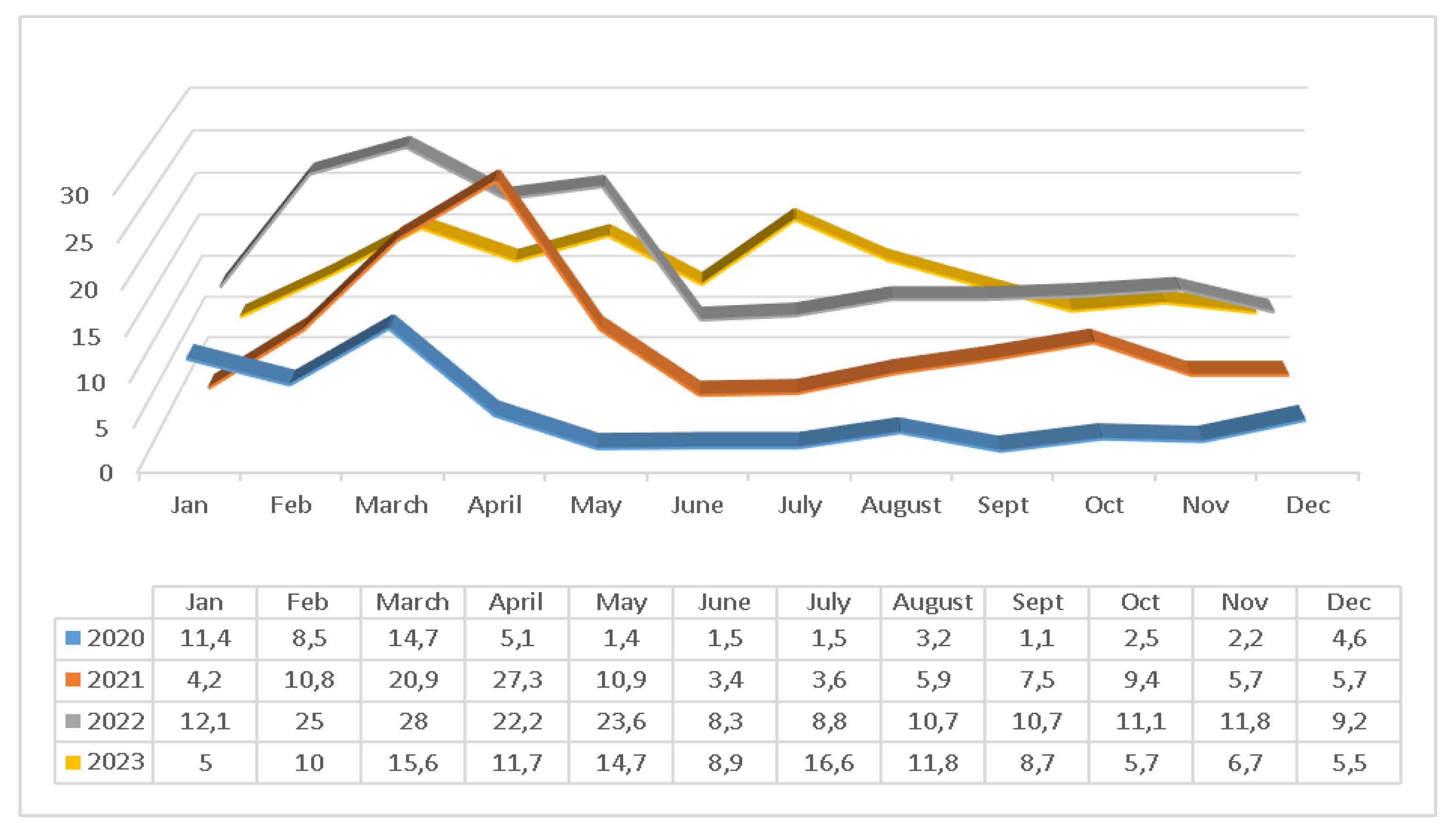
Figure 2.
Distribution of Adenovirus incidence rates by month in 2020-2023.
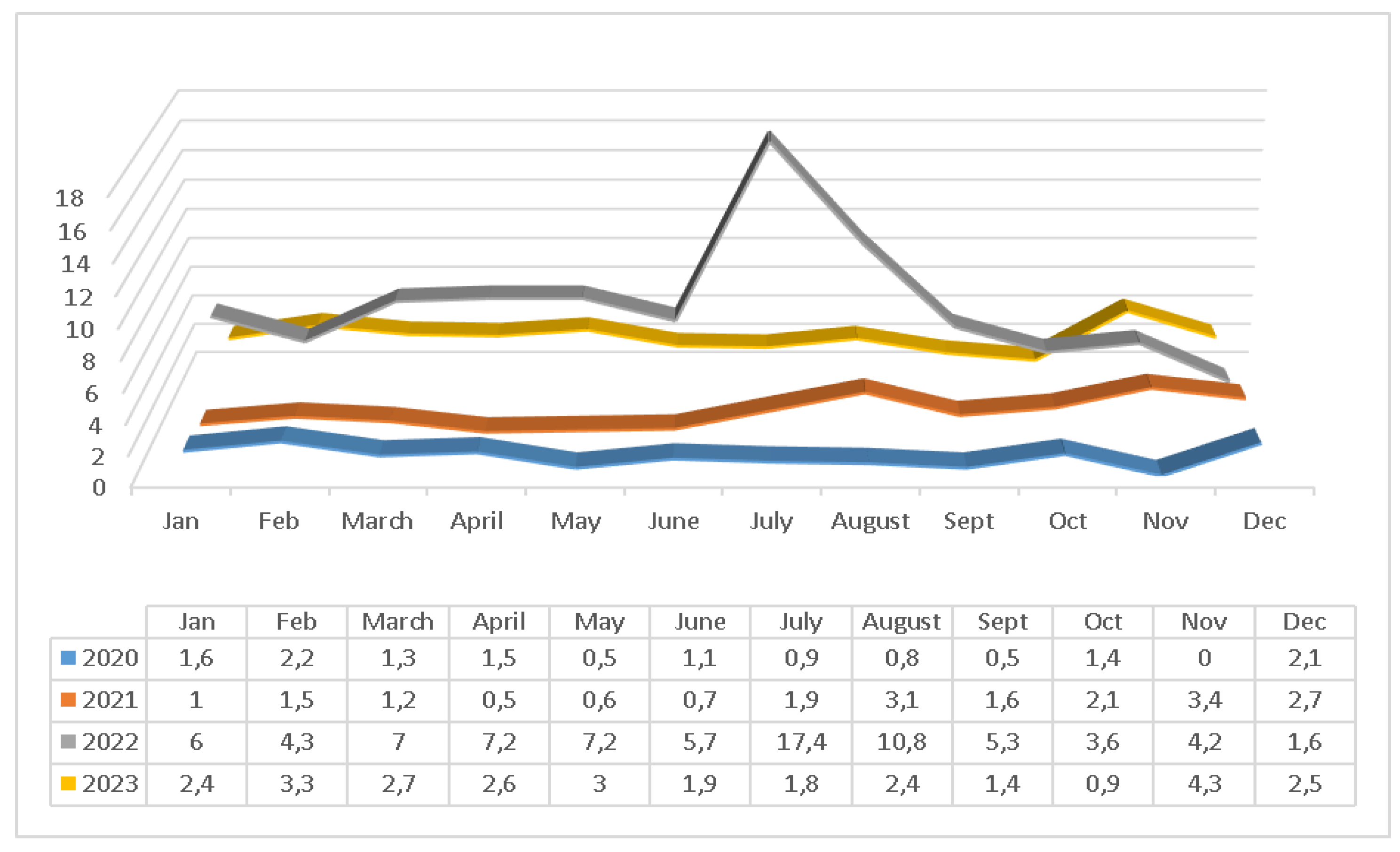

Table 1.
Rotavirus results in samples investigated for viral antigen between 2020 and 2023.
| 2020-2023 | 2020 | 2021 | 2022 | 2023 | |||
|---|---|---|---|---|---|---|---|
| TOTAL | Positive | Negative | Positive | Positive | Positive | Positive | |
| N (%) | N (%) | N (%) | N (%) | N (%) | |||
| Gender | |||||||
| Female | 10423 |
1138 (10.9) |
9285 (89.1) |
112 (5.7) |
231 (9.0) |
456 (15.4) |
339 (11.5) |
| Male | 13337 |
1401 (10.5) |
11936 (89.5) |
120 (5) |
304 (9.2) |
572 (15.4) |
405 (10.4) |
| p* | 0.306 | 0.306 | 0.834 | 0.930 | 0.135 | ||
|
Age Distribution |
|||||||
| 0-12 months | 5547 |
414 (7.5) |
5133 (92.5) |
49 (4.1) |
109 (8.3) |
150 (10.0) |
106 (6.9) |
| 13-24 months | 4446 |
643 (14.5) |
3803 (85.5) |
55 (6.8) |
166 (13.8) |
238 (19.6) |
184 (15) |
| 2-4 years | 6416 |
894 (13.9) |
5522 (86.1) |
59 (5.9) |
166 (9.8) |
400 (21.5) |
269 (14.4) |
| 5-16 years | 6634 |
564 (8.5) |
6070 (91.59 |
66 (5.4) |
89 (5.8) |
234 (12.3) |
175 (8.8) |
| >16 | 717 |
24 (3.3) |
693 (96.7) |
3 (2.3) |
5 (3.8) |
6 (2.9) |
10 (4.0) |
| p* | 0.000 | 0.039 | 0.000 | 0.000 | 0.000 | ||
|
Seasonal Distribution |
|||||||
| Winter | 5270 |
518 (9.8) |
4752 (90.2) |
120 (8.9) |
72 (6.5) |
231 (16.2) |
95 (6.8) |
| Spring | 5673 |
1036 (18.3) |
4637 (81.7) |
67 (9.1) |
278 (20.2) |
440 (24.8) |
251 (14.1) |
| Summer | 7426 |
574 (7.7) |
6852 (92.3) |
26 (2.1) |
89 (4.2) |
159 (9.4) |
300 (12.8) |
| Autumn | 5391 |
411 (7.6) |
4980 (92.4) |
19 (1.9) |
96 (7.5) |
198 (11.2) |
98 (7.3) |
| p* | 0.000 | 0.000 | 0.000 | 0.000 | 0.000 | ||
| TOTAL | 23760 |
2539 (10.7) |
21221 (89.3) |
232 (5.3) |
535 (9.1) |
1028 (15.4) |
744 (10.8) |
*p: Pearson-Chi-Square.
Table 2.
Adenovirus results in samples investigated for viral antigen between 2020 and 2023.
| 2020-2023 | 2020 | 2021 | 2022 | 2023 | |||
|---|---|---|---|---|---|---|---|
| TOTAL | Pozitive | Negative | Pozitive | Pozitive | Pozitive | Pozitive | |
| N (%) | N (%) | N (%) | N (%) | N (%) | |||
| Gender | |||||||
| Female | 9985 |
326 (3.3) |
9659 (96.7) |
24 (1.2) |
44 (1.7) |
198 (6.7) |
60 (2.4) |
| Male | 12714 |
411 (3.2) |
12303 (96.8) |
29 (1.2) |
57 (1.7) |
249 (6.7) |
76 (2.3) |
| p* | 0.892 | 0.964 | 0.995 | 0.988 | 0.845 | ||
| Age Distribution | |||||||
| 0-12 months | 5261 |
168 (3.2) |
5093 (96.8) |
18 (1.5) |
29 (2.2) |
94 (6.3) |
27 (2.2) |
| 13-24 months | 4258 |
155 (3.6) |
4103 (96.4) |
6 (0.7) |
17 (1.4) |
101 (8.3) |
31 (3) |
| 2-4 years | 6138 |
249 (4.1) |
5889 (95.9) |
11 (1.1) |
30 (1.8) |
165 (8.9) |
43 (2.7) |
| 5-16 years | 6315 |
153 (2.4) |
6162 (97.6) |
16 (1.3) |
25 (1.6) |
84 (4.4) |
28 (1.7) |
| >16 | 727 |
12 (1.7) |
715 (98.3) |
2 (1.3) |
0 |
3 (1.4) |
7 (3) |
| p* | 0.000 | 0.622 | 0.291 | 0.000 | 0.170 | ||
| Seasonal Distribution | |||||||
| Winter | 4792 |
129 (2.7) |
4663 (97.3) |
26 (1.9) |
21 (1.9) |
57 (4) |
25 (2.8) |
| Spring | 5702 |
195 (3.4) |
5507 (96.6) |
8 (1.1) |
10 (0.7) |
127 (7.1) |
50 (2.8) |
| Summer | 7443 |
285 (3.8) |
7158 (96.2) |
12 (0.9) |
41 (1.9) |
184 (10.8) |
48 (2) |
| Autumn | 4762 |
128 (2.7) |
4634 (97.3) |
7 (0.7) |
29 (2.3) |
79 (4.4) |
13 (1.9) |
| p* | 0.000 | 0.034 | 0.011 | 0.000 | 0.289 | ||
| TOTAL | 22699 |
737 (3.2) |
21962 (96.8) |
53 (1.2) |
101 (1.7) |
447 (6.7) |
13.6 (2.4) |
*p: Pearson-Chi-Square.
Disclaimer/Publisher’s Note: The statements, opinions and data contained in all publications are solely those of the individual author(s) and contributor(s) and not of MDPI and/or the editor(s). MDPI and/or the editor(s) disclaim responsibility for any injury to people or property resulting from any ideas, methods, instructions or products referred to in the content. |
© 2026 by the authors. Licensee MDPI, Basel, Switzerland. This article is an open access article distributed under the terms and conditions of the Creative Commons Attribution (CC BY) license (http://creativecommons.org/licenses/by/4.0/).
Copyright: This open access article is published under a Creative Commons CC BY 4.0 license, which permit the free download, distribution, and reuse, provided that the author and preprint are cited in any reuse.